radiation equipment safety manual · radiation equipment safety manual . preface . the use of...
TRANSCRIPT

RADIATION EQUIPMENT SAFETY MANUAL
Environment, Health & Safety Services STATE UNIVERSITY OF NEW YORK AT BUFFALO
Revised July 2004

State University of New York at Buffalo Environment, Health & Safety Services
Radiation Safety Division
Radiation Equipment Safety Manual
Preface The use of radiation producing equipment is strictly controlled by the New York State Department of Health under provisions of Title 10, Part 16 of the State Code. Such provisions apply to physical characteristics of the equipment, to the procedures and applications of their use and to the qualifications of the users. This safety manual presents the University's program to ensure the provisions of the State Code are met, and must be adhered to by all users under the University Registration. The purpose of the program is to reduce human exposure to radiation to as low as is reasonably achievable. EH&S also has available a "Radiation Protection Training Manual and Study Guide” for review by those individuals who have little or no background in basic protection techniques. This safety manual is issued to you as an individual for use while working with radiation producing equipment at SUNY at Buffalo. It shall be updated as required. Should you discontinue work with such equipment, this manual should be returned to EH&S or provided to the individual assuming responsibility for the equipment. In either case, this Department should be promptly advised of any such change.
Jeffrey W. Slawson Radiation Safety Officer
Revised July 2004
Environment, Health & Safety Services
Radiation Safety Program Contacts: During normal working hours call 829-3281 Fax 829-2029 Web Site www.ehs.buffalo.edu After 5:00 p.m. or weekends call University Police 2222 (on campus) or 645-2222 (off campus)

RADIATION EQUIPMENT SAFETY MANUAL
TABLE OF CONTENTS
TOPIC PAGE 1.0 Requirements For All Users 1
1.1 Introduction 1 1.2 Injury From High Intensity Radiation 2 1.3 General Operating Requirements 3 1.4 General Radiation Safety Procedures 4 1.5 Personnel Monitoring 6
2.0 Requirements For Research Installations 7
2.1 Veterinary Radiographic General Info 7 2.2 Veterinary Fixed Radiographic Machines 8 2.3 Veterinary Portable and Mobile Equipment 9 2.4 Veterinary Fluoroscopic Installations 9 2.5 Veterinary Mobile Fluoroscopic Equipment 10 2.6 X-ray Diffraction Equipment General Requirements 10 2.7 Enclosed Beam X-Ray Diffraction Equip. Requirements 12 2.8 Open Beam X-Ray Diffraction Equip. Requirements 12 2.9 Electron Beam Devices 13 2.10 Miscellaneous Producing Equip. 14
3.0 Requirements For Human Use Installations 14
3.1 Diagnostic Radiographic Units 14 3.2 QA Programs for Diagnostic Facilities 15 3.3 Dental Radiographic Machine Operation 17 3.4 Dental Facility Requirements 19
4.0 Appendices 22
4.1 Supplementary Training Information 22 4.2 Protective Garments 26 4.3 Bibliography 27
Revised July 2004

1.0 REQUIREMENTS FOR ALL USERS This information applies to all users of radiation generating equipment at the University at Buffalo. 1.1 Introduction Radiation producing equipment refers to any instrument or device that can emit ionizing radiation by virtue of the application of high voltage. Possession and use of radiation producing equipment at the State University of New York at Buffalo (UB) is authorized by the New York State Department of Health (DOH) under the provisions of Title 10, of the Official Compilation of Codes, Rules and Regulations of the State of New York, Chapter I, Part 16 (referred to "Part 16"). The Certificate of Registration issued by the DOH for all campus x-ray devices is posted at the Environment, Health and Safety (EH&S) Services office. All analytic and diagnostic radiation producing devices and their users must be registered with EH&S. Contact EH&S at 829-3281 for the appropriate applications. This safety manual outlines rules and regulations for the safe operation of radiation producing equipment and specifies practices to aid radiation equipment users in minimizing their exposure to radiation. In the final analysis, these measures taken to comply with regulations can succeed only when each user follows both the spirit and the actual rules contained in this manual. Failure to do so may result in the revocation of the privilege of using such equipment. EH&S has the responsibility to implement the Radiation Safety Program at UB. The department performs periodic inspections of campus radiation facilities to assure compliance with provisions of this manual and Part 16. Violations posing an imminent health hazard will result in immediate cessation of use of that equipment. Administrative violations not corrected promptly will result in temporary suspension of the use of equipment and may be referred to the Radiation Safety Committee (RSC) for resolution. Violations of a serious nature could result in suspension of UB's DOH Certificate of Registration. Such action would curtail all equipment use at the university. Copies of the Registration, Notices of Violations, and Part 16 are available for review at the EH&S Office. The goal of each worker shall be to maintain his or her exposure to radiation as low as reasonably achievable (ALARA). This includes the ability to achieve lower exposure rates and minimizing the risk of personnel exposure by taking into account the state of technology, the economics of improvements in relation to the state of technology, the economics of improvements in relation to public health and safety, and other societal and socioeconomic considerations and in relation to utilization of registered sources of radiation in the public interest Provisions of this manual may prove impractical in certain instances and mechanisms of modifying these procedures are available. Such variances must be requested in writing, with sufficient documentation for evaluation by the Radiation Safety Officer (RSO). After appropriate review, an exception may be issued which details such modifications. Operational
1

guidelines for radiation producing equipment not included in this manual will be provided as needed. 1.2 Injury From High Intensity Radiation High intensity radiation can cause severe and permanent injury if any part of the body is exposed for even a few seconds. Serious injury can occur especially by the dismantling of shutters in the presence of the x-ray beam. Scatter radiation from the sample or leakage near the tube shield ports are other sources of potential injury from high intensity ionizing radiation. Injury of the skin typically consists of burns, pigment changes or dermatitis. Severe skin burns usually involve the hands. Incidents have been documented that involve the use of units with inadequate safety devices that have lead to accidents serious enough to result in amputation of fingers. The effect on skin is due to the low penetrating ability of the low energy radiations and occurs in the basil layer of rapidly dividing cells that consistently renew the epidermis. The smallest dose giving rise to visible signs of injury (after an interval of 2-3 weeks) is about 300 rads. Doses in this range may lead to erythema (skin reddening) and epilation (hair loss). Doses in the range of 5,000 rads may lead to tissue necrosis with intense pain and be severe enough to require grafting or amputation. Overexposure of the lens of the eye can lead to the development of lens opacities and cataracts. If the radiation is of very low energy, corneal damage may be a greater factor than cataract formation. Cancer induction from injuries to the skin or chronic exposure is considered possible, but unlikely [Bibliography 5 and 7]. Examples of possible radiation exposure from common radiation producing equipment:
Diagnostic/Dental Radiography: Primary beam: >100 milliRoentgen (mR) per exposure.
Fluoroscope:
Primary beam at tube head: > 5,000 Roentgen (R) per minute. Beam between subject and image receptor: > 0.800 R/hour. Scatter near subject and image receptor: > 0.200 R/hour.
X-ray diffraction:
Unshielded beam at port: 50,000 to 500,000 R/minute. Primary beam to sample: 5,000 to 50,000 R/minute. [Bibliography 7].
Injury typically consists of skin burns; with eye exposure, there is a possibility of cataract formation. Therefore, it is important that each operator of this type of equipment receives proper instructions in the operation of the device and follows basic radiation safety guidelines presented in this manual.
2

1.3 General Operating Requirements Equipment RegistrationAll radiation producing machines must be registered with EH&S. A "Registration Certificate" is issued to the principal investigator (PI) in charge of such equipment. Changes in the installation such as adding a new machine, taking a machine out of service, moving a machine to a new location, modifying an existing machine, etc. must be reported to EH&S at once so that the Registration Certificate can be updated. EH&S must be notified if the PI is to be absent from work for an extended period of time. The PI may request permission to appoint another individual to assume responsibility during the absence, otherwise the use of the radiation producing equipment will be restricted. EH&S must approve plans for new facilities or major modifications of existing installations. Users and those contemplating the use of radiation-producing equipment are urged to contact EH&S for aid in design of facilities, shielding calculations or selection of equipment. Inspections EH&S inspects all radiation producing equipment for compliance with the applicable portions of Part 16 on a biennial (once every two years) basis. Additional inspections are required within 30 days if any machine is moved or altered. Site Specific Responsibilities and Training All users of radiation producing equipment must be under the supervision of an approved PI. Authorization to use radiation producing equipment includes the notification of EH&S by completing an "Application to Use Radiation Generating Equipment" (form RMA-2A) which is available upon request or can be obtained from the EH&S website (www.ehs.buffalo.edu). With the exception of dental school students and other students performing supervised course work, all users of radiation generating equipment must have a completed RMA-2A form on file with EH&S. The PI must have written standard operating procedures available for review by all users. These procedures must include, at a minimum, the information listed below. This information must be reviewed with each new operator:
1. Proper startup and shut down. 2. The mA, kVp and exposure time for each technique. 3. Functions of all interlocks and safety devices. 4. Effects of changes in controls. 5. Response to abnormal occurrences 6. Possible sources of radiation exposure.
It is the responsibility of the PI or supervisor of the equipment to provide the proper instruction and training to personnel working with radiation producing equipment.
3

This training must include:
1. The purposes and functions of protective devices. 2. Precautions to minimize exposure to radiation and the health protection problems
associated with such exposure. 3. Applicable provisions of Part 16 and this manual. 4. Appropriate responses to unusual occurrences that may involve exposure to radiation. 5. Available radiation exposure reports. 6. The individual’s responsibility to report promptly to the PI and EH&S any condition
that may result in unnecessary exposure to radiation or a violation of the regulations of this manual or Part 16.
7. Annual radiation safety refresher training for all users.
EH&S may also require users to take specific training before using the equipment. Users will be notified of additional training requirements when applications are received. Posting Copies of the DOH "Notice to Employees" and the EH&S "Radiation Equipment Registration Certificate" must be conspicuously posted where employees working in or frequenting any portion of an area where radiation producing equipment is used. In addition, a copy of this manual and the site’s standard operating procedures for radiation producing equipment must be available for reference at each machine location. Safety Device Checks All protective devices such as interlocks, timers, panel meters, indicator lights, alarms, switches, fail safe indicators, beam traps, shutter stops, etc., must be maintained in good repair and operating condition and should be tested for proper operation every six months when the instrument is in service. 1.4 General Radiation Safety Procedures All operators of radiation producing equipment must make every effort to maintain radiation exposures at ALARA levels. This can be accomplished by following basic radiation safety procedures:
1. Before operating any radiation producing equipment, receive adequate instructions and be familiar with the equipment operating characteristics as well as the purpose and function of protective devices employed.
2. Notify the PI and EH&S of any unusual occurrence. 3. Promptly report to EH&S any condition which may lead to or cause a violation of
regulations or unnecessary exposure to radiation. 4. Reduce the TIME spent in the area in which radiation is present. 5. Maximize DISTANCE from the radiation source by staying as far away as possible. 6. Interpose SHIELDING between the user and the radiation. Make use of protective
4

barriers such as building materials and leaded walls when present. 7. Secure the area or the operation of the radiation producing equipment when the room
is unattended. 8. Do not allow unauthorized individuals access to radiation areas. Children and pets
are not allowed in the vicinity of energized radiation producing equipment. Pregnancy EH&S has available, upon request, an information booklet entitled "Instructions Concerning Prenatal Radiation Exposure" describing information one should know about the radiation exposure of pregnant women. Current regulations allow a woman to decide whether she wants to formally declare her pregnancy to her employer, thereby taking advantage of the special dose limits (500 millirem for the entire pregnancy) provided to protect the developing embryo/fetus. This booklet also contains a form letter for officially declaring pregnancy in writing. Under normal circumstances, declaring pregnancy should not result in a mandatory change in the declared pregnant woman's work duties. However, a declared pregnant woman is encouraged to discuss her situation with her PI to make sure all possible precautions are being taken to keep all radiation hazards ALARA. Radiation Survey Instruments Open beam x-ray diffraction units require the use of portable radiation detection equipment for the purpose of locating the beam during alignment. Portable survey instruments may also be needed to measure leakage or scatter from closed x-ray diffraction units. All radiation detection equipment must be registered and calibrated by EH&S. All purchases should be cleared through EH&S before ordering and must be registered and calibrated by EH&S before use. Portable survey meters are calibrated once a year. Before using a portable survey meter, follow these requirements:
1. Verify the instrument has a current calibration label. 2. Verify the batteries are operating. 3. Perform an operational check with an appropriate check source. 4. Establish the background reading in a non-radiation area. 5. When in use, pass the instrument slowly over the area to be surveyed. Adjust controls
and apply efficiencies where applicable to correctly measure the radiation present.
Laboratory personnel must also inform EH&S when any significant repair or modification of equipment has been performed so that an exposure rate survey of the area can be performed.
5

1.5 Personnel Monitoring External Radiation Exposure Guides It is the responsibility of each occupational radiation worker to keep all exposures ALARA. For occupationally exposed workers, age 18 years or over, external radiation exposure will be restricted under normal conditions to the limits below [Part 16.6(a)]: Whole Body: 5,000 millirem per year (head and trunk, gonads, active blood-forming organs, arms above the elbow, legs above the knee). Skin of Whole Body: 50,000 millirem per year. Extremities: 50,000 millirem per year (hands, forearms, elbows, knee, lower leg, ankles and feet). Lens of the Eye: 15,000 millirem per year. For individuals (occupational) under age 18 with the prior permission of the RSO: one-tenth (1/10) of the above values. Embryo/fetus of a Declared Pregnant Worker: 500 millirem prorated over the length of the pregnancy. Personnel Monitoring Requirements External personnel monitoring is required whenever an individual enters a controlled area and is likely to receive a dose from external exposure in any calendar year that exceeds ten percent of the annual allowable limit. Personnel monitoring is also required in any area in which radiation exists at such a level that a major portion of an individual’s body could receive a dose from external exposure that exceeds 100 millirem in any hour. Personnel monitoring devices (dosimeters) are provided to fluoroscopy users by EH&S. Personnel using other equipment are generally issued dosimeters for confirmatory purposes or when experience, actual survey results, or hazard evaluation calculations indicate a need. General rules for personnel dosimeter use:
1. Notify EH&S when monitored at another radiation facility. EH&S will provide the information needed to have a copy of the radiation exposure history be sent to UB.
2. Wear the whole body dosimeter between the waist and neck level. If, however, one area of the body is more likely to be exposed than the rest, the dosimeter should be worn in that location. If a lead apron is being worn during an exposure, the badge should be worn on the outside of the apron to assess the whole body dose.
3. The front of the dosimeter should face away from the body. Never allow clothing, buttons, pens, etc. to shield the front of the dosimeter.
4. Extremity (ring) dosimeters must be worn on the hand closest to the useful beam of the machine. They are required to worn by all fluoroscopic personnel and under a lead glove when any animal/patient is held during a radiographic exposure.
5. Dosimeters are to be worn ONLY BY THE PERSON TO WHOM IT IS ISSUED.
6

6. Protect dosimeters from damage by heat, moisture, or pressure. 7. Dosimeters must not be worn during the wearer's patient exposure, such as during
personal treatments with medical and dental x-rays. 8. When not in use, dosimeters should be stored on the racks provided to prevent their
damage or loss. 9. EH&S should be notified whenever an individual's personnel dosimeter is no longer
needed. 10. Dosimeters are exchanged on a regular basis. Always wear the most current one. 11. Personnel who are exposed to fluoroscopic radiation during veterinary studies will be
assigned the exposure from the dosimeter worn outside the apron. 12. Review the Radiation Exposure Report (provided by EH&S) to monitor any exposure
received. All exposure reports are reviewed by EH&S and any unusual exposures are investigated to determine the cause.
Reports to Individuals Radiation Exposure Reports are posted for all individuals in laboratories where personnel dosimetry is used. Workers and their PI will be notified of any exposures that exceed Level 1 or Level 2 notification levels. All Level 2 exposures will be investigated by EH&S and reviewed by the RSC for ALARA purposes. These levels are set well below the allowable legal exposure limits but serve as a guide to the radiation user concerning current exposure. EH&S will be furnish a radiation exposure report upon a worker's formal request. 2.0 REQUIREMENTS FOR RESEARCH INSTALLATIONS This information applies to all users of radiation generating equipment involving non-human related research at UB. Other types of equipment not described here must conform to the requirements of Part 16. Operational guidelines for radiation producing equipment not included in this manual will be provided when needed. 2.1 Veterinary Radiographic and Fluoroscopic Machines General Information Radiographic or fluoroscopic units used in veterinary imaging have operational requirements similar to units used for human imaging. The radiation equipment quality assurance program's goal is reducing human operator and ancillary personnel's exposure to ALARA levels. Veterinary operational procedures are more restrictive in assigning a clinician's whole body exposure. The dose equivalent obtained from the whole body dosimeter on the outside of the lead apron is assigned as that person's whole body exposure. This differs from the reported algorithmic adjusted whole body exposure when the clinician is exposed during human diagnostic imaging. Operational procedures are part of a PI's experimental protocol. Radiation protection procedures are also part of this protocol. The use of equipment and personnel radiation safety are reviewed as part of the campus laboratory radiation safety program. All new facilities and equipment must be registered and approved before operating. EH&S must approve any experimental protocol
7

which uses radiation producing equipment. Standard operating procedures for personnel operating this equipment must be included in the experimental protocol. Operational parameters for common veterinary imaging equipment are found below. 2.2 Veterinary Fixed Radiographic Machines Operational Requirements A device must terminate the exposure after a preset time interval or exposure. The exposure switch must be of the dead-man type, and shall be so arranged that it cannot be operated outside of a shielded area. Collimating devices capable of restricting the useful beam to the area of clinical interest must be used and shall provide the same degree of protection as is required of the tube housing. The x-ray film must show substantial evidence of beam delineation. Collimation must be shown on all films. This is especially important if an animal is being held. Collimation also improves radiographic quality by reducing scattered radiation. Excess scattered radiation causes general fogging of the film and reduces image contrast. The total filtration in the useful beam shall be no less than
1. 0.5 mm Al for machines operating below 50 kVp; 2. 1.5 mm Al for machines operating between 50-70 kVp; 3. 2.5 mm Al for machines operating above 70 kVp.
Shielding The control apparatus must be located in an adjacent room or in a fixed booth within the same room provided such booth is composed of radiation shielding to a minimum height of seven feet. The control booth must be either arranged that the radiation has to be scattered at least twice before entering the booth, or it shall be provided with a protective door that is interlocked so that the x-ray tube cannot be energized unless the door is in the closed position. The operator must be able to see the animal patient by means of a mirror or through a window of lead equivalent sufficient for the required protection and so placed that the operator is always in a shielded position. Holding of Subjects Only persons required for the x-ray procedure shall be in the x-ray room during the exposure. When an animal subject must be held in position during exposures, mechanical supporting or restraining devices shall be used. Occupationally exposed individuals should hold animal patients only when clinically necessary under extreme conditions. If it is necessary to hold an animal during an exposure, the individual doing the holding must be monitored and wear leaded protective garments.
8

The holder must wear:
1. A whole body dosimeter outside of a lead apron and thyroid shield of at least 0.25 mm lead equivalent.
2. An extremity dosimeter under lead gloves having at least 0.5 mm lead equivalent. 3. Lead impregnated glasses.
The person holding an animal must
1. Keep all parts of his/her body out of the useful beam and never hold a film during an exposure.
2. Not be a pregnant women or under 18 years of age under any condition. 3. Never regularly support or hold animals or film during x-ray exposures.
2.3 Veterinary Portable and Mobile Equipment Portable or mobile equipment are subject to the following additional requirements:
1. A dead-man type of exposure switch shall be provided with a cord sufficiently long so that the operator can stand at least six feet from the animal patient, the x-ray tube, and the useful beam.
2. The operator must wear a whole body dosimeter outside of a lead apron of at least 0.25 mm lead equivalent.
2.4 Veterinary Fluoroscopic Installations Use of fluoroscopy equipment on campus entails the highest potential of risk of health hazards associated with exposure to radiation. Exposure rates in the range of 5,000 mR/minute may exit the fluoroscopic tube. Exposure rates of 800 – 1,000 mR/hour can be found in the area of the image receptor. Scatter radiation near the primary beam may be 200 mR/hour. This risk can be minimized if safety precautions are followed. Personnel radiation exposure can be reduced by decreasing fluoroscopic exposure time, increasing the distance from the radiation source, wearing personal protective equipment (lead aprons, thyroid shields, lead glass eye shields), using mobile shielding walls, and selecting low dose exposure settings. Operational Requirements Equipment shall be so constructed so that the entire cross section of the useful beam is always intercepted by an image intensifier. The exposure shall automatically terminate if the image intensifier is removed from the useful beam. The fluoroscopic exposure switch shall be of the dead-man type. Collimators, and adjustable diaphragms, or shutters used to restrict the size of the useful beam shall provide the same degree of protection as is required of the tube housing. The total filtration in the useful beam shall be no less than:
1. 0.5 mm Al for machines operating below 50 kVp; 2. 1.5 mm Al for machines operating between 50-70 kVp; 3. 2.5 mm Al for machines operating above 70 kVp.
9

Protective gloves, aprons and thyroid shields of at least 0.25 mm lead equivalent each shall be available and worn by the fluoroscopist and other persons involved in every examination. Hands and face must be kept away from the central beam, image receptor and sources of direct scatter. All personnel present in the examination room while the tube is energized must wear:
1. A whole body dosimeter (worn on the outside of the lead apron at the area where the highest exposure is expected).
2. An extremity dosimeter on the hand most likely to receive the highest radiation exposure.
When appropriate, operators should use low dose rate settings. This exposure setting increases the energy of the beam and reduces the exposure rate from the beam and associated scatter radiation. Only persons needed in the fluoroscopic room shall be present during the exposure. Only necessary personnel should be near the unit when it is operating. All appropriate personnel must stand behind leaded shields or wear lead impregnated glasses or face shields to reduce eye exposure when they are less than six feet away from the central beam or primary sources of scatter radiation. Other personnel should stay at least six feet from the unit or behind a shielding panel to avoid exposure to unnecessary scattered radiation. In addition, the fluoroscopic room shall be free of extraneous light that interferes with the examination. 2.5 Veterinary Mobile Fluoroscopic Equipment Operational Requirements In addition to the operating procedures stated above, mobile fluoroscopic equipment is subject to the above following requirements:
1. In the absence of a tabletop, a cone or spacer frame shall limit the source to skin distance to not less than 12 inches.
2. It shall be impossible to operate a machine unless the useful beam is intercepted by
the image intensifier. 2.6 X-ray Diffraction Equipment General Requirements Beams associated with the use of x-ray diffraction machines are of high intensity and small cross section. The exposure rate in the primary beam can be as high as 500,000 R/minute, with the scattered beams of 300 R/hour. Physical limitations in beam dimension reduce the risk of high exposure being received by significant volumes of tissue. However, an exposure of only a few seconds can result in a local injury. The most serious injuries are associated with exposure from very high dose rates in the primary beam close to the tube head or in the specimen chamber during non-routine repair and alignment. This would include inserting fingers into the sample chamber and adjusting shutters and filters in the presence of the x-ray beam. Scattered radiation around the ports may also be high enough to cause injury.
10

Safety procedures for the use of x-ray diffraction equipment must include the following requirements:
1. Operating procedures, including alignment techniques, must be posted at each machine location, or otherwise available for reference. It is the responsibility of the PI to develop, post, and keep these procedures current.
2. All operations involving removal of covers, shielding materials or tube housings or modifications to shutters, collimators or beam stops shall be performed with the tube off. The main switch, rather than safety interlocks, shall be used for routine shut down in preparation for repairs. After reassembly, the PI shall perform a safety check of the x-ray system.
3. Radiation leakage surveys should be performed after non-routine operations such as repair and alignment. The registrant must provide an appropriate survey meter to detect the presence of unwanted radiation leakage. EH&S can provide information on the types of survey instruments available for this purpose.
4. A "Caution - This Equipment Produces X-rays When Energized" label (available from EH&S) shall be attached near any switch that energizes an x-ray tube.
5. Normal operation procedures shall be such that a qualified operator following instructions will not receive, in any one hour, a dose equivalent in excess of 37.5 mrem to the hands and forearms or 2.5 mrem to the whole body, gonads, blood forming organs or lens of the eye. EH&S must initially perform radiation measurements in order to determine if there is any radiation exposure rate that conforms to these levels. Radiation measurements are also necessary when equipment or shielding has been repaired or reconfigured.
6. The dose due to unwanted radiation from components such as high voltage rectifiers shall not exceed 10 mrem in a week in any accessible region 5 cm from the outside surface of the generator cabinet. Assuming that an individual may be in the vicinity of the equipment while it is operating for as long as 40 hours per week, the dose rate should not exceed 0.25 mrem per hour.
Operational Requirements Fail safe warning and on/off lighting are extremely important safety features that must be part of any type of x-ray diffraction unit. Fail-safe means a failure of the indicator will cause the equipment to fail in a mode such that personnel are safe from exposure to radiation. For example, if a light indicating "X-ray On" fails, the production of x-rays shall be prevented. If a shutter status indicator fails, the shutter shall close. Both enclosed and open beam systems require two types of operational indicators:
1. A warning light or device labeled of fail-safe design is required near any switch that energizes the x-ray tube in addition to the "Caution - This Equipment Produces X-rays When Energized" label.
2. A readily visible fail-safe light or indicator stating "X-RAYS ON” or other
appropriate message that operates when the shutter is open is required near the source housing to signify when the x-ray tube is energized and the primary beam is being projected [Bibliography 2].
11

2.7 Enclosed Beam X-ray Diffraction Equipment Requirements X-ray diffraction units with all possible x-ray paths fully enclosed have the following requirements:
1. Access ports to the sample chamber must be provided with interlocks of fail-safe design that prevent x-ray generation or entry of the x-ray beam into the chamber when any port is open.
2. The inherent shielding of the chamber walls shall be sufficient to limit the dose rate in all regions 5 centimeters from its outer surface to 0.25 mrem/hour during normal operations.
3. The radiation source, sample, detector, and analyzing crystal (if used) shall be enclosed in a chamber or coupled chambers that cannot be entered by any part of the body during normal operation.
2.8 Open Beam X-ray Diffraction Equipment Requirements X-ray diffraction units with one or more x-ray paths not fully enclosed have the following requirements:
1. Each port of the radiation source housing must be provided with a shutter that can be opened only when accessory apparatus or a collimator is coupled to the port.
2. A guard or interlock must be provided which prevents exposure of any part of the body to the primary beam.
3. Shutters at unused ports must be secured to prevent casual opening. 4. All shutters shall be provided with a fail-safe indicator of open/shut status. 5. Radiation levels external to the x-ray tube housing with all shutters closed shall not
exceed 2.5 mrem/hour as measured at 5 centimeters from the surface of the housing when the x-ray tube is operating at full rated power (maximum rated accelerating potential).
6. Radiation exposure levels in the vicinity of controls and adjustments of the x-ray accessory apparatus used during normal operation shall not exceed 37.5 mrem/hour to the hands or 2.5 mrem/hour to the whole body, gonads, blood forming organs, or lens of the eye.
7. A system barrier will be provided so that the dose received by individuals in the controlled area is a low as reasonably achievable, but does not exceed 5 mrem in any one hour or 100 mrem in any five consecutive days.
8. Shutters should be periodically checked by lab personnel to lessen the chance of failure. Older shutters that are not kept clean or have lost tension in a spring mechanism are subject to failure. Situations like these could result in significant exposure to the extremities.
12

Alignment and Repair During normal operation or following repair or modification of a unit the following guidelines must be followed when aligning a beam:
1. No x-ray tube shall be operated without a suitable housing to restrict the radiation to a well defined beam.
2. No individual shall bypass a safety device or interlock unless they have the approval of the registrant of the device. If the safety device or interlock shall remain bypassed for a period of time during which the registrant or a designated person will not be in visual contact of the device, a readily discernible sign indicating that the safety features are not functioning shall be posted on the source housing.
3. If an alignment procedure may result in raising the dose rate above the limits of the area designation, the aligner shall erect temporary barriers and signs as required and keep the area under surveillance until normal operating conditions are resumed.
4. Alignment procedures should be such that a qualified worker aware of the radiation hazards will not receive, in any one hour, a dose equivalent in excess of 37.5 mrem to the hands and forearms or 2.5 mrem to the whole body, gonads, blood forming organs or lens of the eye while following these instructions. If either of these dose rates is likely to be exceeded, a definite warning shall be included in the alignment instructions. Alignment of the x-ray beam in many instances requires extreme care.
In order to keep exposures ALARA during beam alignment:
1. Use minimum kVp. 2. Use long handled tools. 3. Use temporary shielding. 4. Use protective glasses. 5. Reduce ambient lighting to increase ease of seeing the glow screen (where
applicable). After each change in configuration or repair of the device, a survey shall be performed and recorded by the PI, a designated operator, or by EH&S to ensure that exposure rates are within the applicable range. 2.9 Electron Beam Devices Electron beam devices, such as electron microscopes and electron beam instruments used for chemical or structural analysis, generate unwanted x-rays through interactions of charged particles accelerated under high voltages. Some units are capable of using these x-rays to identify species through characteristic radiation analysis. These units generally require the same level of fail-safe lighting and shielding that x-ray diffraction unit’s use. Operating procedures including alignment techniques must be posted at each machine location. It is the responsibility of the PI to develop, post, and keep these procedures current. Users should be aware that radiation at the electron gun, camera, ports, high voltage power supply unit, and other accessible locations could exist.
13

It is important that manufacturer's instructions are followed whenever disassembling or adjusting the unit. All movable shielding should be in place prior to the operation of the unit. Radiation leakage surveys must be performed after non-routine operations such as repair and alignment. EH&S will perform exposure rate surveys upon request. 2.10 Miscellaneous and Special Types of Radiation Producing Equipment Other types of analytical systems that use ionizing radiation producing equipment not covered in this manual will be governed by special inspections or surveys by EH&S. As mentioned above, each unit must have written standard operating procedures that include operational and alignment techniques. These procedures must be posted at each machine location or otherwise be available for reference. 3.0 REQUIREMENTS FOR HUMAN USE INSTALLATIONS Human exposure under UB's Certificate of Registration is for diagnostic purposes only. Research involving human exposure to ionizing radiation requires special licensing by EH&S and approval by the Institutional Review Board (IRB). EH&S must approve all initial plans and any changes to diagnostic radiographic facilities. All new radiation producing equipment must be registered and inspected before it is used on any human patient. This manual does not cover the provisions necessary for the operation of diagnostic fluoroscopy, portable radiographic, or mammography units. As a result, EH&S must be consulted prior to obtaining this equipment. Performing any procedures associated with diagnostic fluoroscopy, portable radiographic, or mammography units without prior written approval will be considered a serious violation of UB's radiation equipment policy. 3.1 Diagnostic Radiographic, Bone Densitometry, and Celpholmetric Units Performing radiographic studies is limited to orders from:
1. A professional practitioner as defined by the DOH. 2. A physician’s assistant working under Article 37 of the Public Health Law. 3. A certified nurse practitioner in accordance with Article 139 of the Educational Law,
within the practice agreement with a physician or in an Article 28 facility
No other person, except for a professional practitioner, licensed radiologic technologist or a student radiologic technologist, under the direct supervision of the above, may position patients, set techniques, or apply radiation to a human being when using the equipment listed above. Operation of any radiation installation requires the following conditions:
1. Occupational doses and doses to members of the public are kept as low as is reasonably achievable (ALARA). Efforts to reduce public radiation exposure shall include, where practicable, the use of:
a. Procedures (e.g., documentation of positioning and exposure techniques), and
14

b. Engineering controls, (e.g., patient and personnel shielding) based on sound radiation protection principles.
2. A radiation protection program that is appropriate to the radiation activities engaged in.
3. A Radiation Safety Officer (RSO) and a Radiation Safety Committee (RSC) that approves and ensures the safe use of radiation-producing equipment. The RSC periodically reviews the radiation protection program and the RSO is responsible to assure day-to-day practice of radiation safety.
4. There is a written quality assurance program for diagnostic uses of radiation producing equipment pursuant to DOH regulations (i.e., Parts 16.19, 16.23, 16.50, and 16.56).
5. All personnel involved in planning or using radiation producing equipment are supervised, adequately instructed (as described in Part 16.13(c)) and are competent to safely operating radiation producing equipment.
6. Radiation equipment is used only for those procedures for which it is designed. 7. All medical diagnostic equipment is registered and inspected by EH&S or its
contracted representative before the first use on humans. 3.2 Quality Assurance Programs for Diagnostic Facilities When applying radiation to humans, a written quality assurance (QA) program must be present to serve a means to keep radiation exposure to patients and staff at ALARA levels. A QA program is a system of plans, actions, reviews, reports and records whose purpose is to ensure that diagnostic facilities achieve consistent high quality imaging and other diagnostic results, while maintaining radiation output and personnel doses within limits prescribed by the DOH. As a result, each radiation facility conducting diagnostic x-rays must implement a documented QA program that includes:
1. A copy of the EH&S "Radiation Equipment Safety Manual." 2. A site specific manual of written policies and procedures for radiation protection,
describing the facility's QA program. The policies and procedures in the manual must be consistent with the types of equipment and services provided.
The written QA program must describe the use of gonad shielding; personnel monitoring and the methods used to protect pregnant workers and patients. At a minimum, the site specific manual must address shielding, holding of patients, and the following information (based on Part 16.56) to satisfy ALARA concerns:
1. Descriptions of the tests performed in sufficient detail to ensure that they will be performed properly.
2. Machine settings and exposure factors in order to produce studies that provide repeatable, appropriate high quality diagnostic information and keep patient exposure ALARA.
3. Written procedures to assure consistent equipment operation, the means to monitor adequate film storage and provide information for new and substitute radiographers. Exposure factors at a minimum must include normal tube head to
15

film distance, time, milliamperage (mAs), kilovoltage (kVp), and correction factors for patient size. These factors may include film type, grid factors, photo timing or other variables that are common at the site. Machine settings and exposure factors should also be posted at control panel for quick reference.
4. Detailed information to insure quality performance of radiation producing and ancillary equipment as well as answer patients questions as to the amount of radiation received during routine examinations.
5. Instructions on how to perform quality control tests and how deficiencies will be corrected for radiation producing equipment, film developing, and any other systems that affects the application of radiation to humans and its resulting diagnostic information.
6. Equipment records for each diagnostic imaging system, containing test results, records of equipment repairs and other pertinent information.
7. Documentation of formal in-service training programs for employees, including, but not limited to, quality assurance and radiation safety procedures; (includes annual retraining and any other training deemed necessary by EH&S and the site's clinical staff).
8. The measurement of radiation output at the point of skin entry for common x-ray examinations.
9. The provision to provide, upon request of any patient, the measurement of radiation output at the point of skin entry for common x-ray examinations.
10. The procedures used to conduct an ongoing analysis of repeated, rejected or misadministered diagnostic studies, which is designed to identify and correct problems and to optimize quality.
11. Film storage and darkroom procedures. These procedures need to prevent or identify problems that may result in poor quality radiographs that have little diagnostic value but also may result in exposing the patient additional radiation exposure if the x-ray has to be retaken. At a minimum, film storage and darkroom procedures need to include the following standard practices:
a. Keep x-ray films away from all sources of radiation to prevent fogging. Lead lined film bins in dark rooms should be used to safely store films.
b. Elimination of light leaks which can cause fogging of films during processing, resulting in substantial loss in detail and contrast.
c. Install and maintain adequate safelights. Proper safelights provide ample lighting during processing, yet is safe for sensitive films. The wrong safelight or light filter will result in fogged films.
12. Any other instructions, details or information that site personnel have found through experience is needed to maintain diagnostic quality, and reduce patient and personnel exposure to ionizing radiation.
Shielding Patients who have not passed reproductive age must have their gonads shielded with not less than 0.5 mm lead equivalent during radiographic procedures in which gonads are in the useful beam, except for cases in which this would interfere with the diagnostic procedure. All patient's torsos should be shielded with a 0.5 mm lead equivalent apron except for cases in which this would interfere with the diagnostic procedure.
16

The radiographs shall show substantial evidence of beam cut-off, i.e., the use of collimation. The control apparatus shall be located in an adjacent room or in a fixed booth within the same room, provided such booth is composed of radiation shielding at a minimum height of seven feet. The control booth shall be arranged that the radiation has to be scattered at least twice before entering the booth, or shall be provided with a protective door that is interlocked so that the x-ray tube cannot be energized unless the door is in the closed position. The operator must be able to see the patient by means of a mirror or through a lead equivalent window sufficient for the required protection and placed so that the operator is always in a shielded position. Only persons required for the x-ray procedure shall be in the x-ray room during the exposure. Holding of Patients Most studies performed in the university's clinics are on ambulatory patients who are able to maintain appropriate positioning on their own. Therefore, occupationally exposed individuals should hold a patient only when clinically necessary under extreme conditions. When a patient must be held in position during exposures, mechanical supporting or restraining devices must always be used except under extreme circumstances. Any person holding a patient during a radiographic study must follow the procedures below. The holder must:
1. Wear a whole body dosimeter outside of a lead apron and thyroid shield of at least 0.25 mm lead equivalent.
2. Wear an extremity dosimeter under lead gloves having at least 0.5 mm lead equivalent.
3. Wear lead impregnated glasses. 4. Keep all parts of his/her body out of the useful beam and never hold a film during an
exposure. 5. Not be a pregnant women or under 18 years of age under any condition. 6. Never be regularly used to support or hold patients during x-ray exposures.
3.3 Dental Radiographic Machine Operation Only a professional practitioner may direct, order or apply radiation to humans. However, regulations allow dental assistants or dental students to take x-rays while under the supervision of a licensed dentist or x-ray technologist as long as conventional dental radiographic equipment or panoramic equipment is used in which the diameter of the x-ray beam at the patients face is limited to not more than three inches. Only a professional practitioner or radiographic technologist may operate cephalometric equipment, since the beam from this unit exposes the entire patient's head. The State also limits the use of x-rays, performed in the course of dental services, solely for diagnostic purposes and prohibits their use for the purpose of verifying services performed for which requests for payment were submitted to third party payers.
17

The following radiation safety procedures are required for the use of dental x-ray equipment:
1. Before operating any equipment, all users must receive instructions and be familiar with the equipment’s operating procedures.
2. The proper machine settings must be used for each radiographic technique. Do not change settings unless other radiation control factors are compensated for. For example, an increase of 15 kVp requires the exposure time to be cut in half to produce the same film density.
3. During exposures, the operator must stand behind an appropriately positioned protective barrier or in a designated area least 6 feet away from the tube head that is perpendicular from the useful beam.
4. Only the persons required shall be in the room during the exposure. 5. The film, the tube head, or collimating cone cannot be held by any person during an
exposure. 6. During an exposure, all patients must be provided a lead apron and thyroid shield for
protection against scatter radiation. Operational Requirements The following operational procedures are required for the use of dental x-ray equipment:
1. The exposure switch must be of the dead-man type, and have a cord long enough to stand at least 6 feet from the patient, x-ray tube and useful beam. When the operator cannot stand 6 feet from the patient, x-ray tube and useful beam, then the switch must be arranged so that it cannot be operated outside of a shielded area.
2. The timer must terminate the exposure after a preset time interval. 3. The source-to-skin-distance must be at least 7 inches for equipment operating at
above 50 kVp; at least 4 inches for equipment operating at 50 kVp or below. 4. The collimation of the useful beam for intra-oral radiography (except Panoramic)
must not exceed 3 inches in diameter at the patient's face. For extra-oral panoramic machines, the useful beam must be completely intercepted by the film cassette carrier. The beam must be no more than 1 inch longer than the slit and no more than 1/8 inch wider than the slit. For extra-oral radiography (except Panoramic), the x-ray film must show substantial evidence of beam delineation.
5. The total filtration in the useful beam shall be no less than: 0.5 mm Al for machines operating below 50 kVp; 1.5 mm Al for machines operating between 50-70 kVp; 2.5 mm Al for machines operating above 70 kVp. 6. The tube head must remain stationary when placed in the exposure position. 7. The patient’s torso and gonads must be shielded with a lead apron when taking x-rays
of the mouth and teeth. 8. EH&S must approve all initial or changes to dental radiographic facilities where the
operator cannot stand 6 feet from the radiographic tube or the scatter or leakage from the x-ray tube and useful beam of the dental unit may expose other patients, employees, or the general public.
18

PORTABLE/MOBILE DENTAL MACHINES In addition to the regulations above, mobile dental equipment must have an exposure switch of the dead-man type and be provided with a cord sufficiently long that the operator can stand at least six feet from the patient, x-ray tube and the useful beam. Whenever a portable unit is used in a new location, EH&S should be notified so that appropriate radiation measurements can be taken. Operating instructions must be attached to the unit in keeping with the general provisions of the site quality assurance program. 3.4 Dental Facility Requirements The site quality assurance program requires that the following procedures are documented and followed:
1. Dental x-ray film needs to be kept from all sources of radiation to prevent fogging. Lead lined boxes can be used to safely store films in just about any location.
2. Proper exposure settings must be established. Exposure times for the kilovoltage and milliamperage settings must be correct for the exam, type of film, and patient characteristics. Attempting to compensate the developing of overexposed films by under-processing will result in a poor quality film and unnecessary radiation to the patient. Using exposure factors that under-expose the film will expose the patient to additional radiation from retakes. Settings must be recorded and posted at each machine for reference. Changes in image quality must be reported to appropriate personnel.
Darkroom Requirements and Procedures
Improper darkroom techniques may result not only in poor quality radiographs that have little diagnostic value, but also additional exposure to the patient since the x-ray will have to be retaken. At the minimum, the site quality assurance program must document film developing and equipment quality control procedures by using these established practices:
1. Eliminate light leaks which can cause fogging of films during processing, resulting in
substantial loss in detail and contrast. 2. Install adequate safelights. A proper safelight provides ample lighting during
processing, yet is safe for sensitive films. Poor safelighting will fog the films. 3. Follow manufacturer's recommendations for proper preparation of developing/fixing
solutions. Assure that under normal conditions solutions are changed at least every 3 to 4 weeks or more often, if required by the manufacturer. Heavy workloads may require more frequent change. Maintain solution life by proper storage. Improper storage will reduce chemical potency. Keep solutions covered to slow down the oxidation, evaporation and noxious odors. Exposure to air and light causes oxidation of the chemicals, extreme heat can cause chemicals to deteriorate, and low temperatures can deactivate them. Storage tanks and developer mechanisms should be routinely and thoroughly cleaned and rinsed according to manufacture's recommendations. Developer and fixing holding tanks must not be interchanged.
4. Take routine readings of the developing solution temperature to determine the proper
19

developing time by using an accurate thermometer. A floating or hanging type is recommended, since the thermometer should remain in the developing tank throughout the day.
5. If performing tank developing, the darkroom must also have an interval timer with an alarm device to be used routinely in developing, fixing, and washing procedures.
6. Provisions must be made for control and temperature regulation of solutions in tanks and developing systems.
7. Practice good housekeeping. Wipe up spilled solutions, wash hangers after each use, etc. Organize chemicals for safe storage, have adequate cleaning supplies and an appropriate chemical spill kit in the darkroom at all times.
8. Used darkroom chemicals shall be disposed of according to EH&S policy. Sink disposal is not permitted.
Automatic Processor Quality Assurance Automatic processors require a conscientious quality assurance program. To assure maximum performance, a regular maintenance schedule should be set up to maintain proper solution temperature, levels, and activity. Working parts need to be clean and routine preventative maintenance needs to scheduled and performed. The manufacture's suggested maintenance procedures need to be followed to assure proper equipment operation and high quality diagnostic information. Manual Film Processing Techniques If hand processing is routinely performed or there is the potential to use manual film processing, a quality control procedure needs to be documented. If manual processing is infrequently used, it is important to review the procedure and practice quality control procedures on a regular basis to assure the process can be effectively used when required. The manual film processing procedure should include the following step (specific chemical and developing information may be different depending on manufacturer's requirements):
1. Before processing films, stir solutions thoroughly to establish even temperature and concentration. Always check the temperature of the developing solution. If possible, adjust the temperature between 68 and 70 degrees Fahrenheit.
2. Unwrap and clip films carefully. Handle films by edges to avoid finger marks and abrasions. Attach films securely to hangers.
3. Immerse films smoothly and completely in the developing solution, gently raising and lowering the film rack several times to prevent air bubbles from clinging to the film surface. Do not allow films to touch the tank walls or other films. Consult the time-temperature chart for the particular solutions you are using for the recommended developing time. Do not remove the film from the developer until the full development time has been reached.
4. After development, rinse in running water for 20 seconds. Rinsing will prevent contamination of the fixer. After fixing for two minutes, safelight use is no longer required. Films may be viewed at this time and then returned to the solution for complete fixing (at least 10 minutes).
5. Wash films for at least 20 minutes in running water to remove the remaining
20

chemicals. This will prevent later discoloration. Allow films to dry completely before mounting.
6. Keep hands clean and dry to avoid scratching or marring the image. Label mounts properly for identification.
21

4.0 APPENDICES 4.1 Supplementary Training Information All x-ray devices emit radiation in the following ways:
1. Central Beam (useful beam) -- Radiation that passes through the source or tube housing and the aperture of the collimating device when the exposure switch or timer is activated.
2. Tube Head Leakage -- All radiation coming from within the source or tube housing except for the useful beam.
3. Scatter -- Radiation that has been altered in passage through matter.
USEFUL orCENTRAL BEAM
SCATTER
TUBE LEAKAGE
SCATTER
22
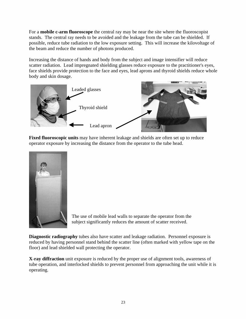
For a mobile c-arm fluoroscope the central ray may be near the site where the fluoroscopist stands. The central ray needs to be avoided and the leakage from the tube can be shielded. If possible, reduce tube radiation to the low exposure setting. This will increase the kilovoltage of the beam and reduce the number of photons produced. Increasing the distance of hands and body from the subject and image intensifier will reduce scatter radiation. Lead impregnated shielding glasses reduce exposure to the practitioner's eyes, face shields provide protection to the face and eyes, lead aprons and thyroid shields reduce whole body and skin dosage.
Leaded glasses
Thyroid shield
Lead apron Fixed fluoroscopic units may have inherent leakage and shields are often set up to reduce operator exposure by increasing the distance from the operator to the tube head.
Diagnostic radiograreduced by having pefloor) and lead shield X-ray diffraction untube operation, and inoperating.
The use of mobile lead walls to separate the operator from the subject significantly reduces the amount of scatter received.
phy tubes also have scatter and leakage radiation. Personnel exposure is rsonnel stand behind the scatter line (often marked with yellow tape on the ed wall protecting the operator.
it exposure is reduced by the proper use of alignment tools, awareness of terlocked shields to prevent personnel from approaching the unit while it is
23

Analytical X-ray Diffraction (XRD) with shielding window open andinterlocks are on. No exposure can be made.
XRD with windows closed, failsafe interlocks are off. Beam port(s) can be opened. Exposure can be made.
24
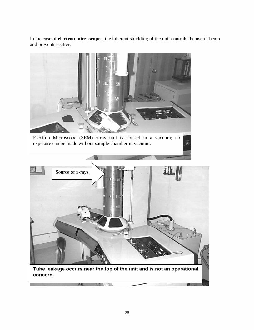
In the case of electron microscopes, the inherent shielding of the unit controls the useful beam and prevents scatter.
Electron Microscope (SEM) x-ray unit is housed in a vacuum; noexposure can be made without sample chamber in vacuum.
Source of x-rays
Tube leakage occurs near the top of the unit and is not an operational concern.
25

4.2 Protective Garments Leaded aprons, thyroid shields and safety glasses are the most frequently used methods to protect personnel during fluoroscopy. At 70 kVp the transmission factor of a 0.5 mm lead equivalent garment is less than one percent. This means that leaded garments are made to be worn to protect against scatter radiation. At and above 90 kVp the transmission factor is ten percent or greater. Proper care of leaded protective garments is important. They are expensive and apart from deterioration due to aging, they are very often damaged by mishandling and improper storage. Lead aprons will develop cracks at fold lines and will be punctured or torn when hung on pointed objects.
• Do not throw aprons on the floor, bench tops, over the backs of chairs or folded in any way, even for temporary storage.
• When not in use, these garments should be hung carefully on designated hangers or on reinforced bars.
While not required by law, EH&S regularly checks leaded garments for integrity. If there are any significant flaws in the shielding capabilities of an apron, glove, or thyroid shield, it must be taken out of service. Contact our office for the proper disposal of damaged or deteriorated lead garments.
26

4.3 Bibliography
1. Codes, Rules and Regulations of the State of New York: Title 10, Health (A), Chapter I, State Sanitary Code, Part 16, "Ionizing Radiation," as amended; Chapter II, Administrative Rules and Regulations, Part 89, "Practice of X-ray Technology".
2. Conference of Radiation Control Program Directors. Inc, Suggested State Regulations,
SSRCR Volume 1, January 1991, Part H, Radiation Safety Requirements for Analytical X-Ray Equipment.
3. Gkanatsios N.A., Huda W. "Effective Dose and Energy Imparted in Diagnostic
Radiology," Medical Physics, 24: 1311-1316 (1997).
4. Gkanatsios, N.A., Huda W, Peters, K.R., Freeman, J. "Evaluation of an On-line Patient Exposure Meter in Neurology," Radiology, 203: 837-842.1997.
5. Grosch, Daniel S., Hopwood, Larry E. Biological Effects of Radiations, second edition,
New York: Academic Press, 1979.
6. HEW Publication (FDA) 78-8039, "Exposure and Processing Guide for Dental Radiography."
7. University of Illinois at Champaign Urbana, Research Safety, Radiation Safety.
Analytical X-ray Safety on Line Tutorial (http://www.ehs.uiuc.edu/rss/), November 25, 2003.
National Council on Radiation Protection and Measurements (NCRP), 7910 Woodmont Avenue, Washington, DC 20014:
8. Report Number 33, "Medical X-ray and Gamma Ray Protection for Energies up to 10 MeV - Equipment Design and Use".
9. Report Number 35, "Dental X-ray Protection". 10. Report Number 36, "Radiation Protection in Veterinary Medicine". 11. Report Number 39, "Basic Radiation Protection Criteria". 12. Report Number 49, "Structural Shielding Design and Evaluation for Medical use of X-
rays and Gamma Rays of Energies up to 10 MeV". 13. Report Number 51, "Radiation Protection Design Guidelines for 0.01 - 100 MeV Particle
Accelerator Facilities".
14. Report Number 57, "Instrumentation and Monitoring Methods for Radiation Protection".
27

15. State of New York, Department of Health: Public Health Law, Article 35 - Practice of X-ray Technology, Title I, "General Provisions and Public Policy"; Title II, "Licensing and Registration".
16. SUNY at Buffalo, Radioactive Materials Safety Manual, August 2003; "Radiation
Protection Training Manual and Study Guide", April 1990. "Instructions Concerning Prenatal Radiation Exposure", August 2003.
17. United States Department of Commerce: National Bureau of Standards Handbook III,
American National Standard N43.2 – "Radiation Safety X-ray Diffraction and Fluorescence Analysis Equipment," 1977.
28