radiation safety for x-ray equipment operators
TRANSCRIPT
ONLINE SELF-STUDY
Radiation Safety for X-Ray Equipment Operators

Getting Going
The intent of this training module is to provide radiation safety
training to employees and students of The University of North
Carolina at Chapel Hill who operate diagnostic x-ray
equipment in clinical and/or research settings.
To document your participation in this self-study, we have
provided a short multiple choice test. A score of 80% or better
must be achieved to receive credit for this safety training.
Click on the forward button at the end of the module to take the
test.
Use the buttons below to navigate through the tutorial or click on
the hyper-link on the index page to advance to topic of
interest.
What is Radiation?
Radiation, simply put, is energy, and comes in many different
forms. It can come from unstable atoms, or can be electrically
(machine) produced.
Radiation is useful in medicine because it allows for the imaging
and non-surgical treatment of internal structures and diseases.
UNC uses many forms of radiation for diagnosis, treatment, and
research, including:
X-Ray Machines from Radiology
Radiopharmaceuticals in Nuclear Medicine/PET
Linear Accelerators and Sealed Radiation Sources in Radiation
Oncology
Certain Hospital Labs
Radiation Units and Terms
Terms used to describe radiation and radioactive materials include:
Exposure (R – Roentgen)
Electrical charge per unit mass of air produced by x or gamma rays.
Absorbed Dose (RAD – Radiation Absorbed Dose / Gray (Gy))
The amount of energy per unit mass absorbed by an irradiated object.
Note: 1 rad = 0.01 Gy.

Radiation Units and Terms
Terms used to describe radiation and radioactive materials include:
Dose Equivalent (REM – Roentgen Equivalent Man / Sievert (Sv))
Regulatory dose reporting unit
Modified absorbed dose taking into account the biological impact of the
different types of radiation (i.e. beta particle, x-ray, neutron).
Note: 1,000 millirem (mrem) = 1 rem = 0.01 Sv.
Activity (Curie – Ci / Becquerel - Bq)
The number of nuclear transformations occurring per unit of time for a
radioactive nuclide – describes the amount of radioactivity of a nuclide.
Note: 1 mCi = 37 mBq.
Background Radiation
We are all exposed to radiation everyday.
The average US Citizen receives about 600 mrem per
year from naturally-occurring and man-made
sources of radiation.
That's the equivalent of approximately 20 two-view
chest x-rays procedures at UNC.
This is mostly from natural sources of radiation, such as
radon, cosmic radiation, and natural deposits in the
earth.
Even our bodies contain natural radioactivity!
Radioactive Materials
Diagnostic Radiopharmaceuticals (such as Tc-99m, F-18, Tl-201, I-131, and
I-125) are used in Nuclear Medicine for diagnostic procedures and emit
gamma rays, which are a penetrating radiation, like x-rays.
These radionuclides remain in the patient after the study is over, but have
short half-lives, so the patient and the people around him or her are not
exposed for a long period of time.
Although radiation exposures may arise from the radiation emitted by
radionuclides in patients, by accidental contamination of skin with
radioactive materials, or by accidental ingestion of these materials
(possibly through smoking or eating when hands are contaminated), there is,
in general, no radiation hazard from these patients who have received
diagnostic doses of radioactive materials.
No special precautions are needed in caring for them, and there are no
restrictions on patient activities or contacts with other people.

Radioactive Materials, con’t.
When Therapeutic Radiopharmaceuticals
or Sealed Sources are used, relatively
large doses are involved, the patient can
be a significant source of radiation
exposure to staff, family and visitors.
When such procedures require that
radiation precautions be put into effect,
a radiation sign and a precaution sheet
(like this one) will be posted on the door
to the patient's room.

Radioactive Materials, con’t.
Research and medical laboratories use radionuclides
that may emit beta particles and low-energy
gamma rays.
Beta particles are not nearly as penetrating as
gamma rays or x-rays. Weak and moderate energy
betas will not even penetrate the skin.
The most important safety precaution for most of
these radionuclides is to keep the material from
contaminating the skin thereby avoiding the
possibility of ingestion or absorption.
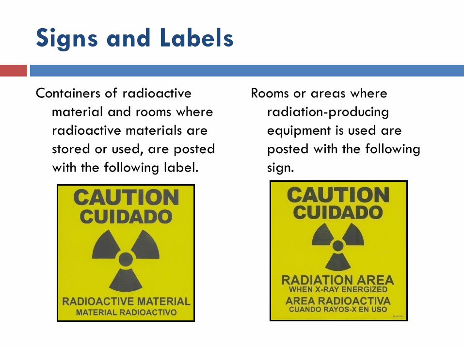
Signs and Labels
Containers of radioactive
material and rooms where
radioactive materials are
stored or used, are posted
with the following label.
Rooms or areas where
radiation-producing
equipment is used are
posted with the following
sign.

Radiation Risk Assessment
Effects of large doses of radiation are well-documented and understood. The
effects of the very low doses of radiation (like those encountered in the
hospital setting) are not, however, so well understood.
When a large dose of radiation is increased to an even larger dose, the
adverse effects become greater or more prevalent.
This dose vs. effect relationship can be thought of as linear, with confirmed and
documented effects beginning at a certain "threshold" level of radiation
dose.
But since this "threshold" level is far greater than any allowable occupational
dose, health physicists "extend" what is known about higher doses of
radiation down to "zero" dose.
In other words, any radiation dose is assumed to have some effect. This
is a conservative model of the risk.
Consider that for very low doses of radiation the effect of most concern is
cancer.
Radiation Risk Assessment, con’t.
It is estimated that approximately 20% (1 in 5) of all deaths in the United
states are due to some type of cancer.
If every member of a population of 1 million were to receive 10 mrem of
radiation, it is possible that 5 additional deaths would be observed.
Remember that out of this population of 1 million, about 200,000 will die
of cancer, making these few additional deaths statistically impossible to
detect.
Additionally, the risk of cancer death is 0.08% per rem for doses received
rapidly (acute) and might be 2 times (0.04%, or 4 in 10,000) less that that
for doses received over a long period of time (chronic).
All activities carry some element of risk. Flying in an airplane, driving a car,
smoking cigarettes, eating certain foods, and drinking alcoholic beverages
are everyday activities that carry some risk. Many of us are willing to
accept the risk from these activities. The risk is very small for the amounts of
radiation encountered by employees at UNC Health Care System.
Radiation Protection at UNC
Health Care System
The Radiation Safety section of the UNC-Chapel Hill Department
of Environment, Health & Safety (EHS) acts as an "agent" for
the Radiation Safety Subcommittee, and manages the Health
Care System’s radiation protection program.
Information on radiation safety may be obtained from the
Radiation Safety Officer (RSO) at 919-962-5507.
A link to Radiation Safety's website is provided here: UNC
Radiation Safety
For more information about radiation, click here: What You Need
to Know About Radiation
Radiation Protection at UNC
Health Care System, con’t.
Radioactive materials are used by the UNC Health Care System
under a "broad medical license" issued by the state radiation
protection regulatory agency -the North Carolina Radiation
Protection Section (visit their website at: NC Radiation
Protection)
UNC Health Care has been issued a "license" by the NCRPS to
possess and operate linear accelerators in Radiation Oncology.
Additionally, all x-ray machines are "registered" with the NCRPS.
The UNC Health Care Radiation Safety Subcommittee oversees
and approves all use of radioactive materials at this institution.
Refer to the "Notice to Employees" form issued by the NCRPS for
important information (posted where radiation sources are used):
Notice to Employees
Basic Radiation Safety Practices
North Carolina State regulations require that UNC Health Care
System have an ALARA program.
The main purpose of this program is to ensure that radiation
exposures are maintained:
As Low As Reasonably Achievable.
The ALARA concept is based on the assumption that any radiation
dose, no matter how small, can have some adverse effect.
Under the ALARA program, every reasonable means of lowering
exposure is used.
Radiation exposure can be minimized (ALARA) by utilizing three
basic principles:
Basic Radiation Safety Practices
Time: Time is used in radiation protection to limit the time spent near a
radiation source. Reducing the time decreases the radiation dose received.
Distance: Distance plays an important role in radiation protection. Increasing
the distance from a source of radiation significantly reduces the radiation
dose.
Doubling the distance from a radiation source means one-fourth the
dose rate.
Tripling the distance gives one-ninth the rate.
Shielding:
The use of appropriate shielding greatly reduces dose.
The material used and thickness of the shield depends on the source of
the radiation.
Lead is a common material used to shield radiation.
Basic Radiation Safety Practices,
con’t.
Radioactive Spills: When a spill of radioactive
material is encountered, do not clean it up.
Remember that small droplets may have splashed
away from the spill.
If liquid is running, try to contain it with a paper towel
or other absorbent material.
Isolate the area and notify the Radiation Safety
Officer.
All persons involved in a spill should be monitored for
contamination.
Dose Limits and Monitoring
Requirements
The amount of radiation received by persons exposed occupationally should
not exceed the dosages specified in the North Carolina Regulations For
Protection Against Radiation:
15A NCAC 11 Annual Dose Limits:
Whole Body: 5,000 mrem
Skin/Extremities: 50,000 mrem
Lens of Eye: 15,000 mrem
A member of the general public is allowed only 100 millirem per year from all
licensed and registered radiation activities at UNC.
The average annual dose of a radiation worker at UNC is about 100
millirem.
A radiation worker is required to be monitored if he/she is likely to receive in
excess of 10% of the dose limits.

Dose Limits and Monitoring
Requirements, con’t.
Radiation doses are monitored with either a Luxel®
OSL (Optically Stimulated Luminescence) whole
body badge, or a TLD (Thermoluminescent
dosimeter) extremity badge.
The devices are processed monthly or quarterly.
Action levels have been set which trigger
investigations to determine if the exposures were as
low as reasonably achievable.
If not, recommendations are made to ensure that
future exposures are ALARA.
Conceptus Protection Policy
Recent studies have shown that the risk of childhood leukemia and other
cancers increases if the mother experienced a significant radiation
exposure during pregnancy.
The N.C. regulations limit the dose of the conceptus to 500 mrem over the
course of the pregnancy, if the worker declares her pregnancy in writing to
the employer.
If an employee decides to declare her pregnancy, she should notify her
supervisor who will arrange for her to meet with the Radiation Safety
Officer to discuss possible precautions to limit radiation exposure.
The Radiation Safety Officer will review work assignments and radiation
exposure history, and may recommend limitations in work assignment if
necessary.
Radiation doses will be reviewed monthly.
If radioactive materials are used, the employee may also be placed on a
periodic bioassay program.
X-Ray Equipment Operator
QualificationsDiagnostic equipment (other than dental x-ray equipment) shall be used under the
direction or supervision of a qualified physician.
Only the following individuals are authorized to make radiographic (other than dental)
and fluoroscopic exposures:
qualified physicians,
engineering and physics staff members,
technologists and radiation therapists who are ARRT-registered, have graduated
from an accredited educational program, or in-training for ARRT registration.
non-ARRT registered individuals may operator bone densitometry equipment but
must meet established training requirements.
exceptions to the above requirements must be approved by the Radiation Safety
Subcommittee (RSS).
Dental x-ray equipment shall only be used under the direction or supervision of a
qualified physician or dentist.
Indications for Use
Examinations involving radiation should be requested by a Licensed
Independent Practitioner (Physician, Dentist, Physician Assistant and Nurse
Practitioner)
request should reflect the practitioners' knowledge of the clinical condition
exams should not be repeated merely for the practitioners' convenience
retakes may be performed upon the discretion of the qualified x-ray
equipment operator or if requested by a LIP
Keep in mind - not exposing the patient gives the largest dose reduction.
Radiology Department Clinical Staff are available for consultation before and
after diagnostic radiology examinations.
Guidelines for Safe Operation of
X-Ray Equipment
Personnel Monitoring:
Wear only YOUR assigned monitoring device
Wear your device for the current time period
Return your device in a timely fashion
Only persons whose presence is necessary should be
in the radiographic or fluoroscopic room during
exposures
Guidelines for Safe Operation of
X-Ray Equipment, con’t.
Protect all persons subject to direct scatter radiation
with whole body lead aprons (such as a skirt AND
vest) or whole body protective barriers
Includes individuals using or around "mini" c-arms
A 0.25 mm lead equivalence apron reduces
scattered x-rays by 95%
Shielding integrity of protective shielding devices is
evaluated annually
Guidelines for Safe Operation of
X-Ray Equipment, con’t.
Operators must stand behind protective barriers during
radiographic exposures at permanent radiographic
installations
Exemptions to this requirement - Cysto/Urology Suite
Operator remains in the room during radiographic exposure
Wears a lead apron
DEXA equipment operators
Remain at least 6 feet from the patient during exposures, or
as far away as practical due to room space limitations

Guidelines for Safe Operation of
X-Ray Equipment, con’t.
Make exposures with doors to the x-ray room closed
Exceptions include:
Corridor doors of the Dental Clinic
Doors leading to the Tech Work Area of Main
Radiology
Avoid making exposures when individuals are in or
near these doorways
Guidelines for Safe Operation of
X-Ray Equipment, con’t.
Holding Patients:
Use mechanical supporting devices when a patient or cassette must be held
If a patient must be held by an individual:
Protect holders with appropriate shielding devices (such as a lead apron)
Protect body part exposed to the primary x-ray with at least 0.5 mm lead
equivalence (lead glove, lead apron)
The individual holding the patient should be a member of the patient's family
Do not use minors
Do not use pregnant females
Operator shall provide appropriate instructions to the holder to maintain
doses ALARA
No individual shall be used routinely for holding!
Although necessary under exceptional circumstances, x-ray equipment
operators should not routinely hold patients
Guidelines for Safe Operation of
X-Ray Equipment, con’t.
Exposure Control and Technique Charts:
Keep exposure to the patient minimal practical amount
consistent with clinical objectives
Automatic Exposure Control (AEC) feature should be
appropriately utilized for all exposures when available
If not available, manual techniques must be utilized
Technique Charts must be available for radiographic and
dental units:
Indicate set of exposure factors (kVp, mAs, SID) which normally yield an
optimal image for a body part of specific size and orientation
Consider using dose reduction techniques (like high tube
potential (kVp) and low current (mA)), as long as image quality
is not compromised

Guidelines for Safe Operation of
X-Ray Equipment, con’t.
Limitation of the Useful Beam:
Collimate the x-ray beam to the smallest area consistent with
clinical requirements
Align the beam accurately with the patient and image
receptor
Use the smallest practical field sizes and shortest exposure
times
Exception: long exposure times may be required for breathing technique
studies
Position all individuals such that no part of their body will be
exposed to the useful beam unless protected by at least 0.5
mm lead equivalence lead apron or whole body protective
barrier
Guidelines for Safe Operation of
X-Ray Equipment, con’t.
Gonadal Shielding:
Used for potentially procreative patients during radiographic
procedures when the gonads are in the direct beam
Except when clinical objectives would be compromised
Never use as a substitute for careful patient positioning and limitation of
the beam
Consider gonadal shielding when the gonads lie within close proximity
(2 inches) of the primary beam
Using shielding for procedures with the primary beam >2 inches from
the reproductive organs has minimal effect on patient dose
It does reassure patients that consideration has been given to protecting them
from unnecessary radiation exposure
Guidelines for Safe Operation of
X-Ray Equipment, con’t.
Mobile Radiographic Equipment Procedures:
Use mobile equipment only used when impractical to transfer patients to
permanent installations
Operator of mobile equipment must ask individuals whose presence is not
required to leave the room until the exposure is complete
It may not be possible for all individuals to leave the room in areas such as
the PACU or ED, however the operator is responsible for positioning
individuals as far away as practical and protecting them in accordance with
ALARA policy
Individuals subject to direct scatter radiation, including operators, must be
protected by lead aprons or whole body protective barriers
The operator shall also:
Stand as far as possible (at least 6 feet) from the x-ray tube head and the
nearest edge of the image receptor
Give an audible warning before exposure is made

Pregnant or Potentially-Pregnant
Patients
Special precautions, consistent with clinical needs, shall be taken to minimize
exposure of the embryo or fetus in patients known to be or suspected of
being pregnant.
It is the responsibility of the referring physician to determine the pregnancy
status of patients of childbearing age, and to write a note in the chart
describing the indication for the study and confirming that this was discussed
with the patient. Exceptions to this include any study involving body parts
above the abdomen or below the hips.
When the x-ray procedure does not include the abdomen or pelvis of the
pregnant or potentially-pregnant patient, the abdominal region should be
shielded with at least 0.25 mm lead equivalence whenever feasible, and
the examination performed without regard to pregnancy.
Pregnant or Potentially-Pregnant
Patients, con’t.
When the x-ray procedure includes the abdominal region of the pregnant or
potentially-pregnant patient, the examination shall not be performed
without approval from the physician responsible for the procedure involving
radiation. Written informed consent should be obtained for all procedures
involving direct exposure of the conceptus and/or whenever conceptus dose
is likely to exceed 1 rem and shall be obtained whenever dose to the
conceptus is likely to exceed 5 rem.
Consent forms are available in English and Spanish. Procedures involving the
abdomen or pelvis with the conceptus in the field of view that are likely to
deliver a conceptus dose greater than 1 rem include, but are not limited to,
CT, fluoroscopy in excess of 1 minute, and radiographic procedures
requiring multiple imaging (>3) of the conceptus region.

Pregnant or Potentially-Pregnant
Patients, con’t.Although it is the responsibility of the referring physician to determine pregnancy
status, those operating diagnostic x-ray equipment shall ask all patients of
childbearing age whether or not they are pregnant and the date of their last
menstrual period. This information is to be recorded prior to the procedure. When
pregnancy status is unclear, or when the date of the last menstrual period is greater
than two weeks (14 days), a urine pregnancy test must be performed to exclude
pregnancy, unless the delay necessary to perform the pregnancy test would
jeopardize the patient's health.
Radiation exposure must be used judiciously and kept to a minimum, and imaging
techniques not involving radiation should be considered. The imaging techniques
used, such as fluoroscopic time, kVp and mA, as well as the number of images taken
and the abdominal thickness measurements are to be recorded. A radiation
physicist should be contacted to assist with dose estimates. A formal dose calculation
will be performed for procedures that are likely to deliver a conceptus dose in
excess of 15 rem. Radiation Safety is available for consultation at 919-962-5507,
Monday – Friday 8 am to 5 pm, and after-hours and on weekends and holidays by
calling 919-962-6565 and asking to have Radiation Safety paged.
Fluoroscopy-Guided
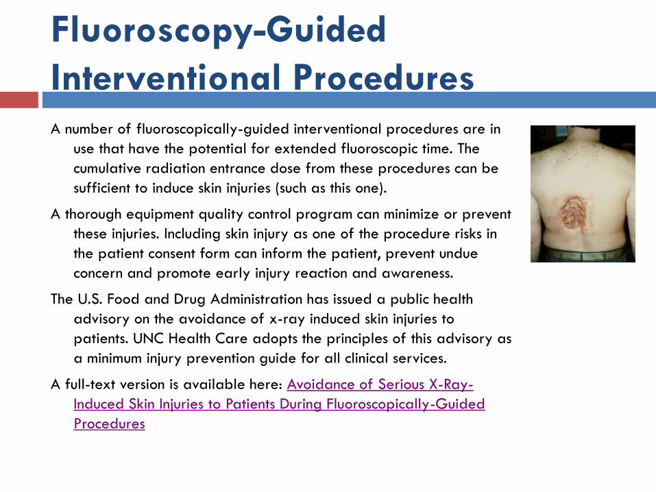
Interventional ProceduresA number of fluoroscopically-guided interventional procedures are in
use that have the potential for extended fluoroscopic time. The
cumulative radiation entrance dose from these procedures can be
sufficient to induce skin injuries (such as this one).
A thorough equipment quality control program can minimize or prevent
these injuries. Including skin injury as one of the procedure risks in
the patient consent form can inform the patient, prevent undue
concern and promote early injury reaction and awareness.
The U.S. Food and Drug Administration has issued a public health
advisory on the avoidance of x-ray induced skin injuries to
patients. UNC Health Care adopts the principles of this advisory as
a minimum injury prevention guide for all clinical services.
A full-text version is available here: Avoidance of Serious X-Ray-
Induced Skin Injuries to Patients During Fluoroscopically-Guided
Procedures

Fluoroscopy-Guided
Interventional Procedures, con’t.
The exposure rate used in fluoroscopy should be as low as is consistent with the
fluoroscopic requirements and should not normally exceed 10 R/min
(measured in air) at the position where the beam enters the patient (in
normal mode – high level mode must not exceed 20R/min).
It is usually best to operate fluoroscopic machines with the "automatic
brightness system" engaged to optimize performance and minimize patient
dose.
Fluoroscopy should not be used as a substitute for radiography but should be
reserved for the study of dynamics or spatial relationships or for guidance
in spot-film recording of critical details.
Medical fluoroscopy should be performed only by or under the immediate
supervision of physicians or physician extenders properly trained in
fluoroscopic procedures.
Fluoroscopy-Guided
Interventional Procedures, con’t.
Protective drapes may only be removed during procedures granted a waiver
for removal, and must be reattached immediately after the procedure.
The hand of the fluoroscopist should not be placed in the useful beam unless
the beam is attenuated by the patient and a protective glove of at least
0.5 mm lead equivalent.
The hand of the fluoroscopist should not be placed in the useful beam unless
the beam is attenuated by the patient and a protective glove of at least
0.5 mm lead equivalent.
In digital/image acquisition, take special care to limit patient exposure as this
mode uses 8-10x higher tube currents than normal fluoroscopy.
Utilize pulsed or low-dose fluoroscopy whenever possible.
Record fluoroscopy time and/or reported patient dose for each procedure.
Important
Report any unusual or unsafe condition involving sources of
radiation to the Radiation Safety Office immediately.
Radiation Safety is available during normal duty hours at
919-962-5507.
Radiation Safety can be reached after normal working hours
through Campus Police at 919-962-6565.
Radioactive Materials licenses, x-ray registrations,
regulations, inspection reports and exposure reports are
available for review in the UNC Department of
Environment, Health and Safety, Radiation Safety Section,
1120 Estes Drive Extension, CB# 1650, 919-962-5507.