"vision in dogs" by miller & murphy - smart paws
TRANSCRIPT
LEADING EDGE OF MEDICINE—A REVIEW I
Vision in dogs
Paul E. Miller, DVM, and Christopher J. Murphy DVM, PhD
A lmost every dog owner, veterinary student, and vet-erinarian has wondered, on one occasion or an-
other, how well dogs see. The question is more than amatter of intellectual curiosity, however, because a dog'svisual capabilities directly affect its ability to engage inhigh performance, visually orientated activities, such asguiding the blind, police work, schutzhund, obediencetraining, racing, and hunting. Nevertheless, none of thecurrently available textbooks of veterinary ophthalmol-ogy provide a concise summary of the visual abilities ofdogs, and the pertinent literature on canine vision iswidely scattered throughout such diverse fields as psy-chology, physiology, optics, neuroanatomy, and electro-physiology. The purpose of this review was to collectand interpret the relevant literature on the visual abilitiesof normal dogs.
Fundamentals of VisionBecause a multitude of factors are involved in the
sensation of vision, the outwardly simple question ofhow well dogs see is, in reality, quite complicated. Thequestion can be partially answered by describing the vi-sual acuity of dogs, their abilities to detect light or color,or the features of other individual visual parameters, butthe complete visual experience is a synthesis of all of theseconstituent parts into a unified perception of the world.Because our current understanding of many componentsof canine vision is imperfect, so, therefore, will be anyattempt to describe them. It is also important to recognizethat, because of species differences in visual parameters,dogs probably perceive the world differently from the waythat people do. Descriptions of the visual abilities of non-human species are, of necessity, couched in terms ofhuman visual capabilities and, therefore, may not be per-fectly accurate representations of how animals see.
The ability to perceive light and motion are gener-ally regarded as the fundamental aspects of vision. How-ever, other factors, such as visual perspective, visual fieldof view, depth perception, visual acuity, and the abilitiesto perceive color and form, also play important roles inhow animals see.1
Sensitivity to LightThe canine visual system has adapted to exploit a
particular ecological niche by enhancing visual perform-
From the Comparative Ophthalmology Research Laboratories, De-partment of Surgical Sciences, School of Veterinary Medicine, Univer-sity of Wisconsin, 2015 Linden Dr W, Madison, WI 53706-1102.
The authors thank Marie Nelson for assistance with figures, andDr. Gustavo Aquirre (James A. Baker Institute for Animal Health, Cor-nell University), Dr. Donald Mutti (School of Optometry, University ofCalifornia-Berkeley), Dr. Jay Neitz (Department of Cellular Biology andAnatomy, Medical College of Wisconsin), Dr. Jacob Sivak (School ofOptometry, University of Waterloo), Dr. Peter Spear (Department ofPsychology ar\ Ceniei (OT Neuroscience, University of Wisconsin-Madison), Dr. James Ver Hoeve (Department of Ophthalmology,School of Medicine, University of Wisconsin-Madison), and Dr. KarlaZadnik (School of Optometry, University of California-Berkeley) forreview and suggestions.
ance under low light conditions but still retaining goodfunction under a wide array of lighting conditions, in-cluding daylight. Therefore, the dog's visual system isnot highly adapted for strictly diurnal or nocturnal con-ditions but, rather, has evolved for an arrhythmic photicexistence.1'2 The minimum threshold of light for visionin cats is approximately 6 times lower than that for nor-mal human beings.3 Although the minimum thresholdof light for vision in dogs is assumed to be somewhatgreater than that for cats, Pavlov concluded in the earlypart of this century that the ability of dogs to analyzethe intensity of low-level illumination is so well devel-oped that human experimenters are unable to determineits limits using their own senses.1
Dogs employ several methods of improving visionin dim light. Both dogs and humans use rod photore-ceptors to function in dim light, but the central 25° ofthe retina in dogs consists predominantly of rods.4 Inpeople, this region consists predominately of cones,which are important for color vision and vision in brightlight. The rod photopigment, rhodopsin, is also slightlydifferent between dogs and humans. In dogs, rhodopsinhas a peak sensitivity to light of wavelengths between506 and 510 nm and, as is typical of species adapted tofunction well in dim light, takes over an hour to com-pletely regenerate after extensive exposure to brightlight.4-6
Rhodopsin in people, on the other hand, has a peaksensitivity to light of somewhat shorter wavelengths (ap-prox 496 nm) and regenerates more quickly after ex-posure to bright light. The range of wavelengths towhich rhodopsin in dogs is sensitive4'6 suggests that thevisible spectrum for dogs in dim light is similar to thatof human beings, and that the enhanced night vision indogs, relative to humans, is not due to differences in therange of wavelengths of light to which dogs and peopleare sensitive.
The superiorly located reflective tapetum lucidumalso enhances the dog's ability to detect objects in dimlight. Presumably, it does so by reflecting light that hasalready passed through the retina back through it a sec-ond time, thus providing the photoreceptors at least 2chances at capturing each quantum of light. This reflec-tion of light, however, has its price, as scattering of lightduring this process may reduce the ability of the eye toprecisely resolve the details of an image.2
Anatomically, the tapetum lucidum in dogs is ahighly cellular structure that is between 9 and 20 layersthick at its center and is rich in zinc and cysteine.7'12
The variety of colors seen in the region of the tapetumlucidum during ophthalmoscopy result from the differ-ential interaction of light with the tapetum's physicalstructure rather than from the inherent spectral com-position, or color, of its pigments.13 The tapetum is anefficient reflector of light; 1 study has suggested that thefeline eye reflects about 130 times more light than doesthe human eye.14 Because of anatomic differences, the
JAVMA, Vol 207, No. 12, December 15, 1995 Scientific Reports Leading Edge of Medicine—A Review 1623
canine tapetum is probably less efficient at reflectinglight than is that of the cat, but its light-reflecting prop-erties are still undoubtedly substantial.
The tapetum may not only reflect light. It has beensuggested in cats and lemurs that tapetal riboflavin ab-sorbs light in the shorter wavelengths (blue, approx 450nm) and shifts it via fluorescence to a longer wavelength(520 nm) that more closely approximates the maximalsensitivity of rhodopsin in the rod photoreceptors.15"17
This shift may brighten the appearance of a blue-blackevening or night sky and, thereby, enhance the contrastbetween other objects in the environment and the back-ground sky.16 Additionally, it has been proposed thatregional differences in reflection spectra (seen ophthal-moscopically as differences in regional coloration of thetapetum) may lead to local variations in the retinal spec-tral sensitivity. However, it is unclear whether this isimportant in living animals.18 In a strain of Beagles inwhich a hereditary tapetal degeneration was identified,it was observed during electrophysiologic testing that af-fected dogs had a slightly reduced sensitivity to whitelight, compared with unaffected dogs.9 In our experi-ence, however, dogs that lack a tapetum do not appearclinically to have impaired vision in dim lighting circum-stances. Similarly, dogs that lack pigmentation in thenon-tapetal zone and, therefore, presumably haveexaggerated scattering of light inside the eye do not ap-pear to have clinically significant reduced visual acuityin bright light. However, the visual abilities of dogs withatapetal and subalbinotic fundi have not yet been pre-cisely measured, and it is possible that differences invisual abilities associated with variations in the appear-ance of the tapetum may be discernable in the futurewith more rigorous testing.
Albinism, the complete absence of light-absorbingpigment, is well documented in many species and is as-sociated with impaired visual function.19 True albinismis rare in dogs, but an Australian Shepherd with ocularalbinism did have photophobia, presumably from in-creased light scattering within the eye.20 Abnormalitiesin the fovea (in species that possess one) and centralvisual pathways have also been reported in albinos.19
The effect of albinism on the visual system in dogs hasnot been critically evaluated.
The canine visual system has also adapted to per-form adequately in bright light and when there is amarked difference in luminance between different regionsof the retina. Typically, the superior part of the retinareceives light from the darker ground, and the inferiorpart receives light from the brighter sky. In some in-stances, however, the sky is darker and the ground ishighly reflective (eg, when it is covered with snow orsand) and, therefore, brighter than the sky. Severalmechanisms allow dogs (and people) to maintain visualfunction under these situations of greatly varying light-ing intensities. Reflexive adjustment of pupil size; un-conscious alteration of the overall, and perhaps regional,sensitivity of the rods; and recruitment of cone photo-receptors, which are adapted for use in bright light, im-prove the dog's visual performance in bright light. Ad-ditionally, it has been suggested that specialized retinalamacrine cells, which bridge the inferior and superiorportions of the retina,21 help to equilibrate differences in
incident illumination among the areas of the retina. Theextent of this equilibration, if it does occur, is unclear,and equilibration may be confined to only a narrow zonein the horizontal meridian. The superiorly located re-flective tapetum lucidum may also enhance the view ofthe usually darker ground, and the inferiorly located,usually darkly pigmented, tapetum nigrum may reducelight scattering originating from the usually brighter sky.
Sensitivity to MotionAlthough little work has been done on the motion-
detecting abilities of dogs, it is probable that the percep-tion of movement is a critical aspect of canine vision1
and that dogs, like people, are much more sensitive tomoving objects than they are to stationary ones. Thedominant photoreceptor in dogs, the rods, are particu-larly well suited for detecting motion and shapes. In a1936 study of the visual performance of 14 police dogs,the most sensitive dogs could recognize moving objectsat a distance of 810 to 900 m, but could recognize thesame object, when stationary, at a distance of only 585m or less.2
Sensitivity to Flickering LightsAlthough not related to motion detection, the point
at which rapidly flickering light appears to fuse into aconstantly illuminated light (flicker fusion) provides in-sight into the functional characteristics of the rods andcones in dogs. The flicker frequency at which fusion oc-curs varies with the intensity and wavelength of the stim-ulating light.22'25 Results of electroretinographic studiesof anesthetized dogs suggest that canine rods can detectflickering up to a maximum of approximately 20 Hz,22-23
which is similar to the maximum for human rods.24'25
With more intense light, cone photoreceptors are acti-vated, and flicker fusion occurs at around 70 Hz in dogsas measured electroretinographically.22'23 Behavioral test-ing of 4 Beagles trained to press a key when the test lightwas perceived to be flickering demonstrated that una-nesthetized dogs could detect flicker at moderatelyhigher frequencies (70 to > 80 Hz) and at lower levelsof light intensity than the results of electroretinographywould suggest.26 The flicker fusion frequency for humancones (approx 50 to 60 Hz for luminous spots27) is re-portedly lower than that for canine cones, but some peo-ple are capable of detecting flicker up to approximately70 Hz.24'25 Because of this heightened sensitivity toflicker, a television program, in which the screen is up-dated 60 times/s and appears as a fluidly moving storyline to most humans, may appear to rapidly flicker todogs.
Visual PerspectiveObviously, height of the eyes above the ground has
a major impact on the perception any animal has of itsenvironment, and visual perspective in dogs is orientedconsiderably closer to the ground than it is in people.On the other hand, shoulder height in dogs ranges from< 8 inches to > 34 inches.28 A field of tall grass mayappear to be a daunting array of impenetrable brush toa Shih Tzu, whereas an Irish Wolfhound may experienceno difficulty in visually orienting itself in the same field(Fig 1). Some breeds, such as the English Springer Span-
1624 Leading Edge of Medicine—A Review Scientific Reports JAVMA, Vol 207, No. 12, December 15, 1995

Figure 1—The effect of visual perspective on vision. Thesame scene as viewed by a small dog with eyes located8 inches above the ground (top), a tall dog with eyes 34inches above the ground (middle), and a person witheyes 66 inches above the ground (bottom).
iel, appear to have developed behavioral traits, such asleaping into the air while visually searching for objects,that perhaps enhance their visual perspective. The lackof morphologic standardization among breeds and in-dividuals is doubtless a contributing factor as to why thevisual system of dogs has not been more intensivelystudied.
Visual Field of ViewThough not critically evaluated, the extent of the
visual field in dogs (ie, the area that can be seen by aneye when it is fixed on 1 point) also varies by breed, asthere are marked variations among breeds in the place-ment of the eyes in the skull and, thereby, in the orbital
Figure 2—The extent of the monocular and binocular vi-sual fields in a typical mesocephalic dog.
axis.13 In the brachycephalic breeds, the eyes are morelaterally directed, and the extent of the visual field andamount of binocular overlap are probably different fromthe visual field and binocular overlap in mesocephalicbreeds, in which the eyes are directed more forward.13
Length of the nose would also interfere with the amountof binocular overlap. In dogs, the eyes typically areplaced so as to deviate approximately 20° lateral to themidline, whereas in people, the eyes do not deviate butlook straight ahead.1 When the 2 eyes are consideredtogether, the visual field of the typical dog has been es-timated, on the basis of calculations from morphologicdata, to be approximately 250°.2 In 1 study,29 two 20kg mixed-breed dogs with average length noses weretrained to fixate on 1 visual stimulus and react to a sec-ond stimulus introduced in a limited area of their pe-ripheral visual field. Visual fields for these dogs wasestimated to be 240°.29 These estimates suggest that,with each eye, the typical dog can see from 120° ipsi-lateral to between 15 and 30° contralateral, for a totalmonocular field of view of 135 to 150° (Fig 2).29 There-fore, the field of view of the average dog is approximately60 to 70° greater than that of people, and this providesdogs with a greater ability to scan the horizon. However,the degree of binocular overlap is greater in people thanit is in dogs.
Depth PerceptionDepth perception is enhanced in those regions in
which the visual fields of the 2 eyes overlap. Reportedestimates of the degree of binocular overlap in dogs varywidely, and binocular overlap may differ by breed. Theextent of binocular overlap was estimated to be between30 and 60° in behavioral studies,29 was calculated to bebetween 35 and 40° on the basis of ganglion cell den-sity,30 and was reported to be approximately 80 to 116°on the basis of optical considerations.1'2 It is probablethat calculations based on optical considerations over-estimate the extent of binocular overlap in dogs, because
JAVMA, Vol 207, No. 12, December 15, 1995 Scientific Reports Leading Edge of Medicine—A Review 1625

Figure 3—Ray diagrams depicting emmetropia (ie, properfocusing of images on the retina; top), myopia (near-sight-edness, or focusing of images in front of the retina; mid-dle), and hyperopia (far-sightedness, or focusing ofimages behind the retina; bottom). German Shepherds,Rottweilers, and Miniature Schnauzers are predisposedto myopia.36
the nose blocks more of the temporal retina's view ofthe nasal visual field than anticipated,29 and that the ex-tent of binocular overlap is most likely in the range of30 to 60° for the average dog. For comparison, field ofview in people is approximately 180°, and the degree ofbinocular overlap is approximately 1400.2 In dogs, themonocular visual fields are wide, but the binocular fieldhas been suggested to be tall, narrow, and pear-shaped.2Depth perception is probably greatest when dogs lookstraight ahead and is probably blocked by the nose inmost breeds when dogs look below the horizontal.
However, merely viewing an object with both eyessimultaneously does not guarantee improved perceptionof depth. Stereopsis (binocular depth perception) resultswhen the 2 eyes view the world from slightly differentvantage points, and the resulting image is blended orfused into a single image. If the 2 images are not fused,double vision may result. It is the disparity between the2 resulting retinal images that, when fused, providesclues that allow accurate discrimination of depth.31 Non-conjugate movements of the eyes probably also providesclues for depth perception.31
Although binocular depth perception is superior ifthe images can be blended into 1, monocular depth per-
ception is also possible.31 Monocular clues relating todepth include relative brightness, contour, areas of lightand shadows, object overlay, linear and aerial perspec-tive, and density of optical texture.31'33 In addition,movement of the head results in an apparent change inthe relative positions of the objects viewed (a phenom-enon known as parallax) and, thereby, produces the sen-sation that the elements in the visual environment aremoving at different speeds, allowing depth to be esti-mated. Puppies were shown to have excellent monocularand binocular depth perception in a controlled experi-ment evaluating avoidance of a visual cliff.33 Adult dogsprobably have even better visual abilities than youngpuppies, because the canine retina and tapetum do notmature until several weeks to months of age.34
On the basis of studies of retinal ganglion cell to-pography, it has been hypothesized (although not ex-perimentally verified) that depth perception may beimpaired in the peripheral 15° of the right and left por-tions of the area of binocular overlap in dogs because ofa lack of alpha (also referred to as "Y") ganglion cells inthe corresponding areas of the retina.30 Therefore, thearea of the retina available for high-quality depth per-ception may be smaller than the area estimated on thebasis of binocular overlap. Nevertheless, depth percep-tion in dogs is clearly sufficient for their lifestyle. Theyeasily judge distances visually, as evidenced by their abil-ity to catch fast-moving objects and clear hurdles.
Visual AcuityWhen dogs are said to see well in dim light, what
is meant is that canine visual sensitivity in reduced levelsof light is quite high,2 and that dogs have relatively goodvisual acuity under those circumstances. Visual acuity,however, is different from sensitivity to dim light, andrefers to the ability to see the details of an object sepa-rately and unblurred.2 Visual acuity depends on the op-tical properties of the eye (ie, the ability of the eye togenerate a precisely focused image), the retina's abilityto detect and process images, and the ability of highervisual pathways to interpret images sent to them.2 Ingeneral, visual acuity in dogs is believed to be limitedby the retina and not by the optical properties of the eyeor by postretinal neural processing in the brain.35 Nev-ertheless, these 2 factors can become the limiting step invisual discrimination in many pathological conditionssuch as myopia, when corrective lenses are required tosee clearly, or when higher CNS visual pathways are im-paired. Postretinal neural processing has been exten-sively investigated in cats, but much less so in dogs, andis beyond the scope of this review.
Optical factors in visual acuity—The optical qual-ities of the canine eye have been investigated by severalauthors,36'40-3 and a schematic eye that allows mathe-matical predictions of the optical capabilities of the ca-nine eye has been constructed.41 The optical media ofthe eye, namely the cornea, aqueous humor, lens, andvitreous humor, are responsible for creating a properlyfocused image on the retina. In a normally focused (em-metropic) eye, parallel rays of light (such as those froma distant object) are focused on the retina. If parallel raysof light are focused in front of the retina, myopia Ineai-
1626 Leading Edge of Medicine—A Review Scientific Reports JAVMA, Vol 207, No. 12, December 15, 1995

sightedness) results. If they are focused behind the ret-ina, hyperopia (farsightedness) results (Fig 3). Sucherrors in refraction are usually expressed in units of op-tical power called diopters. The extent of the error canbe expressed by the formula diopters = 1/f, where f =the focal length (in meters) of either the lens or opticalsystem as a whole. Therefore, if an eye is said to be 2diopters myopic at rest, it is focused at a plane located'/2 m in front of the eye. Similarly, an eye that is em-metropic at rest, but can accommodate (change focus) 3diopters is capable of clearly imaging objects on the ret-ina that range from as far away as the visual horizon(infinity) to as near as 'A m in front of the eye.
Sensory clues such as smell or sound, in additionto vision, may help dogs to better characterize nearbyitems in their environment. This penchant for usingsmell to identify objects that are very close can lead tothe mistaken belief that dogs are normally nearsightedor myopic. This is clearly not the case. In a survey of240 dogs, it was found that the average resting refractivestate was within 0.25 diopter of emmetropia.36 Therewere individuals in this large population, however, thatwere significantly myopic, and there was a distinct ten-dency towards development of myopia with greater ageand development of nuclear sclerosis.36 This mild shift inresting focus of the eye needs to be distinguished fromage-related presbyopia, which is the loss of focusing (ac-commodative) ability that affects all middle-aged humans(and probably dogs). The prevalence, degree, and impor-tance of presbyopia in dogs has not been determined.
Breed predispositions to myopia also were found.In 1 study, 53% of German Shepherds in a veterinaryclinic population were myopic by —0.5 diopters ormore, and 64% of all Rottweilers were myopic.36 In con-trast, only 15% of German Shepherds in a guide dogprogram were myopic, suggesting that dogs with visualdisturbances such as nearsightedness do not perform aswell as normally sighted dogs, and are often withdrawnfrom intense training programs by observant handlers.36
For comparison, people with -0.75 to -2.0 diopters ofmyopia will typically complain of visual impairment andreport improved vision with corrective lenses. Defocus-sing a human being by 2 diopters reduces visual acuityfrom 20/20 to 20/100,42 and a comparable reduction inacuity has been reported for dogs.43 Therefore, it maybe reasonable to screen dogs performing visually de-manding tasks, or those on which human life relies, forabnormalities in their refractive state (ie, whether theyare nearsighted or farsighted, or have significant irregu-larities in their focusing abilities, such as astigma-tism).36'43
In addition to myopia or hyperopia, other opticalaberrations may result from imperfections in the refrac-tive media and lead to degradation of the image formedon the retina. These aberrations may be quite simple orvery complex in nature. Astigmatism occurs when dif-ferent regions of the optical system fail to focus parallelrays of light in a uniform fashion. This can occur becauseof regional irregularities in the curvature of the corneaor lens that permit a light ray in 1 meridian to be focuseddifferently from a light ray in another meridian. The re-sult is warping of the image, an extreme example ofwhich can be found in the irregular mirrors found at
Figure 4—Illustration of spherical aberration. With posi-tive spherical aberration, the more highly curved periph-eral portion of the lens focuses light in front of rays thatpass centrally (top). With negative spherical aberration,peripheral rays are brought to focus behind the more cen-tral rays (middle). Photograph of an isolated dog lensthrough which parallel helium-neon laser beams havebeen passed (bottom). Beams that pass through the pe-riphery of the lens are focused farther away from the lensthan are beams that pass through the central portion ofthe lens, suggesting that dog lenses have negativespherical aberration. It has been postulated that this ar-rangement may compensate for positive spherical aber-ration of the peripheral portion of the canine cornea.(Photo courtesy of Dr. Jakob Sivak)
many fairs and carnivals. Astigmatism is generally un-common in dogs, but has been observed in a variety ofbreeds since the early 1900s.36/H)'a In a recent survey,astigmatism, which ranged from 0.5 to 3.0 diopters, wasfound in only 10 of 240 dogs, and was unilateral in 8of the 10 dogs.36
Spherical aberrations of the lens result in unevenbending of light rays across its convex optical surface(Fig 4). Positive spherical aberration occurs when lightrays passing through the more highly curved peripheryof the lens are brought into focus in front of the raysthat pass more centrally. Many species of animals havecompensated for this effect, and in these species, thereis a gradient of refractive indices throughout the lenssuch that the more peripheral cortical fibers have a lesserrefractive index (ie, a lesser ability to bend light) thando the more central fibers. Dogs actually have negativespherical aberration, and the peripheral rays are broughtto focus behind the more central rays.44 It has been sug-gested that negative spherical aberration of the lens indogs may serve to compensate for positive spherical ab-erration of the peripheral portion of the cornea. The op-tics of the peripheral region of the cornea in dogs,however, remain to be investigated.
JAVMA, Vol 207, No. 12, December 15, 1995 Scientific Reports Leading Edge of Medicine—A Review 1627
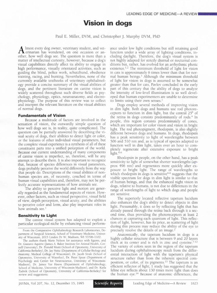
Figure 5—Photograph illustrating chromatic aberration.Red and green lasers have been superimposed and sub-sequently passed through an isolated lens of a fish. Theshorter wavelength light (green) is focused in front of thelonger wavelength light (red). (Photo courtesy of Dr. JakeSivak)
Another common type of aberration in many ver-tebrate eyes is chromatic aberration, whereby light ofshort wavelengths (blue) is focused in front of light oflong wavelengths (red; Fig 5). In a survey of the degreeof chromatic aberration in vertebrates, it was found thatmost eyes had a relatively constant amount of chromaticaberration amounting to 4.6% of the equivalent focallength.45 Surprisingly, dogs were found to have thegreatest amount of chromatic aberration in this survey(5.7% of the equivalent focal length).45 Although theclinical importance of chromatic aberration in dogs re-mains unclear, it was suggested that this high degree ofchromatic aberration may reflect fundamental differencesin the composition of the lens between dogs and theother species that were studied, in such factors as watercontent, protein distribution, or packing of lens fibers.Additionally, the relative insensitivity of canine cones tothe longer (ie, red) wavelengths of light may minimizethe impact of this aberration on visual performance.
Although visual acuity requires that the optical por-tions of the eye be transparent and that optical blur fromrefractive errors or astigmatism be limited, an adjustablefocusing (accommodative) mechanism is also needed ifobjects at different distances are to be seen with equalclarity.1 In dogs, accommodation may be brought aboutby altering the curvature of the lens surface or by movingthe lens anteriorly, as has been demonstrated in rac-coons.46 Dogs generally have a limited accommodativerange that does not exceed 2 to 3 diopters.1'13 This sug-gests that dogs are capable of accurately imaging on theretina objects that are within 50 to 33 cm of their eyes,but that objects nearer than this will be blurred. Hence,dogs must use other senses, such as smell or taste, toaugment vision in the investigation of very near objects.For comparison, young children are capable of accom-modating approximately 14 diopters or to about 7 cm.47
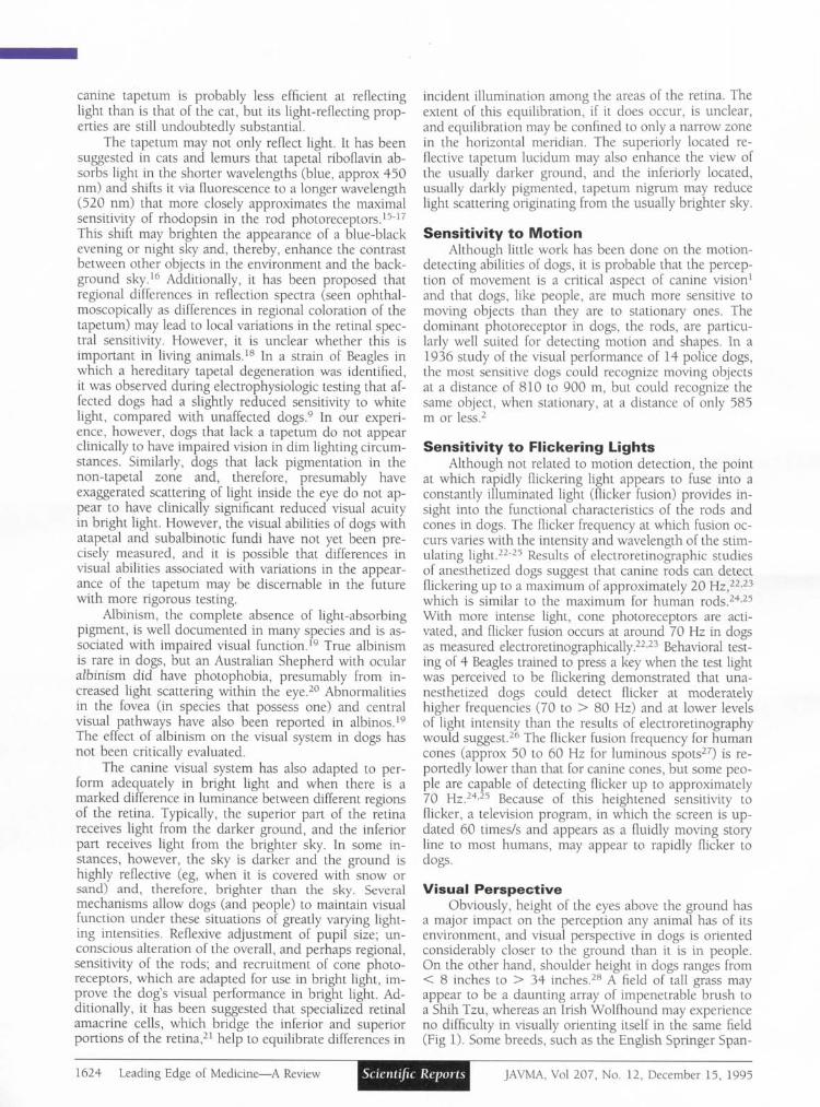
With age, the ability to accommodate declines (ie, pres-byopia develops), perhaps as a result of increased resis-tance of the lens to changes in shape, or as a result ofalterations in the excursions of the ciliary body muscu-lature.47 Loss of the lens, as occurs during cataract sur-gery, obviously creates a significant change in therefractive state of the eye.38'48 The result is severe hy-peropia (farsightedness), with objects being focused ap-proximately 14 diopters behind infinity, and a reductionin visual acuity to 20/800 or worse.43 This means thataphakic eyes are unable to image any object clearly,
Figure 6—Simulation of the optical impact of 1 diopter ofmyopia and 14 diopters of hyperopia. A typical scene asit would appear to an individual with normal vision (top).The same scene with the camera focused at 1 meter,which simulates 1 diopter of myopia (middle). The samescene as it would appear to an individual with 14 dioptersof hyperopia as occurs following lensectomy in dogswithout optical correction (bottom). Notice that this illus-tration differs from true canine vision in regards to actualvisual acuity, field of view, and the color spectrum per-ceived.
whether near or far away, and are unable to accommo-date (Fig 6).48 Although aphakic dogs are farsighted, itmust be kept in mind that, for objects of similar size,objects that are closer to the dog will create a largerimage on the retina than will objects that are located faraway. Therefore, aphakic dogs may be able to better vi-sually orient to near objects, despite being farsighted.
1628 Leading Edge of Medicine—A Review Scientific Reports JAVMA, Vol 207, No. 12, December 15, 1995

Figure 7—Diagram of retinal ganglion cell densities from the right retinas of a German Shepherd with a very pro-nounced wolf-like visual streak (left) and a Beagle with a moderately pronounced visual streak (right). Retinas werecut radially to flatten them and are displayed at the same magnification. The intensity of the dots reflects varyingganglion cell densities. The irregular shape in the center of each retina is the region of the optic nerve head. Ganglioncells could not be seen in this area because of thick, overlying nerve fiber layer. (Modified with permission from"The topography of ganglion cells in the dog and wolf retina" by Leo Peichl, J Comp Neurol 1992;324:603-620)
The degree of hyperopia associated with aphakiacan be approximated for a human observer by setting adirect ophthalmoscope to —14 diopters and viewing theroom through the view-port. Surprisingly, although thisdegree of hyperopia is markedly debilitating to somedogs, most dogs are still able to adequately orient them-selves visually in their environment without correction.They would not, however, be able to perform visuallychallenging tasks. Because of the dominance of fovealvision in people, a similar loss of optical power is ex-tremely debilitating and renders a patient functionallyblind. Recently, corrective intraocular lenses have beendesigned specifically for use in dogs. These lenses areused in an effort to maximize visual recovery by restor-ing emmetropia following cataract removal.48
Retinal factors in visual acuity—The retina maybe the limiting factor in visual acuity for normal dogs,35
and its architecture may provide clues to the potentialvisual abilities of the canine eye. Enhanced vision in dimlight, as occurs in dogs, typically necessitates that agreater number of photoreceptors (primarily rods) syn-aptically converge on a single ganglion cell. This tendsto result in reduced visual acuity, however, just as highspeed camera film produces a grainy image in brightdaylight. On the other hand, retinas with excellent re-solving power have a high ratio of ganglion cells:pho-toreceptors,1 a large number of ganglion cells and opticnerve fibers, and a high density of photoreceptors. Forexample, the human optic nerve contains 1.2 millionnerve fibers versus 167,000 in the canine optic nerveand 116,000 to 165,000 in that of cats.49'53 In primates,the fovea has 1 ganglion cell per cone; in cats,21 thereare 4 cones for each ganglion cell in the retinal areacapable of greatest resolution. Dogs are probably similarto cats, although the ratio of rods or cones to ganglion
cells has not been determined for dogs. The size of aspecific type of retinal ganglion cell, the beta/X cell, inthe central portion of the retina limits the resolvingpower of the ganglion cell system, because it is the small-est ganglion cell with the smallest dendritic field.21'54
These cells are critical in determining the limits of visualacuity and are approximately the same size in dogs andcats, suggesting that these 2 species potentially may havesimilar visual acuity.54
Dogs lack a fovea, which is found in people andother primates, but instead have a visual streak, whichis the area of highest visual acuity.30'55'56 The visualstreak is oval and located superior and temporal to theoptic nerve. It has short temporal and longer nasal ex-tensions that are approximately linear (Fig 7).30'55 Indogs, the visual streak is located in the tapetal portionof the retina, suggesting that vision in dim light may beenhanced, but that resolution of images in bright lightmay be degraded by scattered light.55 The oval temporalpart of the visual streak is generally free of blood vesselslarger than capillaries (although some larger vessels mayencroach into this area), and nerve fibers take a curvedcourse to the optic disc dorsal and ventral to the visualstreak, presumably to avoid reducing visual acuity in thisregion by interfering with light reaching the photorecep-tors.4'30'55 Detailed maps of visual pigment concentra-tions demonstrate that this area also has high concen-trations of rhodopsin.4 The oval temporal part of thevisual streak also probably plays a role in enhancing bin-ocular vision.30'55 The nasal linear extension of the streakmay facilitate scanning of the horizon, thereby allowingthe dog to better use its wider field of view.30-55
There are fewer ganglion cells in the periphery ofthe retina than there are in the region of the visualstreak. In monkeys, the ratio of cones:ganglion cells in
JAVMA, Vol 207, No. 12, December 15, 1995 Scientific Reports Leading Edge of Medicine—A Review 1629

Figure 8—Drawings of retinal blood vessel patterns ofthe left eyes from 2 Beagles. Only the larger vesselshave been drawn. One of the Beagles (A) had a moder-ately pronounced visual streak, and blood vessels in thetemporal portion of the retina radially converge towardsthe central area of the visual streak (star). The other Bea-gle (B) had a very pronounced visual streak (dotted line).Most vessels in the temporal and nasal portions of theretina do not radially converge towards the streak, butinstead, approach it superiorly or inferiorly and do notcross it. (Reprinted with permission from "The topogra-phy of ganglion cells in the dog and wolf retina" by LeoPeichl, J Comp Neurol 1992;324:603-620)
the fovea is 1:1, but at 10 mm eccentricity from theoptical center of the retina, the ratio is 16:1. In cats, theratio in the area centralis is 4:1 but increases to 20:1 inthe peripheral portion of the retina.21 Equivalent valuesfor dogs could not be identified in the literature, butdogs probably are closer to cats than they are to mon-keys. To compensate for reduced numbers of ganglioncells, the dendritic field size of the ganglion cells in theperiphery is increased,21 perhaps to permit increasedsensitivity to light. Up to 20 different classes of ganglion
cells may cover the mammalian retina,21 suggesting thatstill poorly understood qualitative, as well as quantita-tive, differences in retinal function may exist in differentregions of the retina.
Wolves, which presumably are the ancestral speciesof modern-day dogs, have a pronounced visual streakwith a dense central area and extensions far into thetemporal and nasal portions of the retina.30 Such a streakmay allow wolves to examine the visual horizon withrelatively high visual acuity.30 Domesticated dogs, incontrast, have been found to have either a pronouncedvisual streak, similar to that seen in wolves, or a smaller,less densely packed, moderately pronounced visualstreak (Fig 7).30'56 Wolves also generally have a greatermaximum density of ganglion cells (12,000 to 14,0007mm2) than do most dogs (6,400 to 14,400/mm2).30'56
This implies that the visual acuity in wolves may be bet-ter than that in dogs, and that the constancy of form ofthe visual streak in wolves may be a result of environ-mental pressures in their natural state. Similarly, the var-iation in appearance of the visual streak in domesticateddogs may be the result of breeding programs that placelittle selective pressure on maximizing visual perform-ance.30
Different breeds of dogs have considerable differ-ences in retinal ganglion cell topography (and thereforepresumably in visual acuity),30 and pronounced varia-tions can also be found among individuals of a singlebreed. For instance, most of 1 strain of Beagles had avery pronounced streak, whereas most of another strainof Beagles had only a moderately pronounced streak.30
Insufficient numbers of animals were studied to deter-mine if differences exist between breeds that have beendeveloped to hunt by sight (sight hounds) and breedsthat have been developed to hunt by smell (scenthounds), although finding that a large number of Beagles(a scent hound) had a pronounced visual streak30 wouldsuggest that there may not be significant differences be-tween these 2 groups of dogs, despite their uses. Thepronounced and moderate forms of the visual streak maybe differentiated during careful ophthalmoscopy of livingdogs (Fig 8), although in the authors experience this isoften very difficult to do with only a direct ophthalmo-scope.30 In dogs with a moderately pronounced visualstreak, the retinal blood vessels covering the temporalportion of the retina converge towards the central areafrom all sides; in dogs with a very pronounced streak,the vessels approach the streak from the inferior andsuperior aspects of the retina and generally do not crossthe streak.30 Whether employing these criteria would aidin the selection of dogs with enhanced visual acuity,however, is still far from certain.
Estimates of visual acuity—The most familiar in-dicator of visual acuity in human beings is the Snellenfraction, which relates the ability of a subject to distin-guish between letters or objects at a fixed distance (usu-ally 20 feet or 6 meters) with a standard response.Snellen fractions of 20/20, 20/40, 20/100, for instance,mean that the test subject needs to be 20 feet away froma test image to discern the details that the average personwith normal vision could resolve from 20, 40, or 100feet away, respectively. For human beings, this test ac-
1630 Leading Edge of Medicine—A Review Scientific Reports JAVMA, Vol 207, No. 12, December 15, 1995

Table 1—Comparison of terms used to describe visualacuity. Derived from Borish60
Snellenvalue
20/2020/2520/3020/3520/4020/4520/5020/5520/6020/6520/7020/7520/8020/8520/9020/9520/10020/15020/200
Numericalvalue
1.00.800.6670.5710.500.4440.400.3640.3330.3080.2860.2670.250.2350.2220.210.200.1330.10
Minutes of arc
1.01.251.51.752.02.252.52.753.03.253.53.754.04.254.54.755.07.5
10.0
Min/sec of arc
V 0"V15-V301
V45-2' 0-2'15"2'30"2' 45'3' 0'3'15'3' 30"3' 45"4' 0"4'15'4'30'4' 45"5' 0"7' 30"
10' 0"
Cycles perdegree
30.024.020.017.215.013.412.010.910.09.28.68.07.57.16.76.36.04.03.0
tually measures the ability of the area of greatest visualacuity (ie, the fovea) to discriminate between objects.Peripheral visual acuity in people is typically poor (ie,20/100, 20/200, or worse),42 presumably because thephotoreceptor density is lower and the ratio of photo-receptors to ganglion cells is higher in these regions ofthe retina than in the fovea.
Estimates of canine visual acuity vary widely, per-haps because they have been obtained by various meth-ods, including behavioral testing,57 measurement ofvisually evoked cortical potentials35'43 or pattern electro-retinography,35'58 and assessment of the optokinetic re-sponse.59 Each method has its own units for expressingvisual acuity, although these units are comparable (TableI).60 One unit is the minimum angle of resolution, thatis, the minimum distance by which 2 lines need to beseparated to be distinguished as separate. This distanceis typically expressed in minutes of arc of the visual fieldthat separates the lines. Another method of determiningvisual acuity uses repeating patterns, such as alternatinglight and dark bars,35 and expresses visual acuity in cy-cles of alternation per degree of visual field.35
In behavioral tests, the visual acuity at high lightintensity (37 lux) of a medium-sized dog was 4 minutes50 seconds of arc, or approximately 20/95 with the Snel-len chart.57 When estimated by means of visual evokedpotentials (the electrical response generated in the brainwhen the retina is stimulated by illuminated patterns),the visual acuity of 2 dogs was determined to be 12.6cycles/degree, or approximately 20/50, although this wasdetermined by extrapolation and therefore has the po-tential to overestimate visual acuity.35 With a more so-phisticated sweep visual evoked potential procedure, themaximal visual acuity of 3 Beagles was determined to be7.0 to 9.5 cycles/degree (20/85 to 20/65 on the Snellenchart).43 When electrical response of the retina ratherthan response of the cerebral cortex was used, a meanthreshold of 11.6 cycles/degree (approx 20/50) was ob-tained for 4 dogs.35 Another study using pattern electro-retinography estimated mean acuity of the central 15° ofthe canine retina to be 6.9 minutes of arc/phase (approx20/140), and the mean acuity of the toroidal 15° of ret-
ina around this central area to be 11.8 minutes of arc/phase (approx 20/235).58
Testing the optokinetic reflex to determine visualacuity involves projecting a grating pattern of horizon-tally or vertically arranged bars of light and dark on ascreen placed a fixed distance away from the eyes.59 Thebars are made to appear to move across the screen, in-ducing a nystagmus in the test animal. By determiningthe minimum distance separating the bars required toproduce nystagmus, the threshold of visual acuity canbe estimated.59 Although this method has several limi-tations, 1 study using horizontally moving bars in dogssuggests that the visual acuity of the dog is approxi-mately 5 minutes of arc or about 20/100.59
If one assumes from all these studies that visualacuity in the typical dog is about 20/75, then from 20feet away, dogs could only begin to distinguish the de-tails of an object that a person with normal vision coulddifferentiate from 75 feet away (Fig 9). It should bepointed out that the most commonly used proceduresto determine vision in dogs (eg, determination of menaceresponses by moving a hand across the dog's visual fieldor the ability to follow a moving cotton ball) are testingthe motion sensitivity of virtually the entire retina, andpositive responses are still present even though visualacuity may be worse than 20/400 (a person with visualacuity of 20/400 would be considered legally blind). Itmust be remembered, however, that visually distinguish-ing the fine details in objects is less important for a dog'slifestyle than it is for most people (even for workingdogs), and the trade-off of improved vision in dim lightversus less acute vision in bright light allows dogs toexploit an ecological niche inaccessible to us.
Form PerceptionIn general, few carefully controlled studies have
been performed on the abilities of dogs to perceiveshapes,61 although form perception in dogs is reportedto be good.1 Pavlov found that conditioned reflexes thatdepended on discriminating a circle from an ellipse withsemi-axes in a ratio of 8:9 could be developed in dogs.1Another study61 found that dogs rapidly learned to dis-criminate between horizontal and vertical lines, butlearned more slowly to differentiate between upright andinverted triangles. Once learned, however, these distinc-tions were found to be independent of the size of theobject, and whether the figure was given as an outlineor completely filled in.61
Color VisionThe ability of dogs to distinguish color has been the
subject of several studies with often conflicting results.62'65
Many early behavioral studies indicated either that dogslacked color vision, or that if they could discriminatehue, it was without importance to dogs, and form andbrightness were more important.1'64'65 Many of theseearly studies, however, were poorly controlled and morerecent, well controlled studies have clearly documentedthat dogs possess and use color vision.5'64'65
Color sensitive cones are found in the canine retina;therefore, there is, at least, an anatomic potential forcolor vision in dogs.66 Studies using antibodies to coneouter segments indicate that, morphologically, there ap-
JAVMA, Vol 207, No. 12, December 15, 1995 Scientific Reports Leading Edge of Medicine—A Review 1631

Figure 9—A depiction of 20/20 visual acuity of the normal human (left), and 20/75 visual acuity of the normal dog(right). The normal human with 20/20 vision can resolve the details of the fine lines on the right from 2 meters away,whereas the normal dog cannot. With visual acuity of 20/75, the human (and normal dog) can only resolve the lineson the left from the same distance.
pear to be 2 types of cones in dogs.c However, theycomprise only a minority of the photoreceptors in thecentral area of the canine retina (probably < 10%),whereas in people, cones occupy virtually 100% of thecentral visual field's fovea.67'68 Although 1 study68 sug-gested that cones were slightly more concentrated in thecentral than in the peripheral portion of the retina indogs, 2 other studies4-67 have suggested that the distri-bution pattern is virtually uniform. Additional studies ofthe distribution of cones in the canine retina using moremodern morphometric techniques are required beforeany definitive conclusions about regional variations incone density can be made in dogs.
Behavioral discrimination testing and electroretino-gram flicker photometry support the morphologic evi-dence for 2 types of canine cones.5'65 One cone type ismaximally sensitive to light with a wavelength of about429 to 435 nm, which appears violet to people withnormal color vision. The other type has a maximal sen-sitivity to light with a wavelength of about 555 nm,which appears yellow-green to people with normal colorvision.5'65 Although it is not known whether dogs per-ceive these 2 colors in the same way people do, it issuggested that the visible spectrum in dogs is dividedinto 2 hues: 1 in the violet and blue-violet range (430to 475 nm wavelengths), which is probably seen as blueby dogs, and 1 in the greenish-yellow, yellow, and redrange (500 to 620 nm wavelengths), which is probablyseen as yellow by dogs (Fig 10).65 Dogs probably alsohave a narrow region of the visible spectrum that ap-pears colorless (a spectral neutral point). Light thatranges in wavelength from approximately 475 nm to 485nm (blue-green to people) probably appears to be whiteor a shade of gray to dogs (Fig 10).65 Wavelengths atthe 2 ends of the spectrum (blue at 1 end and yellow atthe other) probably provide the most saturated colors.Intermediate wavelengths are less intensely colored, ap-pearing as if they were blends with white or gray. There-fore, in contrast to people, who are classically describedas having trichromatic vision and see all wavelengths inthe visible spectrum as hundreds of discriminable colors,the dog's color vision is dichromatic with a spectral neu-tral point.
Behavioral measures of wavelength discrimination
Figure 10—Comparison of the visible spectrum in individ-uals with trichromatic (top) or dichromatic (bottom) colorvision. People with normal color vision are trichromatic;dogs are believed to be dichromatic. (Illustration courtesyof Dr. Jay Neitz)
have shown that dogs were able to differentiate wave-lengths from about 440 to 520 nm (the region that in-cludes what appears as violet, blue, blue-green, andgreen to people).65 As stated previously, however, thisdoes not mean that light in these wavelengths appearsto be the same color to dogs and people. In fact, the
1632 Leading Edge of Medicine—A Review Scientific Reports JAVMA, Vol 207, No. 12, December 15, 1995

appearances are probably different. The most strikingdifferences in color vision between dogs and people isthe dogs' inability to differentiate among middle to longwavelengths of light, which appear to people as green,yellow-green, yellow, orange, or red, and their inabilityto distinguish greenish-blue from gray. This pattern ofcolor recognition is similar to that of a person with deu-teranopia65 (a type of dichromasy) who lacks cones sen-sitive to green light and characteristically tends toconfuse red and green colors (red-green color blind).The dog differs, however, in that it has fewer cones, ingeneral, than do human beings, and the colorless spec-tral neutral point in dogs is shifted toward the blue re-gion of the spectrum (480 nm), whereas in people withdeuteranopia, the spectral neutral point is in the green(505 nm) region of the spectrum. This difference inspectral neutral point may be the result of yellow pig-ment in the human lens that blocks short wavelength(blue) light and significantly reduces sensitivity to violetand blue light. Dogs lack such a yellow pigment in theirlens.
Restrictions in color vision are probably of limitedconsequence in dogs, as it is likely that dogs react onlyto colors of biological importance to them.64 Problemsmay arise, however, when people attempt to teach hunt-ing and working dogs to distinguish among red, orange,yellow, and green objects solely on the basis of color.Additionally, a guide dog would be unable to differen-tiate among the signals at a stop light on the basis ofcolor alone. In these cases, dogs must use clues otherthan color, such as smell, taste, texture, or other visualclues such as relative brightness and position, to differ-entiate between these similarly colored objects. On theother hand, Orbeli reported in 1909 that dogs are ableto differentiate perfectly among closely related shades ofgray that are indistinguishable to the human eye.1 Thisability would be a greater aid in increasing visual dis-crimination among animals adapted to function in lowlight levels than would enhanced color vision, becausein low light conditions, there may be insufficient lightto stimulate the cones.
SummaryCompared with the visual system in human beings,
the canine visual system could be considered inferior insuch aspects as degree of binocular overlap, color per-ception, accommodative range, and visual acuity. How-ever, in other aspects of vision, such as ability tofunction in dim light, rapidity with which the retina canrespond to another image (flicker fusion), field of view,ability to differentiate shades of gray, and, perhaps, abil-ity to detect motion, the canine visual system probablysurpasses the human visual system. This has made thedog a more efficient predator in certain environmentalsituations and permits it to exploit an ecological nicheinaccessible to humans.
aBoden R, University of Berlin, Germany: Inaugural dissertation,1909.
bMurphy CJ, School of Veterinary Medicine, University of Wiscon-sin, Madison, Wis: Unpublished data, 1993.
cGropp K, Szel A, Aquirre G, James A Baker Institute for Animal
Health, New York State College of Veterinary Medicine, Cornell Uni-versity, Ithaca, NY: Personal communication, 1993.
References1. Duke-Elder S. System of ophthalmology. Vol 1. The eye in evo-
lution. St Louis: CV Mosby Co, 1958;605-706.2. Walls GL. The vertebrate eye and its adaptive radiation. New
York: Hafner Publishing Co, 1963.3. Gunter R. The absolute threshold for vision in the cat. J Physiol
1951;114:8-15.4. Kemp CM, Jacobson SG. Rhodopsin levels in the central ret-
inas of normal miniature poodles and those with progressive rod-conedegeneration. Exp Eye Res 1992;54:947-956.
5. Jacobs GH, Deegan JF, Crognale MA, et al. Photopigments ofdogs and foxes and their implications for canid vision. Vis Neurosci1993;10:173-180.
6. ParkesJH, Aquirre G, RockeyJH, et al. Progressive rod-conedegeneration in the dog: characterization of the visual pigment. InvestOphthalmol Vis Sci 1982;23:674-678.
7. Wen GY, Sturman JA, Shek JW. A comparative study of thetapetum, retina, and skull of the ferret, dog, and cat. Lab Anim Sci1985;35:200-210.
8. Lesiuk TP, Braekevelt CR. Fine structure of the canine tape-tum lucidum. J Anat 1983;136:157-164.
9. Burns MS, Bellhorn RW, Impellizzeri CW, et al. Developmentof hereditary tapetal degeneration in the beagle dog. Curr Eye Res 1988:7:103-114.
10. Weitzel G, Buddecke E, Fretzdorff AM, et al. Struktur derim tapetum lucidum von hund und fuchs enthaltenen zinkverbindung.Z Physio! Chem 1955:299:193-213.
11. Hebel R. Entwicklung und struktur der retina und des ta-petum lucidum des hundes. Ergeb Anat Entwicklungsgesch 1971:45:3-92.
12. Wyman M, Donovan EF. The ocular fundus of the normaldog. J Am Vet Med Assoc 1965;147:17-26.
13. Murphy CJ, Pollock RVS. The eye. In: Evans HE, ed. Miller'sanatomy of the dog. 3rd ed. Philadelphia: WB Saunders Co, 1993; 1009-1057.
14. Rodieck RW. The vertebrate retina: principles of structure andJunction. San Francisco: WH Freeman Co, 1973:259.
15. Pirie A. Crystals of riboflavin making up the tapetum luci-dum in the eye of the lemur. Nature 1959:183:985-986.
16. Elliot JH, Futterman S. Fluorescence in the tapetum of thecat's eye. Arch Ophthalmol 1963;70:531-534.
17. Pedler C. The fine structure of the tapetum cellulosum. ExpEye Res 1963:2:189-195.
18. Coles JA. Some reflective properties of the tapetum lucidumof the cat's eye. J Physiol (Lond) 1971;212:393-409.
19. Leventhal AG, Creel DJ. Retinal projections and functionalarchitecture of cortical areas 17 and 18 in the tyrosinase negative al-bino cat. J Neurosd 1985:5:795-807.
20. Rubin LF. Inherited eye diseases in purebred dogs. Baltimore:The Williams & Wilkins Co, 1989.
21. Wassle H, Boycott BB. Functional architecture of the mam-malian retina. Physiol Rev 1991:71:447-480.
22. Aquirre G. Retinal degenerations in the dog. I. Rod dysplasia.Exp Eye Res 1978:26:233-253.
23. Aquirre GD, Rubin LF. The electroretinogram in dogs withinherited cone degeneration. Invest Ophthalmol Vis Sci 1975:14:840-847.
24. Dodt E, Wadensten L. The use of flicker electroretinographyin the human eye: observations on some normal and pathological ret-inae. Acta Ophthalmol 1954:32:165-180.
25. Wadensten L. The use of flicker electroretinography in thehuman eye: observations on clinical cases. Acta Ophthalmol 1956;34:311-340.
26. Coile DC, Pollitz CH, Smith JC. Behavioral determination ofcritical flicker fusion in dogs. Physiol Behav 1989;45:1087-1092.
27. Hart WM. The temporal responsiveness of vision. In: HartWM, ed. Adler's physiology of the eye: clinical application. 9th ed. StLouis: Mosby Year-Book Inc, 1992:548-578.
JAVMA, Vol 207, No. 12, December 15, 1995 Scientific Reports Leading Edge of Medicine—A Review 1633

28. The complete dog book. 18th ed. New York: Howell BookHouse Inc, 1992.
29. Sherman SM, Wilson JR. Behavioral and morphological evi-dence for binocular competition in the postnatal development of thedog's visual system. J Comp Neurol 1975;161:183-195.
30. Peichl L. Topography of ganglion cells in the dog and wolfretina. J Comp Neurol 1992;324:603-620.
31. Bishop PO. Binocular vision. In: Moses RA, Hart WM, eds.Adler's physiology of the eye: clinical application. 8th ed. St Louis: CVMosby Co, 1987;619-689.
32. Weale RA. Focus on vision. Cambridge: Harvard UniversityPress, 1982;153-167.
33. Walk RD, Gibson EJ. A comparative and analytic study ofvisual depth perception. Psycho/ Monogr 1961;75:l-44.
34. Aquirre G, Rubin LF, Bistner SI. Development of the canineeye. Am J Vet Res 1972;33:2399-2414.
35. Odom JV, Bromberg NM, Dawson WW. Canine visual acu-ity: retinal and cortical field potentials evoked by pattern stimulation.Am J Physiol 1983;245:R637-R641.
36. Murphy CJ, Zadnik K, Mannis MJ. Myopia and refractiveerror in dogs. Invest Ophthalmol Vis Sci 1992;33:2459-2463.
37. Nowak MR, Neumann W. Refraktion des hundeauges. KlinMonatsbl Augenheilkd 1987;191:81-83.
38. Pollette L. Refraction of normal and aphakic canine eyes. JAm Anim Hosp Assoc 1982;18:323-326.
39. Worfold RL. Canine optics. Aust J Optom 1965;48:164-174.40. Dubar MJ, Thieulin MG. L'etat de refraction des yeux des
mammiferes domestiques. Rev Gen Med Vet 1927;36:561-566.41. Coile DC, O'Keefe LP. Schematic eyes for domestic animals.
Ophthalmic Physiol Opt 1988;8:215-220.42. Westheimer G. Visual Acuity. In: Hart WM, ed. Adler's phys-
iology of the eye: clinical application. 9th ed. St Louis: Mosby Year-BookInc, 1992;531-547.
43. Murphy CJ, Mutti DO, Zadnik K, et al. The effect of opticaldefocus on visual acuity in the dog. Am J Vet Res 1996; in press.
44. Sivak JG, Kreuzer RO. Spherical aberration of the crystallinelens. Vision Res 1983;23:59-70.
45. Kreuzer RO, Sivak JG. Chromatic aberration of the vertebratelens. Ophthalmic Physiol Opt 1985;5:33-41.
46. Rohen JW, Kaufman PL, Eichhorn M, et al. Functional mor-phology of accommodation in the raccoon. Exp Eye Res 1989;48:523-537.
47. Kaufman P. Accommodation and presbyopia: neuromuscularand biophysical aspects. In: Hart WM, ed. Adler's physiology of the eye:clinical application. 9th ed. St Louis: Mosby Year-Book Inc, 1992;391-411.
48. Davidson MG, Murphy CJ, Nasisse MP, et al. Refractive stateof aphakic and pseudophakic eyes of dogs. Am J Vet Res 1993;54:174-177.
49. Potts AM, Hodges D, Shelman CB, et al. Morphology of the
primate optic nerve: method and total fiber count. Invest OphthalmolVis Sci 1972;! 1:980-988.
50. Arey LB, Gore M. The numerical relationship between theganglion cells of the retina and the fibers in the optic nerve of the dog.J Comp Neurol 1942;76:609-617.
51. Stone J. The number and distribution of ganglion cells in thecat's retina. J Comp Neurol 1978;180:753-772.
52. Williams RW, Cavada C, Reinoso-Suarez F. Rapid evolutionof the visual system: a cellular assay of the retina and dorsal lateralgeniculate nucleus of the Spanish wildcat and the domestic cat. J Neu-rosci 1993;13:208-228.
53. Williams RW, Bastiani MJ, Lia B, et al. Growth cones, dyingaxons, and developmental fluctuations in the fiber population of thecat's optic nerve. J Comp Neurol 1986;246:32-69.
54. Peichl L. Morphological types of ganglion cells in the dogand wolf retina. J Comp Neurol 1992;324:590-602.
55. Hebel R. Distribution of retinal ganglion cells in 5 mam-malian species (pig, sheep, ox, horse, dog). Anat Embryol (fieri) 1976;150:45-51.
56. Krinke A, Schnider K, Lundbeck E, et al. Ganglionic celldistribution in the central area of the beagle dog retina. Zentralbl Ve-terinaermed C Anat Histol Embryol 1981;10:26-35.
57. Neuhaus W, Regenfuss E. Uber die sehscharfe des haushun-des bei verschiedenen helligkeiten. Z Vgl Physiol 1967;57:137-146.
58. Ofri R, Dawson WW, Gelatt KN. Visual resolution in normaland glaucomatous dogs determined by pattern electroretinogram. ProgVet Comp Ophthalmol 1993;3:111-116.
59. Ezeh PI, Myers LJ, Cummins KA, et al. Utilizing an optoki-netic device in assessing the functional visual acuity of the dog. ProgVet Neurol 1990;l:427-432.
60. Borish IM. Clinical refraction. Volume J. 3rd ed. Chicago: TheProfessional Press Inc, 1975;373.
61. Karn WH, Munn NL. Visual pattern discrimination in thedog. J Genet Psycho! 1932;40:363-374.
62. Jacobs GH. The distribution and nature of color visionamong the mammals. Biol Rev 1993;68:413-471.
63. Schmidt-Morand D. Vision in the animal kingdom. Vet Int1992;l:3-32.
64. Rosengren A. Experiments in colour discrimination in dogs.ActaZool Fenn 1969;121:1-19.
65. Neitz J, Geist T, Jacobs GH. Color vision in the dog. VisNeurosci 1989;3:119-125.
66. Jacobs GH. Colour vision in animals. Endeavour 1983;7:137-140.
67. Parry HB. Degenerations of the dog retina 1. Structure anddevelopment of the retina of the normal dog. Br J Ophthalmol 1953;37:385-404.
68. Koch SA, Rubin LF. Distribution of cones in retina of thenormal dog. Am J Vet Res 1972;33:361-363.
Correction: Histologic appearance of axial osteochondral frag-ments from the proximoplantar/proximopalmar aspect of theproximal phalanx in horses
In "Histologic appearance of axial oesteochondral fragments from the proxi-moplantar/proximopalmar aspect of the proximal phalanx in horses" (JAVMA, Oct15, 1995, pp 1076-1080), the last sentence beginning on page 1078, which endsat the top of page 1079, should read, "The position of these fragments betweenthe base of the sesamoid bone and the plantar/palmar perimeter of the proximalphalanx suggests that lameness may have been a result of stretching of the synovialand fibrous attachments of these fragments to adjacent structures during full ex-tension of the metatarsophalangeal/metacarpophalangeal joint." An extra phrase wasinadvertently added to the sentence. The JAVMA regrets the error.
1634 Leading Edge of Medicine—A Review Scientific Reports JAVMA, Vol 207, No. 12, December 15, 1995