quinolinium 8-hydroxy-7-iodoquinoline-5-sulfonate...
TRANSCRIPT

Quinolinium 8-hydroxy-7-iodoquinoline-5-sulfonate 0.8-hydrate
Graham Smith
Science and Engineering Faculty, Queensland University of Technology, GPO Box
2434, Brisbane, 4001, Australia
Correspondence e-mail: [email protected]
Received 30 October 2012; accepted 8 November 2012
Key indicators: single-crystal X-ray study; T = 200 K; mean �(C–C) = 0.009 A;
disorder in main residue; R factor = 0.040; wR factor = 0.082; data-to-parameter
ratio = 13.1.
In the crystal structure of the title hydrated quinolinium salt of
ferron (8-hydroxy-7-iodoquinoline-5-sulfonic acid), C9H7N+�-
C9H5INO4S��0.8H2O, the quinolinium cation is fully disor-
dered over two sites (occupancy factors fixed at 0.63 and 0.37)
lying essentially within a common plane and with the ferron
anions forming �–�-associated stacks down the b axis
[minimum ring centroid separation = 3.462 (6) A]. The cations
and anions are linked into chains extending along c through
hydroxy O—H� � �O and quinolinium N—H� � �O hydrogen
bonds to sulfonate O-atom acceptors which are also involved
in water O—H� � �O hydrogen-bonding interactions along b,
giving a two-dimensional network.
Related literature
For the crystal structure of ferron, see: Balasubramanian &
Muthiah (1996). For analytical applications of ferron, see:
Vogel (1964). For the crystal structures of other non-zwitter-
ionic compounds of ferron, see: Hemamalini et al. (2004);
Smith et al. (2004, 2007).
Experimental
Crystal data
C9H8N+�C9H5INO4S��0.8H2O
Mr = 494.69Orthorhombic, Pca21
a = 16.2403 (5) A
b = 7.1539 (3) Ac = 15.2458 (5) AV = 1771.28 (11) A3
Z = 4
Mo K� radiation� = 1.96 mm�1
T = 200 K0.32 � 0.25 � 0.12 mm
Data collection
Oxford Diffraction Gemini-S CCD-detector diffractometer
Absorption correction: multi-scan(CrysAlis PRO; Agilent, 2012)Tmin = 0.906, Tmax = 0.980
6143 measured reflections3207 independent reflections2709 reflections with I > 2�(I)Rint = 0.028
Refinement
R[F 2 > 2�(F 2)] = 0.040wR(F 2) = 0.082S = 1.183207 reflections244 parameters1 restraint
H-atom parameters constrained��max = 0.65 e A�3
��min = �0.66 e A�3
Absolute structure: Flack (1983),789 Friedel pairs
Flack parameter: 0.01 (3)
Table 1Hydrogen-bond geometry (A, �).
D—H� � �A D—H H� � �A D� � �A D—H� � �A
N1A—H1A� � �O53i 0.86 1.97 2.783 (10) 157N1B—H1B� � �O53i 0.86 1.88 2.725 (16) 166O8—H8� � �O52ii 0.81 2.13 2.769 (7) 135O1W—H11W� � �O52 0.89 2.18 3.066 (9) 179O1W—H12W� � �O51iii 0.90 2.18 3.080 (8) 178
Symmetry codes: (i) �xþ 1;�yþ 1; zþ 12; (ii) �xþ 1;�yþ 2; zþ 1
2; (iii) x; yþ 1; z.
Data collection: CrysAlis PRO (Agilent, 2012); cell refinement:
CrysAlis PRO; data reduction: CrysAlis PRO; program(s) used to
solve structure: SIR92 (Altomare et al., 1993); program(s) used to
refine structure: SHELXL97 (Sheldrick, 2008) within WinGX
(Farrugia, 2012); molecular graphics: PLATON (Spek, 2009); soft-
ware used to prepare material for publication: PLATON.
The author acknowledges financial support from the
Science and Engineering Faculty and the University Library,
Queensland University of Technology.
Supplementary data and figures for this paper are available from theIUCr electronic archives (Reference: SU2523).
References
Agilent (2012). CrysAlis PRO. Agilent Technologies, Yarnton, England.Altomare, A., Cascarano, G., Giacovazzo, C. & Guagliardi, A. (1993). J. Appl.
Cryst. 26, 343–350.Balasubramanian, T. & Muthiah, P. T. (1996). Acta Cryst. C52, 2072–2073.Farrugia, L. J. (2012). J. Appl. Cryst. 45, 849–854.Flack, H. D. (1983). Acta Cryst. A39, 876–881.Hemamalini, M., Muthiah, P. T., Bocelli, G. & Cantoni, A. (2004). Acta Cryst.
C60, o284–o286.Sheldrick, G. M. (2008). Acta Cryst. A64, 112–122.Smith, G., Wermuth, U. D. & Healy, P. C. (2004). Acta Cryst. C60, o600–o603.Smith, G., Wermuth, U. D. & Healy, P. C. (2007). Acta Cryst. C63, o405–o407.Spek, A. L. (2009). Acta Cryst. D65, 148–155.Vogel, A. I. (1964). Textbook of Macro and Semi-Micro Qualitative Inorganic
Analysis, 4th ed., p. 266. London: Longmans.
organic compounds
Acta Cryst. (2012). E68, o3349 doi:10.1107/S1600536812046247 Graham Smith o3349
Acta Crystallographica Section E
Structure ReportsOnline
ISSN 1600-5368
supporting information
sup-1Acta Cryst. (2012). E68, o3349
supporting information
Acta Cryst. (2012). E68, o3349 [doi:10.1107/S1600536812046247]
Quinolinium 8-hydroxy-7-iodoquinoline-5-sulfonate 0.8-hydrate
Graham Smith
S1. Comment
Ferron (8-hydroxy-7-iodoquinoline-5-sulfonic acid) is a bidentate complexing agent which has analytical applications as
a selective colour reagent for the detection of iron(III) but not iron(II) (Vogel, 1964). The crystal structure of ferron
(Balasubramanian & Muthiah, 1996) has shown that the molecule exists as a sulfonate-quinolinium zwitterion. As a
sulfonic acid, ferron is potentially capable of protonating most Lewis bases, but the crystal structures of only a small
number of such salts have been reported. With 8-hydroxyquinoline, a 1:1 sesquihydrate is formed (Smith et al., 2004) and
with bifunctional 4,4′-bipyridine (Hemamalini et al., 2004) a monoprotonated 1:1 dihydrate is found. A common
structural feature in these ferron proton-transfer salts is the presence of R22(10) cyclic hydrogen-bonded ferron···ferron
dimers involving the 8-hydroxy donor and hetero-N acceptor groups. Reaction of ferron with quinoline gave the title
chemically stable 1:1 hydrated salt, whose crystal structure is reported on herein.
In the title compound, Fig. 1, the quinolinium cation is fully disordered over two sites A and B with occupancy factors
fixed at 0.63 and 0.37, lying essentially within a common plane. These cations are linked to the anions through both
quinolinium N—H···O and hydroxyl O—H···O and hydrogen bonds to sulfonate O-atom acceptors (Table 1), forming
chains extending along c. Water O—H···Osulfonate hydrogen-bonding interactions together with cation–anion ring π–π
associations [minimum ring centroid separation = 3.462 (6) Å] link the chains down the b axial direction, giving a two-
dimensional network structure (Figs. 2 and 3). The ferron–ferron dimeric association is not present. In the crystal, there
are relatively short intra-anionic I7···O51iv interactions [3.027 (5) Å] [symmetry code (iv): x + 1/2,-y, z].
With the ferron anion, the short intra-anionic O8—H8···N1 association [2.693 (7) Å] is present, similar to that found in
other non-zwitterionic compounds of ferron (Hemamalini et al., 2004; Smith et al., 2004, 2007). Also the common
aromatic ring C6–H6···O51sulfonate association [2.827 (8) Å] maintains the S5–O51 bond close to the extended plane of the
aromatic ring [torsion angle C10—C5—S5—O51, 171.1 (5) °].
S2. Experimental
The title compound was synthesized by heating a solution containing 1 mmol of 8-hydroxy-7-iodoquinoline-5-sulfonic
acid (ferron) and 1 mmol of quinoline in 50 ml of 50% ethanol-water for 10 min under reflux. After concentration to ca.
40 ml, partial room temperature evaporation of the hot-filtered solution gave yellow flat prisms of the title compound
(m.p. 460.6–462.3 K) from which a specimen was cleaved for the X-ray analysis.
S3. Refinement
Hydrogen atoms on the water molecule and the hydroxyl group were located in a difference-Fourier synthesis but were
subsequently allowed to ride in the refinement with Uiso(H) = 1.5Ueq(O). Other H-atoms were included in the refinement
in calculated positions with N—H = 0.86 Å or C—H = 0.93 Å and were also treated as riding, with Uiso(H) = 1.2Ueq(C).
The site occupancy of the water molecule was determined as 0.801 (12) and was subsequently fixed as 0.80. The
supporting information
sup-2Acta Cryst. (2012). E68, o3349
quinolinium cation was completely disordered laterally within a common plane and the minor component (B) was
subsequently located and its occupancy determined as 0.373 (14). Because of the instability in the anisotropic
displacement parameters for both components, these were refined isotropically. The maximum difference peak was 0.64 e
Å-3 1.07 Å from I7.
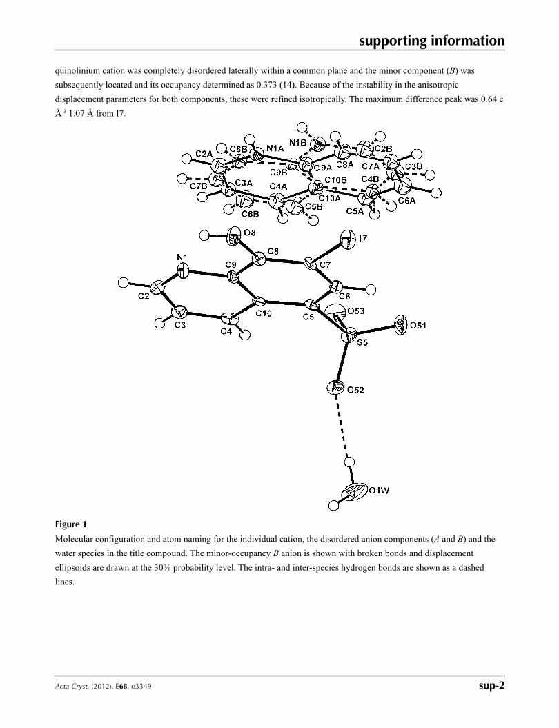
Figure 1
Molecular configuration and atom naming for the individual cation, the disordered anion components (A and B) and the
water species in the title compound. The minor-occupancy B anion is shown with broken bonds and displacement
ellipsoids are drawn at the 30% probability level. The intra- and inter-species hydrogen bonds are shown as a dashed
lines.
supporting information
sup-3Acta Cryst. (2012). E68, o3349
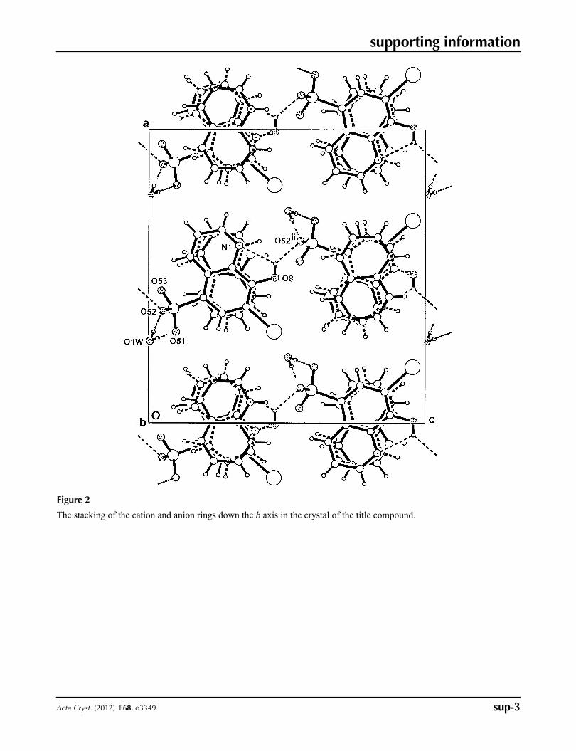
Figure 2
The stacking of the cation and anion rings down the b axis in the crystal of the title compound.
supporting information
sup-4Acta Cryst. (2012). E68, o3349
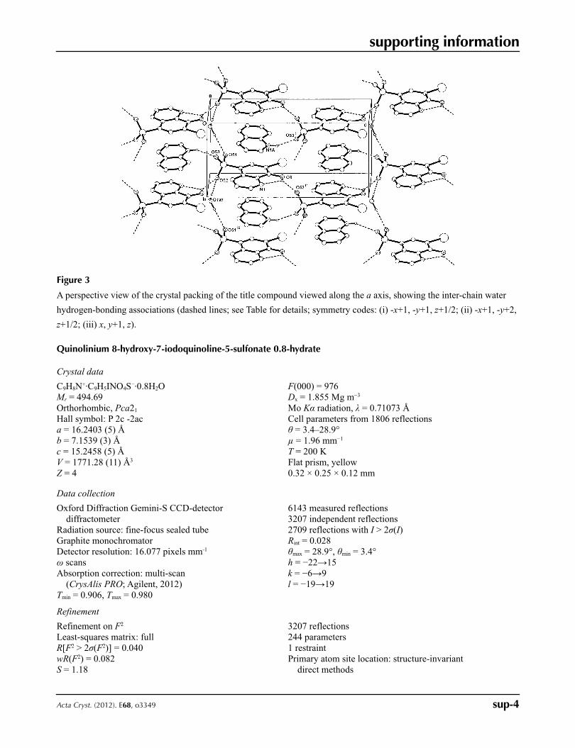
Figure 3
A perspective view of the crystal packing of the title compound viewed along the a axis, showing the inter-chain water
hydrogen-bonding associations (dashed lines; see Table for details; symmetry codes: (i) -x+1, -y+1, z+1/2; (ii) -x+1, -y+2,
z+1/2; (iii) x, y+1, z).
Quinolinium 8-hydroxy-7-iodoquinoline-5-sulfonate 0.8-hydrate
Crystal data
C9H8N+·C9H5INO4S−·0.8H2OMr = 494.69Orthorhombic, Pca21
Hall symbol: P 2c -2aca = 16.2403 (5) Åb = 7.1539 (3) Åc = 15.2458 (5) ÅV = 1771.28 (11) Å3
Z = 4
F(000) = 976Dx = 1.855 Mg m−3
Mo Kα radiation, λ = 0.71073 ÅCell parameters from 1806 reflectionsθ = 3.4–28.9°µ = 1.96 mm−1
T = 200 KFlat prism, yellow0.32 × 0.25 × 0.12 mm
Data collection
Oxford Diffraction Gemini-S CCD-detector diffractometer
Radiation source: fine-focus sealed tubeGraphite monochromatorDetector resolution: 16.077 pixels mm-1
ω scansAbsorption correction: multi-scan
(CrysAlis PRO; Agilent, 2012)Tmin = 0.906, Tmax = 0.980
6143 measured reflections3207 independent reflections2709 reflections with I > 2σ(I)Rint = 0.028θmax = 28.9°, θmin = 3.4°h = −22→15k = −6→9l = −19→19
Refinement
Refinement on F2
Least-squares matrix: fullR[F2 > 2σ(F2)] = 0.040wR(F2) = 0.082S = 1.18
3207 reflections244 parameters1 restraintPrimary atom site location: structure-invariant
direct methods

supporting information
sup-5Acta Cryst. (2012). E68, o3349
Secondary atom site location: difference Fourier map
Hydrogen site location: inferred from neighbouring sites
H-atom parameters constrainedw = 1/[σ2(Fo
2) + (0.0181P)2 + 3.2291P] where P = (Fo
2 + 2Fc2)/3
(Δ/σ)max = 0.004Δρmax = 0.65 e Å−3
Δρmin = −0.66 e Å−3
Absolute structure: Flack (1983), 789 Friedel pairs
Absolute structure parameter: 0.01 (3)
Special details
Geometry. Bond distances, angles etc. have been calculated using the rounded fractional coordinates. All su's are estimated from the variances of the (full) variance-covariance matrix. The cell e.s.d.'s are taken into account in the estimation of distances, angles and torsion anglesRefinement. Refinement of F2 against ALL reflections. The weighted R-factor wR and goodness of fit S are based on F2, conventional R-factors R are based on F, with F set to zero for negative F2. The threshold expression of F2 > σ(F2) is used only for calculating R-factors(gt) etc. and is not relevant to the choice of reflections for refinement. R-factors based on F2 are statistically about twice as large as those based on F, and R- factors based on ALL data will be even larger.
Fractional atomic coordinates and isotropic or equivalent isotropic displacement parameters (Å2)
x y z Uiso*/Ueq Occ. (<1)
N1A 0.5706 (6) 0.4412 (12) 0.3693 (6) 0.020 (2)* 0.630C2A 0.6389 (9) 0.4939 (16) 0.3279 (9) 0.025 (3)* 0.630C3A 0.6447 (7) 0.4807 (15) 0.2359 (9) 0.019 (2)* 0.630C4A 0.5784 (8) 0.4161 (17) 0.1888 (8) 0.026 (3)* 0.630C5A 0.4375 (7) 0.2937 (15) 0.1920 (9) 0.023 (3)* 0.630C6A 0.3681 (8) 0.2443 (17) 0.2377 (9) 0.030 (2)* 0.630C7A 0.3664 (8) 0.2600 (17) 0.3288 (10) 0.026 (2)* 0.630C8A 0.4325 (10) 0.3261 (17) 0.3750 (8) 0.026 (3)* 0.630C9A 0.5015 (8) 0.3760 (17) 0.3272 (8) 0.021 (3)* 0.630C10A 0.5084 (11) 0.356 (3) 0.2307 (12) 0.022 (5)* 0.630C8B 0.6037 (15) 0.466 (3) 0.3434 (14) 0.020 (4)* 0.370C9B 0.5273 (13) 0.388 (3) 0.3220 (12) 0.013 (4)* 0.370C10B 0.4986 (15) 0.362 (3) 0.2426 (16) 0.006 (6)* 0.370C3B 0.3682 (13) 0.254 (3) 0.2889 (17) 0.029 (4)* 0.370C4B 0.4180 (12) 0.288 (3) 0.2210 (14) 0.020 (4)* 0.370C5B 0.5542 (14) 0.390 (3) 0.1688 (15) 0.032 (5)* 0.370C6B 0.6308 (13) 0.451 (3) 0.1885 (15) 0.030 (4)* 0.370C7B 0.6552 (12) 0.496 (3) 0.2747 (15) 0.022 (4)* 0.370N1B 0.4736 (11) 0.341 (2) 0.3879 (10) 0.022 (3)* 0.370C2B 0.4005 (15) 0.285 (3) 0.3719 (14) 0.028 (5)* 0.370I7 0.30996 (2) 0.75195 (7) 0.45473 (4) 0.0259 (1)S5 0.38785 (10) 0.7547 (3) 0.09027 (10) 0.0232 (4)O8 0.4943 (2) 0.8886 (6) 0.4574 (4) 0.0265 (11)O51 0.3101 (3) 0.6580 (7) 0.0999 (3) 0.0327 (16)O52 0.3807 (3) 0.9388 (7) 0.0506 (3) 0.0280 (16)O53 0.4497 (3) 0.6391 (7) 0.0466 (3) 0.0323 (16)N1 0.6037 (3) 0.9565 (7) 0.3285 (4) 0.0220 (17)C2 0.6580 (4) 0.9911 (9) 0.2656 (5) 0.027 (2)C3 0.6418 (4) 0.9684 (9) 0.1760 (5) 0.0233 (19)

supporting information
sup-6Acta Cryst. (2012). E68, o3349
C4 0.5659 (4) 0.9043 (9) 0.1505 (4) 0.0223 (19)C5 0.4255 (4) 0.7936 (7) 0.1981 (4) 0.0150 (17)C6 0.3719 (4) 0.7600 (9) 0.2665 (4) 0.0183 (17)C7 0.3948 (4) 0.7926 (7) 0.3545 (4) 0.0147 (17)C8 0.4722 (4) 0.8578 (8) 0.3737 (4) 0.0173 (17)C9 0.5289 (4) 0.8920 (8) 0.3042 (4) 0.0157 (17)C10 0.5063 (4) 0.8627 (8) 0.2143 (4) 0.0167 (17)O1W 0.2775 (5) 1.2846 (9) 0.0055 (5) 0.057 (3) 0.800H4A 0.58090 0.41330 0.12790 0.0310* 0.630H5A 0.43630 0.28440 0.13110 0.0270* 0.630H6A 0.32210 0.20010 0.20780 0.0360* 0.630H7A 0.31910 0.22470 0.35900 0.0310* 0.630H8A 0.43110 0.33690 0.43570 0.0310* 0.630H1A 0.56960 0.44830 0.42560 0.0240* 0.630H2A 0.68320 0.53980 0.35990 0.0300* 0.630H3A 0.69290 0.51530 0.20730 0.0220* 0.630H1B 0.48960 0.34940 0.44150 0.0260* 0.370H2B 0.36610 0.26310 0.41960 0.0330* 0.370H3B 0.31450 0.21280 0.28100 0.0350* 0.370H4B 0.40210 0.26440 0.16340 0.0240* 0.370H5B 0.53800 0.36630 0.11140 0.0380* 0.370H6B 0.66890 0.46380 0.14340 0.0360* 0.370H7B 0.70690 0.54790 0.28460 0.0270* 0.370H8B 0.61840 0.49450 0.40080 0.0250* 0.370H2 0.71010 1.03290 0.28190 0.0320*H3 0.68190 0.99650 0.13450 0.0280*H4 0.55400 0.88840 0.09130 0.0270*H6 0.31940 0.71470 0.25440 0.0220*H8 0.54190 0.92330 0.45790 0.0390*H11W 0.30700 1.18390 0.01810 0.0850* 0.800H12W 0.28600 1.39490 0.03310 0.0850* 0.800
Atomic displacement parameters (Å2)
U11 U22 U33 U12 U13 U23
I7 0.0229 (2) 0.0360 (2) 0.0189 (2) −0.0052 (2) 0.0053 (2) −0.0028 (2)S5 0.0250 (8) 0.0290 (8) 0.0156 (6) −0.0017 (8) −0.0034 (6) 0.0023 (7)O8 0.025 (2) 0.036 (2) 0.0184 (18) −0.0057 (18) −0.006 (3) −0.007 (3)O51 0.030 (3) 0.043 (3) 0.025 (2) −0.017 (2) −0.010 (2) 0.003 (2)O52 0.028 (3) 0.031 (3) 0.025 (2) 0.000 (2) −0.003 (2) 0.010 (2)O53 0.045 (3) 0.031 (3) 0.021 (2) 0.006 (3) 0.000 (2) −0.009 (2)N1 0.017 (3) 0.022 (3) 0.027 (3) −0.005 (2) −0.003 (2) −0.001 (3)C2 0.018 (3) 0.023 (4) 0.041 (4) 0.002 (3) −0.007 (3) 0.007 (3)C3 0.018 (3) 0.017 (3) 0.035 (4) 0.006 (3) 0.005 (3) 0.003 (3)C4 0.028 (4) 0.016 (3) 0.023 (3) 0.005 (3) 0.004 (3) 0.004 (3)C5 0.025 (3) 0.008 (3) 0.012 (3) 0.000 (2) 0.001 (2) 0.000 (2)C6 0.018 (3) 0.019 (3) 0.018 (3) −0.003 (3) −0.002 (2) 0.004 (3)C7 0.013 (3) 0.011 (3) 0.020 (3) −0.001 (2) 0.006 (2) 0.001 (2)

supporting information
sup-7Acta Cryst. (2012). E68, o3349
C8 0.020 (3) 0.014 (3) 0.018 (3) 0.001 (2) 0.001 (3) −0.004 (3)C9 0.013 (3) 0.009 (3) 0.025 (3) 0.000 (2) 0.001 (2) −0.001 (2)C10 0.018 (3) 0.012 (3) 0.020 (3) −0.001 (2) 0.000 (3) 0.008 (3)O1W 0.070 (5) 0.027 (4) 0.073 (5) 0.023 (4) −0.034 (4) −0.010 (4)
Geometric parameters (Å, º)
I7—C7 2.078 (6) C8A—C9A 1.38 (2)S5—O52 1.454 (5) C8B—C9B 1.40 (3)S5—O51 1.447 (5) C9A—C10A 1.48 (2)S5—O53 1.462 (5) C9B—C10B 1.31 (3)S5—C5 1.776 (6) C2A—H2A 0.9300O8—C8 1.344 (8) C2B—H2B 0.9300O8—H8 0.8100 C3A—H3A 0.9300O1W—H12W 0.9000 C3B—H3B 0.9300O1W—H11W 0.8900 C4A—H4A 0.9300N1A—C2A 1.331 (17) C4B—H4B 0.9300N1A—C9A 1.374 (16) C5A—H5A 0.9300N1B—C9B 1.37 (3) C5B—H5B 0.9300N1B—C2B 1.28 (3) C6A—H6A 0.9300N1A—H1A 0.8600 C6B—H6B 0.9300N1B—H1B 0.8600 C7A—H7A 0.9300N1—C2 1.326 (9) C7B—H7B 0.9300N1—C9 1.351 (8) C8A—H8A 0.9300C2A—C3A 1.409 (19) C8B—H8B 0.9300C2B—C3B 1.39 (3) C2—C3 1.401 (11)C3A—C4A 1.374 (17) C3—C4 1.371 (9)C3B—C4B 1.34 (3) C4—C10 1.404 (9)C4A—C10A 1.37 (2) C5—C6 1.380 (9)C4B—C10B 1.45 (3) C5—C10 1.424 (9)C5A—C10A 1.37 (2) C6—C7 1.412 (9)C5A—C6A 1.371 (18) C7—C8 1.372 (9)C5B—C6B 1.35 (3) C8—C9 1.425 (9)C5B—C10B 1.46 (3) C9—C10 1.434 (9)C6A—C7A 1.39 (2) C2—H2 0.9300C6B—C7B 1.41 (3) C3—H3 0.9300C7A—C8A 1.37 (2) C4—H4 0.9300C7B—C8B 1.36 (3) C6—H6 0.9300
O51—S5—O52 113.9 (3) C4B—C3B—H3B 122.00O51—S5—O53 112.1 (3) C10A—C4A—H4A 120.00O51—S5—C5 106.3 (3) C3A—C4A—H4A 120.00O52—S5—O53 112.2 (3) C3B—C4B—H4B 122.00O52—S5—C5 105.7 (3) C10B—C4B—H4B 122.00O53—S5—C5 105.9 (3) C10A—C5A—H5A 118.00C8—O8—H8 108.00 C6A—C5A—H5A 118.00H11W—O1W—H12W 122.00 C10B—C5B—H5B 122.00C2A—N1A—C9A 123.7 (10) C6B—C5B—H5B 122.00

supporting information
sup-8Acta Cryst. (2012). E68, o3349
C2B—N1B—C9B 121.9 (17) C5A—C6A—H6A 120.00C9A—N1A—H1A 118.00 C7A—C6A—H6A 120.00C2A—N1A—H1A 118.00 C5B—C6B—H6B 119.00C9B—N1B—H1B 119.00 C7B—C6B—H6B 119.00C2B—N1B—H1B 119.00 C8A—C7A—H7A 119.00C2—N1—C9 117.6 (6) C6A—C7A—H7A 119.00N1A—C2A—C3A 120.6 (12) C8B—C7B—H7B 120.00N1B—C2B—C3B 125 (2) C6B—C7B—H7B 120.00C2A—C3A—C4A 119.4 (11) C7A—C8A—H8A 121.00C2B—C3B—C4B 117 (2) C9A—C8A—H8A 122.00C3A—C4A—C10A 120.7 (13) C9B—C8B—H8B 122.00C3B—C4B—C10B 116 (2) C7B—C8B—H8B 122.00C6A—C5A—C10A 123.8 (14) N1—C2—C3 124.0 (6)C6B—C5B—C10B 116 (2) C2—C3—C4 119.0 (6)C5A—C6A—C7A 120.1 (12) C3—C4—C10 119.6 (6)C5B—C6B—C7B 123 (2) C6—C5—C10 120.7 (6)C6A—C7A—C8A 121.7 (12) S5—C5—C6 117.1 (5)C6B—C7B—C8B 120.7 (19) S5—C5—C10 122.2 (5)C7A—C8A—C9A 117.0 (12) C5—C6—C7 121.6 (6)C7B—C8B—C9B 115.4 (19) I7—C7—C8 119.9 (4)C8A—C9A—C10A 124.0 (13) I7—C7—C6 120.1 (5)N1A—C9A—C10A 115.8 (12) C6—C7—C8 120.0 (6)N1A—C9A—C8A 120.2 (11) O8—C8—C9 120.4 (5)N1B—C9B—C8B 119.4 (17) O8—C8—C7 120.2 (5)C8B—C9B—C10B 126 (2) C7—C8—C9 119.5 (6)N1B—C9B—C10B 115 (2) N1—C9—C10 122.8 (6)C4A—C10A—C9A 119.6 (15) C8—C9—C10 121.3 (6)C4A—C10A—C5A 126.7 (16) N1—C9—C8 115.8 (6)C5A—C10A—C9A 113.3 (14) C4—C10—C9 117.0 (6)C5B—C10B—C9B 118 (2) C4—C10—C5 126.1 (6)C4B—C10B—C5B 116 (2) C5—C10—C9 116.9 (6)C4B—C10B—C9B 126 (2) N1—C2—H2 118.00C3A—C2A—H2A 120.00 C3—C2—H2 118.00N1A—C2A—H2A 120.00 C4—C3—H3 121.00C3B—C2B—H2B 117.00 C2—C3—H3 120.00N1B—C2B—H2B 118.00 C3—C4—H4 120.00C2A—C3A—H3A 120.00 C10—C4—H4 120.00C4A—C3A—H3A 120.00 C5—C6—H6 119.00C2B—C3B—H3B 122.00 C7—C6—H6 119.00
O53—S5—C5—C6 −130.4 (5) C8A—C9A—C10A—C4A 177.6 (14)O53—S5—C5—C10 51.8 (5) N1—C2—C3—C4 −0.9 (10)O52—S5—C5—C6 110.4 (5) C2—C3—C4—C10 −0.1 (9)O51—S5—C5—C6 −11.0 (5) C3—C4—C10—C5 −179.3 (6)O51—S5—C5—C10 171.1 (5) C3—C4—C10—C9 0.5 (9)O52—S5—C5—C10 −67.4 (5) S5—C5—C6—C7 −177.8 (5)C2A—N1A—C9A—C10A 3.5 (18) C10—C5—C6—C7 0.0 (9)C9A—N1A—C2A—C3A −1.6 (17) S5—C5—C10—C4 −1.5 (8)

supporting information
sup-9Acta Cryst. (2012). E68, o3349
C2A—N1A—C9A—C8A −179.2 (11) S5—C5—C10—C9 178.7 (4)C9—N1—C2—C3 1.4 (9) C6—C5—C10—C4 −179.2 (6)C2—N1—C9—C8 −179.6 (5) C6—C5—C10—C9 1.0 (8)C2—N1—C9—C10 −0.9 (9) C5—C6—C7—I7 177.1 (4)N1A—C2A—C3A—C4A 1.3 (17) C5—C6—C7—C8 −0.5 (9)C2A—C3A—C4A—C10A −3 (2) I7—C7—C8—O8 2.6 (7)C3A—C4A—C10A—C5A 178.0 (15) I7—C7—C8—C9 −177.6 (4)C3A—C4A—C10A—C9A 5 (2) C6—C7—C8—O8 −179.9 (5)C6A—C5A—C10A—C4A −176.6 (16) C6—C7—C8—C9 0.0 (8)C6A—C5A—C10A—C9A −4 (2) O8—C8—C9—N1 −0.4 (8)C10A—C5A—C6A—C7A 1 (2) O8—C8—C9—C10 −179.1 (5)C5A—C6A—C7A—C8A 0.7 (19) C7—C8—C9—N1 179.8 (5)C6A—C7A—C8A—C9A −0.3 (19) C7—C8—C9—C10 1.1 (9)C7A—C8A—C9A—C10A −2 (2) N1—C9—C10—C4 0.0 (9)C7A—C8A—C9A—N1A −179.1 (11) N1—C9—C10—C5 179.8 (5)N1A—C9A—C10A—C4A −5 (2) C8—C9—C10—C4 178.6 (6)C8A—C9A—C10A—C5A 4 (2) C8—C9—C10—C5 −1.5 (8)N1A—C9A—C10A—C5A −179.0 (13)
Hydrogen-bond geometry (Å, º)
D—H···A D—H H···A D···A D—H···A
N1A—H1A···O53i 0.86 1.97 2.783 (10) 157N1B—H1B···O53i 0.86 1.88 2.725 (16) 166O8—H8···N1 0.81 2.23 2.693 (7) 117O8—H8···O52ii 0.81 2.13 2.769 (7) 135O1W—H11W···O52 0.89 2.18 3.066 (9) 179O1W—H12W···O51iii 0.90 2.18 3.080 (8) 178C4—H4···O53 0.93 2.55 3.110 (8) 119C6—H6···O51 0.93 2.39 2.827 (8) 108C8A—H8A···O53i 0.93 2.58 3.251 (14) 130
Symmetry codes: (i) −x+1, −y+1, z+1/2; (ii) −x+1, −y+2, z+1/2; (iii) x, y+1, z.