quinidine€¦ · somequantitative aspects of thebinding of quinidine andrelated quinoline...
TRANSCRIPT
SOMEQUANTITATIVE ASPECTSOF THE BINDING OF QUINIDINEANDRELATEDQUINOLINE COMPOUNDSBY HUMAN
SERUMALBUMIN*
By HADLEYL. CONN, JR. AND ROBERTJ. LUCHI t
(From the Robinette Foundation, Department of Medicine, University of Pennsylvania Schoolof Medicine, Philadelphia, Pa.)
(Submitted for publication August 18, 1960; accepted November 21, 1960)
The interaction of quinidine with various bodyproteins may well be the fundamental processthrough which quinidine exerts its many effects.For example, quinidine binding with membranelipoprotein may lead to decreased membrane per-meability to passive ion movement. This perme-ability change has been held responsible for thequinidine-induced alterations in sodium and po-tassium exchange in cardiac muscle (1-4). Quin-idine binding with critical enzyme proteins maybe responsible for the alterations in intermediarymetabolism which have been ascribed to quinidine(5). In our experience, glucose-6-phosphate de-hydrogenase activity is reduced in vitro by quini-dine. Finally, the known binding of quinidine toalbumin may protect the cell against the action ofquinidine by making less drug available at cellu-lar sites. The nature of the interaction with pro-tein of quinidine and of other compounds having asimilar chemical structure seemed, therefore, towarrant an investigation.
Albumin is readily available in adequate amountsand in relative purity. As noted above, it isknown to complex with quinidine. Further, thestate of the quinidine-albumin interaction mightbe considered as a prototype of the "protective"form and perhaps characteristic of all forms ofquinidine binding to protein. For these reasonsour initial investigations were concerned with thereactions of quinidine and related quinoline com-pounds with albumin. Experiments were de-signed to permit determination of the stoichiom-etry, the association constant, and the energies ofbinding for the reactions; and to show the contri-
* This study was supported by grants from the AtomicEnergy Commission and the American Heart Associa-tion.
t Supported in part by United States Public HealthService Grant H5663 and a grant from the Pharmaceuti-cal Manufacturers Association.
butions to binding by the various reactive sites orareas in the quinidine molecule.
METHODS
A dialysis equilibrium technique was used to evaluatethe in vitro binding of quinidine and of similar chemicalsto human serum albumin.' A limited investigation ofquinidine binding to human y-globulin was carried outin the same manner.2 A cellulose dialysis membrane3was prepared by an initial wash with 1 per cent sodiumcarbonate and a subsequent wash with distilled water.Twenty ml of 0.2 M phosphate buffer solution contain-ing 250 mg of human serum albumin was placed in adialysis bag. Fifty ml of the same buffer solution wasplaced around the bag in a plastic beaker. To this, ex-ternal solution quinidine and other substances were addedas desired. The beaker was then covered tightly withSaran wrap and shaken mechanically at 25° C untilequilibrium had been achieved. In practice this wasfound to occur within 18 hours.
Quinidine may be considered structurally as 4-hydroxy-methyl-6-methoxy-quinoline connected by the secondaryalcohol bridge to a quinuclidine ring. Therefore, notonly was the effect of quinidine binding to albumin stud-ied but also the binding to albumin of 4-hydroxymethyl-6-methoxy-quinoline,4 6-methoxy-quinoline, 4-hydroxy-methyl quinoline,5 quinoline, and naphthalene. The hy-drochloride salt was used in all cases except that ofnaphthalene. These compounds were used as a meansof evaluating the contribution of binding of the variousparts of the quinoline molecule. In addition, the com-petitive or inhibitory effects of all of the above sub-stances, except naphthalene, on quinidine-albumin bind-ing were studied during the presence both of quinidineand of one of the other compounds in the system. Thepart played in the binding reaction by the quinuclidine
1 Obtained from the Red Cross in solution form andfrom Pentex Co., Kankakee, Ill., in crystalline form.On paper electrophoresis both migrated as homogeneouspeaks corresponding to albumin when barbital buffer(pH 8.6) and an ionic strength of 0.075 were used.
2 Obtained from Sharp and Dohme, division of Merck,Inc., Philadelphia, Pa.
3A. H. Thomas Company, Philadelphia, Pa.4 5 Synthesized with the generous aid of R. W. Greeff
and Co., Nederlandsche Kininefabriek, Maarsen, Holland.
509
510 HADLEYL. CONN, JR. AND ROBERTJ. LUCHI
TABLE I
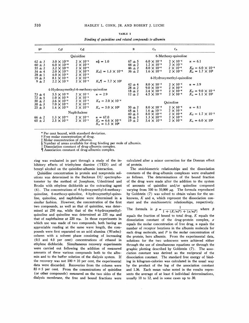
Binding of quinidine and related compounds to albumin
B* Cqt Cat B Cq Ca
Quinidine 6-Methoxy-quinoline62 ± 3 3.0 X 10⁻⁶ 2 X 10⁻⁴ n§ = 1.0 67 5 6.0 X 10⁻⁶ 2 X 10⁻⁴ n = 6.160 3 6.0 X 10⁻⁶ 2 X 10⁻⁴ 66 2 1.2 X 10⁻⁵ 2 X 10-455 2 3.2 X 10⁻⁵ 2 X 10⁻⁴ 46 2 8.0 X 10⁻⁴ 2 X 10⁻⁴ Kd = 6.0 X 10-434 ± 4 2.0 X 10⁻⁴ 2 X 10⁻⁴ Kdl= 1.3 X 10⁻⁴ 36 ± 2 1.6 X 10⁻³ 2 X 10⁻⁴ K, = 1.7 X 10328±1 4.0X10⁻⁴ 2X10-419 3 8.1 X 10⁻⁴ 2 X 10⁻⁴ 4-Hydroxymethyl-quinoline9 2 2.2 X 10⁻³ 2 X 10⁻⁴ KaT = 7.7 X 10³
42 ± 4 8.0 X 10⁻⁶ 2 X 10⁻⁴ n = 3.94-Hydroxymethyl-6-methoxy-quinoline 28 ± 2 9.0 X 10⁻⁴ 2 X 10-418±==2 2.4 X10⁻³ 2 X10⁻⁴ Kd =9.0 X10⁻⁴
73 ± 4 5.5 X 10⁻⁶ 2 X 10⁻⁴ n = 2.9 12 ± 2 4.5 X 10⁻³ 2 X 10⁻⁴ Ka = 1.1 X 10372 ± 4 1.0 X 10⁻⁵ 2 X 10-450 ±-~ 2 3.6 X 10⁻⁴ 2 X 10⁻⁴ Kd = 2. 0 X 10-Qio4n38 ± 3 7.0 X 10⁻⁴ 2 X 10⁻⁴ Quinoline25 ± 3 1.4 X 10⁻³ 2 X 10⁻⁴ Ka = 5.0 X 10³ 50 ± 2 8.0 X 10⁻⁶ 2 X 10⁻⁴ n = 8.1
48 i 1 1.6 ± 10-5 2 X 10⁻⁴Naphthalene 40 1 3 8.0 X 10⁻⁴ 2 X 10⁻⁴ Kd = 1.7 X 10-366 2 1.3 X 10⁻⁴ 2 X 10⁻⁴ n = 67.0 34 ±t 3 1.3 X 10⁻³ 2 X 10-465 2 2.5 X 10⁻⁴ 2 X 10⁻⁴ Kd = 6.6 X 10⁻³ 19 ± 2 5.4 X 10⁻³ 2 X 10⁻⁴ Ka = 6.0 X 10²
Ka = 1.5 X 10²
* Per cent bound, with standard deviation.t Free molar concentration of drug.t Molar concentration of albumin.§ Number of areas available for drug binding per mole of albumin.
Dissociation constant of drug-albumin complex.¶ Association constant of drug-albumin complex.
ring was evaluated in part through a study of the in-hibitory effects of triethylene diamine (TED) and ofbenzyl alcohol on the quinidine-albumin interaction.
Quinidine concentration in protein and nonprotein sol-utions was determined in the Beckman DU spectropho-tometer by the method of Josephson, Udenfriend andBrodie with ethylene dichloride as the extracting agent(6). The concentrations of 4-hydroxymethyl-6-methoxy-quinoline, 6-methoxy-quinoline, 4-hydroxymethyl-quino-line, quinoline, and naphthalene were determined in asimilar fashion. However, the concentration of the firsttwo compounds, as well as that of quinidine, was deter-mined at 250 miu, while that of the 4-hydroxymethyl-quinoline and quinoline was determined at 235 mju andthat of naphthalene at 220 myA. In those experiments inwhich use was made of two compounds, both having anappreciable reading at the same wave length, the com-pounds were first separated on an acid alumina (Woelm)column with a solvent phase consisting of increasing(0.5 and 4.0 per cent) concentrations of ethanol inethylene dichloride. Simultaneous recovery experimentswere carried out following the addition of measuredamounts of these various compounds both to the albu-min and to the buffer solution of the dialysis system. Ifthe recovery was not 100 + 10 per cent, the experimentaldata were discarded. Recoveries from the column were85 ± 5 per cent. From the concentrations of quinidine(or other compounds) measured on the two sides of the
dialysis membrane, the free and bound fractions were
calculated after a minor correction for the Donnan effectof protein.
The stoichiometric relationships and the dissociationconstants of the drug-albumin complexes were evaluatedas follows. The determinations of the bound fractionof the drug were made after the addition to the systemof amounts of quinidine and/or quinoline compoundvarying from 100 to 50,000 yg. The formula reproducedby Goldstein (7) was solved to obtain values for the un-knowns, K and n, which represent the dissociation con-stant and the stoichiometric relationships, respectively.
The formula is 0 = 1 + (K/nP) + (x/nP)whereequals the fraction of bound to total drug, K equals thedissociation constant of the drug-protein complex, xequals the molar concentration of free drug, n equals thenumber of receptor locations in the albumin molecule foreach drug molecule, and P is the molar concentration ofthe protein, here albumin. From the experimental data,solutions for the two unknowns were achieved eitherthrough the use of simultaneous equations or through thegraphic plotting described by Goldstein (7). The asso-ciation constant was derived as the reciprocal of thedissociation constant. The standard free energy of bind-ing in kilogram-calories was calculated in the usual wayby the product of the log of the association constantand 1.36. Each mean value noted in the results repre-sents the average of at least 6 individual determinations,usually 10 to 12, and in some cases up to 30.
QUINIDINE-ALBUMIN BINDING
RESULTS
Stoichiometry and the dissociation constants ofthe quinidine-albumin complex. The stoichio-metric relationship and the dissociation constants
were obtained at pH values of 7.4, although theyhave been obtained at other pH values in experi-ments reported in another communication (8).The mean value for amount of drug bound at vari-ous free concentrations, the actual dissociationconstants and the stoichiometric value are shownin Table I. The calculated results indicate thatthere is one receptor area for quinidine in eachmole of albumin (n = 1.04). The dissociationconstant is 1.3 X 10⁻⁴ at pH 7.4, although fromother studies it is known to vary inversely withpH (8). Expressed as an association constant,this value becomes 7.7 x 10³ at pH 7.4. Thermo-dynamically, this gives a standard free energy ofbinding of about 5.3 kg-cal per mole of quinidine.
The binding of quinidine to y-globulin. To theextent that these solutions resemble plasma, the Kand n values obtained at pH 7.4 may be used to
predict the protein-bound fraction of quinidine inthe serum of patients given commonly employeddoses of quinidine. With an average serum albu-min concentration of 4 g per cent and a therapeu-tic serum-quinidine concentration of 5 mg per L(total concentration) the predicted value for thebound fraction would be about 80 per cent. Anearly identical value (0.78) has been reportedto be the fraction of quinidine and also quininenormally bound in human serum (9, 10). Thesimilarity of these figures suggested that quini-dine is not bound to any significant extent by se-
rum globulin and led us to test this hypothesisexperimentally. As the data from the albuminexperiments would suggest, the amount of quini-dine bound to y-globulin in a dialysis equilibriumsystem was not significantly different from zero.
The binding of 4-hydroxymethyl-6-methoxy-quinoline to albumin. The results of the bindingstudies carried out with this drug are shown inTable I. The calculated n indicates that thereare three receptor locations for 4-hydroxymethyl-6-methoxy-quinoline in each mole of albumin.The dissociation constant is 2 x 10⁻⁴ and the as-
sociation constant is 5 x 10³. The calculated freeenergy of binding is 5.0 kg-cal, only slightly lessthan that calculated for quinidine-albumin bind-
ing. Consequently, by inference the quinuclidinering contributes only slightly to the binding.
The binding of 6-methoxy-quinoline to albu-min. Results for the interaction of 6-methoxy-quinoline with albumin are shown in Table I.The results indicate that 6 moles of this drug arebound by each interacting molecule of albumin.The dissociation constant is 6 x 10⁻⁴. The asso-ciation constant is therefore 1.7 x 10³, and thefree energy of binding 4.4 kg-cal.
The binding of 4-hydroxymethyl-quinoline toalbumin. The results for the binding of this drugto albumin are shown in Table I. Calculations in-dicate four receptor locations for this drug ineach molecule of albumin. The dissociation con-stant is 9 x 10⁻³. This is equivalent to an associa-tion constant of 1.1 X 10³. The free energy ofbinding is calculated at 4.1 kg-cal.
The binding of quinoline to albumin. The re-sults for the binding of quinoline to albumin areshown in Table I. They indicate that there areeight receptor areas for quinoline in the albuminmolecule. The dissociation constant is 1.7 x 10⁻³,equivalent to an association constant of 6 X 10².The calculated standard free energy of binding is3.5 kg-cal.
The binding of naphthalene to albumin. Be-cause of the extremely low solubility of naphtha-lene in aqueous solutions it was impossible tovary the free concentration so as to determinewith adequate accuracy either the number of al-bumin receptor areas for naphthalene or the dis-sociation constant for the reaction. From the fig-ures for per cent naphthalene bound to albuminobtained under these restricting conditions an nof about 67 and a dissociation constant of about6.6 x 10-3 were calculated. This is equivalent toan association constant of 1.5 x 10², indicating anapproximate free energy value of 2.9 kg-cal. De-spite the recognized inadequacy of the data andtherefore the uncertainty of the extrapolations,they are presented as matters of interest in that1) the calculated free energy value is similar tothat found for the association of albumin withnon-polar substituents (personal communicationfrom Dr. Fred Karush) and 2) the free energyvalue is in general agreement with that predictedindirectly from our other experiments.
The effect of quinoline compounds in inhibitingquinidine-albumin binding. In order to show
511
HADLEYL. CONN, JR. AND ROBERTJ. LUCHI
that the several related quinoline compounds were
actually combining at the same receptor locationon the albumin molecule as quinidine, some stud-ies were carried out with both quinidine and one
quinoline compound present in the dialysis sys-
tem. As described under Methods these sub-stances were subsequently separated by columnchromatography, and each compound was readat its proper spectral peak. On the basis of thepreviously determined K and n values for thequinoline compounds, an amount of each (oneper experiment) was added to the system thatwould be expected to reduce quinidine bindingvery significantly under the existing experimentalconditions, provided the two drugs competed forthe same reaction area in the albumin molecule.From the measured concentrations of unboundquinidine and quinoline drug and from other per-
tinent data, a predicted value for the bound frac-tion of quinidine was calculated.6 These values
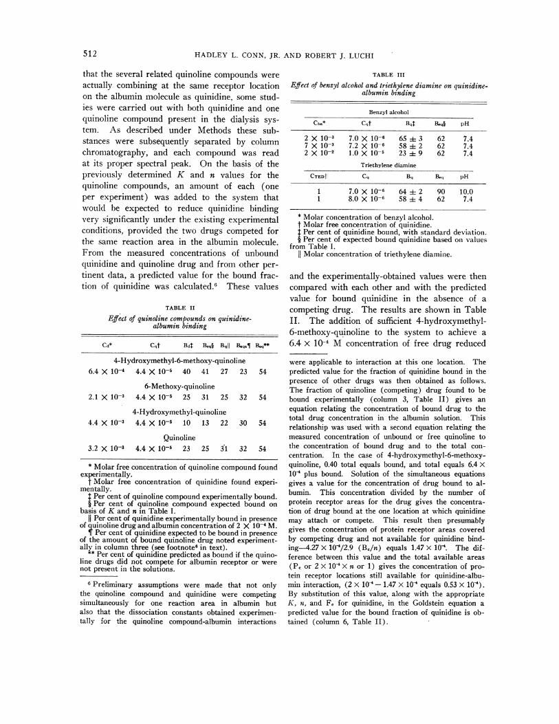
TABLE II
Effect of quinoline compounds on quinidine-albumin binding
Cd* Cqt BdT Be§ Bqj Beqa¶l Beq**
4-Hydroxymethyl-6-methoxy-quinoline6.4 X 10-4 4.4 X 10⁻⁵ 40 41 27 23 54
6-Methoxy-quinoline2.1 X 10-3 4.4 X 10⁻⁵ 25 31 25 32 54
4-Hydroxymethyl-quinoline4.4 X 10-s 4.4 X 10⁻⁵ 10 13 22 30 54
Quinoline3.2 X 10-3 4.4 X 10⁻⁵ 23 25 31 32 54
* Molar free concentration of quinoline compound foundexperimentally.
t Molar free concentration of quinidine found experi-mentally.
Per cent of quinoline compound experimentally bound.§ Per cent of quinoline compound expected bound on
basis of K and n in Table I.
1i Per cent of quinidine experimentally bound in presenceof quinoline drug and albumin concentration of 2 X 10⁻⁴ M.
Per cent of quinidine expected to be bound in presenceof the amount of bound quinoline drug noted experiment-ally in column three (see footnote6 in text).
** Per cent of quinidine predicted as bound if the quino-line drugs did not compete for albumin receptor or werenot present in the solutions.
6 Preliminary assumptions were made that not onlythe quinoline compound and quinidine were competingsimultaneously for one reaction area in albumin butalso that the dissociation constants obtained experimen-tally for the quinoline compound-albumin interactions
TABLE III
Effect of benzyl alcohol and triethylene diamine on quinidine-albumin binding
Benzyl alcohol
Cba* Cqt Bqt Bpq§ pH
2 X 10⁻³ 7.0 X 10⁻⁶ 65 3 62 7.47 X10⁻³ 7.2 X 10⁻⁶ 58 2 62 7.42 X 10⁻² 1.0 X 10⁻⁵ 23 9 62 7.4
Triethylene diamine
CTEDI| Cq B, Beq pH
1 7.0 X 10⁻⁶ 64 ± 2 90 10.01 8.0 X 10⁻⁶ 58 ± 4 62 7.4
* Molar concentration of benzyl alcohol.t Molar free concentration of quinidine.$ Per cent of quinidine bound, with standard deviation.§ Per cent of expected bound quinidine based on values
from Table I.1I Molar concentration of triethylene diamine.
and the experimentally-obtained values were thencompared with each other and with the predictedvalue for bound quinidine in the absence of acompeting drug. The results are shown in TableII. The addition of sufficient 4-hydroxymethyl-6-methoxy-quinoline to the system to achieve a6.4 X 10-4 M concentration of free drug reduced
were applicable to interaction at this one location. Thepredicted value for the fraction of quinidine bound in thepresence of other drugs was then obtained as follows.The fraction of quinoline (competing) drug found to bebound experimentally (column 3, Table II) gives anequation relating the concentration of bound drug to thetotal drug concentration in the albumin solution. Thisrelationship was used with a second equation relating themeasured concentration of unbound or free quinoline tothe concentration of bound drug and to the total con-centration. In the case of 4-hydroxymethyl-6-methoxy-quinoline, 0.40 total equals bound, and total equals 6.4 X10' plus bound. Solution of the simultaneous equationsgives a value for the concentration of drug bound to al-bumin. This concentration divided by the number ofprotein receptor areas for the drug gives the concentra-tion of drug bound at the one location at which quinidinemay attach or compete. This result then presumablygives the concentration of protein receptor areas coveredby competing drug and not available for quinidine bind-ing-4.27 X 10'/2.9 (Be/n) equals 1.47 X 10'. The dif-ference between this value and the total available areas(PR or 2 X 10' X n or 1) gives the concentration of pro-
tein receptor locations still available for quinidine-albu-min interaction, (2 X 10 - 1.47 X 10⁻⁴ equals 0.53 X 10').By substitution of this value, along with the appropriateK, n, and F, for quinidine, in the Goldstein equation apredicted value for the bound fraction of quinidine is ob-tained (column 6, Table II).
512
QUINIDINE-ALBUMIN BINDING
the percentage of quinidine bound (unboundquinidine concentration of 4.4 x 10⁻⁵ MI) from54 to 27, close to the predicted value of 23 percent. With a concentration of free 6-methoxy-quinoline of 2.1 x 10⁻³ nearly the same result wasachieved, a reduction in the quinidine bound from54 to 25 per cent. This may be compared withthe predicted result of 32 per cent bound. A re-duction of the bound fraction of quinidine from54 to 22 per cent was achieved with a 4.4 x 10⁻³M concentration of 4 hydroxymethyl quinoline.The 22 per cent figure is similar to the predictedvalue of 30 per cent. A free quinoline concentra-tion of 3.2 x 10⁻³ M\1 reduced the fraction ofquinidine bound to albumin from 54 to 31 percent, almost exactly as predicted. The data con-sequently provide strong evidence that there iscompetition between quinidine and the relatedcompounds tested for one specific reactive areain albumin.
The effects of triethylene diamine and benzylalcohol on quinidine binding With albumin. Anattempt to learn something further about the im-portance of the quinuclidine ring and the alcoholicOH site to the binding reaction was carried outthrough the addition of varying amounts of TEDor of benzyl alcohol along with quinidine to thedialysis system. Addition of benzyl alcohol, whichpresumably might compete for an albumin sitewith the combined quinuclidine ring and adja-cent alcohol fragment of quinidine, led to the re-sults indicated in Table III. In the presence offree quinidine, concentrations of slightly less than1 X 10-5 M benzyl alcohol in 2 x 10⁻³ M concen-trations had little effect on binding. In the pres-ence of a 7 x 10⁻³ M alcohol concentration, boundquinidine was reduced slightly from the 62 percent expected if no alcohol were present to 58per cent. With concentrations of benzyl alcoholat 2 x 10⁻² M the inhibition of quinidine-albuminbinding was very marked. An average of only23 per cent was calculated for the bound fractionas compared with an expected 62 per cent. Thisis compatible with a contribution by the quinu-clidine ring and the secondary alcohol of some-thing like 1.5 kg-cal to the total energy of bind-ing, a value close to that predicted from the previ-ously noted experimental results. On the otherhand, TED which presumably is similar to thebasic quinuclidine structure, failed to reduce ap-
preciably quinidine binding at pH 7.4 even whenpresent in concentrations as great as 1 AI. Onlya slight reduction from an expected value of 62per cent to a calculated value of 58 per cent wasfound with 1 1\1 concentrations. Since the twopK's of triethylene diamine are 5 and 8.4, at pH7.4 one of the nitrogens is mainly positivelycharged, as is the nitrogen in the quinuclidine ring(pK, 8.6). Therefore, further evidence is ad-duced that the quinuclidine ring per se does notseem to play an important part in the binding ofquinidine to albumin, at least when the nitrogengroup is positively charged, but that the alcoholicbridge fragment contributes something like 1kg-cal to the reaction.
In view of these conclusions, we were some-what surprised to find that in the presence of car-bonate buffer at pH 10, the same concentration ofTED reduced the binding to 64 per cent from anexpected 90 per cent. This significant reductionin the binding, however, occurs at a pH at whichthe two nitrogens in the triethylene diamine andthe nitrogen of the quinuclidine ring of quinidineare essentially all noncharged. We have foundthat at this pH the increased binding, found nor-mally, relates mainly to an increase in the num-ber of quinidine receptor areas in albumin from1 to 3 (8), possibly because of the loss of the polareffect of the positively charged nitrogen. Thisincreased n value (3.0) is the same as that cal-culated for 4-hydroxymethyl-6-methoxy-quinoline.It is suggested that neutral TED in 1 M concen-tration may compete with neutral quinidine forthese additional two albumin binding areas.
DISCUSSION
These data reveal a moderately large (Ka 7.7 x10³) association constant for the quinidine-albu-min reaction, and an uncommon, one-for-onemolar interaction-i.e., one receptor area forquinidine attachment in each albumin molecule.The basic quinoline ring structure apparently con-tributes a major portion of the binding energyabout 2.9 out of 5.3 kg-cal. The neutral nitrogenof the quinoline ring (pK, 4.0), the methoxygroup, and the secondary bridging alcohol groupalso contribute to the energy of binding andtherefore presume to be sites of interaction wvith
513
HADLEYL. CONN, JR. AND ROBERTJ. LUCHI
the albumin molecule. The specificity of thequinidine-albumin reaction is contributed to by allthe three reactive groups as is evident by the in-creasing number of binding areas in albumin in-volved and the decreasing association constantvalues resulting as these groups are deleted fromthe molecule. Either hydrogen bonding or Vander Waals forces could account for these interac-tions. Formation of hydrogen bonds seems moreprobable in view of the virtually identical bindingenergies (0.5 to 1.0 kg-cal) concerned with eachof the three reactive sites, and particularly be-cause the experimental data appear to show thatthe energy of binding is the same for all albuminlocations involved in binding both quinidine andrelated quinoline compounds. Equal Van derWaals interactions in all cases would imply nearlyidentical spatial complementarity of albumin anddrug at the critical areas. It is improbable thatthe eight areas concerned in albumin would havethe required structural identity. On the otherhand, albumin sites at which hydrogen bond link-age to the quinoline nitrogen, the methoxy, andthe hydroxymethyl groups of quinidine might oc-cur have not been identified. Our recent unre-ported studies are compatible with the concept thatthe quinuclidine nitrogen forms a hydrogen bondwith an imidazole nitrogen in a histidine residueof albumin. The methoxy group may form a hy-drogen bond with a hydroxyl group in serine, and
7The term site as commonly used in characterizingdrug-protein interaction has at least two connotationsand needs to be defined for purposes of the present dis-cussion. It has been used to refer to the point of molecu-lar interaction between a reactive group or atom in thedrug and a corresponding group in an amino acid of pro-tein. Electrostatic interaction between a positivelycharged nitrogen atom in a drug and a negativelycharged hydroxyl group in protein is one such example.We have reserved site for use in describing this formof interaction. Site or receptor site has also been com-monly used to refer to the general area at which a drugis attached to a protein, irrespective of whether one ormany drug-protein reactive group interactions take place,or whether, in our terminology, one or many sites ofinteraction are involved. Thus, in common parlance nis defined as the number of drug receptor sites per mole-cule of protein. In order to avoid needless confusion be-tween the former (single) and the latter (in this case,multiple interaction), we have arbitrarily substituted theterms area or location for use in the second connotation.We have defined n as the number of receptor locations(or areas) for drug attachment to the albumin molecule.
the suggestion has been made that a similar reac-tion may link the secondary alcohol to a hydroxylgroup in tyrosine (personal communication fromDr. I. M. Klotz).
At pH 7.4, the quinuclidine ring does not seemto play a very great part in the binding of quini-dine to albumin. Its most important role may re-late to the action of the charged nitrogen in pre-venting binding at more than one reactive area inalbumin. This role is suggested 1) by the factthat the simpler compound without the quinuclidinering-4-hydroxymethyl-6-methoxy-quinoline-ap-pears to react at three binding areas in albuminand 2) by the fact that when the positive nitrogenin the quinuclidine ring becomes neutral with in-creasing pH, the number of receptor areas forquinidine in albumin is also three. In brief, theuncharged form of quinidine may be able to attachitself at two additional areas in albumin where thecharged form is unable to penetrate or attach.While this hypothesis may appear more attractive,an additional consideration is deserving of men-tion-competition for quinidine between the waterand protein phases. Water solubility of quinidinevaries markedly with pH in an inverse fashion.This relationship might be anticipated if thecharged nitrogen groups (pK's of 4.0 and 8.6)were undergoing polar interactions with water.At a pH of 10, quinidine is nearly all in the un-charged form, quinidine solubility is slight, andwith increasing pH, it rapidly becomes negligible.With decreasing pH below 7, quinidine solubilityincreases markedly, the acidic salts-quinidinehydrochloride and quinidine sulfate-being thecompounds commonly used to prepare concen-trated aqueous solutions of quinidine. Quinidine'shigh aqueous solubility at low pH, reflecting pre-dominance of the most polar forms of the molecule,might be partially responsible for the reciprocallylow degree of attachment to protein we have ob-served in unreported studies. In contrast, lowaqueous solubility of the neutral form of the drugat high pH may account partially for the markeddrug affinity for albumin noted under these con-ditions. To the extent that this process is opera-tive, a plot of quinidine binding by albumin againstpH should resemble a quinidine acid titrationcurve. Unfortunately, the former plot as ob-tained experimentally is more complex than the
514
QUINIDINE-ALBUMIN BINDING
latter. Interpretation is not so simple as pro-posed and the issue remains unresolved.
The similarity of association constants for thequinidine-albumin and the (4-hydroxymethyl-6-methoxy-quinoline)-albumin reactions, and thefinding of a receptor area in albumin available toboth drugs led us to test whether these drugshave any common cardiovascular effects (11).The substituted quinoline does have a peripheralvasodilation action like that of quinidine. Thehypotension produced by both drugs is similarilyreversed by sympathomimetic amines. Finallythe effective "hypotensive" dose of 4-hydroxy-methyl-6-methoxy-quinoline is three or four timesthat of quinidine, a relationship predictable bytranslation of the relative quantitative aspects ofthe drugs binding with albumin to their bindingvia a similar hypothetical receptor in arteriolarprotein. A comparison of the action of the twodrugs on the heart gives a different picture. Thesubstituted quinoline compound possesses mini-mal antiarrythmic and apparently none of theusual myocardial depressant properties of quini-dine. The indirect implication is that the phar-macologic effects of quinidine on the heart aredependent on the presence of the quinuclidine ring.From these considerations two questions areposed, neither of which is currently answerableexcept in an indirect and speculative fashion.
The first question is concerned with the extentto which the characteristics of quinidine-albumininteraction can be considered to reflect the natureof quinidine binding by cell protein. Studies suchas those of Wegria: and Boyle (12) tell us simplythat there must be a considerable amount of cel-lular quinidine binding to protein. We areaware of only one quantitative study concerningquinidine binding to isolated purified cell pro-tein. This is one of our unreported studies in-volving the use of Escherichia coli endotoxin, amaterial thought to be membrane lipoprotein al-though perhaps modified in preparation. Fromthe results of that study we calculated that thenumber of receptor areas for quinidine per mole-cule of endotoxin (based on an estimated 106molecular weight of endotoxin) is about 1.5.While this n value is similar to that for the reac-tion between quinidine and albumin, the associa-
-tion constant at pH 7.4 is approximately 100-fold
greater, being 8.0 x 10a. It may well turn outthat quinidine is highly associated with certainfractions of cell protein and negligibly associatedwith others. Our findings of an apparentlyrather specific interaction between quinidine andalbumin and a negligible or absent one betweenquinidine and y-globin makes this speculationplausible.
The second question is: Why should the pres-ence of the quinuclidine ring be critical to the car-diac but not to the peripheral vascular action ofquinidine? The cinchona alkaloids have beenknown for many years to have the properties ofa surface film agent. Accordingly, the cardiac cellsurface has been proposed as a critical site withrespect to quinidine action. The results indicatinga quinidine-induced alteration in ionic transferrates and ion distribution are compatible with thisconcept (1-4). Other surface active agents de-rive their activity from the hydrophilic nature ofone portion of the molecule and a nonaqueousphase affinity of another. The substance is thusmolecularly oriented so as to link aqueous andnonaqueous phases. From the observed charac-teristics of quinidine-albumin binding and of quin-idine solubility in water, it appears that under ap-propriate circumstances quinidine can play this role.The quinoline portion of the molecule is albumino-philic and binds to protein, while the charged quinu-clidine ring is hydrophilic and interacts with water.If this kind of reaction were required at the cellmembrane interfaces in order to bring about themyocardial effects of quinidine, the necessarypresence of the charged quinuclidine ring wouldbe explained. We should therefore like to sug-gest the possibility that the cardiac effects ofquinidine can be at least in part explained by itsmolecular orientation at cell interfaces.
SUMMARY
The quantitative aspects of quinidine bindingto albumin were investigated in an in vitro dialy-sis equilibrium system. Similar investigationswere made using related compounds-4-hydroxy-methyl-6-methoxy-quinoline, 6-methoxy-quinoline,4-hydroxymethyl-quinoline, quinoline, and naph-thalene. The inhibitory effects on quinidine-albu-min binding of these latter drugs, save naphthalene,
515
HADLEYL. CONN, JR. AND ROBERTJ. LUCHI
and the effects of benzyl alcohol and triethylenediamine were evaluated. Quinidine-y-globulinbinding was also evaluated and found to benegligible.
The quinidine-albumin reaction is characterizedby the presence of one receptor area for quinidineper protein molecule and a moderately large as-sociation constant (7.7 x 10³). The interactionof albumin and the quinoline compounds relatedto quinidine is characterized by an increasingnumber of available binding sites in albumin anddecreasing association constants as the chemicallyactive sites in the quinoline molecule are elimi-nated. All the quinoline drugs tested appear tocompete with quinidine for a presumed commonreceptor area in albumin. This competition oc-curs in a manner which is at least semiquanti-tatively predictable from quinoline relationshipswith albumin, relative to those of quinidine withalbumin.
Quinidine binding by albumin seems to dependupon protein interaction with the basic quinolinering and a more or less equal interaction withthe quinoline nitrogen, the methoxy, and thesecondary alcohol groups. The last three groupsprobably interact with protein sites as a result ofhydrogen bond formation. Minor interaction ofthe quinuclidine ring structure and albumin couldnot be excluded.
Observations on the similarities of 4-hydroxy-methyl-6-methoxy-quinoline and of quinidinebinding to albumin, on their similarities of struc-ture, and on their comparative effects on the car-diovascular system may be considered in agree-ment with two hypotheses. Quinidine-albuminbinding reflects the nature of the combination ofquinidine with a "critical" receptor in the proteinof arteriolar smooth muscle. Cardiac action ofquinidine may result from a special orientation ofthe molecule at interfaces, with binding of thequinoline ring to interface lipoprotein and of thecharged quinuclidine ring to water.
ACKNOWLEDGMENT
The authors wish to express their thanks to Dr. FredKarush and Dr. I. M. Klotz for advice during the courseof these studies, and to Dr. Karush for criticisms of themanuscript. The authors also wish to acknowledge withthanks the technical assistance of Miss Eve Kritcher andMiss Jean Cannon.
REFERENCES
1. Holland, W. C. A possible mechanism of action ofquinidine. Amer. J. Physiol. 1957, 190, 492.
2. Holland, W. C., and Klein, R. L. Effects of tem-perature, Na and K concentration and quinidineon transmembrane flux of K'4 and incidence ofatrial fibrillation. Circulat. Res. 1958, 6, 516.
3. Conn, H. L., Jr., and Wood, J. C. Quinidine anddigitalis effects on cation kinetics and circulationof the isolated heart. Fed. Proc. 1958, 17, 28.
4. Conn, H. L., Jr., and Wood, J. C. Acute effects ofquinidine on K exchange and distribution in thedog ventricle. Amer. J. Physiol. 1960, 199, 151
5. Uyeki, E. M., Geiling, E. M. K., and Dubois, K. P.Studies on the effects of quinidine on intermediarycarbohydrate metabolism. Arch. int. Pharmacodyn.1954, 97, 191.
6. Josephson, E. S., Udenfriend, S., and Brodie, B. B.Estimation of basic organic compounds in biologi-cal material. VI. Estimation by ultraviolet spec-trophotometry. J. biol. Chem. 1947, 168, 341.
7. Goldstein, A. The interactions of drugs and plasmaproteins. Pharmacol. Rev. 1949, 1, 102.
8. Conn, H. L., Jr., and Luchi, R. J. Some studies onthe nature of quinidine-albumin interaction (ab-stract). J. clin. Invest. 1960, 39, 978.
9. Davis, B. D. The binding of chemotherapeutic agentsto proteins and its effect on their distribution andactivity in Symposium of Section on Microbiology,N. Y. Acad. Med., no. 2. New York, ColumbiaUniv. Press, 1949, p. 44.
10. Hiatt, E. P., and Suhrie, V. Renal excretion ofcinchona alkaloids and some quaternary base deriva-tives and their effect on renal hemodynamics. Fed.Proc. 1946, 5, 46.
11. Conn, H. L., Jr., Helwig, J. H., and Luchi, R. J.The circulatory effects of a quinidine homolog, 4hydroxymethyl 6 methoxy quinoline. Physiologist1960, 3, 39.
12. Wegria, R., and Boyle, M. N. Correlation betweenthe effect of quinidine sulfate on the heart and itsconcentration in the blood plasma. Amer. J. Med.1948, 4, 373.
516