quantitative studies of pinocytosis i. kinetics of uptake
TRANSCRIPT

QUANTITATIVE STUDIES OF PINOCYTOSIS
I. Kinetics of Uptake of [~sI]Polyvinylpyrrolidone by Rat
Yolk Sac Cultured In Vitro
KENNETH E. WILLIAMS, ELIZABETH M. KIDSTON, FELIX BECK,
and JOHN B. LLOYD
From the Biochemistry Research Unit, Keele University, Staffordshire ST5 5BG, and the Anatomy Department, University of Leicester, LEI 7RH, England
ABSTRACT
A method is described for the in vitro culture of 17.5-day rat visceral yolk sac. Tissue survival was good as judged by light and electron microscopy. The rate of pinocytic uptake of ~25I-labeled polyvinylpyrrolidone by the tissue was constant both within and between experiments. Within the concentrat ion range 0 .15-24 ~tg/ml, the a2~I-labeled polyvinylpyrrolidone neither stimulated nor inhibited pinocytosis. The system offers many advantages in the quantitative study of the physical basis of pinocytosis.
Many important questions concerning pinocytosis remain unanswered. For example, do pinocyticaily ingested solutes enter chiefly in free solution, or adsorbed to the plasma membrane? By what means and to what extent can pinocytosis be stimulated and inhibited? Such questions can be answered only by experiments in which pinocytosis of substances is studied quantitatively. Unfortu- nately, although theoretical analyses of the pino- cytic process have been made (l, 2), the experimen- tal data available are inadequate to test their validity. Some data, such as those gathered on the clearance of macromolecules from the blood- stream of intact animals or from organ perfusates, are difficult to interpret because of the many unmeasurable physiological variables inherent in such systems. Studies with isolated cells or unicel- lular organisms cultured in vitro, although attrac- tive because potentially simpler to interpret, have in general proved disappointing since the rate of pinocytic uptake has frequently been observed to change markedly within the course of a single
experiment and the reproducibility of experiments has been poor. This is true of Gosselin's (1) data on the uptake of colloidal gold by rabbit peritoneal macrophages, regarded by Jacques (2) as the most complete analysis available, and of the data of Chapman-Andresen (3, 4) on uptake of several solutes by Amoeba species. Some authors have approached the quantitation of pinocytosis by measuring morphological parameters such as the number of pinocytic vesicles visible in a ~zell. In this way, Cohn (5, 6) has produced evidence on the stimulation of pinocytosis in macrophages by a range of chemicals, and Chapman-Andresen (3, 4) has published similarly on Amoeba. But, as Ryser (7) and Jacques (2) point out, changes in the rate of pinocytosis of a solute do not necessarily parallel changes in morphological parameters.
In this and the accompanying paper we present an experimental method that appears to have considerable advantages over previously described methods for studying quantitative aspects of pino- cytosis. The tissue used is the visceral yolk sac of
THE JOURNAL OF CELL BIOLOGY . VOLUME 64, 1975 . pages 113-122 113
Dow
nloaded from http://rupress.org/jcb/article-pdf/64/1/113/1386949/113.pdf by guest on 01 April 2022
the 17.5-day p regnan t rat , ma in t a ined in organ culture. S o m e of the work has been repor ted briefly e l sewhere (8, 9, 10).
M A T E R I A L S A N D M E T H O D S
Organ Culture of Yolk Sac Wistar rats, aged 3-6 too, from an inbred laboratory
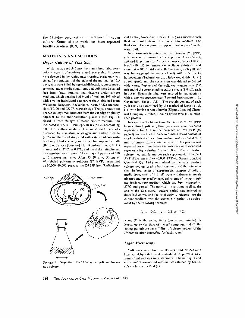
colony were brother-sister mated overnight. If sperm were detected in the vagina next morning, pregnancy was timed from midnight of the night of the mating. At 17.5 days, rats were killed by cervical dislocation, conceptuses removed under sterile conditions, and yolk sacs dissected free from fetus, amnion, and placenta under culture medium, which consisted of 9 vol of medium 199 mixed with ! vol of inactivated calf serum (both obtained from WeUcome Reagents, Beckenham, Kent, U.K.; prepara- tions TC 20 and CS 07, respectively). The yolk sacs were opened out by small incisions from the cut edge originally adjacent to the chorioallantoic placenta (see Fig. 1), rinsed in three changes of sterile culture medium, and incubated in sterile Erlenmeyer flasks (50 ml) containing 9.0 ml of culture medium. The air in each flask was displaced by a mixture of oxygen and carbon dioxide (95:5) and the vessel stoppered with a sterile silicone-rub- ber bung. Flasks were placed in a Unitemp water bath (Baird & Tatlock [London] Ltd., Romford, Essex, U.K.) maintained at 37.0 ~ • 0.2~ and the shaker attachment was regulated to a stroke of 3.4 cm at a frequency of 100 • 5 strokes per min. After 15-20 min, 30 /~g of 12Sl-labeled polyvinylpyrrolidone ([125]PVP; mean tool wt 30,000 40,000; preparation IM 33P from Radiochem-
AMNION ~ - -- PLACENTA
FIGURE 1 Dissection of a 17.5-day rat yolk sac for or- gan culture.
ical Centre, Amersham, Bucks., U.K.) was added to each flask as a solution in 1.0 ml of culture medium. The flasks were then regassed, stoppered, and replaced in the water bath.
In experiments to determine the uptake of [ '~q]PVP, yolk sacs were removed after a period of incubation, agitated three times for 2 min in changes of ice-cold 0.9% NaCI (30 ml) to remove extracellular substrate, and stored at - 2 0 ~ until assay. Before assay, each yolk sac was homogenized in water (2 ml) with a Virtis 45 homogenizer (Techmation Ltd., Edgware, Middx., U.K.) at top speed, and the suspension was diluted to 5.0 ml with water. Portions of the yolk sac homogenates (I.0 ml) and of the corresponding culture media ( 1.0 ml), each in a 3-ml disposable tube, were assayed for radioactivity with a gamma spectrometer (Packard Instruments Ltd., Caversham, Berks., U.K.). The protein content of each yolk sac was determined by the method of Lowry et al. (1 I) with bovine serum albumin (Sigma [London] Chem- ical Company Limited, London SWS; type I1) as refer- ence protein.
In experiments to measure the release of [~z~I]PVP from cultured yolk sac, three yolk sacs were incubated separately for 6 h in the presence of [~51]PVP (40 ~g/ml), and each was transferred into a 10-ml portion of sterile, substrate-free culture medium and incubated for 2 min to remove extracellular substrate. This process was repeated twice more before the yolk sacs were incubated separately for a further 6 h in 10.0 ml of substrate-free culture medium. In another such experiment, 1% wt/vol PVP of average mol wt 40,000 (PVP-40; Sigma [London] Chemical Co. Ltd.) was added to the substrate-free culture medium used in both the wash and the reincuba- tion. in both series of experiments, samples of culture media (two, each of 1.0 ml) were withdrawn in sterile pipettes and replaced by an equal volume of the appropri- ate fresh culture medium which had been warmed to 37~ and gassed. The activity in the tissue itself at the end of the 12-h overall culture period was assayed as described above, and the total activity released into the culture medium over the second 6-h period was calcu- lated by the following formula:
Tn = IOC,~,=, ,+ 2Z~=0. C,
where T~ is the radioactivity (counts per minute) re- leased up to the time of the n th sampling, and C~ the counts per minute per milliliter of culture medium of the i th sample after correcting for background.
Light Microscopy
Yolk sacs were fixed in Bouin's fluid or Zenker's fixative, dehydrated, and embedded in paraffin wax. Bouin-fixed sections were stained with hematoxylin and eosin, and Zenker-fixed material was stained by Mallo- ry's trichrome method (12).
114 THE JOURNAL OF CELL BIOLOGY �9 VOLUME 64, 1975
Dow
nloaded from http://rupress.org/jcb/article-pdf/64/1/113/1386949/113.pdf by guest on 01 April 2022
Electron Microscopy Small pieces of yolk sac were fixed at room tempera-
ture for 3 h in glataraldehyde solution (4% vol/vol), washed at 4~ for 16 h in sucrose solution (7% wt/vol), then postfixed for 1.0 h in osmium tetroxide (1% wt/vol); all three solutions were made up in the same buffer (sodium cacodylate-HCl, pH 7.3). Araldite-embedded ultrathin sections were stained with uranyl acetate and lead citrate and examined in a Siemens Elmiskop I.
RESULTS
Light Microscopy
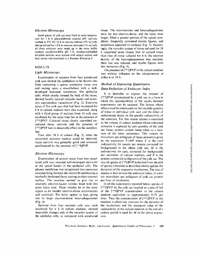
Examination of sections from four uncultured yolk sacs showed the epithelium to be thrown into folds containing a sparse connective tissue core and resting upon a mesothelium with a well- developed basement membrane. The epithelial cells, which clearly formed the bulk of the tissue, showed basally located vesicular nuclei and exten- sive supranuclear vacuolation (Fig. 2). Extensive areas of five yolk sacs that had been incubated for 6 h in culture medium were also examined, along with a third group of sections from 16 yolk sacs incubated for the same time but in the presence of [125I]PVP. Cultured tissue closely resembled un- cultured tissue controls and the presence of [~2sl]PVP had no detectable effect on the morphol- ogy.
Even after 24 h of culture (Fig. 3), when the occasional pycnotic nucleus could be observed, tissue survival was generally good and remained uninfluenced by the presence of [I~51]PVP.
Electron Microscopy
Examination of several areas from two uncul- tured yolk sacs revealed well-developed microvilli at the apical border of the epithelial cells. The plasma membrane was invaginated into numerous caveolae lying between the microvilli and bearing a markedly thickened fuzzy coating on their external surface. The caveolae seemed to give rise to relatively electron-lucent vesicles lined with this same fuzzy coat. These vesicles lie in the same region as do smaller electron-dense microvesicles and canaliculi. The latter appear to fuse, giving rise to large electron-dense heterophagosomes (Fig. 4).
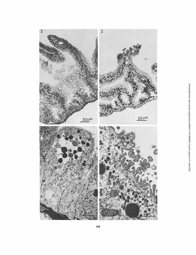
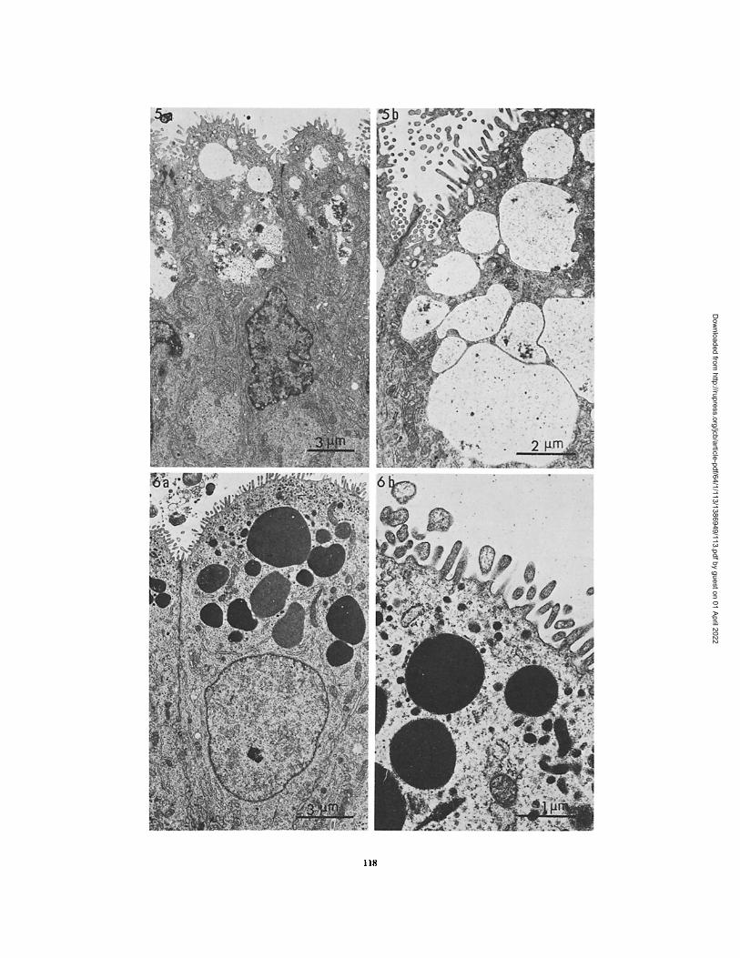
Sections from four separate yolk sacs, each incubated for 6 h in culture medium, showed detectable changes only in the vacuolar system of the epithelial cells, as compared with uncultured
tissue. The microvesicles and heterophagosomes were far less electron-dense, and the latter were larger, filled a greater portion of the apical cyto- plasm, frequently contained myelin figures, and sometimes appeared to coalesce (Fig. 5). Surpris- ingly, the vacuolar system of tissue cultured for 24 h resembled more closely that of control tissue than that of tissue cultured for 6 h; the electron density of the heterophagosomes was restored, their size was reduced, and myelin figures were less numerous (Fig. 6).
The presence of [t~I]PVP in the culture medium was without influence on the ultrastructure at either 6 or 24 h.
Method of Expressing Quantitative Data:Definition o f Endocytic Index
It is desirable to express the amount of [12~I]PVP accumulated by a yolk sac in a form in which the reproducibility of the results between experiments can be assessed. The factors whose effects must be eliminated are the variable quantity of tissue in individual yolk sacs and the effect of radioisotope decay on the specific radioactivity of the substrate. For this reason uptake is expressed as the volume of culture medium whose contained substrate is captured by unit quantity of the yolk sac tissue, protein content being taken as a mea- sure of the latter parameter. This volume (in microliters per milligram of tissue protein) is given by the expression Y/MP where Y is the total radioactivity (in counts per minute corrected for background) in the whole yolk sac, M is the radioactivity (in cpm, corrected for background) per microliter of culture medium, and P is the protein content (in milligrams) of the yolk sac. The rate of uptake of [I~5I]PVP is derived from the plot of uptake (obtained as described above) against the durations of the respective incubations. The rate of uptake is here termed the endocytic index; its units are microliters per milligram of yolk sac protein per hour of incubation.
In all the experiments reported below, uptake of [~251]PVP by the yolk sac resulted in a rate of fall of the ['~5I]PVP concentration in the culture medium equivalent to approximately 0.1% per hour. Thus the concentration of [~25I]PVP in the medium is effectively constant for the duration of the incubations and the measured value of the radioactivity in the culture medium at the end of a culture period is used for M in the above expres- sion.
WILLIAMS, KIDSTON, BECK, AND LLOYD Quantitative Studies ofPinocytosis. I 115
Dow
nloaded from http://rupress.org/jcb/article-pdf/64/1/113/1386949/113.pdf by guest on 01 April 2022
116
Dow
nloaded from http://rupress.org/jcb/article-pdf/64/1/113/1386949/113.pdf by guest on 01 April 2022

Quantitative Data on the Uptake o f
[ ~ 2 ~I ]Labeled Polyvinylpyrrolidone
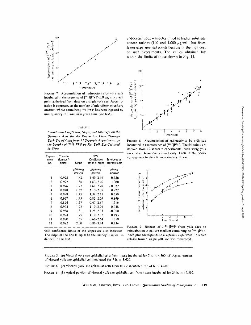
Fig. 7 shows the result of a typical experiment in which six yolk sacs from the same rat were incubated in culture medium containing 3 #g [12~I]PVP/ml, incubations being terminated at intervals up to 8 h. The rate of uptake is constant over this period. The results of 12 similar experi- ments were each subjected to a linear regression analysis and Table I indicates the correlation coefficient, the slope, and the intercept on the ordinate axis of the computer-fitted regression line through each set of experimental points. The slope of the regression line gives the endocytic index, i.e. the rate of uptake of [t~I]PVP from the culture medium by the tissue.
A series of statistical tests (13) was applied to the data of Table I to determine whether the variation between the 12 sets of data, each of which had been derived from observations on tissues from a single pregnant animal, was greater than the variation within each of the separate sets of data considered individually. The tests employed constituted an analysis of covariance in which variance ratios were calculated and used to deter- mine whether either the slope or the intercept from each of the 12 separate plots of accumulation against time possessed individually distinct values for these two parameters, or whether interexperi- mental variation was no greater than intraexperi- mental variation. The variance ratios calculated indicated that, at the 10% significance level, there was no detectable difference in values of the slopes and intercepts. Hence it was legitimate to pool the uptake data from the 12 experiments to give a single overall plot, from which a single value of both the slope and intercept could be derived, along with associated confidence limits.
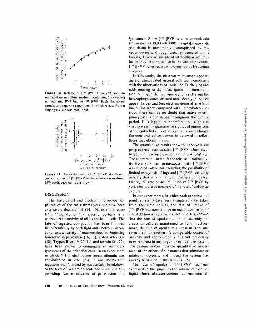
The regression line for the 64 experimental points (Fig. 8) of the 12 experiments had a slope of 1.71 ul/h/mg yolk sac protein (95% confidence limits 1.61, 1.83) and an intercept on the ordinate axis of 0.73 pA/mg yolk sac protein (95% confi- dence limits 0.26, 1.20).
Relationship o f Rate o f Accumulation o f
[125I]PVP to Rate of Its Endocytic Uptake
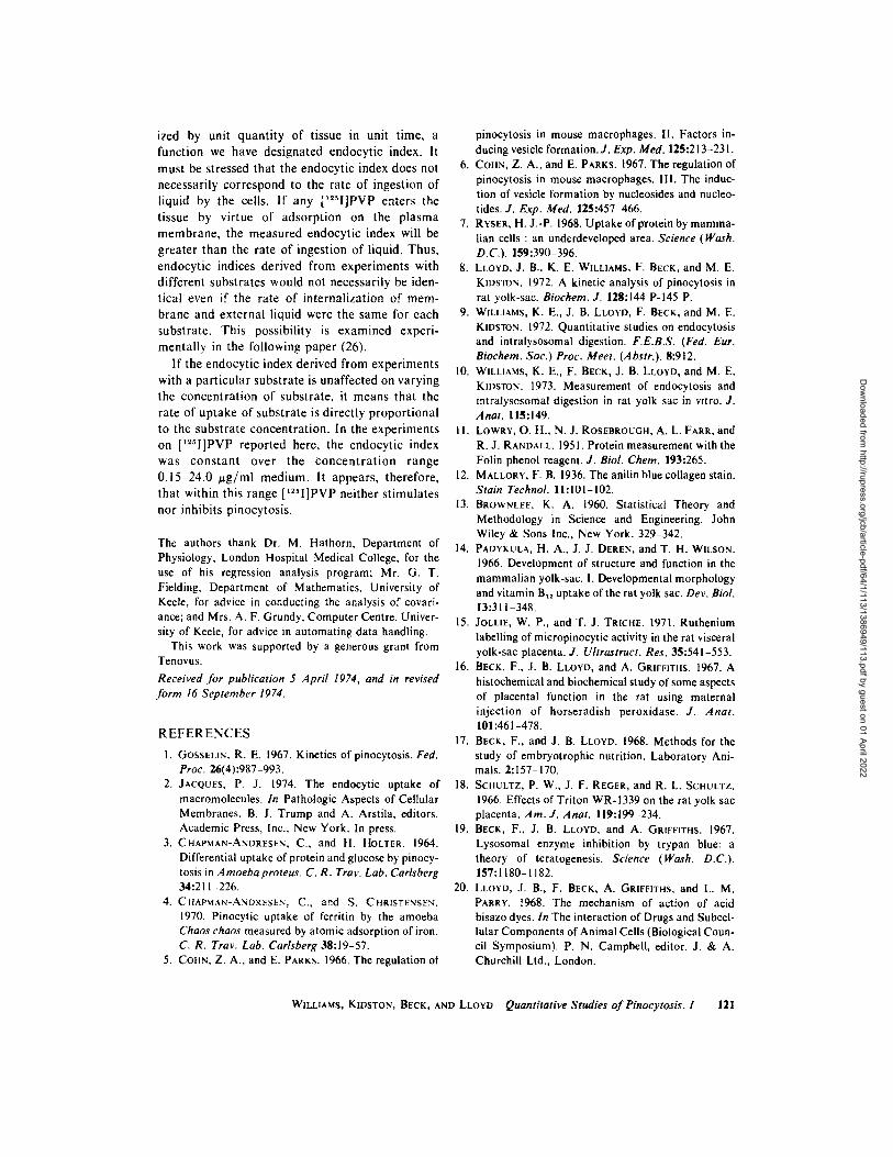
The rate of accumulation of [t25I]PVP as deter- mined in the above experiments will be equal to the rate of endocytic uptake only if captured [~25I]PVP is not released from the tissue. Fig. 9 shows the release of radioactivity from yolk sacs that had accumulated [~25I]PVP during a 6-h period in culture, been washed three times for 2 min in substrate-free culture medium, then been rein- cubated for 6 h in substrate-free culture medium. From 2 to 3% of the total radioactivity remaining in the tissue after the washing procedure was released within 30 rain and a further 2-3% was released progressively over the next 5.5 h. Even when a high concentration (1% wt/vol) of non- iodinated PVP was included in the reincubation culture medium (Fig. 10), the extent and pattern of release of radioactivity was essentially unchanged.
The 2 3% loss of radioactivity within 30 rain of reincubation is most easily explained as the wash- ing off of occluded or adsorbed extracellular radioactivity that remains after the standard washing procedure. This explanation is compati- ble with the observation that the regression line through the pooled data for uptake of [I~q]PVP (Fig. 8) has a small but positive intercept on the ordinate axis. The slow progressive release that followed must include the contributions from cell death or detachment, from exocytosis, and from further desorption, although it is unlikely that the latter is a significant component in view of the rate of release being unaffected by the presence of high concentrations of noniodinated PVP.
Relationship o f Endocytic Index to
[ 1 ~ ~ I ]P VP Concentration
Further experiments were performed in stan- dard culture medium but with different concentra- tions of [~51]PVP in the medium. At each sub- strate concentration, uptake proceeded at a con- stant rate up to 7 h, and Fig. 11 shows that the endocytic index is independent of substrate con- centration within the range 0.15-24 t~g/ml. The
FIGURE 2 Unincubated visceral layer of yolk sac from 17.5-day pregnant rat. • 230.
FIGURE 3 Visceral layer of yolk sac from 17.5-day pregnant rat incubated for 24 h. Occasional pycnotic nuclei may be seen. • 230.
FiGur~e 4 (a) Visceral yolk sac epithelial cell from unincubated tissue. • 3,050. (b) Apical portion of vis- ceral yolk sac epithelial cell from unincubated tissue. • 17,000.
WILLIAMS, KIDSTON, BECK, AND LLOYD Quantitative Studies ofPinocytosis. 1 117
Dow
nloaded from http://rupress.org/jcb/article-pdf/64/1/113/1386949/113.pdf by guest on 01 April 2022
118
Dow
nloaded from http://rupress.org/jcb/article-pdf/64/1/113/1386949/113.pdf by guest on 01 April 2022
t2- c
endocy t ic index was d e t e r m i n e d at h igher subs t ra te concen t r a t i ons (100 and 1,000 ~,g/ml) , but f rom
fewer expe r imen ta l poin ts because o f the high cost
o f such exper imen t s . The values ob ta ined lay within the l imits o f those shown in Fig. 1 I.
20
< E /
Time(hours) ~ E 15
FIGURE 7 Accumulation of radioactivity by yolk sacs o~
incubated in the presence of ['2~I]PVP (3.0 ug/ml). Each ~ point is derived from data on a single yolk sac. Accumu- ~ ~ io lation is expressed as the number of microliters of culture -~ ~' medium whose contained [1251]PVP has been ingested by = ~ unit quantity of tissue in a given time (see text). < - :L 5
TABLE I
Correlation Coefficient, Slope, and Intercept on the Ordinate Axis for the Regression Lines Through Each Set of Data from 12 Separate Experiments on the Uptake of['zs1]PVP by Rat Yolk Sac Cultured in Vitro
Experi- Correla- 95% ment tion coef- Confidence Intercept on no. ficient Slope limits of slope ordinate axis
~l/h/rng lal/h/mg td/mg protein protein protein
1 0.995 1.82 1.49-2.16 0.156 2 0.997 1.86 1.63-2.10 1.080 3 0.996 1.93 1.68-2.20 -0.072 4 0.978 1 .57 1.10-2.05 0.972 5 0.989 1 .75 1.38-2.11 0.359 6 0.957 1.43 0.82-2.05 0.849 7 0.894 1 .57 0.47-2.67 1.716 8 0.974 1 .73 1.19-2.29 0.748 9 0.988 1 .81 1.28-2.33 -0.010
10 0.994 1.75 1.19 2.32 0.193 1 I 0.980 1 .65 0.66-2.64 1.350 12 0.982 2.00 0.86-3.14 0.134
95% confidence limits of the slopes are also indicated. The slope of the line is equal to the endocytic index, as defined in the text.
FIGURE 8
', 2 3 4 ~ 6 7 i Time (min)
Accumulation of radioactivity by yolk sac incubated in the presence of ['~51]PVP. The 64 points are derived from 12 separate experiments, each using yolk sacs taken from one animal only. Each of the points corresponds to data from a single yolk sac.
6
o x3 E4
~ o3 ~E
~g
Time (hours)
FIGURE 9 Release of [I~I]PVP from yolk sacs on reincubation in culture medium containing no [1251]PVP. Each plot corresponds to a separate experiment in which release from a single yolk sac was monitored.
FIGURE 5 (a) Visceral yolk sac epithelial cells from tissue incubated for 7 h. x 4,500. (b) Apical portion of visceral yolk sac epithelial cell incubated for 7 h. • 8,620.
FIGURE 6 (a) Visceral yolk sac epithelial cells from tissue incubated for 24 h. 5< 4,600.
FIGURE 6 (b) Apical portion of visceral yolk sac epithelial cell from tissue incubated for 24 h. • 17,350.
WILLIAMS, KIDSTON, BECK, AND LLOYD Quantitative Studies ofPinocytosis. 1 119
Dow
nloaded from http://rupress.org/jcb/article-pdf/64/1/113/1386949/113.pdf by guest on 01 April 2022
% - m o 3
Z ~ z co
~ G
g I 2 5 a 5 Co
Time (hours)
FIGURE 10 Release of [~251]PVP from yolk sacs on reincubation in culture medium containing 1% (wt/vol) noniodinated PVP but no [x~sI]PVP. Each plot corre- sponds to a separate experiment in which release from a single yolk sac was monitored.
5 0 -
,1i t E c~ I0"
tJ _ E
5 IO 15 20 25 Concentration of [125I] PVP
in culture medium (~g per ml medium)
FIGURE I1 Endocytic index of [12H]PVP at different concentrations of [~2~I]PVP in the incubation medium. 95% confidence limits are shown.
DISCUSSION
The histological and electron microscope ap- pearances of the rat visceral yolk sac have been extensively documented (14, 15), and it is clear from these studies that micropinocytosis is a characteristic activity of all its epithelial cells. The fate of ingested compounds has been followed histochemically by both light and electron micros- copy, and a variety of macromolecules, including horeseradish peroxidase (16, 17), Triton WR-1339 (18), Trypan Blue (19, 20, 21), and ferritin (21, 22), have been shown to congregate in secondary lysosomes of the epithelial cells. In an experiment in which 1251-labeled bovine serum albumin was adminsitered in vivo (23), it was shown that ingestion was followed by intracellular breakdown to the level of free amino acids and small peptides, providing further evidence of penetration into
lysosomes. Since [t2sl]PVP is a macromolecule (mean mol wt 30,000-40,000), its uptake into yolk sac tissue is presumably accomplished by mi- cropinocytosis, although direct evidence of this is lacking. Likewise, the site of intracellular accumu- lation may be supposed to be the vacuolar system, [12H]PVP being resistant to digestion by lysosomal enzymes.
In this study, the electron microscope appear- ance of unincubated visceral yolk sac is consistent with the observations of Jollie and Triche (15) and adds nothing to their description and interpreta- tion. Although the micropinocytic vesicles and the heterophagosomes situated more deeply in the cell appear larger and less electron dense after 6 h of incubation when compared with unincubated con- trols, there can be no doubt that active micro- pinocytosis is continuing throughout the culture period. It is legitimate, therefore, to use this in vitro system for quantitative studies of pinocytosis of the epithelial cells of visceral yolk sac although the measured values cannot be assumed to reflect those that obtain in vivo.
The quantitative results show that the yolk sac progressively accumulates [t~H]PVP when incu- bated in culture medium containing this substrate. The experiments in which the release of radioactiv- ity from yolk sacs preincubated with [I~I]PVP was studied, while not excluding the possibility of limited exocytosis of ingested [~251]PVP, certainly indicate that it is of no quantitative significance. Hence, the rate of accumulation of [~zH]PVP by yolk sacs is a true measure of the rate of endocytic capture.
in our experiments, in which each experimental point represents data from a single yolk sac taken from the same animal, the rate of uptake of [~51]PVP was constant for an incubation period of 8 h. Additional experiments, not reported, showed that the rate of uptake did not measurably de- crease in cultures maintained to 12 h. Further- more, the rate of uptake was constant from one experiment to another. A comparable degree of linearity and reproducibility has not previously been reported in any organ or cell culture system. The system makes possible quantitative assess- ment of the effects of substances that stimulate or inhibit pinocytosis, and indeed the system has already been used in this way (24, 25).
The rate of uptake of [~I ]PVP has been expressed in this paper as the volume of external liquid whose substrate content has been internal-
1 2 0 T H E J O U R N A L OF C E L L BIOLOGY - V O L U M E 64, 1 9 7 5
Dow
nloaded from http://rupress.org/jcb/article-pdf/64/1/113/1386949/113.pdf by guest on 01 April 2022
ized by unit quant i ty of tissue in unit time, a function we have designated endocytic index. It
must be stressed that the endocytic index does not necessarily correspond to the rate of ingestion of liquid by the cells. If any [~2q]PVP enters the tissue by virtue of adsorpt ion on the plasma membrane , the measured endocytic index will be greater than the rate of ingestion of liquid. Thus, endocytic indices derived from exper iments with different substrates would not necessarily be iden- tical even if the rate of internal izat ion of mem- brane and external liquid were the same for each substrate. This possibility is examined experi- mental ly in the following paper (26).
If the endocytic index derived from experiments with a par t icular substrate is unaffected on varying the concentra t ion of substrate, it means tha t the rate of uptake of substrate is directly propor t ional to the substrate concentra t ion. In the exper iments on [ I ~ I ] P V P reported here, the endocytic index was c o n s t a n t over the c o n c e n t r a t i o n range 0.15 24.0 u g / m l medium. It appears, therefore, tha t within this range [12q]PVP nei ther s t imulates nor inhibits pinocytosis.
The authors thank Dr. M. Hathorn, Department of Physiology, London Hospital Medical College, for the use of his regression analysis program; Mr. G. T. Fielding, Department of Mathematics, University of Keele, for advice in conducting the analysis of covari- ance; and Mrs. A. F. Grundy, Computer Centre, Univer- sity of Keele, for advice in automating data handling.
This work was supported by a generous grant from Tenovus.
Received for publication 5 April 1974, and in revised form 16 September 1974.
R E F E R E N C E S
1. GOSSELIN, R. E. 1967. Kinetics of pinocytosis. Fed. Proc. 26(4):987-993.
2. JACQUES, P. J. 1974. The endocytic uptake of macromolecules. In Pathologic Aspects of Cellular Membranes. B. J. Trump and A. Arstila, editors. Academic Press, Inc., New York. In press.
3. CHAPMAN-ANDRESEN, C., and H. HOLTER. 1964. Differential uptake of protein and glucose by pinocy- tosis in Amoeba proteus. C. R. Tray. Lab. Carlsberg 34:211-226.
4. CHAPMAN-ANDRESEN, C., and S. CHRISTENSEN. 1970. Pinocytic uptake of ferritin by the amoeba Chaos chaos measured by atomic adsorption of iron. C. R. Trav. Lab. Carlsberg 38:19-57.
5. COHN, Z. A., and E. PARKS. 1966. The regulation of
pinocytosis in mouse macrophages. II. Factors in- ducing vesicle formation. J. Exp. Med. 125:213-231.
6. COHN, Z. A., and E. PARKS. 1967. The regulation of pinocytosis in mouse macrophages. II1. The induc- tion of vesicle formation by nucleosides and nucleo- tides. J. Exp. Med. 125:457 466.
7. RYSER, H. J.-P. 1968. Uptake of protein by mamma- lian cells : an underdeveloped area. Science (Wash. D.C.). 159:390-396.
8. LLOYD, J. B., K. E. WILLIAMS, F. BECK, and M. E. KIOSTON. 1972. A kinetic analysis of pinocytosis in rat yolk-sac. Biochem. J. 128:144 P-145 P.
9. WILLIAMS, K. E., J. B. LLOYO, F. BECK, and M. E. KIDSTON. 1972. Quantitative studies on endocytosis and intralysosomal digestion. F.E.B.S. (Fed. Eur. Biochem. Soc.) Proc. Meet. (Abstr.). 8:912.
10. WILLIAMS, K. E., F. BECK, J. B. LLOYD, and M. E. KIDSTON. 1973. Measurement of endocytosis and intralysosomal digestion in rat yolk sac in vitro. J. Anat. 115:149.
11. LOWRY, O. H., N. J. ROSEBROUGS, A. L. FARR, and R. J. RANDALL. 1951. Protein measurement with the Folin phenol reagent. J. Biol. Chem. 193:265.
12. MALLORY, F. B. 1936. The anilin blue collagen stain. Stain Technol. 11:101-102.
13. BROWNLEE, K. A. 1960. Statistical Theory and Methodology in Science and Engineering. John Wiley & Sons Inc., New York. 329-342.
14. PADYKULA, H. A., J. J. DEREN, and T. H. WILSON. 1966. Development of structure and function in the mammalian yolk-sac. I. Developmental morphology and vitamin Blz uptake of the rat yolk sac. Dev. Biol. 13:311 348.
15, JOLLIE, W. P., and T. J. TRICHE. 1971. Ruthenium labelling of micropinocytic activity in the rat visceral yolk-sac placenta. J. Ultrastruct. Res. 35:541-553.
16. BECK, F., J. B. LLOYD, and A. GRIEFITHS. 1967. A histochemical and biochemical study of some aspects of placental function in the rat using maternal injection of horseradish peroxidase. J. Anat. 101:461-478.
17. BECK, F., and J. B. LLOYD. 1968. Methods for the study of embryotrophic nutrition. Laboratory Ani- mals. 2:157-170.
18. SCHULTZ, P. W., J. F. REGER, and R. L. SCHULTZ. 1966. Effects of Triton WR- 1339 on the rat yolk sac placenta. Am. J. Anat. 119:199-234.
19. BECK, F., J. B. LLOYD, and A. GRIrFITnS. 1967. Lysosomal enzyme inhibition by trypan blue: a theory of teratogenesis. Science (Wash. D.C.). 157:1180-1182.
20. LLOYD, J. B., F. BECK, A. GRIFFITHS, and L. M. PARRY. 1968. The mechanism of action of acid bisazo dyes. In The interaction of Drugs and Subcel- lular Components of Animal Cells (Biological Coun- cil Symposium). P. N. Campbell, editor. J. & A. Churchill Ltd., London.
WILLIAMS, KIDSTON, BECK, AND LLOYD Quantitative Studies ofPinocytosis. 1 121
Dow
nloaded from http://rupress.org/jcb/article-pdf/64/1/113/1386949/113.pdf by guest on 01 April 2022

21. KRZYZOWSgA-GaucA, S., and T. H. SCHIEBLER. 1967. Experimentelle Untersuchungen am Dotter- sackepithel der Ratte. Z. Zellforsch. Mikrosk. Anat. 79:157-171.
22. LAMBSON, R. O. 1966. An electron microscopic visualization of transport across rat visceral yolk- sac. A m. J. Anat. 115:21-30.
23. WILLIAMS, K. E., J. B. LLOYD, M. DAVIES, and F. BECg. 1971. Digestion of an exogenous protein by rat yolk-sac cultured in vitro. Biochem. J. 125:303-308.
24. LLOYD, J. B., M. DAVIES, K. E. WILLIAMS, and F. BECK. 1971. Protein digestion within lysosomes and
25.
26.
the effect of endocytosed inhibitors. In Tissue Pro- teinases. A. J. Barrett and J. T. Dingle, editors. North-Holland Publishing Co., Amsterdam. 255-262. WILLIAMS, K. E , J. B. LLOYD, M. E. KtDSTON, and F. BECg. 1973. Biphasic effect of trypan blue on pinocytosis. Biochem. Soc. Trans. 1:203 206. WILLIAMS, K. E., E. M. KIDSTON, F. BECK, and J. B. LLOYD. 1975. Quantitative studies on pino- cytosis. 1I. Kinetics of protein uptake and digestion by rat yolk-sac cultured in vitro. J. Cell Biol. 64: 123-134.
122 THE JOURNAL OF CELL BIOLOGY - VOLUME 64, 1975
Dow
nloaded from http://rupress.org/jcb/article-pdf/64/1/113/1386949/113.pdf by guest on 01 April 2022