quantitative peptidomics study reveals that a wound ... · pdf filequantitative peptidomics...
TRANSCRIPT

Quantitative Peptidomics Study Reveals That aWound-Induced Peptide from PR-1 RegulatesImmune Signaling in TomatoW OPEN
Ying-Lan Chen,a,b Chi-Ying Lee,a Kai-Tan Cheng,a Wei-Hung Chang,a Rong-Nan Huang,c Hong Gil Nam,d
and Yet-Ran Chena,e,1
a Agricultural Biotechnology Research Center, Academia Sinica, Taipei 11529, TaiwanbDepartment of Life Science and Institute of Plant Biology, National Taiwan University, Taipei 10617, TaiwancDepartment of Entomology and Research Center for Plant Medicine, National Taiwan University, Taipei 10617, TaiwandDepartment of New Biology, Daegu Gyeongbuk Institute of Science and Technology, Daegu 711-873, Republic of KoreaeDepartment of Bioscience and Biotechnology, National Taiwan Ocean University, Keelung 20224, Taiwan
ORCID ID: 0000-0002-2409-6655 (Y.-R.C.)
Many important cell-to-cell communication events in multicellular organisms are mediated by peptides, but only a few peptideshave been identified in plants. In an attempt to address the difficulties in identifying plant signaling peptides, we developed a novelpeptidomics approach and used this approach to discover defense signaling peptides in plants. In addition to the canonicalpeptide systemin, several novel peptides were confidently identified in tomato (Solanum lycopersicum) and quantified to beinduced by both wounding and methyl jasmonate (MeJA). A wounding or wounding plus MeJA-induced peptide derived from thepathogenesis-related protein 1 (PR-1) family was found to induce significant antipathogen and minor antiherbivore responses intomato. This study highlights a role for PR-1 in immune signaling and suggests the potential application of plant endogenouspeptides in efforts to defeat biological threats in crop production. As PR-1 is highly conserved across many organisms and theputative peptide from At-PR1 was also found to be bioactive in Arabidopsis thaliana, our results suggest that this peptide may beuseful for enhancing resistance to stress in other plant species.
INTRODUCTION
All multicellular organisms have evolved mechanisms to perceiveand respond to extracellular chemical signals. External signalsinclude endogenous hormones and cues from the environment,pathogens, and symbiotic organisms. In animal systems, inter-cellular communications are mostly mediated by steroids, pep-tides, and other small compounds (Ryan et al., 2002). Amongthem, peptides are the most common mediators of cell-to-cellinteractions in animals because they provide great variety in theirsequences, lengths, and/or posttranslational modifications to rep-resent different physiological responses (Boller, 2005). In contrast topeptide discovery in animals, only a few signaling peptides havebeen identified in plants (Farrokhi et al., 2008a; Butenko et al.,2009). The completion of the Arabidopsis thaliana genome hasrevealed that plants have up to 10 times as many predictedpeptide transporters (Initiative, 2000) and receptors (Shiu andBleecker, 2003) as animals. Therefore, it is expected that mostof the endogenous plant signaling peptides that play prominentroles in cell-to-cell communication are still undiscovered. Amongthe currently identified peptides in plants, only relatively few have
been found to function in defense signaling. This may be becausedefense signaling peptides are mostly derived from the selectiveaction of proteases on larger precursor proteins, are expressed atlow levels, and are highly dynamic. The identification of defensesignaling peptides in plants has the potential not only to advanceplant stress biology, but also to aid in the development of alter-native ways to improve stress tolerance or resistance for bettercrop productivity and minimization of the use of agrochemicals(Pearce et al., 1991, 2001a; Huffaker et al., 2006). So far, defensesignaling peptides have been discovered mainly by bioassay-guided screening. However, this approach requires large amountsof plant tissue and complicated purification of bioactive peptides(Pearce et al., 1991, 2001a). Furthermore, the detection of thesignaling peptides can be limited by the selection of bioassay.One type of stress may produce several defense and physio-logical responses, which may be regulated by different sig-naling peptides. Therefore, the use of a single bioassay maynot be sufficient to identify all the defense signaling peptidesinduced by a stress.Peptidomics approaches can provide comprehensive and un-
biased detection of transient amounts of signaling peptide andeven identify their modification status (Fricker et al., 2006). In an-imal tissues, peptidomics approaches have been used to eluci-date novel bioactive peptides and their functions (Svensson et al.,2003; Che et al., 2005; Sasaki et al., 2009; Tinoco and Saghatelian,2011). However, very few successful cases of discovery of plantsignaling peptides have been reported. This may be due to thehigh complexity and low abundance of plant signaling peptides.More sensitive and reliable peptidomics approaches are required
1Address correspondence to [email protected] author responsible for distribution of materials integral to the findingspresented in this article in accordance with the policy described in theInstructions for Authors (www.plantcell.org) is: Yet-Ran Chen ([email protected]).W Online version contains Web-only data.OPENArticles can be viewed online without a subscription.www.plantcell.org/cgi/doi/10.1105/tpc.114.131185
This article is a Plant Cell Advance Online Publication. The date of its first appearance online is the official date of publication. The article has been
edited and the authors have corrected proofs, but minor changes could be made before the final version is published. Posting this version online
reduces the time to publication by several weeks.
The Plant Cell Preview, www.aspb.org ã 2014 American Society of Plant Biologists. All rights reserved. 1 of 14

(Murphy et al., 2012). Recent developments in mass spectrometry(MS) have significantly improved the accuracy and sensitivity ofpeptide detection. MS is routinely applied to proteomics analysisthat analyzes the total peptides generated from the digestion ofcomplex protein mixtures using tandem mass spectrometry (MS/MS) (Mallick and Kuster, 2010). A reliable protein identification re-sult can be obtained by matching peptide MS/MS spectra totheoretical fragment spectra (Aebersold and Mann, 2003). Un-fortunately, proteomics approaches cannot be directly applied tothe detection of signaling peptides because it is difficult to createan endogenous peptide library for MS/MS spectra matching, ashow plants utilize proteases to produce peptides from proproteinsis still unclear (Yamaguchi and Huffaker, 2011). Identification ofendogenous peptides using a protein database and consider-ation of all the possible peptide cleavages during MS/MSspectra matching would generate large quantities of false pos-itive hits (Kapp et al., 2005). Therefore, more stringent matchingcriteria are required to accept significant peptide hits to reducethe detection sensitivity (Ding et al., 2008).
In this study, an MS-based peptidomics approach using ahypothetical peptide database combining a target-decoy searchstrategy and differential database match scoring was devel-oped to discover defense signaling peptides. This platform wasdemonstrated by the identification of defense peptides inducedby wounding plus methyl jasmonate (MeJA) treatment in tomato(Solanum lycopersicum). It is already known that tomato woundingcan induce an antiherbivore response, which is regulated by thepeptide hormone systemin, and the small molecule hormonejasmonic acid (JA) and its methyl ester, MeJA (Pearce et al., 1991;Orozco-Cardenas et al., 2001). Antipathogen responses are acti-vated in plants through signals known as damage-associatedmolecular patterns (DAMPs) (Huffaker et al., 2006; Boller and Felix,2009a; Malinovsky et al., 2014). DAMPs are endogenous mole-cules produced by the wounding or damaging of the tissue (whichcan occur through pathogen or herbivore attack). Systemin wasthe first identified signaling peptide and also the first confirmedpeptide elicitor of DAMPs in plants. It is expected that severalsignaling peptides are involved in combating herbivore and path-ogen attack (Cheong et al., 2002; Francia et al., 2007; Chassotet al., 2008), but the details of the regulation of antiherbivore andantipathogen responses by peptides during wounding stress stillawait elucidation. Several DAMP peptides have been discovered inother plant species and suggested to be bioactive in tomato (Bollerand Felix, 2009b; Campos et al., 2014); these include HypSys(Pearce et al., 2001a; Narvaez-Vasquez et al., 2007), RALF (Pearceet al., 2001b), and Pep1 (Huffaker et al., 2006; Trivilin et al., 2014).Pep1 was clearly identified to be pathogen related in Arabidopsis,and its putative precursor in tomato was recently found to involvein the antipathogen response (Trivilin et al., 2014). However, itsendogenous level in tomato has not yet been proved to be in-duced by tissue damage or MeJA, a potent inducer of systemicwound signaling and response in tomato (Scheer and Ryan, 1999).To our knowledge, no study to date has quantitatively profiled theglobal change in cellular peptide expression in plants beforeand/or after the induction of stress responses. Using this platform,several peptides including systemin were identified and quantifiedto be wounding plus MeJA induced. One of the peptides inducedby wounding only or wounding plus MeJA was found to activate
immune signals for defense against biological threats. The setup ofthis platform and functional studies of this peptide are describedhere.
RESULTS
Platform for the Discovery of Defense-Related Peptides
A MS-based platform using a hypothetical database was de-veloped to discover peptides involved in defense that are inducedby stress, as outlined in Figure 1. Global endogenous peptideswere profiled and compared before and after stress induction. Theendogenous peptide mixtures from unstressed and stressedplants were extracted and fractionated. Each of the fractions wasanalyzed by nanoflow ultrahigh performance liquid chromato-graphy mass spectrometry (nanoUHPLC-MS) using the data-dependent acquisition (DDA) mode. The acquired MS/MS spectrawere processed by UniQua (Chang et al., 2013) and searchedagainst a peptide database using Mascot MS/MS ion search. Toidentify the endogenous peptides with higher sensitivity, it is im-portant to reduce the false positive rate in MS/MS ion searching.This is because there is no specific prediction method for thegeneration of endogenous peptides from the precursor proteinsby plants. Therefore, the identification of endogenous peptidesignals using MS/MS ion search requires considering all possiblecleavage events. Although the protein database of tomato is notlarge, considering that all subsequences can be degraded fromthe proteins without any specificity would generate several ordersof magnitude more candidate sequences to match the MS/MSspectra, in comparison with identification of peptides with specificprotease cleavage sites (Zhou et al., 1999; Svensson et al., 2003;Farrokhi et al., 2008a). The large number of candidate sequencesfor peptide MS/MS ion matching would generate more falsepositive hits and increase the matching score to accept confidentpeptide hits. Therefore, in this study, a hypothetical peptide data-base (HT database) using partial protein sequences was used asthe target database for the peptide identification. To date, mostdefense-related peptides have been identified to be derived fromthe C-terminal end of proproteins (Farrokhi et al., 2008b). There-fore, the HT database was composed of 50 amino acid sequencesfrom the C-terminal end of protein sequences in the proteindatabase. To evaluate the criteria for positive peptide identi-fication, the target-decoy database search strategy was ap-plied (Elias and Gygi, 2007). The randomized hypotheticalpeptide database (RanHT database) was used as the decoydatabase.The changes in abundance level of each identified peptide be-
fore and after induction of stress were quantified by the extractedion chromatogram (XIC) peak area of the peptide precursor ion. Ifone peptide was detected in several gel filtration fractions, all ofthe XIC peak areas were summarized and compared. To normalizethe sample recovery during the sample preparation and columntrapping efficiency, the relative peptide abundances in stressedand unstressed samples were normalized by the signal ratio ofdoped internal control peptides from stressed and unstressedsamples. In addition, when peptides are fractionated before per-forming nanoUHPLC-MS, quantitation accuracy can be impaired
2 of 14 The Plant Cell

by the retention reproducibility during peptide fractionation. Toconfirm that the observed peptide was responding to stress, ananoUHPLC-MS operated in selected reaction monitoring (SRM)was used to quantify the peptide of interest in a more sensitive andreliable way without prefractionation of the extracted peptides.
Identification of Wounding- plus MeJA-Induced Peptides inTomato Leaves
To discover the defense peptides in tomato that are induced bywounding, the endogenous peptides extracted from tomatoplants with or without wounding plus MeJA treatment were pro-filed and compared. To assign significant peptide hits, the distri-bution of Mascot andMascot Delta (MD) scores using randomizedtomato protein (RanTom) and hypothetical (RanTomHT) data-bases were used as the model for the null hypothesis. As shown inFigure 2A, the Mascot scores for P value < 0.05 using RanTomand RanTomHT databases were >49 and >38, respectively. Asshown in Figure 2B, the MD score was >21 with P value < 0.01using RanTom and RanTomHT databases. To obtain confidentpeptide identification results, in this study, Mascot score > 38 andMD score > 21 were considered as true positive hits using theTomHT database. The Mascot and MD score distribution for all ofthe identified peptides in unwounded and wounding plus MeJA-treated tomato is illustrated in Figure 2C. In this analysis, a total of46 unique peptides derived from 25 proproteins were identifiedand quantified in tomato leaves using the TomHT database(Supplemental Table 1). However, only 25 unique peptides were
identified with the use of the Tom database because the Mascotscore for P < 0.05 and MD score for P < 0.01 were >49 and >21,respectively.Quantitative analysis revealed 14 novel peptides and a known
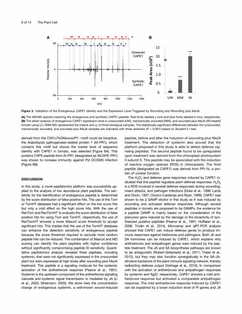
peptide (systemin) with expression levels more than 2-fold upre-gulated after wounding plus MeJA treatment (Table 1). Systeminshowed no significant expression in the unwounded plant but washighly expressed after wounding plus MeJA treatment. One novelpeptide derived from PATHOGENESIS-RELATED PROTEIN1b(PR-1b) showed a similar expression response to systemin. SincePR-1b is classified as a member of the cysteine-rich secretoryproteins, antigen 5, and pathogenesis-related 1 proteins (CAP)superfamily (Gibbs et al., 2008), this peptide was designated asCAP-derived peptide 1 (CAPE1). In CAPE1 identification, with theexception of the y1 and b1 ions, most of the y and b fragment ionswere matched to the theoretical fragments of the CAPE1 sequence,as shown in Figure 3A. Using this approach, the tissue quantityused for global peptide identification was <150 g. To confirm thematched sequences, synthetic CAPE1 was analyzed by MS/MSand the resulting spectrum was totally matched to the endogenousCAPE1 (Figure 3A). Without peptide prefractionation, total peptidesextracted from the unwounded, mechanically wounded, andwounded plus MeJA-treated tomato plants were directly analyzedby nanoUHPLC-SRM-MS targeted on the specific CAPE1 colli-sional induced dissociation reaction (Supplemental Figure 1). Thequantitation result showed that CAPE1 was expressed in low levelin unwounded plants but significantly induced after wounding orwounding plus MeJA treatments (Figure 3B).
Figure 1. Hypothetical Database-Assisted Peptidomics Platform for the Discovery of Stress-Induced Peptides.
Arrows show experimental priority for endogenous peptide extraction and analysis, and dashed arrows show the SRM method for target peptidequantitation.
A Wound-Induced Peptide from PR-1 3 of 14

Bioactivity of CAPE1
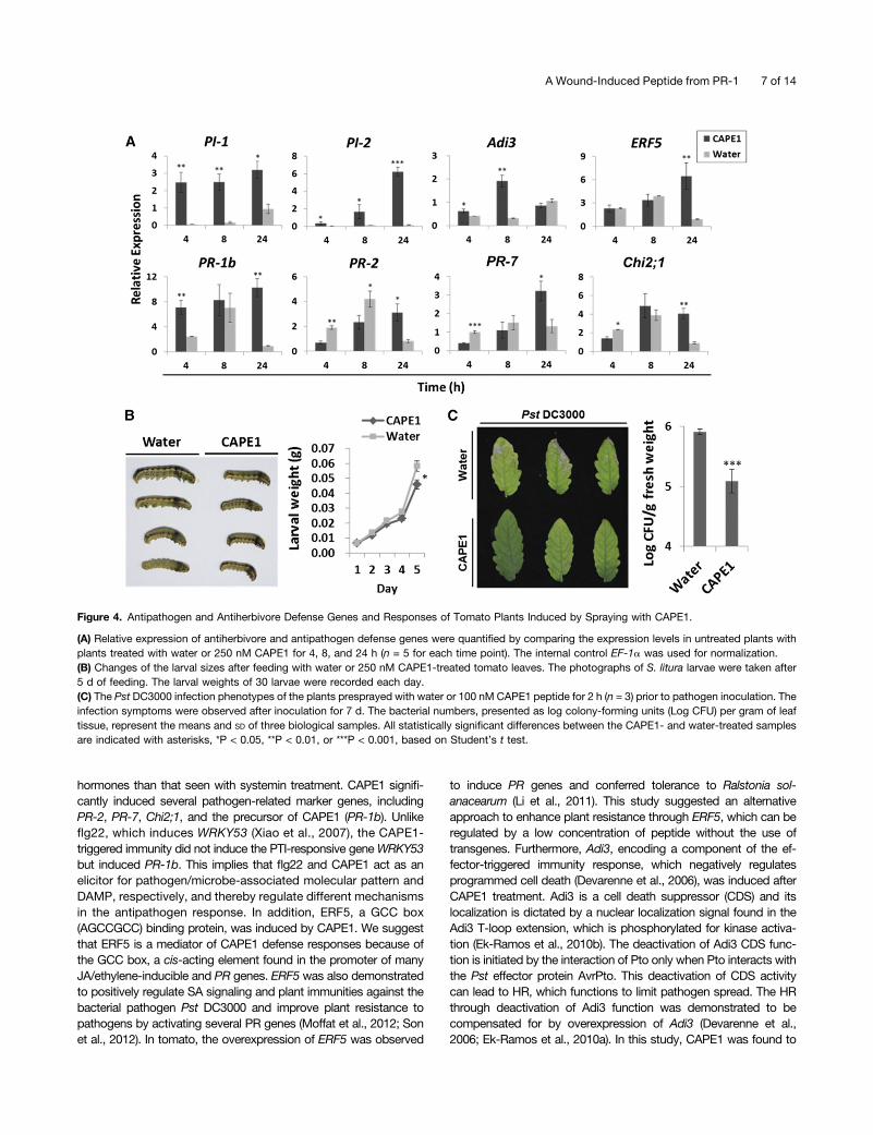
CAPE1 treatment induces H2O2 formation in tomato leaves asdetected by 3,3-diaminobenzidine (DAB) staining (SupplementalFigure 2). The profiles of induced genes obtained by microarrayanalysis (Supplemental Data Set 1) suggest that CAPE1 elevatesthe expression of several genes known to be involved in theantiherbivore and antipathogen defense response. CAPE1 mainlyinduced genes involved in the stress response, defense response,innate immune response, bacterial defense, and systemic ac-quired resistance (Supplemental Figure 3). Quantitative RT-PCR(qRT-PCR) analysis further confirmed that the antiherbivore genesPROTEINASE INHIBITOR1 (PI-1) and PI-2 and pathogen-relatedgenes PR-1b (CAPE1 precursor gene), BETA-1,3-GLUCANASE(PR-2), CYS PROTEASE (PR-7), CLASS II CHITINASE (Chi2;1),ETHYLENE RESPONSE FACTOR5 (ERF5), and AvrPto-DEPENDENTPto-INTERACTING PROTEIN3 (Adi3) were activated after CAPE1treatment (Figure 4a).
The antiherbivore response was evaluated by average larvalweights of 30 Spodoptera litura larvae fed with tomato leavespretreated with water or CAPE1. Tomato plants presprayed withCAPE1 suppressed larval growth and reduced larval weight by;20% (Figure 4B). To demonstrate that plant resistance canalso be enhanced by CAPE1, two groups of tomato plants werepresprayed with water or synthetic CAPE1 for 2 h. After the
treatment, the two plant groups underwent challenge with thepathogen Pseudomonas syringae pv tomato DC3000 (Pst DC3000).As shown in Figure 4C, the water pretreated tomato plants showedsevere pathogen infection symptoms. However, the plants pre-treated with CAPE1 showed no significant symptoms of infection orhypersensitive response (HR) (Heath, 2000) after the pathogenchallenge. Although the HR was not induced to limit pathogenspread, CAPE1 was still able to reduce the bacterial population inthe plant through activating a defense response (Figure 4C).To compare the antiherbivore response of systemin with
CAPE1, excised tomato plants were used, since the excised planttreated with peptide solution has been used previously to test thebioactivity of systemin (Schaller et al., 1995; Howe et al., 1996). Acomparison of systemin and CAPE1 showed that with CAPE1,a higher PI-1 and PI-2 expression level was induced after 1 h oftreatment, but expression decreased rapidly after 2 h of treatment(Figure 5A). With systemin, PI-1 was induced after 2 h and PI-2was induced after 1 h. Both PI-1 and PI-2 were observed to beinduced by systemin at 2 h and the effect lasted for more than 4 h(Figure 5A). JA and JA-IIe were observed to be significantly in-duced by treatment for 2 h with systemin but was induced after4 h treatment with CAPE1, and the expression level was ;4-foldlower than that of systemin (Figure 5B). As the results suggestedthat CAPE1 may be a novel DAMP signal for the induction ofimmunity to pathogenesis, next, CAPE1 was compared with the
Figure 2. Score Distribution of the Peptides Identified in Unwounded and Wounded plus MeJA-Treated Tomato.
(A) and (B) The Mascot (A) and MD (B) score distribution using a target-decoy search based on tomato (Tom) and randomized tomato (RanTom) proteindatabase or tomato hypothetical (TomHT) and randomized tomato hypothetical (RanTomHT) peptide database, respectively. W, wounded; UW, un-wounded.(C)Mascot versus MD score distribution using a target-decoy search based on TomHT and RanTomHT database, respectively. The red circles and bluedots indicated the peptide hits from TomHT and RanTomHT, respectively.
4 of 14 The Plant Cell

canonical pathogen/microbe-associated molecular pattern pep-tide flg22 (Hayashi et al., 2001). As shown in Figure 6A, bothCAPE1 and flg22 significantly induced salicylic acid (SA) whensupplied to excised plants. In Figure 6B, the flg22 highly inducedWRKY TRANSCRIPTION FACTOR53 (WRKY53) expression butnot PR-1b in tomato. This result was consistent with the publicRNA-seq data in the Tomato Functional Genomics Database (Feiet al., 2011), which are based on the experiment “transcriptomesequencing of tomato leaves treated with different bacteria andPAMPs” (Rosli et al., 2013). The RNA-seq data showed thatWRKY53 could be induced but PR-1b, Adi3, and ERF5, but couldnot be significantly induced using 30 min or 6 h treatment of flg22on the tomato. However, the CAPE1 did not induce WRKY53 buthighly induced its precursor gene PR-1b. Spraying of plants for2 h with either CAPE1 or flg22 resulted in plant resistance to PstDC3000 infection (Figure 6C).
CAPE1 Proprotein
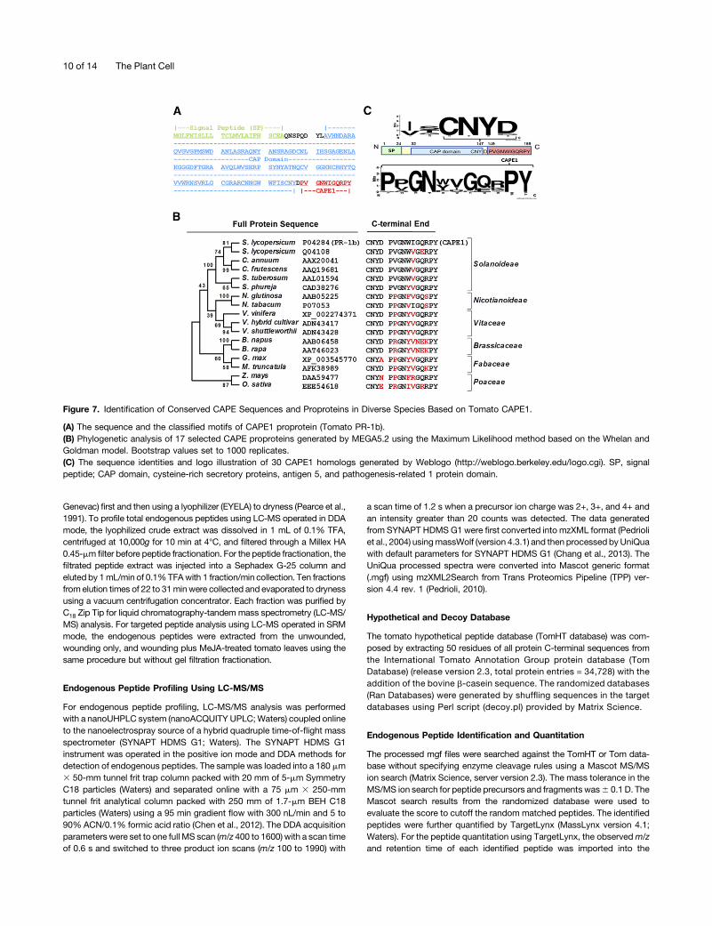
The mature CAPE1 peptide is derived from the C-terminal end oftomato PR-1b. This proprotein consists of an N-terminal signalpeptide, a CAP domain, and an extended C-terminal end (Figure7A). The phylogenetic analysis using Molecular EvolutionaryGenetics Analysis version 5.2 (MEGA5.2)(Tamura et al., 2011)and C-terminal alignment of the PR-1b protein demonstrated thatthe full protein and extended C-terminal end are highly conservedacross different flowering plants ranging from monocots to dicots(Figure 7B; Supplemental Data Set 2). It is interesting that thePxGNxxxxxPY- motif was conserved in the CAPE1 sequence andthat the three residue sequences before the cleavage site hada conserved CNYx motif (Figure 7C). This suggests that CNYx.PxGNxxxxxPY- could be a functional motif that may mark bio-active peptides in other species. To demonstrate that a peptide
Table 1. Endogenous Peptides Induced by Wounding plus MeJA Treatment
Proprotein Information Peptide IdentificationPeptide Ratio(W/UW)a
AccessionNumber Description Peptide Sequence MD Score
MASCOTScore
Observedm/z Z
ObservedMW
Theor.MW
MassError
ScoringIons RT (min) Ratio
Norm.Ratio
InternalStandard
b-Casein F.LLYQEPVLGPVR.G 36.84;61.66 51.47;78.69 692.42 2 1382.83 1382.79 60.08 6;10 38.75;40.20 1.63 1.00
Solyc05g051750.2.1
Prosystemin L.AVQSKPPSKRDPPKMQTD.N(systemin)
21.35 38.18 1005.54 2 2009.07 2009.04 0.04 18 21.52 9999b 9999b
Solyc05g025600.1.1
Chloroplastphotosystem IIsubunit X
G.VSNFDPVKR.T 21.71 40.69 531.28 2 1060.55 1060.57 20.01 5 26.16 9999b 9999b
Solyc00g174340.1.1
Pathogenesis-relatedprotein 1b
D.PVGNWIGQRPY.-(CAPE1)
41.91 53.37 643.86 2 1285.7 1285.66 0.05 8 36.84 9999b 9999b
Solyc07g044860.2.1
Oxygen-evolvingenhancerprotein 2,chloroplastic
K.KFVENAATSFSI.A 39.23 49.38 657.35 2 1312.69 1312.67 0.03 8 40.20 111.62 68.48
Solyc01g007350.2.1
Photosystem Ireactioncentersubunit VIII
M.ASLFLHVQKNK.I 38.52 56.85 642.88 2 1283.75 1283.73 0.01 7 29.04 19.08 11.71
Solyc12g035280.1.1
Photosystem IICP47chlorophyllapoprotein
K.LGDPTTKRQAA.- 26.31 45.66 579.33 2 1156.65 1156.62 0.03 5 21.47 7.70 4.72
Solyc02g038690.1.1
Histone H2B E.IQTAVRLVLPGE.L 29.20 38.23 648.41 2 1294.8 1294.76 0.04 6 41.81 5.35 3.28
Solyc11g071640.1.1
b-D-Glucosidase D.SHNDPLFHFGFGLTTKPVK.A
45.86 46.63 714.73 3 2141.16 2141.11 0.05 14 41.75 4.43 2.72
Solyc01g104170.2.1
Ankyrin repeatdomain-containingprotein 2
Q.DVLKLLEKDAFL.- 21.47;32.00 38.68;42.18 702.42 2 1402.82 1402.81 0.01 8 50.47 4.04 2.48
Solyc06g063370.2.1
Chlorophyll a/bbindingprotein 1A,chloroplastic
L.LTVIGGASERVPT.L 22.63;62.90 41.92;86.53 650.39 2 1298.77 1298.72 60.05 6;10 33.38;35.15 3.73 2.29
Solyc01g087520.2.1
Ferredoxin-thioredoxinreductasevariable chain
L.EGRSTPVKFSAHLKED.E
27.28 39.74 600.97 3 1799.9 1799.92 20.02 8 26.58 29.71 18.23
L.EGRSTPVKFSAHLKEDE.F
24.72;59.75 37.34;67.73 644.01 3 1929.01 1928.96 60.05 8;9 26.67;27.01 6.23 3.82
Solyc09g063130.2.1
Photosystem Ireactioncentersubunit IV A
R.FNKVNYANVSTNNYALDEVEEVK.-
31.18;85.88 40.12;91.04 887.46 3 2659.37 2659.28 60.09 8;20 38.14;38.75 20.97 12.87
D.PKTRYPVVVR.F 20.95;27.72 37.64;46.28 607.86 2 1213.71 1213.73 20.02 6 25.38 9.90 6.07F.NKVNYANVSTNNY
ALDEVE.E56.12;79.63 57.96;86.34 1079.02 2 2156.03 2156 60.02 12;14 36.34;36.41 5.70 3.50
aRatio of the peptide intensity observed in unwounded (UW) and wounding plus MeJA (W) treated plants.bThe peptide was only detected in wounding plus MeJA-treated plants (S/n < 10 in unwounded sample).
A Wound-Induced Peptide from PR-1 5 of 14
derived from the CNYx.PxGNxxxxxPY- motif could be bioactive,the Arabidopsis pathogenesis-related protein 1 (At-PR1), whichcontains this motif but shows the lowest level of sequenceidentity with CAPE1 in tomato, was selected (Figure 8A). Thisputative CAPE peptide from At-PR1 (designated as AtCAPE-PR1)was shown to increase immunity against Pst DC3000 infection(Figure 8B).
DISCUSSION
In this study, a novel peptidomics platform was successfully ap-plied to the analysis of low abundance plant peptides. The sen-sitivity for the identification of endogenous peptide is determinedby the score distribution of false positive hits. The use of the Tomor TomHT database had a significant effect on the low score hitsbut only a mild effect on the high score hits. With the use ofRanTom and RanTomHT to evaluate the score distribution of falsepositive hits for using Tom and TomHT, respectively, the use ofRanTomHT showed a lower Mascot score threshold to acceptsignificant hits. This implies that the use of the TomHT databasecan enhance the detection sensitivity of endogenous peptidebecause the score threshold required to exclude most randompeptide hits can be reduced. The combination of Mascot and MDscoring can identify the plant peptides with higher confidencewithout significantly compromising peptide ID sensitivity. Quanti-tative peptidomics analysis revealed three peptides, includingsystemin, that were not significantly expressed in the unwoundedplant but were expressed at high levels after wounding plus MeJAtreatment. This peptide is a signaling molecule for the systemicactivation of the antiherbivore response (Pearce et al., 1991).Systemin is the upstream component of the antiherbivore signalingcascade and systemic signal transmission is mediated by JA (Liet al., 2003; Stratmann, 2003). We show here the concentrationchange of endogenous systemin, a well-known wound-induced
peptide, before and after the induction of wounding plus MeJAtreatment. The detection of systemin also proved that theplatform proposed in this study is able to detect defense sig-naling peptides. The second peptide found to be upregulatedupon treatment was derived from the chloroplast photosystemII subunit X. This peptide may be associated with the inductionof reactive oxygen species (ROS) in chloroplasts. The thirdpeptide (designated as CAPE1) was derived from PR-1b, a pro-tein of unclear function.The H2O2 and defense gene responses induced by CAPE1 in-
dicated that this peptide regulates plant defense responses. H2O2
is a ROS involved in several defense responses during wounding,insect attacks, and pathogen infections (Doke et al., 1996; Lamband Dixon, 1997; Orozco-Cardenas and Ryan, 1999). CAPE1 wasshown to be a DAMP elicitor in this study as it was induced bywounding and activated defense responses. Although severalpeptides in tomato are proposed to be DAMPs, the evidence fora peptide DAMP is mainly based on the consideration of theprecursor gene induced by the damage or the bioactivity of syn-thesized putative peptides (Pearce et al., 1991; Huffaker et al.,2006; Trivilin et al., 2014). Microarray and qRT-PCR analysisshowed that CAPE1 can induce defense genes to produce im-mune responses against herbivores and pathogens. Both JA andSA hormones can be induced by CAPE1, which explains whyantiherbivore and antipathogen genes were induced by the pep-tide treatment. The JA and SA biosynthesis pathways are knownto be antagonistic (Robert-Seilaniantz et al., 2011; Thaler et al.,2012), but they may also function synergistically in the SA-JA-ethylene backbone of the plant immune signaling network, therebyredirecting defense output (Verhage et al., 2010). In comparisonwith the activation of antiherbivore and antipathogen responsesby systemin and flg22, respectively, CAPE1 showed a mild anti-herbivore response but activated a comparable antipathogenresponse. The mild antiherbivore response induced by CAPE1can be explained by a lower induction level of PI genes and JA
Figure 3. Validation of the Endogenous CAPE1 Identity and the Expression Level Triggered by Wounding and Wounding plus MeJA.
(A) The MS/MS spectra matching the endogenous and synthetic CAPE1 peptide. Red fonts labeled y ions and blue fonts labeled b ions, respectively.(B) The direct analysis of endogenous CAPE1 expression level in unwounded (UW), mechanically wounded (MW), and wounded plus MeJA (W) treatedtomato using LC-SRM-MS represented the means and SD of three biological samples. The statistically significant differences between the unwounded,mechanically wounded, and wounded plus MeJA samples are indicated with three asterisks (P < 0.001) based on Student’s t test.
6 of 14 The Plant Cell
hormones than that seen with systemin treatment. CAPE1 signifi-cantly induced several pathogen-related marker genes, includingPR-2, PR-7, Chi2;1, and the precursor of CAPE1 (PR-1b). Unlikeflg22, which induces WRKY53 (Xiao et al., 2007), the CAPE1-triggered immunity did not induce the PTI-responsive geneWRKY53but induced PR-1b. This implies that flg22 and CAPE1 act as anelicitor for pathogen/microbe-associated molecular pattern andDAMP, respectively, and thereby regulate different mechanismsin the antipathogen response. In addition, ERF5, a GCC box(AGCCGCC) binding protein, was induced by CAPE1. We suggestthat ERF5 is a mediator of CAPE1 defense responses because ofthe GCC box, a cis-acting element found in the promoter of manyJA/ethylene-inducible and PR genes. ERF5 was also demonstratedto positively regulate SA signaling and plant immunities against thebacterial pathogen Pst DC3000 and improve plant resistance topathogens by activating several PR genes (Moffat et al., 2012; Sonet al., 2012). In tomato, the overexpression of ERF5 was observed
to induce PR genes and conferred tolerance to Ralstonia sol-anacearum (Li et al., 2011). This study suggested an alternativeapproach to enhance plant resistance through ERF5, which can beregulated by a low concentration of peptide without the use oftransgenes. Furthermore, Adi3, encoding a component of the ef-fector-triggered immunity response, which negatively regulatesprogrammed cell death (Devarenne et al., 2006), was induced afterCAPE1 treatment. Adi3 is a cell death suppressor (CDS) and itslocalization is dictated by a nuclear localization signal found in theAdi3 T-loop extension, which is phosphorylated for kinase activa-tion (Ek-Ramos et al., 2010b). The deactivation of Adi3 CDS func-tion is initiated by the interaction of Pto only when Pto interacts withthe Pst effector protein AvrPto. This deactivation of CDS activitycan lead to HR, which functions to limit pathogen spread. The HRthrough deactivation of Adi3 function was demonstrated to becompensated for by overexpression of Adi3 (Devarenne et al.,2006; Ek-Ramos et al., 2010a). In this study, CAPE1 was found to
Figure 4. Antipathogen and Antiherbivore Defense Genes and Responses of Tomato Plants Induced by Spraying with CAPE1.
(A) Relative expression of antiherbivore and antipathogen defense genes were quantified by comparing the expression levels in untreated plants withplants treated with water or 250 nM CAPE1 for 4, 8, and 24 h (n = 5 for each time point). The internal control EF-1a was used for normalization.(B) Changes of the larval sizes after feeding with water or 250 nM CAPE1-treated tomato leaves. The photographs of S. litura larvae were taken after5 d of feeding. The larval weights of 30 larvae were recorded each day.(C) The Pst DC3000 infection phenotypes of the plants presprayed with water or 100 nM CAPE1 peptide for 2 h (n = 3) prior to pathogen inoculation. Theinfection symptoms were observed after inoculation for 7 d. The bacterial numbers, presented as log colony-forming units (Log CFU) per gram of leaftissue, represent the means and SD of three biological samples. All statistically significant differences between the CAPE1- and water-treated samplesare indicated with asterisks, *P < 0.05, **P < 0.01, or ***P < 0.001, based on Student’s t test.
A Wound-Induced Peptide from PR-1 7 of 14

activate a “defense-no-death” phenotype to enhance plant re-sistance against the bacterial pathogen Pst DC3000 without in-duction of the HR (Yu et al., 1998). This phenotype could beexplained by the elevated level of transcription of antipathogen andcell death suppressor genes as well as the level of SA. It alsosuggests that systemically induced immune responses can beactivated by CAPE1, since SA and JA are essential hormones forthe induction of systemic acquired resistance and induced sys-temic resistance, respectively (Pieterse et al., 2009). Plant insectsand pathogens are responsible for substantial crop losses world-wide every year, and amid increasing environmental concerns, theuse of agrochemicals to defeat the biological stress is more andmore restricted. CAPE1 may potentially be used to activate re-sistance against biological threats in tomato. Furthermore, thehighly conserved sequence of CAPE1 and its proprotein suggeststhat CAPE1 may also exist and be biologically active in otherspecies. This study demonstrated the role of PR-1b in tomatodefense signaling and demonstrated that a putative CAPE peptidewith a PxGNxxxxxPY- motif derived from At-PR1 induces re-sistance against Pst DC3000 in Arabidopsis. Although At-PR1 isconsidered to be a common marker gene for the antipathogenresponse, its function was unclear previously. This study highlightsthe biological role of PR1 and CAP proteins in defense signaling.
METHODS
Chemicals, Enzymes, and Materials
Tris (2-carboxyethyl) phosphine hydrochloride, methyl methanethiosulfonate,DAB, KOH, NaOH, HCl, 103 Murashige and Skoog basal salt micronutrientmixture, King Agar B medium, isopropanol, ethanol, chloroform, MeJA (95%solution), Triton X-100, b-casein, SA, 2-hydroxybenzoic acid-[2H6] (d6-SA),and JA were purchased from Sigma-Aldrich. Dihydrojasmonic acid (H2JA)was purchased from OlChemim. Analytical grade methanol, acetonitrile(ACN), and trifluoroacetic acid (TFA) were purchased from Merck. Liquidchromatography-mass spectrometry (LC-MS) grade ACN with 0.1% formicacid was from J.T. Baker. Deionized water (18.1 MVccm resistivity) fromMilli-Q system (Millipore) was used throughout this work. C18 Zip Tip andMillex HA 0.45-mm filters were purchased from Millipore. The TriPure RNAIsolation Reagent and FastStart Universal SYBRGreenMaster (ROX) kit werepurchased from Roche. The RNA purification reagent RNAmate was fromBioChain. The SuperScript III Reverse Transcriptase Kit was purchased fromInvitrogen. Fast-Run HotStart PCR Mix was from Postech. Miracloth waspurchased from Calbiochem. The customized Sep-Pak C18 Cartridge 60 cc(20 g) was purchased from Waters. Gel filtration XK 16/40 column andpacking gel (SephadexG-25Fine) were purchased fromGEHealthcare Bio-Sciences. Tryptic enolase and [Glu1]-Fibrinopeptide (GFP) was purchasedfrom Waters. Trypsin (modified, sequencing grade) was purchased fromPromega. Systemin (AVQSKPPSKRDPPKMQTD),CAPE1 (PVGNWIGQRPY),and AtCAPE-PR1 (PRGNYVNEKPY) were synthesized and purified to >95%
Figure 5. Induction of PI Genes and Jasmonate Accumulation by CAPE1 and Systemin in Excised Plants.
(A) The relative expression of PI-1 and PI-2 was quantified by comparing the expression levels in untreated detached leaves with detached leavestreated with water, 250 nM CAPE1, or 250 nM systemin (Sys) for 1, 2, and 4 h (n = 4 for each time point). The internal control Ubi3 was used fornormalization.(B) The level of JA and JA-Ile induced by CAPE1 and systemin. The plants treated with 10 mM phosphate buffer (buffer), 365 nM systemin (Sys), or 365nM CAPE1 in buffer through the cut stem for 2 and 4 h (n = 3). The quantities of JA and JA-Ile were quantified by LC-SRM-MS and calculated by theabundance of spiked standard H2JA. Data represent the means and SD of three independent biological replicates. A statistically significant differencecompared with the correspondingly treated water (or buffer) samples is indicated with asterisks, *P < 0.05, **P < 0.01, or ***P < 0.001, based onStudent’s t test.
8 of 14 The Plant Cell

puritybyYao-HongBiotechnology. Theflg22 (QRLSTGSRINSAKDDAAGLQIA)with purity >95%purity was purchased fromKareBay Biochem. The purityof synthetic CAPE1 peptide was further checked to be 97.68% usingnanoUHPLC-MS (Supplemental Figure 4).
Plant Materials and Growth Conditions
Tomato (Solanum lycopersicum cv CL5915) seeds were provided byAVRDC-The World Vegetable Center (Tainan, Taiwan). The tomato plants
were kept at 25°C d/20°C night temperature under a 12-h-light/12-h-darkphotoperiod. Tomato seeds were germinated in soil and grown in a growthchamber for 2 weeks. For detection of endogenous peptides, the 2-week-old plants were transferred and maintained in a phytotron for 6 weeks. Thetomato plants for peptide treatments were continuously grown in a growthchamber for 5 weeks. To examine the peptide activity in Arabidopsisthaliana,Arabidopsis (ecotypeColumbia) seedswere germinated in soil andgrown in a growth chamber at 22°C d/20°C night temperature under a 8-h-light/16-h-dark photoperiod for 4 weeks.
Plant Treatments
To extract the wound-induced peptides, the tomato plants were mechan-ically wounded by cutting across the surface of the mesophyll with a pair ofscissors and spraying with 1.25 mMMeJA in 0.1% Triton X-100 solution for15 h (Pearce et al., 2001a). For direct quantitation of CAPE1 in tomato,unwounded, wounded, or wounded plus MeJA-treated plants for 15 h wereused to studypeptide induction. To examine thepossible function of peptideusing cDNAmicroarray analysis, detached tomato leaves were immersed inwater or 250 nMCAPE1 in aqueous solution for 1, 2, 4, and 8 h, respectively.To confirm the gene expression induced by the peptide, the tomato plantswere collected after spraying with 250 nM CAPE1 or water for 0, 4, 8, and24 h. To compare the ROS induced by different treatments, detachedtomato leaves were treated with water (control), mechanical wounding,1.25 mM MeJA, 250 nM systemin, or 250 nM CAPE1 for 4 h. To test theantiherbivore activity induced by the peptide, the tomato plants were col-lected after spraying with 250 nM CAPE1 or water for 24 h before feedingwith insects. To compare the PI genes induced by the peptides, detachedtomato leaves were immersed in 250 nMCAPE1, 250 nM systemin, or waterfor 1, 2, and 4 h. To compare the jasmonates induced by the peptides, theexcised tomato plants were treated with 10 mM phosphate buffer, 365 nMsystemin, or 365 nM CAPE1 in buffer through the cut stem for 2 and 4 h(Schaller et al., 1995; Howe et al., 1996). To compare the salicylic acidinduced by peptides, the tomato plants were treated with 10mM phosphatebuffer, 365 nM flg22, or 365 nM CAPE1 in buffer through the cut stems ofexcised plants for 8 h. To compare the WRKY53 and PR-1b genes inducedby the peptides, detached tomato leaves were immersed in 250 nMCAPE1,250 nM flg22, or water for 2 h. To test the antipathogen activity induced bypeptides, three groups of the tomato plants were sprayed with 100 nMCAPE1, 100 nM flg22, or water, respectively, for 2 h prior to pathogenchallenge. To test the antipathogen activity induced by peptides in Arabi-dopsis, the plants were sprayed with 100 nMAtCAPE-PR1, 100 nM flg22, orwater, respectively, for 2 h prior to pathogen challenge.
Endogenous Peptide Extraction
The unwounded and wounding plus MeJA-treated tomato leaves werecollected and individually ground into powder under liquid nitrogen bya blender (Waring Commercial). Frozen leaf powder (150 g) was dissolvedin 200mL of 1% TFA and homogenized to leaf juice by a blender for 2 min.The leaf juice was filtered through four layers of cheesecloth and one layerof Miracloth. The filtrated leaf juice was then centrifuged at 10,000g for20 min at 4°C. The supernatant was adjusted to pH 4.5 with 10 N NaOHand centrifuged at 10,000g for 20 min at 4°C. Then the supernatant wasreadjusted to pH 2.5 using TFA and 150mg tryptic b-casein peptides wereadded to the supernatant as an internal control for peptide quantity beforepurification. To avoid the trypsin residue reacting with the endogenousproteins or peptides, the tryptic b-casein peptides were acidified by TFAand purified using C18 Zip Tip. Before purifying the supernatant usinga Sep-Pak cartridge, the stationary phase was first equilibrated by 60 mLof 0.1% TFA. The supernatant was loaded into the Sep-Pak cartridge,washed with 120mL 0.1% TFA, and eluted by 200mL of 60%methanol in0.1% TFA. The eluted solution was vacuum-evaporated to remove meth-anol using a vacuum centrifugation concentrator (miVac Duo Concentrator;
Figure 6. The SA Accumulation, Antipathogen Genes, and ResponsesInduced by CAPE1 and flg22 in Tomato.
(A) The level of SA induced by CAPE1 and flg22. The excised plants werepretreated with 10 mM phosphate buffer (buffer) or 365 nM CAPE1 or365 nM flg22 in buffer through the cut stem for 8 h (n = 3). The quantity ofSA was quantified by LC-SRM-MS and calculated based on the abun-dance of spiked SA isotopic standard (d6-SA).(B) The relative expression of WRKY53 and PR-1b were quantified bycomparing the expression levels in untreated detached leaves with thedetached leaves treated with water, 250 nM CAPE1, or 250 nM flg22 for2 h (n = 4). The internal control Ubi3 was used for normalization.(C) The Pst DC3000 infection phenotypes for plants presprayed with water,100 nM CAPE1, or 100 nM flg22 peptide for 2 h (n = 3) prior to the pathogeninoculation. The infection symptoms were observed 4 d after inoculation. Thebacterial numbers were calculated 4 d after inoculation and represented aslog colony-forming units (Log CFU) per gram leaf tissue. Data represent themeans and SD of three biological samples. A statistically significant differencecompared with the correspondingly treated water (or buffer) samples is in-dicated with asterisks, **P < 0.01 or ***P < 0.001, based on Student’s t test.
A Wound-Induced Peptide from PR-1 9 of 14
Genevac) first and then using a lyophilizer (EYELA) to dryness (Pearce et al.,1991). To profile total endogenous peptides using LC-MS operated in DDAmode, the lyophilized crude extract was dissolved in 1 mL of 0.1% TFA,centrifuged at 10,000g for 10 min at 4°C, and filtered through a Millex HA0.45-mm filter before peptide fractionation. For the peptide fractionation, thefiltrated peptide extract was injected into a Sephadex G-25 column andeluted by 1mL/min of 0.1%TFAwith 1 fraction/min collection. Ten fractionsfrom elution times of 22 to 31minwere collected and evaporated to drynessusing a vacuum centrifugation concentrator. Each fraction was purified byC18 Zip Tip for liquid chromatography-tandem mass spectrometry (LC-MS/MS) analysis. For targeted peptide analysis using LC-MS operated in SRMmode, the endogenous peptides were extracted from the unwounded,wounding only, and wounding plus MeJA-treated tomato leaves using thesame procedure but without gel filtration fractionation.
Endogenous Peptide Profiling Using LC-MS/MS
For endogenous peptide profiling, LC-MS/MS analysis was performedwith a nanoUHPLC system (nanoACQUITY UPLC; Waters) coupled onlineto the nanoelectrospray source of a hybrid quadruple time-of-flight massspectrometer (SYNAPT HDMS G1; Waters). The SYNAPT HDMS G1instrument was operated in the positive ion mode and DDA methods fordetection of endogenous peptides. The sample was loaded into a 180 mm3 50-mm tunnel frit trap column packed with 20 mm of 5-mm SymmetryC18 particles (Waters) and separated online with a 75 mm 3 250-mmtunnel frit analytical column packed with 250 mm of 1.7-mm BEH C18particles (Waters) using a 95 min gradient flow with 300 nL/min and 5 to90% ACN/0.1% formic acid ratio (Chen et al., 2012). The DDA acquisitionparameters were set to one full MS scan (m/z 400 to 1600) with a scan timeof 0.6 s and switched to three product ion scans (m/z 100 to 1990) with
a scan time of 1.2 s when a precursor ion charge was 2+, 3+, and 4+ andan intensity greater than 20 counts was detected. The data generatedfrom SYNAPT HDMS G1 were first converted into mzXML format (Pedrioliet al., 2004) usingmassWolf (version 4.3.1) and then processed byUniQuawith default parameters for SYNAPT HDMS G1 (Chang et al., 2013). TheUniQua processed spectra were converted into Mascot generic format(.mgf) using mzXML2Search from Trans Proteomics Pipeline (TPP) ver-sion 4.4 rev. 1 (Pedrioli, 2010).
Hypothetical and Decoy Database
The tomato hypothetical peptide database (TomHT database) was com-posed by extracting 50 residues of all protein C-terminal sequences fromthe International Tomato Annotation Group protein database (TomDatabase) (release version 2.3, total protein entries = 34,728) with theaddition of the bovine b-casein sequence. The randomized databases(Ran Databases) were generated by shuffling sequences in the targetdatabases using Perl script (decoy.pl) provided by Matrix Science.
Endogenous Peptide Identification and Quantitation
The processed mgf files were searched against the TomHT or Tom data-base without specifying enzyme cleavage rules using a Mascot MS/MSion search (Matrix Science, server version 2.3). The mass tolerance in theMS/MS ion search for peptide precursors and fragments was6 0.1 D. TheMascot search results from the randomized database were used toevaluate the score to cutoff the random matched peptides. The identifiedpeptides were further quantified by TargetLynx (MassLynx version 4.1;Waters). For the peptide quantitation using TargetLynx, the observedm/zand retention time of each identified peptide was imported into the
Figure 7. Identification of Conserved CAPE Sequences and Proproteins in Diverse Species Based on Tomato CAPE1.
(A) The sequence and the classified motifs of CAPE1 proprotein (Tomato PR-1b).(B) Phylogenetic analysis of 17 selected CAPE proproteins generated by MEGA5.2 using the Maximum Likelihood method based on the Whelan andGoldman model. Bootstrap values set to 1000 replicates.(C) The sequence identities and logo illustration of 30 CAPE1 homologs generated by Weblogo (http://weblogo.berkeley.edu/logo.cgi). SP, signalpeptide; CAP domain, cysteine-rich secretory proteins, antigen 5, and pathogenesis-related 1 protein domain.
10 of 14 The Plant Cell

software to obtain the peptide abundance according to the peak area ofthe XIC. The XIC peak areas were summarized if one peptide was de-tected across several gel filtration fractions. The abundance of trypticb-casein peptides was used as the internal control to normalize theexpression level of the endogenous peptides.
Phytohormone Extraction
After peptide treatment, the metabolites were extracted from leaf tissuesfor phytohormone quantitation. The extraction procedure was modifiedfrom a previously published protocol (Pan et al., 2010). The leaf tissues(;0.6 g fresh weight) were ground into powder under liquid nitrogen and
transferred to a 50-mL screw-cap tube. The frozen leaf powder wasdissolved in 6 mL extraction solvent and d6-SA (3 ng to 0.6 g leaf tissue)and H2JA (15 ng to 0.6 g leaf tissue) were added as internal standards. Thesamples were extracted by shaking at a speed of 100 rpm at 4°C for30 min and then 12 mL dichloromethane was added to each sample andshaken at 100 rpm at 4°C for 30 min. The samples were centrifuged at13,000g at 4°C for 5 min, and two phases were formed. The lower phasewas transferred carefully into a new tube and evaporated to dryness bya vacuum centrifugal concentrator for ;1 h. The dried samples weredissolved in 300 mL methanol, mixed well, and centrifuged at 10,000g at4°C for 5 min and then the supernatant was transferred to the sample vialfor targeted quantitation analysis using LC-MS/MS.
Figure 8. Identification of CAPE Homologs in Arabidopsis and the Antipathogen Activity of AtCAPE-PR1.
(A) The putative AtCAPE peptides derived from the CAP proteins in Arabidopsis containing a conserved cleavage (CNYx) and a signaling peptide(PxGNxxxxxPY-) motif. One of the putative AtCAPEs is derived from At-PR1, designated AtCAPE-PR1. Red characters indicated different amino acidscompared with SolCAPE1.(B) The Pst DC3000 infection phenotypes for the plants presprayed with water, 100 nM AtCAPE-PR1, or 100 nM flg22 peptide for 2 h (n = 3) prior to pathogeninoculation. The infection symptoms were observed 4 d after inoculation. The bacterial numbers were calculated 4 d after inoculation and are represented aslog colony-forming units (Log CFU) per gram of leaf tissue. Data represent the means and SD of three biological samples. A statistically significant differencecompared with the corresponding water-treated samples is indicated with asterisks, **P < 0.01 or ***P < 0.001, based on Student’s t test.
A Wound-Induced Peptide from PR-1 11 of 14

Targeted Peptide and Phytohormone Quantitation Using LC-MS/MS
For targeted peptide quantitation, the nanoUHPLC method was thesame as for endogenous peptide profiling and the mass spectrometer(LTQ Velos Pro; Thermo Fisher Scientific) was set to one full MS scan(m/z 400 to 1600) with enhanced scan speed and switched to one SRMscan with normal scan speed. For SRM targeted on CAPE1, the doublycharged CAPE1 precursor ion m/z was selected (m/z 643.84) for frag-mentation and product ions m/z of 620.34, 733.37, and 1090.57 weremonitored. The relative abundances of CAPE1 in wounded and un-wounded samples were estimated by combining SRM peak areas ofproduct ions.
For phytohormone quantitation, a linear ion trap-orbitrap mass spec-trometer (Orbitrap Elite; Thermo Fisher Scientific) coupled online witha UHPLCsystem (ACQUITYUPLC;Waters) was used. The phytohormoneswere separated by a HSS T3 column (Waters) using gradients of 0.5 to 25%ACNat 0 to 2min, 25 to 75%ACN at 2 to 7min, and 75 to 9.5%ACNat 7 to7.5 min. The mass spectrometer was operated in the negative ion modeand set to one full FT-MS scan (m/z 100 to 600) with 60,000 resolutionand switched to five FT-MS product ion scans (in 30,000 resolution) forfive precursors:m/z of 137.02 (for SA), 209.12 (for JA), 322.20 (for JA-Ile),141.05 (for d6-SA dissociated to d4-SA), and 211.13 (for H2JA). Thefragmentation reactions of m/z 137.02 to 93.03 for SA, 209.12 to 59.01for JA, 322.20 to 130.09 for JA-Ile, 141.05 to 97.06 for d6-SA, and 211.13to 59.01 for H2JA were selected for quantitation. The absolute abun-dances of JA, JA-Ile, and SA were calculated by the abundance of d6-SAand H2JA.
cDNA Microarray and qRT-PCR
The transcriptomes of the tomato leaf tissues after treatment with 250 nMCAPE1 for 1, 2, 4, and 8 h were compared with the transcriptomes fromfour independent water-treated tomato leaf tissues using two-color cDNAmicroarray. The two-color gene expression microarray chip for tomatowas purchased from Agilent (Agilent-022270). The hybridization of Cy3-and Cy5-labeled cDNA for each of CAPE1/water sample pair and slidewashing were performed according to the manufacturer’s instructionsand reagent kits (Agilent Technologies). The array was scanned with amicroarray scanner (Agilent Technologies) using 535 nm for Cy3 and635 nm for Cy5. The images were analyzed by Feature Extraction Softwareversion 10.7.1.1 (Agilent Technologies). The microarray data were furtherinterpreted by GeneSpring GX11.5.1 (Agilent Technologies). A gene withraw intensity >100 was considered as identified and its expression ratio forCAPE1/water >1.5-fold and P < 0.05 was considered as induced byCAPE1. There were 95, 204, 179, and 105 genes found to be induced by1-, 2-, 4-, and 8-h CAPE1 treatments, respectively. A total of 485nonredundant genes were determined to be CAPE1 induced. ThoseCAPE1-induced genes were analyzed by BLAST2GO (Conesa et al.,2005) (http://www.blast2go.de/) to obtain the functional annotation andclassification. Two hundred and twenty four genes out of 485 geneswere annotated and used for the functional categorization based onGene Ontology (http://www.geneontology.org). Functional enrichmentanalysis was then used to assign the biological relevance of theseannotated genes using an integrated Web-based GO toolkit, agriGO (Duet al., 2010).
qRT-PCR was used to validate the expression of some specific genes.Three biological replicates were used for qRT-PCR analyses. The qRT-PCR was performed using SYBR Green reagent and ABI 7500 Real TimePCR systems. The PCR cycling steps were 50°C for 2 min and 94°C for10 min for initial steps and followed by 95°C for 15 s and 60°C for 1 min for40 cycles. The gene expressions across different samples were nor-malized with internal control EF-1a or Ubi3. The primers used are listed inSupplemental Table 2. Themelting curve was used to verify the specificityof the PCR product.
In Vivo Detection of H2O2
DAB was dissolved with 1 N HCl and adjusted to pH 3.8 with NaOH toa final concentration of 1 mg/mL. After plant treatments, the detachedleaves were continuously supplied with DAB solutions for 8 h in the darkand then decolorized by boiling ethanol (96%) for 10 min. The leaves werecooled to room temperature and preserved in fresh ethanol.
Herbivory Treatments
The Spodoptera litura larval eggs were originally obtained from TaiwanAgricultural Chemicals and Toxic Substances Research Institute (TaichungCounty, Taiwan). Thirty uniformly sized larvae of the first instar stage wereused for the anti-insect bioassay study. The larvae were continuously fedwith tomato leaves harvested from water or CAPE1 presprayed plantsevery 24 h for 5 d. All larval weights were recorded for each day and theaverages of larval weights were calculated. The larval sizes were observedafter 5 d of feeding.
Pathogen Growth and Challenge
The bacterial pathogen Pseudomonas syringae pv tomato DC3000 (PstDC3000) was grown on King’s B agar medium containing 100 mg/L ri-fampicin for 2 d at 28°C. Before the challenge, the bacteria were culturedin King’s B liquid medium at 28°C with 230 rpm shaking overnight. Thebacteria were pelleted by centrifugation and resuspended in 10 mMMgSO4 at A600 = 0.25 (;108 colony-forming units/mL). The plants weredipped into a diluted suspension of 105 colony-forming units/mL PstDC3000 in 10 mM MgSO4 containing 0.005% Silwet L-77 under vacuumfor 30 s. Pst DC3000 were grown in water- or peptide-treated plants forseveral days to observe the symptoms and then the bacteria were col-lected from the leaves and evaluated by bacterial titers according toa method outlined previously (Zimmerli et al., 2000).
Accession Numbers
Sequence data from this article can be found in the International TomatoAnnotation Group protein database or GenBank/EMBL databases underthe accession numbers listed in Table 1, Supplemental Tables 1 and 2,and Supplemental Data Sets 1 to 3. Sequence data of putative CAPEpeptides in Arabidopsis from this article can be found in the ArabidopsisGenome Initiative under the following accession numbers: AT4G33730.1,AT4G25780.1, AT4G33720.1, AT4G25790.1, AT5G57625.1, AT4G30320.1,AT2G14580.1 (PRB1), AT5G26130.1, and AT2G14610.1 (PR1).
Supplemental Data
The following materials are available in the online version of this article.
Supplemental Figure 1. Abundances of Selected CAPE1 Transitionsin Unwounded, Mechanically Wounded, and Wounded plus MeJA-Treated Tomato.
Supplemental Figure 2. Study of CAPE1 Bioactivities in the Trigger-ing of Leaf Tissue H2O2 Production.
Supplemental Figure 3. Functional Categorization of the DefenseGenes Induced by CAPE1 Using Hierarchy of GO CategorizationAnalyzed by Blast2GO and agriGO.
Supplemental Figure 4. Purity Evaluation of CAPE1 Peptide UsingNanoUHPLC-MS.
Supplemental Table 1. Total Identified and Quantified EndogenousPeptides Observed in Unwounded and Wounded plus MeJA-TreatedTomato.
Supplemental Table 2. Primers Used in This Study.
12 of 14 The Plant Cell

Supplemental Data Set 1. Time-Course Analysis of the TomatoTranscriptome Regulated by CAPE1 Using Microarray.
Supplemental Data Set 2. Alignments of 17 CAPE ProproteinsSelected from Different Species.
Supplemental Data Set 3. Mascot MS/MS Ion Matching Result ofTotal Identified Peptides.
ACKNOWLEDGMENTS
This work was financially supported by Academia Sinica and the NationalScience Council of Taiwan. The MS analysis was supported by theMetabolomics Facilities of the Scientific Instrument Center at AcademiaSinica. The microarray analysis was supported by the DNA MicroarrayCore Laboratory of the Institute of Plant and Microbial Biology at AcademiaSinica. The larval eggs were obtained from Taiwan Agricultural Chemicalsand Toxic Substances Research Institute, Taichung County, Taiwan.
AUTHOR CONTRIBUTIONS
Y.-L.C. contributed ideas, performed experiments, and analyzed data.C.-Y.L. analyzed peptide analysis result. K.-T.C. performed parts ofexperiments. W.-H.C. processed mass spectrometry spectra. R.-N.H.provided larvae and supported insect experiments. H.G.N. commentedon the biological result prior to submission. Y.-R.C. initiated project,contributed ideas, and wrote the article.
Received August 19, 2014; revised September 22, 2014; acceptedOctober 6, 2014; published October 31, 2014.
REFERENCES
Aebersold, R., and Mann, M. (2003). Mass spectrometry-basedproteomics. Nature 422: 198–207.
Boller, T. (2005). Peptide signalling in plant development and self/non-self perception. Curr. Opin. Cell Biol. 17: 116–122.
Boller, T., and Felix, G. (2009a). A renaissance of elicitors: perceptionof microbe-associated molecular patterns and danger signals bypattern-recognition receptors. Annu. Rev. Plant Biol. 60: 379–406.
Boller, T., and Felix, G. (2009b). A renaissance of elicitors: Perceptionof microbe-associated molecular patterns and danger signals bypattern-recognition receptors. Annu. Rev. Plant Biol. 60: 379–406.
Butenko, M.A., Vie, A.K., Brembu, T., Aalen, R.B., and Bones, A.M.(2009). Plant peptides in signalling: looking for new partners. TrendsPlant Sci. 14: 255–263.
Campos, M.L., Kang, J.H., and Howe, G.A. (2014). Jasmonate-trig-gered plant immunity. J. Chem. Ecol. 40: 657–675.
Chang, W.H., Lee, C.Y., Lin, C.Y., Chen, W.Y., Chen, M.C., Tzou, W.S.,and Chen, Y.R. (2013). UniQua: A universal signal processor for MS-based qualitative and quantitative proteomics applications. Anal. Chem.85: 890–897.
Chassot, C., Buchala, A., Schoonbeek, H.J., Metraux, J.P., and Lamotte,O. (2008). Wounding of Arabidopsis leaves causes a powerful but tran-sient protection against Botrytis infection. Plant J. 55: 555–567.
Che, F.Y., Lim, J., Pan, H., Biswas, R., and Fricker, L.D. (2005).Quantitative neuropeptidomics of microwave-irradiated mousebrain and pituitary. Mol. Cell. Proteomics 4: 1391–1405.
Chen, C.J., Chen, W.Y., Tseng, M.C., and Chen, Y.R. (2012). Tunnel Frit:A nonmetallic in-capillary frit for nanoflow ultra high-performance liquid
chromatography-mass spectrometry applications. Anal. Chem. 84:297–303.
Cheong, Y.H., Chang, H.S., Gupta, R., Wang, X., Zhu, T., and Luan,S. (2002). Transcriptional profiling reveals novel interactions be-tween wounding, pathogen, abiotic stress, and hormonal responsesin Arabidopsis. Plant Physiol. 129: 661–677.
Conesa, A., Gotz, S., Garcia-Gomez, J.M., Terol, J., Talon, M., andRobles, M. (2005). Blast2GO: a universal tool for annotation, visu-alization and analysis in functional genomics research. Bioinformatics21: 3674–3676.
Devarenne, T.P., Ekengren, S.K., Pedley, K.F., and Martin, G.B.(2006). Adi3 is a Pdk1-interacting AGC kinase that negatively reg-ulates plant cell death. EMBO J. 25: 255–265.
Ding, Y., Choi, H., and Nesvizhskii, A.I. (2008). Adaptive discriminantfunction analysis and reranking of MS/MS database search resultsfor improved peptide identification in shotgun proteomics. J. Pro-teome Res. 7: 4878–4889.
Doke, N., Miura, Y., Sanchez, L.M., Park, H.J., Noritake, T.,Yoshioka, H., and Kawakita, K. (1996). The oxidative burst pro-tects plants against pathogen attack: mechanism and role as anemergency signal for plant bio-defence–a review. Gene 179: 45–51.
Du, Z., Zhou, X., Ling, Y., Zhang, Z., and Su, Z. (2010). agriGO: a GOanalysis toolkit for the agricultural community. Nucleic Acids Res.38: W64–W70.
Ek-Ramos, M.J., Avila, J., Cheng, C., Martin, G.B., and Devarenne,T.P. (2010a). The T-loop extension of the tomato protein kinaseAvrPto-dependent Pto-interacting Protein 3 (Adi3) directs nuclearlocalization for suppression of plant cell death. J. Biol. Chem. 285:17584–17594.
Ek-Ramos, M.J., Avila, J., Cheng, C., Martin, G.B., and Devarenne,T.P. (2010b). The T-loop extension of the tomato protein kinaseAvrPto-dependent Pto-interacting protein 3 (Adi3) directs nuclearlocalization for suppression of plant cell death. J. Biol. Chem. 285:17584–17594.
Elias, J.E., and Gygi, S.P. (2007). Target-decoy search strategy forincreased confidence in large-scale protein identifications by massspectrometry. Nat. Methods 4: 207–214.
Farrokhi, N., Whitelegge, J.P., and Brusslan, J.A. (2008a). Plantpeptides and peptidomics. Plant Biotechnol. J. 6: 105–134.
Farrokhi, N., Whitelegge, J.P., and Brusslan, J.A. (2008b). Plantpeptides and peptidomics. Plant Biotechnol. J. 6: 105–134.
Fei, Z., Joung, J.G., Tang, X., Zheng, Y., Huang, M., Lee, J.M.,McQuinn, R., Tieman, D.M., Alba, R., Klee, H.J., and Giovannoni,J.J. (2011). Tomato Functional Genomics Database: a comprehen-sive resource and analysis package for tomato functional genomics.Nucleic Acids Res. 39: D1156–D1163.
Francia, D., Demaria, D., Calderini, O., Ferraris, L., Valentino, D.,Arcioni, S., Tamietti, G., and Cardinale, F. (2007). Wounding inducesresistance to pathogens with different lifestyles in tomato: role of eth-ylene in cross-protection. Plant Cell Environ. 30: 1357–1365.
Fricker, L.D., Lim, J., Pan, H., and Che, F.Y. (2006). Peptidomics:identification and quantification of endogenous peptides in neuro-endocrine tissues. Mass Spectrom. Rev. 25: 327–344.
Gibbs, G.M., Roelants, K., and O’Bryan, M.K. (2008). The CAP super-family: cysteine-rich secretory proteins, antigen 5, and pathogenesis-related 1 proteins–roles in reproduction, cancer, and immune defense.Endocr. Rev. 29: 865–897.
Hayashi, F., Smith, K.D., Ozinsky, A., Hawn, T.R., Yi, E.C., Goodlett,D.R., Eng, J.K., Akira, S., Underhill, D.M., and Aderem, A. (2001).The innate immune response to bacterial flagellin is mediated byToll-like receptor 5. Nature 410: 1099–1103.
Heath, M.C. (2000). Hypersensitive response-related death. PlantMol. Biol. 44: 321–334.
A Wound-Induced Peptide from PR-1 13 of 14

Howe, G.A., Lightner, J., Browse, J., and Ryan, C.A. (1996). An octa-decanoid pathway mutant (JL5) of tomato is compromised in sig-naling for defense against insect attack. Plant Cell 8: 2067–2077.
Huffaker, A., Pearce, G., and Ryan, C.A. (2006). An endogenouspeptide signal in Arabidopsis activates components of the innateimmune response. Proc. Natl. Acad. Sci. USA 103: 10098–10103.
Initiative, T.A.G. (2000). Analysis of the genome sequence of theflowering plant Arabidopsis thaliana. Nature 408: 796–815.
Kapp, E.A., Schutz, F., Connolly, L.M., Chakel, J.A., Meza, J.E., Miller,C.A., Fenyo, D., Eng, J.K., Adkins, J.N., Omenn, G.S., and Simpson,R.J. (2005). An evaluation, comparison, and accurate benchmarking ofseveral publicly available MS/MS search algorithms: sensitivity andspecificity analysis. Proteomics 5: 3475–3490.
Lamb, C., and Dixon, R.A. (1997). The oxidative burst in plant diseaseresistance. Annu. Rev. Plant Physiol. 48: 251–275.
Li, C., Liu, G., Xu, C., Lee, G.I., Bauer, P., Ling, H.Q., Ganal, M.W., andHowe, G.A. (2003). The tomato suppressor of prosystemin-mediatedresponses2 gene encodes a fatty acid desaturase required for thebiosynthesis of jasmonic acid and the production of a systemic woundsignal for defense gene expression. Plant Cell 15: 1646–1661.
Li, C.W., Su, R.C., Cheng, C.P., Sanjaya, You, S.J., Hsieh, T.H.,Chao, T.C. and Chan, M.T. (2011). Tomato RAV transcription factoris a pivotal modulator involved in the AP2/EREBP-mediated de-fense pathway. Plant Physiol. 156: 213–227.
Malinovsky, F.G., Fangel, J.U., and Willats, W.G. (2014). The role ofthe cell wall in plant immunity. Front. Plant Sci. 5: 178.
Mallick, P., and Kuster, B. (2010). Proteomics: a pragmatic per-spective. Nat. Biotechnol. 28: 695–709.
Moffat, C.S., Ingle, R.A., Wathugala, D.L., Saunders, N.J., Knight,H., and Knight, M.R. (2012). ERF5 and ERF6 play redundant rolesas positive regulators of JA/Et-mediated defense against Botrytiscinerea in Arabidopsis. PLoS ONE 7: e35995.
Murphy, E., Smith, S., and De Smet, I. (2012). Small signaling pep-tides in Arabidopsis development: how cells communicate overa short distance. Plant Cell 24: 3198–3217.
Narvaez-Vasquez, J., Orozco-Cardenas, M.L., and Ryan, C.A. (2007).Systemic wound signaling in tomato leaves is cooperatively regulated bysystemin and hydroxyproline-rich glycopeptide signals. Plant Mol. Biol.65: 711–718.
Orozco-Cardenas, M., and Ryan, C.A. (1999). Hydrogen peroxide isgenerated systemically in plant leaves by wounding and systemin viathe octadecanoid pathway. Proc. Natl. Acad. Sci. USA 96: 6553–6557.
Orozco-Cardenas, M.L., Narvaez-Vasquez, J., and Ryan, C.A.(2001). Hydrogen peroxide acts as a second messenger for the in-duction of defense genes in tomato plants in response to wounding,systemin, and methyl jasmonate. Plant Cell 13: 179–191.
Pan, X., Welti, R., and Wang, X. (2010). Quantitative analysis of majorplant hormones in crude plant extracts by high-performance liquidchromatography-mass spectrometry. Nat. Protoc. 5: 986–992.
Pearce, G., Strydom, D., Johnson, S., and Ryan, C.A. (1991). Apolypeptide from tomato leaves induces wound-inducible pro-teinase inhibitor proteins. Science 253: 895–897.
Pearce, G., Moura, D.S., Stratmann, J., and Ryan, C.A. (2001a).Production of multiple plant hormones from a single polyproteinprecursor. Nature 411: 817–820.
Pearce, G., Moura, D.S., Stratmann, J., and Ryan, C.A., Jr. (2001b).RALF, a 5-kDa ubiquitous polypeptide in plants, arrests root growthand development. Proc. Natl. Acad. Sci. USA 98: 12843–12847.
Pedrioli, P.G. (2010). Trans-proteomic pipeline: a pipeline for pro-teomic analysis. Methods Mol. Biol. 604: 213–238.
Pedrioli, P.G., et al. (2004). A common open representation of massspectrometry data and its application to proteomics research. Nat.Biotechnol. 22: 1459–1466.
Pieterse, C.M., Leon-Reyes, A., Van der Ent, S., and Van Wees, S.C. (2009). Networking by small-molecule hormones in plant immu-nity. Nat. Chem. Biol. 5: 308–316.
Robert-Seilaniantz, A., Grant, M., and Jones, J.D.G. (2011). Hormonecrosstalk in plant disease and defense: More than just JASMONATE-SALICYLATE antagonism. Annu. Rev. Phytopathol. 49: 317–343.
Rosli, H.G., Zheng, Y., Pombo, M.A., Zhong, S., Bombarely, A., Fei, Z.,Collmer, A., and Martin, G.B. (2013). Transcriptomics-based screen forgenes induced by flagellin and repressed by pathogen effectors iden-tifies a cell wall-associated kinase involved in plant immunity. GenomeBiol. 14: R139.
Ryan, C.A., Pearce, G., Scheer, J., and Moura, D.S. (2002). Poly-peptide hormones. Plant Cell 14 (suppl.): S251–S264.
Sasaki, K., Satomi, Y., Takao, T., and Minamino, N. (2009). Snapshotpeptidomics of the regulated secretory pathway. Mol. Cell. Proteomics8: 1638–1647.
Schaller, A., Bergey, D.R., and Ryan, C.A. (1995). Induction ofwound response genes in tomato leaves by bestatin, an inhibitor ofaminopeptidases. Plant Cell 7: 1893–1898.
Scheer, J.M., and Ryan, C.A. (1999). A 160-kD systemin receptor onthe surface of Lycopersicon peruvianum suspension-cultured cells.Plant Cell 11: 1525–1535.
Shiu, S.H., and Bleecker, A.B. (2003). Expansion of the receptor-likekinase/Pelle gene family and receptor-like proteins in Arabidopsis.Plant Physiol. 132: 530–543.
Son, G.H., Wan, J., Kim, H.J., Nguyen, X.C., Chung, W.S., Hong, J.C., and Stacey, G. (2012). Ethylene-responsive element-bindingfactor 5, ERF5, is involved in chitin-induced innate immunity re-sponse. Mol. Plant Microbe Interact. 25: 48–60.
Stratmann, J.W. (2003). Long distance run in the wound response–jasmonic acid is pulling ahead. Trends Plant Sci. 8: 247–250.
Svensson, M., Skold, K., Svenningsson, P., and Andren, P.E.(2003). Peptidomics-based discovery of novel neuropeptides. J.Proteome Res. 2: 213–219.
Tamura, K., Peterson, D., Peterson, N., Stecher, G., Nei, M., andKumar, S. (2011). MEGA5: Molecular evolutionary genetics analysisusing maximum likelihood, evolutionary distance, and maximumparsimony methods. Mol. Biol. Evol. 28: 2731–2739.
Thaler, J.S., Humphrey, P.T., and Whiteman, N.K. (2012). Evolution ofjasmonate and salicylate signal crosstalk. Trends Plant Sci. 17: 260–270.
Tinoco, A.D., and Saghatelian, A. (2011). Investigating endogenous pep-tides and peptidases using peptidomics. Biochemistry 50: 7447–7461.
Trivilin, A.P., Hartke, S., and Moraes, M.G. (2014). Components of dif-ferent signalling pathways regulated by a new orthologue of AtPROPEP1in tomato following infection by pathogens. Plant Pathol. 63: 1110–1118.
Verhage, A., van Wees, S.C., and Pieterse, C.M. (2010). Plant im-munity: it’s the hormones talking, but what do they say? PlantPhysiol. 154: 536–540.
Xiao, F.M., He, P., Abramovitch, R.B., Dawson, J.E., Nicholson, L.K.,Sheen, J., and Martin, G.B. (2007). The N-terminal region of Pseudo-monas type III effector AvrPtoB elicits Pto-dependent immunity and hastwo distinct virulence determinants. Plant J. 52: 595–614.
Yamaguchi, Y., and Huffaker, A. (2011). Endogenous peptide elic-itors in higher plants. Curr. Opin. Plant Biol. 14: 351–357.
Yu, I.C., Parker, J., and Bent, A.F. (1998). Gene-for-gene diseaseresistance without the hypersensitive response in Arabidopsis dnd1mutant. Proc. Natl. Acad. Sci. USA 95: 7819–7824.
Zhou, A., Webb, G., Zhu, X., and Steiner, D.F. (1999). Proteolytic pro-cessing in the secretory pathway. J. Biol. Chem. 274: 20745–20748.
Zimmerli, L., Jakab, G., Metraux, J.P., and Mauch-Mani, B. (2000).Potentiation of pathogen-specific defense mechanisms in Arabi-dopsis by beta-aminobutyric acid. Proc. Natl. Acad. Sci. USA 97:12920–12925.
14 of 14 The Plant Cell
DOI 10.1105/tpc.114.131185; originally published online October 31, 2014;Plant Cell
and Yet-Ran ChenYing-Lan Chen, Chi-Ying Lee, Kai-Tan Cheng, Wei-Hung Chang, Rong-Nan Huang, Hong Gil Nam
Immune Signaling in TomatoQuantitative Peptidomics Study Reveals That a Wound-Induced Peptide from PR-1 Regulates
This information is current as of May 3, 2018
Supplemental Data /content/suppl/2014/10/08/tpc.114.131185.DC1.html
Permissions https://www.copyright.com/ccc/openurl.do?sid=pd_hw1532298X&issn=1532298X&WT.mc_id=pd_hw1532298X
eTOCs http://www.plantcell.org/cgi/alerts/ctmain
Sign up for eTOCs at:
CiteTrack Alerts http://www.plantcell.org/cgi/alerts/ctmain
Sign up for CiteTrack Alerts at:
Subscription Information http://www.aspb.org/publications/subscriptions.cfm
is available at:Plant Physiology and The Plant CellSubscription Information for
ADVANCING THE SCIENCE OF PLANT BIOLOGY © American Society of Plant Biologists