quality assurance hematology laboratory. hematology test procedures involve the enumeration and...
TRANSCRIPT

QUALITY ASSURANCEHematology Laboratory
2
• Hematology test procedures involve the enumeration and identification of the blood's various cells
• Leukocytes, erythrocytes, and platelets are living cells that are much more sensitive to the test environment than most chemical components such as sodium or chloride
3
1- Specimen collection and sample quality
• Improper sample collection procedures and sample processing can drastically affect: • the size, shape, number, and distribution of blood cells
• Two methods of collecting samples are used: 1. capillary puncture
2. and venous puncture

4
A- Capillary puncture• Sometimes it is the only available method for obtaining
blood from neonates and some geriatric patients
• The use of automatic lancet devices that control the angle, depth, and force of the puncture helps standardize the process from one person to the next
• During collection the sample, don't squeeze the puncture area and follow the following:
• The first drop formed should not be collected but wiped away• Contain any remaining interstitial fluid released during puncture
• The second drop used for platelet count and blood film
• The third one used for WBCs count

5
A- Capillary puncture
• Notes: • Blood from a skin puncture is a mixture of venous,
arterial, and capillary blood• Some of constituents in the blood will differ between
skin puncture blood and an arterial or venous blood• WBCs counts are high in the first drop (immune defense
action)• Platelets count decrease gradually in each drop
(clumping, platelet activators)• Squeezing of the puncture area introduce tissue fluid
into the blood (dilution)

6
B- Venous puncture• Evacuated collection tube or syringe can
be used• Site of collection:
• veins in the antecubital fossa, the back of the hand, ankle, the femoral vein, and the jugular vein
• A tourniquet is used for collection from the arm and the hand1. The tourniquet is placed on the arm or the
hand and the vein is palpated
2. The site is cleaned with alcohol and the puncture made after the site has dried
3. Once the vein has been punctured and the flow of blood into the syringe is established, the tourniquet should be removed

7
B- Venous puncture
• Blood collected in syringe should be transferred immediately to an anticoagulated tube by carefully injecting the sample into the tube (without the needle attached) allowing it to run down the side
• Slowly depress the plunger so as to avoid foaming or rupture of the cells
• Stopper the tube and mix immediately by inversion to prevent clotting

8
2- Anticoagulants • Ethylenediamine tetraacetate (EDTA)
• The anticoagulant of choice in providing whole blood for most hematology procedures
• Samples collected in EDTA should be tested within four hours • If refrigerated at 4oC, are reliable for white blood cell (WBC)
counts and microhematocrit determinations for up to 48 hours, and for platelet counts for 24 hours
• Sodium citrate• Used as the anticoagulant for most coagulation procedures • Can be used to determine the WBC and platelet count on
patients who form platelet clumps when EDTA is used• It causes RBC shrinking • Inaccurate RBC count and hematocrit

9
2- Anticoagulants
• Heparin• Reliable for Hb, PCV, and ESR• Cause WBC to clump together, and result in a blue
background on Wright-stained smears
• • Ammonium/potassium oxalate• For ESR only

10
3- Errors encountered in specimen collection
1. Inappropriate anticoagulant for the test performed
2. Failure to mix the sample thoroughly
3. Under filling or overfilling the collection tube, resulting in the improper sample: anticoagulant ratio
4. Misidentification of the patient
5. "Milking" of the site of skin puncture to obtain more blood
6. Rough or inappropriate handling of specimen after it is collected,
• e.g., exposure to extreme heat or cold

11
4- Procedure Manual 1. The description of each of the cell types found in the blood and in
other body fluids should be clearly defined in writing
2. Outline how the results of WBC differentials and blood film examinations are to be reported
• Eliminate the use of ambiguous terms such as large, moderate, or small, if semi quantitative terms must be used,
• define a table describing exactly what 1+, 2+ or 3+ constitutes as a percentage of cells found in the oil immersion field
3. The criteria requiring the review of specimen by a hematologist or pathologist should be defined in the technical procedure
4. A definitive procedure for the calibration of cell counting instruments is included in the technical procedure manual
5. Quality control procedures, the tolerance limits, and actions to be taken when the limits are exceeded are also defined in the procedure manual

12
A- Referance Intervals
• There are measurable biases between the various methods employed by hematology analyzers in identifying, counting, and measuring blood cells and hemoglobin,
• There is also significant age and sex differences for hemoglobin, hematocrit, and the percentages of Leukocyte distributions
• Example of the variation of reference intervals of hemoglobin• Newborn: 14-24 g/dl • Infants: 10.5-14.5 g/dl • 1-5 Years: 10.5-14.9 g/dl
• 6 - Adolescent: 11-14.9 g/dl
• Adult Males: 14.0-17.0 g/dl
• Adult Females: 12.5-15.0 g/dl

13
5- Choice of Methods: Hematology Analyzers
• The proper selection of a hematology analyzer is an important decision that can prevent or reduce the number of problems that might occur as instrument is put into full service
• The evaluation of a hematology analyzer's accuracy and precision is a little different than that for a chemistry analyzer
• The evaluation of a new hematology analyzer is carried out by comparing fresh patient samples to: • Manual reference methods• Instruments whose performance characteristics, precision,
and accuracy are well documented

14
A- Precision Evaluation
• Replicate analyses of patient samples (used for measuring within run precision)• 10-20 consecutive times• mean, S.D, %CV are calculated and must be ≤ the
published limits for the instrument

15
B- Use of patient Samples to measure Precision
• Patient samples are readily applicable for repeat analysis to monitor instrument precision
• Blood cells will stay stable for 24 hours if refrigerated & not mishandled
• Samples can be set aside and recounted at intervals to detect any change in precision
• Selected samples can be analyzed every 4 hours or every 40 samples

16
A- Procedure to detect a day-to-day change in precision
• 3 Normal samples are selected • Average results for each parameter counted • Samples are stored in the refrigerator • 24 hours later, samples are allowed to come to room temperature and reanalyzed
• Average of the results are calculated for the 2nd analysis
• The average of 2nd analysis is subtracted from that of 1st analysis; the difference should reflect a change in the instrument's precision from one day to the next

17
A- Procedure to detect a day-to-day change in precision
• The tolerance limits of the change are calculated by the following: • WBC ± 10% of the first run • RBC ± 10% of the first run • HGB ± 0.5 g of the first run • HCT ± 2% of the first run • PLT ± 10% of the first run

18
B- The Properties of a Good Commercial Blood Control
1. The cells should maintain the size and shape of human blood cells
2. The cells should behave in a manner similar to blood cells in:
• Deformability, fragility, index of refraction • Physical properties: conductivity, viscosity, pH, turbidity
3. The material should suspend readily without doublet formation
4. The material should be able to be used directly without any preparation or manipulation other than warming to room temperature and mixing
19
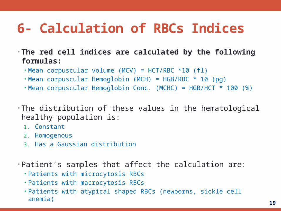
6- Calculation of RBCs Indices • The red cell indices are calculated by the following formulas:
• Mean corpuscular volume (MCV) = HCT/RBC *10 (fl)• Mean corpuscular Hemoglobin (MCH) = HGB/RBC * 10 (pg)• Mean corpuscular Hemoglobin Conc. (MCHC) = HGB/HCT * 100 (%)
• The distribution of these values in the hematological healthy population is:1. Constant
2. Homogenous
3. Has a Gaussian distribution
• Patient’s samples that affect the calculation are:• Patients with microcytosis RBCs• Patients with macrocytosis RBCs• Patients with atypical shaped RBCs (newborns, sickle cell anemia)

20
A- Rule of Thumb Method for Checking Red Cell Values
• A method which can be applied at the instrument results to check validity of red cell count, HGB and HCT • HGB X 3 = HCT (± 2) • HCT + 6 (± 3) = first 2 digits of red cell count
• Example: RBC = 3.62 X 10¹² /L, HGB =11.1 g/dl, HCT = 32.6 %
• HGB X 3 = HCT (± 2) = 11.1 X 3 = 33.3 (30.6 - 34.6)• HCT + 6 (± 3) - first 2 digits of red cell count = 32.6 + 6 ± 3 =35.6-41.6
• These formulas apply to individuals with healthy red cells• If the instrument is not performing well, all of the results
will be consistently outside of those ranges

21
7- Verification of Counts by Peripheral Blood Film
• The use of Blood Film to check the counts is recommended for all abnormal counts
• The blood film can be used for: • WBC estimate • Platelets estimate • RBC count estimate & morphology
• Those can all be determined from the film and compared to the count from the instrument

22
8- Calibration of Haematology Analysers
• Haematology analyzers require calibration and periodic
adjustments
• Preferred method of calibration is to use fresh human
blood with values determined by primary reference
methods such as manual reference procedure or by
counts obtained on a properly adjusted single channel
digital particle counter
• If more than one instrument is to be adjusted, calibrate
one of them using the reference methods
• Then use patient samples analyzed on it as secondary
calibrators to adjust the other instruments

23
8- Calibration of Haematology Analysers
• Calibration of instruments in the absence of reliable hematocytometer counts or particle counters:• In this case whole blood commercial controls with
assigned values can be used • Three separate and independent lots of commercially
prepared whole blood control material with cell concentration in the normal range
• One lot is used to calibrate and the other two lots to confirm the calibration
• Defects in the calibration cannot be detected if the calibrator and the control are the same

24
9- Peripheral Blood Film
• It is one of the older and most important haematological procedures. It serves several purposes: 1. It determines the types and numbers of the various
leukocytes present in the peripheral blood
2. Allows the examination of the morphology of erythrocytes, leukocytes and platelets
3. Provides a means of verifying the results of a cell count from analyzers

25
9- Peripheral Blood Film
• The blood film is difficult procedure to master because: • The leukocyte distribution and erythrocytes are affected
by the method and manner of blood film preparation• The performance of the standard 100 cell differential of
WBC is subject to a great degree of imprecision because of the uneven distribution of leukocytes across the slide
• This is mainly apparent in cell lines in which there are normally few cells present like monocytes, eosinophils and basophils

26
9- Peripheral Blood Film
• Different persons performing a differential on the same blood film will arrive to different results due to the following: • Various techniques are used in examining the film • The choice of working area • The ability to recognize and identify cells • Different films made from the same sample will show more
variation• Each film was made from a different portion of the sample and no two
portions are exactly the same

27
9- Peripheral Blood Film
• Factors that affect the distribution of WBCs and affect the cellular Morphology: 1. The shape of the edge
2. The type of the spreader used
3. The angle of the spreader
4. The speed of the stroke
5. The volume of the drop of blood
6. The erythrocyte concentration

28
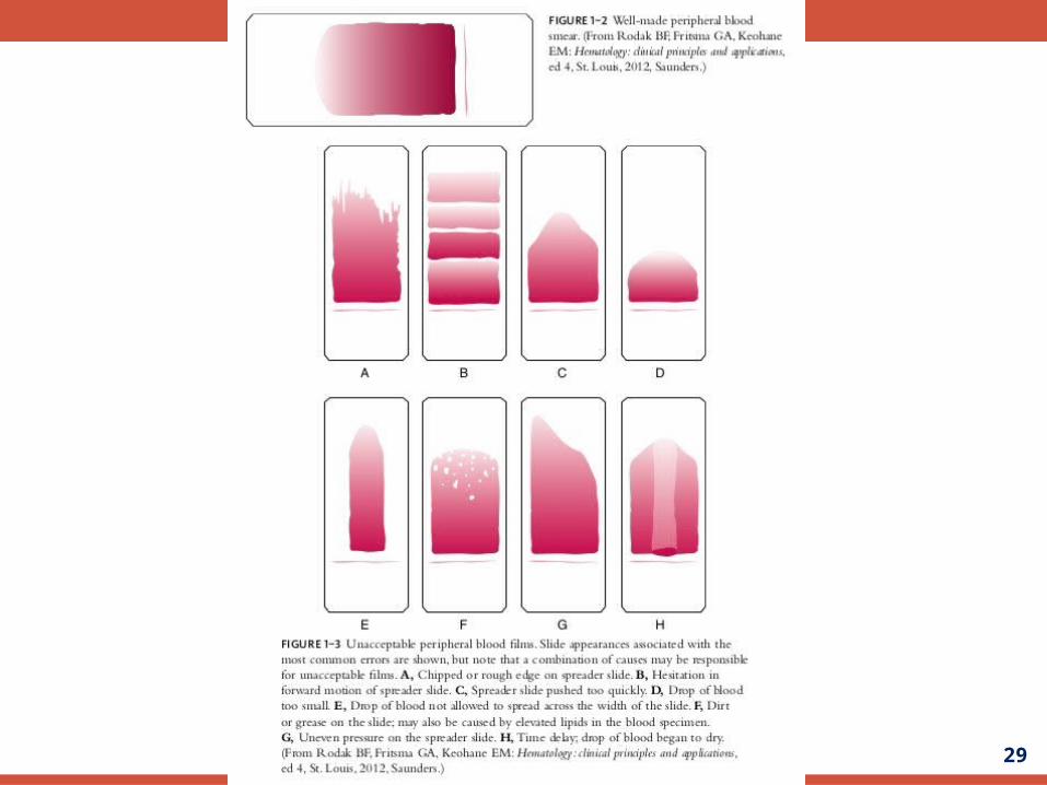
A- Properties of an acceptable blood film
1. Blood film is narrower than the glass slide
2. It has a minimum length of 2.5 cm and ends at least 1 cm from the end of the slide
3. The film should provide sufficient working area, an area where the red cells are touching and not overlapping
4. The film should have a gradual decrease in thickness from thick to thin area
5. The thinner far end should be free of streaks
6. No artifacts of cellular morphology should be introduced by slide -making process
7. There should be minimum distribution disruption of the leukocytes by the spreading process
29
30
B- Intralaboratory Performance of Blood Film Examination
• To ensure that the Intralaboratory precision of the WBC differential and blood film evaluation is acceptable, a small scale proficiency survey can be conducted periodically
• 5 to 10 blood films from freshly drawn EDTA anticoagulated blood are prepared and stained with the routine stain
• All technologists are asked to perform a routine WBC differential, RBC morphology, WBC and platelets estimates on these same slides
• Results of different technologists are compared
• Individuals who are identifying and estimating differently from the expected level of performance can be identified as outliers

31
C- Specimens that should be reviewed by slide examination
• Specimens that should be reviewed by slide examination by a competent technologist:
• Specimens with HGB level < 10 g/dl or > 18 g/dl• check for atypical RBC morphology
• Specimens with WBC counts > 15 X 109 /L or < 3.5 X 10⁹/L• check for abnormal WBC morphology
• Specimen with platelet counts below 100 X 109/L• check for platelet clumping or atypical platelet morphology

32
D- Confirmation of Blood films by a pathologist or haematologist
• Blood films with pathologically significant morphology should be reviewed and confirmed by a pathologist or haematologist. Significant findings include:1. Presence of normoblasts
2. Significant abnormal morphology of red cells
3. Presence of plasma cells in peripheral blood
4. Presence of immature cells from any of the cell lines, especially blast-like cells
5. Presence of atypical cells or unidentifiable cells
6. Thrombocytosis or thrombocytopaenia, presence of giant platelets, or megakaryocytes

33
10- External Quality Control in Haematology
• Proficiency survey offered by the College of American Pathology offers a mean to monitor the accuracy of many haematology procedures
• They include: • Preserved whole blood materials for counting cells and measuring haemoglobin
• Photomicrograph or transparencies of Wright-stained blood cells for identification of various cells found in the blood
• This help the participants to evaluate their own performance relative to other laboratories of similar size and methodology
• The survey is also important in evaluating the performance of the haematology analyser
• Another source of external control is the whole blood commercial controls in which the manufacturer offers a regional quality control program
• Comparison to other laboratories with similar instruments and using the same lot of control material is done
34
11- Continuing Education
• Continuing education programs can be targeted towards problem areas
• They are important in maintaining a high level of quality in the Intralaboratory performance of the blood film examination
• Maintenance of haematology analysers • The best prevention of errors and mistakes is a well-trained and reliable technologist

35
12- Quality assurance of Haematology Instrumentation
• Thermometers, refrigerators, general purpose centrifuges all should be involved in an active quality assurance program with performance and function verification and preventive maintenance performed on a regular basis
• Electronic cell counters: • They require performance and function verification and preventive
maintenance • The manufacturer supplies guidelines for what should be done on a
daily, weekly and monthly basis
36
B- Microhaematocrit Centrifuges
• To measure the packed cell volume accurately: • RCF (radial centrifugal force) must be known • Minimum time necessary for maximum packing of red
cells
• Therefore the speed of centrifugation and the centrifuge timer must be checked at specified intervals

37
B- Microhaematocrit Centrifuges
• Determination of Constant Packing Time• EDTA anticoagulated blood is used
• Aliquot blood into 12 microhaematocit tubes
• First pair of tubes is spun for 1 minute, second pair for
two minutes and so on until all 12 tubes have been spun
• Time is recorded for each pair of tubes
• When two pairs obtain the same PCV, the longer lime is
used as the minimum packing time