purification and characterization of acid trehalase from ... 1988 by the american society for...
TRANSCRIPT
0 1988 by The American Society for Biochemistry and Molecular Biology, Inc. THE JOURNAL OF BIOLOGICAL CHEMISTRY Vol. 263, No. 17, Issue of June 15, pp. 85374543,1988
Printed in U.S.A.
Purification and Characterization of Acid Trehalase from the Yeast suc2 Mutant*
(Received for publication, December 1, 1988)
Klaus MittenbiihlerS and Helmut HolzerSS From the SGesellschuft fur Strahlen- und Umweltforschung, Projekt Inhalation, Zngolstbdter Landstrasse 1, 0-8042 Neuherberg, West Germany and the fBiochemisches Znstitut, Universitat Freiburg, Hermann-Herder-Strasse 7, 0-7800 Freiburg, West Germany
Acid trehalase was purified from the yeast suc2 dele- tion mutant. After hydrophobic interaction chromatog- raphy, the enzyme could be purified to a single band or peak by a further step of either polyacrylamide gel electrophoresis, gel filtration, or isoelectric focusing. An apparent molecular mass of 218,000 Da was cal- culated from gel filtration. Polyacrylamide gel electro- phoresis of the purified enzyme in the presence of sodium dodecyl sulfate suggested a molecular mass of 216,000 Da. Endoglycosidase H digestion of the puri- fied enzyme resulted after sodium dodecyl sulfate gel electrophoresis in one distinct band at 41,000 Da, rep- resenting the mannose-free protein moiety of acid tre- halase. The carbohydrate content of the enzyme was 86%. Amino acid analysis indicated 364 residueslmol- ecule of enzyme including 9 cysteine moieties and only 1 methionine. The isoelectric point of the enzyme was estimated by gel electrofocusing to be approximately 4.7. The catalytic activity showed a maximum at pH 4.6. The activity of the enzyme was not inhibited by 10 mM each of HgCla, EDTA, iodoacetic acid, phenan- throlinium chloride or phenylmethylsulfonyl fluoride. There was no activation by divalent metal ions. The acid trehalase exhibited an apparent K, for trehalose of 4.7 2 0.1 mM and a V,, of 99 pmol of trehalose min” X mg” at 37 “C and pH 4.6. The acid trehalase is located in the vacuoles. The rabbit antiserum raised against acid trehalase exhibited strong cross-reaction with purified invertase. These cross-reactions were removed by affinity chromatography using invertase coupled to CNBr-activated Sepharose 4B. Precipita- tion of acid trehalase activity was observed with the purified antiserum.
Trehalose, a nonreducing disaccharide (a-D-glucopyrano- syl(l+ 1)-a-D-glucopyranoside) was first isolated by Wiggers in 1832 (1). Since then it has been found in a wide variety of organisms including yeast, fungi, bacteria, plants, insects, and other invertebrates. In Saccharomyces cereuisiae, trehalose is one of the major storage carbohydrates, accounting for 1% to more than 23% of the dry weight of the cells, depending on the growth conditions and the stage of the life cycle (2). Interestingly, the large pools of trehalose remain constant in cells starved by incubation in water even though these cells contain high activities of trehalase, the trehalose-hydrolyzing enzyme, first described in yeast by Fischer (3). Such obser- vations led to the postulation of an intracellular compartmen-
* The costs of publication of this article were defrayed in part by the payment of page charges. This article must therefore be hereby marked “advertisement” in accordance with 18 U.S.C. Section 1734 solely to indicate this fact.
talization of trehalase and its substrate in yeast (4,5). Further investigations by Souza and Panek (6) indicated that after disruption and centrifugation of protoplasts, the treha- lase was found in the soluble fraction, whereas trehalose remained in the corresponding sediment. From these results they concluded that the substrate, trehalose, was separated from the enzyme through its binding to special sites on the cytoplasmic membranes. In 1974, van der Plaat and van Solingen (7) reported that the trehalase activity increased substantially and almost instantaneously upon the initiation of growth of stationary yeast cultures. This appeared to in- volve the activation of a preexisting trehalase zymogen by a CAMP-dependent protein kinase (8). In 1982, Wiemken and co-workers (9,lO) demonstrated localization of the trehalase zymogen in the cytosol and localization of a second, perma- nently active trehalase in the vacuoles. Londesborough and Varimo (11) separated these two activities by protein frac- tionation and reported their different physical and catalytic properties using partially purified enzymes. This presented clear evidence for the existence of two different activities in yeast, one assayed at pH 5 that was confined to the vacuoles and the other with a maximum activity at pH 7 that was located in the cytosol and interconverted by phosphorylation/ dephosphorylation (8, 12, 13). In the present paper, the vac- uolar trehalase has been purified to SDS’ gel homogeneity and characterized. This enzyme is designated “acid trehalase’’ due to its maximal activity at pH 4.5. In contrast, the cytosolic trehalase exhibiting a pH optimum at 7.0 is designated “neu- tral trehalase.”
MATERIALS AND METHODS
Reagents
Auxiliary enzymes and biochemicals were purchased from Boehrin- ger (Mannheim, Federal Republic of Germany) and Merck (Darm- stadt, FRG). Orcinol and 2,3,5-triphenyltetrazolium chloride were from Sigma (Taufkirchen, FRG). Concanavalin A, CNBr-activated Sepharose CL-4B, protein A-Sepharose CL-4B, PD-10 columns, and Pharmalyte pH 2.5-5.0 were from Pharmacia LKB Biotechnology Inc. (Freiburg, FRG). The Spherogele TSK phenyl-5PW column for high performance hydrophobic interaction chromatography was from Beckmann (Munchen, FRG).
Organism
The diploid yeast suc2 deletion mutant 2.64.1 Ca producing no external invertase was used in all experiments. In this strain, the SUC2 gene is substituted with the URA3 gene. The strain was a gift from Prof. Dr. Zimmermann and Dr. Stefan Hohmann, Institut fiir
The abbreviations used are: SDS, sodium dodecyl sulfate; Mes, 4- morpholineethanesulfonic acid; Endo H, endo-/3-N-acetylglucosamin- idase H , Hepes, 4-(2-hydroxyethyl)-l-piperazineethanesulfonic acid; 1, liter.
a537

8538 Acid Trehalase from the Yeast suc2 Mutant
Genetik, TH Darmstadt, FRG. Bakers' yeast was obtained from Bako, Darmstadt, FRG.
Growth Conditions suc2 cells, cultivated in YEPD medium (1% Bacto yeast extract,
2% bactopeptone, 50 mg/l adenine, 50 mg/l uracil, and 2% glucose) for 48 at 30 "C (stationary phase), were used for the purification of the enzyme.
Enzyme Assays Acid trehalase was assayed by incubating 30 pl of extract with 270
pl of 200 mM sodium citrate, pH 4.5, containing 1 mM EDTA and 114 mM trehalose. After 15 min, the reaction was stopped by adding 50 pl of the incubation mixture to 950 pl of boiling Hz0 and heating up to 95 "C for 3 min. The controls were stopped immediately. After centrifugation at 10,000 X g for 3 min at 4 "C, the glucose concentra- tion of the supernatant liquid was determined enzymatically by the glucose oxidase-Perid-I1 method using glucose oxidase and peroxidase (14). The measured activity was linear with respect to the amount of enzyme (data not shown). Invertase was assayed in the same way as acid trehalase, except that 114 mM sucrose was used instead of 114 mM trehalose. For studies on Me2+ effects, EDTA was omitted from the incubation mixture. Proteinase B was assayed with azocoll as substrate as described by Saheki and Holzer (15). a-Mannosidase was tested withp-nitrophenyl-a-D-mannopyranoside according to van der Wilden et al. (16). a-Glucosidase was measured as described by Halvorson (17) and Glc-6-P dehydrogenase was assayed as in Ref. 18.
Definition of Units One unit of acid trehalase is defined as the amount of enzyme that
catalyzes the hydrolysis of 1 pmol of trehalose/min at 37 "C and pH 4.5. One unit of invertase catalyzes the hydrolysis of 1 pmol of sucrose/min at 37 "C and pH 4.5.
Carbohydrate Analysis The neutral sugar content of acid trehalase was determined by the
orcinol method as described by Winzler (19) using mannose as stand- ard. The absorption was measured at 505 nm instead of 540 nm (20).
Amino Acid Analysis Amino acid analysis was carried out with a Biotronik LC 6000
amino acid analyzer as previously described (21, 22). Half-cysteine was measured as cysteic acid after performic acid oxidation (23). The tryptophan content in the protein was determined spectrophotomet- rically according to Edelhoch (24).
Absorption Spectra Spectra in the range from 220 to 500 nm were recorded with a
Lambda 7 Perkin-Elmer spectrophotometer equipped with a Perkin- Elmer Lambda computer.
Protein Determination Protein was determined by the method of Bradford (25) using the
commercial Bio-Rad protein assay with crystalline bovine serum albumin as standard.
Electrophoresis Polyacrylamide gel electrophoresis was carried out at pH 8.3 on
10% gels in the presence of SDS as described by King and Laemmli (26). Gels were stained for protein with Coomassie Brilliant Blue R- 250 and destained by diffusion. Isoelectric focusing using Pharmalyte pH 2.5-5.0 was performed in 10-cm gel rods according to Wrigley (27) except that the cathode buffer was 10 mM Hepes and the anode buffer was 10 mM iminodiacetic acid. The 5% gels contained 6.5% carrier ampholyte. The pH was measured by cutting one gel into 2- mm slices and eluting each slice with 750 pl of water. Activity staining was done by the method of Gabriel (28) detecting the released glucose.
Antiserum Preparation and Purification A rabbit was immunized against acid trehalase and boosted 4 and
8 weeks later. The antiserum was titrated and checked for cross- reactivity by Ouchterlony analysis (29) and Western blot as described by Schiifer et al. (30). Nonspecific reactivity with other high-mannose
glycoproteins of yeast was removed by affinity chromatography using invertase coupled to CNBr-activated Sepharose CL-4B.
Isolation of Vacuoles Cells were grown in YEPD medium for 18 h at 30 "C. Spheroplast
formation, lysis of spheroplasts with DEAE-dextran, and purification of vacuoles were performed as described by Mechler et al. (31). Enrichment of vacuoles as determined by vacuolar enzyme activity was 60- to 95-fold.
Purification of Acid Trehalase from the Yeast suc2 Mutant (All Operations Were Performed at 4 "C)
1. Crude Extract-Yeast cells (see "Growth Conditions") were harvested (120 g wet weight), washed twice with buffer A (40 mM sodium citrate, 1 mM EDTA, pH 6.5) by centrifugation (2000 X g for 5 min), and suspended to 50% (wet weight/volume) in the same buffer. Cells were disrupted by passing the suspension three times through a French pressure cell. The supernatant resulting from centrifugation at 10,000 X g for 60 min is referred to as the crude extract.
2. Acid Fractionation-Citric acid (2 M) was added with stirring and with ice bath cooling to adjust the pH of the crude extract to 4.0. Precipitated protein was removed by centrifugation at 10,000 X g for 60 min.
3. Ammonium Sulfate Fractionation-Solid ammonium sulfate (43 g) was added to 100 ml of supernatant from the acid fractionation with stirring for 30 min at 0 "C. Precipitated protein was removed by centrifugation at 10,000 X g for 45 min.
4. Hydrophobic Interaction Chromatography with Phenyl-Sephurose CL-4B"The supernatant of the ammonium sulfate fractionation (150 ml) was applied to a column of phenyl-Sepharose CL-4B (2.5 X 11 cm) equilibrated with buffer B (40 mM sodium citrate, 1 mM EDTA, pH 4.5, and 43 g of solid ammonium sulfate added to 100 ml, equivalent to 69% saturation at 0 "C). The column was washed with 400 ml of buffer B, and the enzyme was eluted with a linear decreasing gradient of 69%, pH 4.5, to 0%, pH 6.5, ammonium sulfate saturation. The enzyme eluted at an ionic strength corresponding to 200 microSiemens.
5. Hydrophobic Interaction Chromatography Using the High Per- formance Liquid Chromatography Column SpherogeP TSK Phenyl- 5PW-The peak fractions from the phenyl-Sepharose CL-4B column were collected and 30% (w/v) solid ammonium sulfate was added. The fractions were then passed through a SpherogeP TSK phenyl- 5PW column (21.5 mm X 15 cm) equilibrated with buffer B and were washed with an additional 100 ml of the same buffer. Acid trehalase was eluted from the hydrophobic column by applying a linear decreas- ing gradient of ammonium sulfate from 69 to 0% saturation using buffer B and Hz0 as final eluant. Fractions with highest activity were pooled and either stored at 4 "C or passed through a PD-10 column and then precipitated with 80% ethanol (final concentration) for 24 h at 0 "C. The precipitate was centrifuged and stored at -20 "C. For further use, the precipitate was dissolved in water.
Crude Extract from Bakers' Yeast Yeast cells were washed three times with 40 mM sodium citrate
buffer, pH 6.5, containing 1 mM EDTA and suspended to 50% (wet weight/volume) in the same buffer. Disruption and centrifugation conditions were as described under "Purification of Acid Trehalase from the Yeast suc2 Mutant, 1. Crude Extract."
RESULTS AND DISCUSSION
As separation of invertase from acid trehalase was found to be extremely difficult and time consuming, trehalase was purified from the yeast suc2 deletion mutant, which contains no external invertase. The five-step procedure is summarized in Table I. An approximately 7,000-fold purification with a yield of 38% was obtained. SDS-polyacrylamide gel electro- phoresis of the crude extract and of aliquots from steps 2-5 of the purification procedure is shown in Fig. 1. The purified enzyme resulting from step 5 was applied in its native form to high performance gel filtration using a Superose 12 HR 10/ 30 column. A molecular mass of 218 kDa was estimated (Fig. 2). SDS-polyacrylamide gel electrophoresis resulted in a single broad band ranging from 167,000 to 265,000 Da; from this an

Acid Trehalase from the Yeast suc2 Mutant 8539 TABLE I
Purification of acid trehalase from the yeast sue2 mutant The purification was performed as described under "Materials and Methods." Starting with 120 g, wet weight,
Total Specific of yeast cells, about 500 pg of purified trehalase were obtained in 7 days.
step Volume Purification Yield Invertase/ activity activity trehalase
ml units unitslmg -fold ?6 1. Crude extract 153 130 0.0138 1 100 0.3 2. Citric acid 133 122 0.115 8 94 0.3 3. Ammonium sulfate 151 117 2.04 148 90 0.25 4. Phenyl-Sepharose chromatogra- 250 102 9.10 659 80 0.03
5. Spherogelm TSK phenyl 5PW 12 49.8 99 7174 38 0.003 PhY
chromatography
1 2 3 4 5 6 7 8 9 10
205 - 116- 97- 66-
4 5 1 ,
29-
FIG. 1. SDS-polyacrylamide gel electrophoresis of acid tre- halase preparations. Samples obtained after different purification steps (see Table I) were solubilized and subjected to SDS-polyacryl- amide gel electrophoresis (see "Materials and Methods"). Lane 1, marker proteins: myosin (M, = 205,000), &galactosidase (MI = 116,000), phosphorylase b (M, = 97,400), albumin (M, = 66,000). ovalbumin (Mr = 45,000), carbonic anhydrase (Mr = 29,000). Lune 2, 150 pg of protein from crude extract. Lane 3, 60 pg of protein after acid precipitation (step 2). Lane 4,60 pg of protein after ammonium sulfate precipitation (step 3). Lane 5, marker proteins as in lane 1. Lane 6, 55 pg of protein after phenyl-Sepharose chromatography (step 4). Lane 7.20 pg of protein after Spherogel" TSK phenyl-5PW chromatography (step 5). Lane 8, marker proteins as in lane 1. Lane 9, acid trehalase (10 pg) treated with endo H (24 h); the protein band at 29,000 Da represents the endoglycosidase H. Lane 10, marker proteins as in lane 1.
average molecular mass of 216 kDa was calculated for acid trehalase. The high carbohydrate content of yeast acid tre- halase is probably responsible for the broad band in SDS-gel electrophoretograms, as it is for external invertase (32, 33). SDS-gel electrophoresis with and without dithiothreitol showed the same molecular weight, indicating that the native enzyme is a monomer lacking intermolecular disulfide bridges. A molecular mass of 215 kDa, which is in good agreement with the values presented here, was estimated by Londesbor- ough and Varimo (11) for a partially purified enzyme prepa- ration by Sephadex G-150 gel filtration. Dependence of puri- fied acid trehalase activity on the pH is shown in Fig. 3. Maximal activity was observed at pH 4.5. A similar pH optimum was obtained with a crude extract from bakers' yeast (Fig. 3) and from stationary suc2 mutant cells (data not shown). The vacuolar pH, which is reported to be about 5.5- 5.9 (34, 35), and the optimal pH of acid trehalase are both in an acid range. Earlier investigations under different condi- tions (11,36,37) showed pH optima between 4 and 5.7. EDTA (6.5 mM), in the tested pH range from 2.5 to 7.7, had no effect on the acid trehalase but completely inhibited the neutral
0 0.2 04 0.8
K,"
FIG. 2. Estimation of the molecular mass of the purified acid trehalase by comparison with marker proteins in high performance gel filtration. 6 pg of purified enzyme (Table I, step 5, specific activity 99 units/mg of protein) were passed through a Sepharose 12 HR 10/30 column (1.0 X 30 cm) equilibrated with 100 mM phosphate buffer, pH 7.0, containing 0.1 M NaC1. Flow rate was 30 ml/h. Fractions of 0.5 ml each were collected. The column was calibrated with (a) ferritin (MI = 440,000); ( b ) catalase (Mr = 232,000); (c) aldolase (M, = 158,000); ( d ) bovine serum albumin (M, = 67,000); (e) ovalbumin (MI = 43,000); ( f ) chymotrypsinogen a (Mr = 25,000); (g) ribonuclease (M. = 13,700); K., = (V, - Vo)/(V, - VO). Position of the marker proteins was determined by measuring the absorption of 280 nm, and the position of acid trehalase was determined by measuring enzymatic activity.
trehalase activity of the extract from bakers' yeast (Fig. 3), as also shown earlier by Londesborough and Varimo (11). In the absence of EDTA, a second pH optimum was observed at around pH 7 (Fig. 3). This peak which corresponds to the neutral trehalase activity was represented only by a shoulder when using crude extract from suc2 mutant (data not shown).
The acid trehalase was assayed for substrate specificity at pH 4.5 using the purified enzyme. Acid trehalase showed a high specificity for trehalose. Other disaccharides such as sucrose, maltose, lactose, cellobiose, melibiose, and the trisac- charide raffinose showed essentially no detectable activity (less than 0.3% of the activity measured with trehalose). Kelly and Catley (37) showed that their partially purified acid trehalase exhibits no action on maltose, sucrose, raffinose, cellobiose, starch, glycogen, glucose 6-phosphate and glucose 1-phosphate. Panek and Souza (36) reported specificity only for trehalose and raffinose among several oligosaccharides. This latter observation is in contrast to the findings of Kelly and Catley (37) and to our observations using the purified

8540 Acid Trehalase from the Yeast suc2 Mutant
PH
FIG. 3. Dependence of trehalase activity on pH. The activity in a crude extract of bakers’ yeast (milliunits/ml) was measured in test mixtures containing 114 mM trehalase, 30 mM citric acid/ Na2HP04 (Mc Ilvaine buffer) without (A) and with (A) 6.5 mM EDTA as described under “Materials and Methods.” The activity of the purified acid trehalase (units/ml) was measured in test mixtures containing 114 mM trehalose, 30 mM citric acid/Na2HP04 (0).
4
1 ’” 0.8 c
FrG. 4. Determination of K, of purified acid trehalase using Lineweaver-Burk plot. Acid trehalase activity was assayed using increasing concentrations of trehalose as substrate. Assay conditions were described under “Materials and Methods.” The S and K,,, values are expressed as mM; V values and V,. are pmol X min” X ml”.
enzyme. Our results indicate that the acid trehalase is highly specific for the crl-a1 bond.
The K,,, for trehalose was 4.7 f 0.1 mM at pi3 4.5, with a corresponding Vmlu of 99 pmol x min” x mg” (Fig. 4). Panek and Souza (36) found a K,,, of 0.4 mM at pH 5.6 while a K,,, of 0.5 mM at pH 5.5 was reported by Kelly and Catley (37). Londesborough and Varimo (11) determined a K,,, for the vacuolar trehalase of 1.4 mM at pH 5.0,5.8, and 6.5 in 50 mM Mes/KOH containing 100 mM KC1. The different test systems and strains used for the determination of K,,, seem to be responsible for the discrepant values obtained.
The amino acid analysis of acid trehalase (Table 11) shows that the enzyme contains 10% aromatic residues and 51% polar residues, which is common for a soluble protein (38). Acid trehalase contains 3% tryptophan and 43% hydrophobic
TABLE I1 Amino acid composition of acid trehalase from suc2 mutant
HCI
24’ 48” 9fY Amino acid Average or extrapo-
lated integer
residueslmoleculeb
Aspartic acid + as- 38.50 39.95 40.88 40 paragine
Threonine Serine Glutamic acid
glutamine Proline Glycine Alanine Valine Methionine Isoleucine Leucine Tyrosine Phenylalanine Lysine Histidine Arginine Half-cysteine Tryptophan Total
+ 24.36 23.02 22.39 38.90 32.57 28.70 18.24 20.70 21.56
26.42 26.38 25.17 33.62 34.60 36.08 33.08 33.91 35.61 26.63 29.19 28.42
1.27 1.21 1.29 20.00 20.94 21.81 21.72 21.97 23.88 8.47 8.34 8.43 9.59 8.92 8.55
13.25 12.95 12.58 4.10 4.18 4.29 4.17 3.28 2.47 8.64 8.84 9.01 7.2Bh
2ff 44’ 20
26 35 34 2 9
1 2 2 24‘ 8 9
13 4 3 9“ 7h
354
a Time of hydrolysis in hours. *Values obtained are based on a molecular mass of 41,000 De,
corresponding to the protein moiety of acid trehalase after endo H treatment.
Extrapolated to zero hydrolysis time. *Values from 48/96-h hydrolysis only are considered. e Values from 96-h hydrolysis only are considered. ’Values from 24-h hydrolysis only are considered.
Determined as cysteic acid according to Ref. 23. Determined spectrophotometrically according to Ref. 24.
PH m ~ . 5. Isoelectric focusing of acid trehalase. Twenty pg of
purified acid trehalase (specific activity 99 units/mg) were applied onto each disc gel (0.6 X 8.5 cm). After focusing at 500 V for 5 h, pH was measured by cutting one gel into 2-mm-thick slices and extracting with water. For determination of trehalase activity, another gel was stained for activity as described under “Materials and Methods” (kft- hand gel). Afterwards, the same gel was stained for protein (right- hand gel). An isoelectric point of 4.7 was estimated.
Acid Trehalase from the Yeast suc2 Mutant 8541
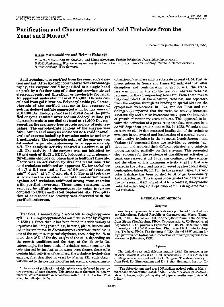
TABLE I11 Distribution of acid trehalase activity and marker proteins in
spheroplast and vacuole preparations The specific activities in the spheroplast lysate are set to be 1. The
protein content of the vacuole preparation was 0.275 mg/ml. The spheroplast lysate contained 8.9 mg protein/ml. Protein determina- tion was performed as described under "Materials and Methods."
Specific activity in
related to sphero- purified vacuoles
plast lysate = 1
Cytosolic markers Glucose-6-P dehydrogenase 0.38 a-Glucosidase 0.55
a-Mannosidase 64 Proteinase B 91
Acid trehalase 93
Vacuolar markers
amino acids. Only one methionine was found among 354 amino acids. The molecular mass of 41,000 Da, obtained after endoglycosidase H treatment (see Fig. l), was used for calcu- lation of amino acid residues per molecule. Maximal absorp- tion of purified acid trehalase was observed at 276 nm. The molar extinction coefficients calculated for acid trehalase where at cm = 98.4 1 x mmol" X cm" and at €276 = 104.3 1 X mmol" X cm" (protein was calculated from amino acid
1 2 3 4 1
-200
1 2 3
+ 97.4
+ 68
443
+ 25,7
analysis). Acid trehalase contains 86% neutral sugars (w/w), as determined with the orcinol-sulfuric acid assay (19, 20) using mannose as a standard. Using the same method, the carbohydrate content of external invertase was determined to be 49.4% which is in agreement with data from Neumann and Lampen (39). Endo H digestion of purified, SDS-denatured acid trehalase in the presence of phenylmethylsulfonyl fluo- ride for 1 h resulted in two distinct bands after SDS-gel electrophoresis at 41,000 and 100,000 Da. Digestion with Endo H for 2, 4, and 8 h resulted in increasing conversion of the 100-kDa to the 41-kDa form with time. Endo H digestions of 24 h or more resulted in the complete absence of the 100 kDa form, leaving only the 41-kDa mannose-free protein moiety (Fig. 1). Similar results were reported after 20-h incubation of denatured external yeast invertase with Endo H, providing a 62,000-Da band (40,41) containing no residual oligosaccha- rides. The purified acid trehalase binds very strongly to con- canavalin A. 38% of the applied activity could be eluted with 40 mM sodium citrate, pH 6.0, containing 0.5 M NaCl and 0.5 M methyl-a-D-mannopyranoside (data not shown). Londes- borough and Varimo (1 1). using a dialyzed protamine super- natant, reported a recovery in the collected eluate from a concanavalin A column of 39%.
The susceptibility of the acid trehalase to classical inhibi- tors was tested using the assay described under "Materials
4 1 2 3 4
.200
97,4
I. 68
* 43
4 974
4 68
4 43
25.7 4 25.7
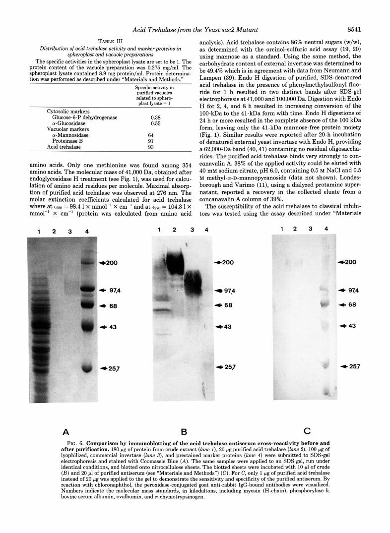
A B C FIG. 6. Comparison by immunoblotting of the acid trehalase antiserum cross-reactivity before and
after purification. 180 pg of protein from crude extract (lane I ) , 20 pg purified acid trehalase (lane 2), 100 pg of lyophilized, commercial invertase (lane 3), and prestained marker proteins (lane 4 ) were submitted to SDS-gel electrophoresis and stained with Coomassie Blue (A) . The same samples were applied to an SDS gel, run under identical conditions, and blotted onto nitrocellulose sheets. The blotted sheets were incubated with 10 pl of crude ( B ) and 20 pl of purified antiserum (see "Materials and Methods") (C). For C, only 1 pg of purified acid trehalase instead of 20 pg was applied to the gel to demonstrate the sensitivity and specificity of the purified antiserum. By reaction with chloronaphthol, the peroxidase-conjugated goat anti-rabbit IgG-bound antibodies were visualized. Numbers indicate the molecular mass standards, in kilodaltons, including myosin (H-chain), phosphorylase b, bovine serum albumin, ovalbumin, and a-chymotrypsinogen.

8542 Acid Trehalase from the Yeast suc2 Mutant
\ \
\Z"" "to" 40 60 8? 1%
"
Serum (PI)
FIG. 7. Immunprecipitation of acid trehalase activity. Affin- ity chromatography-purified antiserum and -purified acid trehalase (see Table I) were incubated for 2 h at 25 "C with (A) and without (0) protein-A-Sepharose. The precipitated material was removed by centrifugation, and the supernatant was tested for acid trehalase activity as described under "Materials and Methods."
and Methods." Except for 10 mM HgC12, which reduced the activity by about 30%, none of the other substances tested (0.1 and 10 mM of each, EDTA disodium salt, iodoacetic acid, o-phenanthrolinium chloride, phenylmethylsulfonyl fluoride and 0.1 mM HgC12) showed a significant effect on the activity of the purified acid trehalase. These results suggest that the active site of the enzyme contains no sulfhydryl, serine, or threonine hydroxyl groups and that its activity is independent of bivalent metal ions. Incubation of the purified enzyme for 1 and 5 h with 0.1 and 10 mM of each inhibitor, prior to determining its activity, also had no significant effect on the acid trehalase activity. The only exception was 10 mM HgC12, which after 1- and 5-h incubations caused 36 and 99% inhi- bition, respectively. The acid trehalase activity was not altered by replacing the 1 mM EDTA in the assay mixtures with 0.1 or 10 mM CaC12, MnC12, MgC12, or ZnS04. Similarly, prein- cubation of the enzyme with 0.1 and 10 mM CaC12, MnC12, MgClz, or ZnS04 (prior to determining its activity) also had no effect. Similar results were reported by Londesborough (11) with partially purified acid trehalase.
The isoelectric point of purified acid trehalase (99 units/ mg) was measured by isoelectric focusing in polyacrylamide gels in a pH range of 2.5-5.0 (Fig. 5). An isoelectric point of 4.7 was determined using activity and protein staining. Iden- tical positions of stained protein and trehalase activity dem- onstrate that the single visible protein band is responsible for the enzymatic activity. The PI of acid trehalase is in the same range as the isoelectric points of three other vacuolar enzymes from yeast: proteinase A (PI = 4.4) (42), carboxypeptidase Y (PI = 3.6) (43), and aminopeptidase I (PI = 4.7 (44). An acid PI value of acid trehalase is also in accord with the results of Londesbrough and Varimo (11).
Subcellular fractionation of lysed spheroplasts by density gradient centrifugation was used to determine the subcellular localization of acid trehalase in yeast. Earlier observations (9) that acid trehalase is confined to the vacuoles were confirmed as shown in Table 111; an additional periplasmic localization, however, cannot be excluded.
Immunodiffusion after Ouchterlony (not shown) and West- ern blot (Fig. 6 ) analysis showed the antiserum raised against acid trehalase in a rabbit to be reactive not only with the acid trehalase, but also with external invertase and with other glycoproteins in the crude extract. This cross-reactivity was removed completely by affinity chromatography with external invertase coupled to CNBr-activated Sepharose in order to bind the high mannose-specific components (45) of the poly- clonal antiserum. Incubation of purified acid trehalase with purified antiserum resulted in complete precipitation of the enzyme activity (Fig. 7).
Acknowledgments-We are grateful to Dr. Henryk Kalisz and Dr. Barry Drees for a critical reading of the manuscript as well as to Prof. Dr. F. K. Zimmermann and Dr. S. Hohmann (TH Darmstadt) for the generous gift of the yeast suc2 mutant strain. We thank Dr. Eulitz, Dr. Konard Maier, and Lieselotte Leuschel (Gesellschaft fin Strah- len- und IJmweltforschung, Munich) for performing the amino acid analysis. We also thank Wolfgang Fritz and Ulrike Kopas for help with the figures and for typing the manuscript.
1. 2.
3. 4. 5. 6.
7.
8.
9.
10.
11.
12.
13.
14.
15. 16.
17.
18.
19. 20.
21.
22. 23. 24. 25. 26. 27. 28. 29.
30.
31.
REFERENCES
Wiggers, H. A. L. (1832) Ann. Phurm. (Pozna) 1, 129-182 Lillie, S. H., and Pringle, J. R. (1980) J. Bocteriol. 143 , 1384-
Fischer, E. (1895) Ber. Dtsch. Chem. Ges. 2 8 , 1429-1438 Myrback, K., and Ortenblad, B. (1937) Biochem. Z. 291,61-69 Avigad, G. (1965) Biochem. J. 97, 715-722 Souza, N. O., and Panek, A. D. (1968) Arch. Biochem. Biophys.
van der Plaat, J. P. (1974) Biochem. Biophys. Res. Commun. 5 6 ,
van Solingen, P., and van der Plaat, J. P. (1975) Biochem.
Keller, F., Schellenberg, M., and Wiemken, A. (1982) Arch. Biophys. Res. Commun. 62,553-560
Microbiol. 13 1 , 298-301 Wiemken, A., and Schellenberg, M. (1982) FEBS Lett. 150,329-
331 Londesborough, J., and Varimo, K. (1984) Bwchem. J. 219,511-
518 Uno, I., Matsumoto, K., Adachi, K., and Ishikawa, T. (1983) J.
Biol. Chem. 258,10867-10872 Ortiz, C. H., Maia, J. C. C., Tenan, M. N., Braz-Padrao, G. R.,
Mattoon, J. R., and Panek, A. D. (1983) J. Bocteriol. 153,644- 651
Bernt, E., and Lachernicht, R. (1974) in Methoden der Enzymu- tischen Analyse (Bergmeyer, H. U., ed) Vol. 2, pp. 1260-1266, Verlag Chemie GmbH, Weinheim, West Germany
Saheki, T., and Holzer, H. (1974) Eur. J . Biochem. 42,621-626 Van der Wilden, W., Matile, P., Schellenberg, M., Meyer, J. and
Wiemken, A. (1973) 2. Naturforsch. 28c, 416-421 Halvorson, H., and Ellias, L. (1958) Biochim. Biophys. Acta 30 ,
28-40 Lohr, G. W., and Waller, H. D. (1974) in Methoden der Enzy-
matischen Analyse (Bergmeyer, H. U., ed), 3rd Ed., pp. 673- 681, Verlag Chemie GmbH, Weinheim, West Germany
Winzler, R. J. (1955) Methods of Biochem. Anal. 2,290-292 Johansen, P. G., Marshall, R. D., and Neuberger, A. (1960)
hackman. D. H.. Stein, W. H., and Moore, S. (1958) Anal. Chem.
1394
125,22-28
580-587
Biochem. J. 77,239-247
-30, 1190-1206' Hamilton. P. B. (1963) Anal. Chem. 35.2055-2064 Moore, s.'(1963)>. B&. Chem. 2 3 8 , 235-237 Edelhoch, H. (1967) Biochemistry 6, 1948-1954 Bradford, M. M. (1976) Anal. Biochem. 7 2 , 248-254 King, J., and Laemmli, U. K. (1971) J. Mol. Biol. 6 2 , 465-477 Wrigley, C. W. (1971) Methods Enzymol. 2 2 , 559-564 Gabriel, O., and Wang, S.-F. (1969) Anal. Biochem. 2 7 , 545-554 Ouchterlony, 0. (1962) in Progress in Allergy (Kallos, P., and
Waksman, B. H., e&) Vol. 6, pp. 30-154, Karger, Basel, swit-
Schafer, W., Kalisz, H., and Holzer, H. (1987) Biochim. Biophys. zerland
Mechler, B., Muller, H., and Wolf, D. H. (1987) EMBO J. 6 , Acta 925,150-155
2157-2163

Acid Trehalase from the Yeast suc2 Mutant 8543
32. Trimble, R. B., and Maley, F. (1977) J. Biol. Chem. 252, 4409- 39. Neumann, N. P., and Lampen, J. 0. (1967) Biochemistry 6,468-
33. Williams. R. S.. Trumblv, R. J., MacColl, R., Trimble, R. B., and 40. Trimble, R. B., and Maley, F. (1984) Anal. Biochem. 141, 515- 4412 475
Maley,'F. (1985) J. Biol. Chek. 260, 13334-13341 34. Slavik, J. (1982) FEBS Lett. 140, 22-26 35. den Hollander, J. A., Ugurbil, K., Brown, T. R., and Shulman, R.
G. (1981) Biochemistry 20,5871-5880 36. Panek, A., and Souza, N. 0. (1964) J. Biol. Chem. 239, 1671-
1673 37. Kelley, P. J., and Catley, B. J. (1976) Anal. Biochem. 72, 353-
358 38. Capaldi, R. A., and Vanderkooi, G . (1972) Proc. Natl. Acad. Sci.
U. S. A. 69,930-932
522 41. Trimble. R. B.. Malev, F., and Chu, F. K. (1983) J. Biol. Chem. - . .
258,2562-2567 42. Meussdoerffer. F.. Tortora. P.. and Holzer. H. (1980) J. Biol. , , . .
Chem. 255,'12087-12093 43. Aibara, S., Hayashi, R., and Hata, T. (1971) Agric. Bid . Chem.
44. Metz, G., and Rohm, K. (1976) Biochim. Biophys. Acta 429,933-
45. Feizi, T., and Childs, R. A. (1987) Biochem. J. 245,l-11
35,658-666
949