pulmonary embolism a revie: w of 200 cases with emphasi osn … · 2019-01-24 · emboli, 1 had a...
TRANSCRIPT
Review Artide
Pulmonary embolism: a review of 200 cases with emphasis on pathophysiology, diagnosis, and treatment1
Richard C. Becker, M.D. Robert Graor, M.D. John Holloway, M.D.
Pulmonary embolism is not ail uncommon occurrence in major medical centers. It is a significant cause of morbidity and mortality in both medical and surgical patients. Clinical and laboratory findings are characteristically nonspecific and may be misleading. Venography, ventilation-perfusion lung scanning, and pulmonary angiography techniques are critical diagnostic tools. Various treat-ment modalities, including the use of heparin, warfarin, and strep-tokinase, are available to the physician and often affect both the short- and long-term medical course. The authors review 200 cases evaluated for the effectiveness of diagnostic procedures and meth-ods of treatment for pulmonary embolism at The Cleveland Clinic Foundation.
Index terms; Pulmonary embolism • Review articles Cleve Clin Q 51:519-529, Fall 1984
1 Department of Peripheral Vascular Disease, The Cleveland Clinic Foundation, Submitted for publica-tion Feb 1984; revision accepted June 1984. lp
0009-8787/84/03/519/11/$3.75/0
Copyright © 1984, The Cleveland Clinic Foun-dation
Pulmonary embolism (PE) continues to be a major cause of morbidity and mortality among the general hospital population. There is evidence that the incidence of PE is increasing as a result of (a) increased life expectancy, particularly in individuals with neoplastic disease; (b) in-creased use of steroidal compounds; (c) the increased num-ber of extensive surgical procedures; and (d) an increased awareness and diagnosis of PE.1
The incidence of PE in the United States approaches 650,000 cases per year, 30% of which are fatal. Unfortu-nately, an alarming number of cases go undiagnosed. De-spite extensive experience and the development of sensitive diagnostic tools, some confusion remains as to the clinical presentation, evaluation, and treatment of PE.
519

520 Cleveland Clinic Quarterly Vol. 51, No. 3
Table 1. Study population Data No. of patients (%)
Age < 5 0 years 68 (34%) > 5 0 years 132(66%)
Sex Males 138 (69%) Females 62 (31%)
Postoperative 144 (72%)
Autopsy 84 (42%)
Oral contraceptives 0 (0%)
We reviewed 200 cases retrospectively and evaluated the effectiveness of diagnostic proce-dures and methods of treatment for PE at The Cleveland Clinic Foundation.
Methods Two hundred patients were studied by retro-
spective chart review. Study population data are described (Table 1). Patients were selected on the basis of a diagnosis of PE. Data were collected to determine the most reliable methods of diagnosis and to evaluate the methods used at our institu-tion (ventilation-perfusion scanning, venography, and pulmonary angiography).
Results The common symptoms of the 200 patients
with pulmonary emboli are listed (Table 2). Fifty-two patients (26%) were diagnosed clinically as having PE without radiographic documentation to substantiate the diagnosis. One hundred forty-eight patients (74%) had a radiographically di-agnosed PE; of these, 62 patients (42%) had ventilation-perfusion lung scans and 48 patients (32%) had perfusion lung scans only. Sixteen patients (11%) underwent pulmonary arteriog-raphy. Sixty-two percent of the lung scans were interpreted as indicating a high probability, 16% low probability, and 22% indeterminate for PE. Six patients (2%) diagnosed as having PE had
Table 2. Symptoms of pulmonary embolism Symptoms No. of patients (%)
Dyspnea 1 70 (85%) Cough 124 (62%) Chest pain 92 (46%) Hemoptysis 8 (4%) Syncope 8 (4%)
only ascending venography to document a source of emboli.
Of the 200 patients studied, 162 patients (81 %) were treated with heparin followed by warfarin, 4 patients (2%) were given streptokinase, and 34 patients (17%) had inferior vena caval interrup-tions.
Forty-eight deaths (24%) occurred. Sixteen (8%) were thought to be directly related to the PE and the remainder died from other causes.
Autopsy was performed on 42 patients (86%). Forty of these patients (95%) had massive PE and 100% had an antemortem diagnosis of suspected PE. Of the 2 patients with submassive pulmonary emboli, 1 had a clinical diagnosis of PE during the antemortem period.
Of the 144 patients in whom PE developed in the postoperative period, 98 patients (68%) had a radiographically documented embolus on either pulmonary angiograms or high-probability perfusion lung scans. The remainder of these patients were clinically diagnosed.
In two patients who underwent lung scanning several days before death, PE was not detected until postmortem examination. The lung scan was interpreted as indicating a low probability two days before one patient's death, and the other was interpreted as indeterminate four days antemortem. The scan indicating low probability was ordered after the abrupt onset of pleuritic chest pain and hypotension. It showed two seg-mental defects in areas of the chest radiograph abnormalities at both lung bases. At autopsy, there were multiple segmental pulmonary em-boli. In retrospect, this patient's scan was re-viewed and was again interpreted as revealing a low probability. For the patient with the indeter-minate lung scan, pulmonary angiography was recommended and obtained three days before the patient's death. The pulmonary angiogram had been correctly interpreted, leading to a di-agnosis of PE.
Discussion The mortality of undiagnosed PE approaches
30%. Once identified and treated, this figure decreases substantially to less than 10%.3 Such statistics dramatically emphasize the importance of early diagnosis and treatment. The diagnosis, however, is often difficult because of the varying presentation of PE.
Critical to the diagnosis of PE is an awareness that the clinical presentation is an outward man-

Fall 1984 Pulmonary embolism 521
ifestation of a series of complex pathophysiolog-ical processes that occur following the embolic event.
The significant incidence, high morbidity, and potential mortality of PE in the hospital setting warrant complete understanding of its predispo-sition, presentation, evaluation, and treatment. The problem should be approached in a swift, logical, and precise manner as in other acute, life-threatening events, such as myocardial infarction, overwhelming sepsis, and the acute abdomen.
Factors predisposing to pulmonary embolism Approximately 90% of pulmonary emboli orig-
inate from thrombi within the deep venous sys-tems of the pelvis and thighs. Exceptions include tumor emboli, air emboli, amniotic fluid emboli, fat emboli, and thrombi originating in the right side of the heart.4
The risk of embolization is approximately 40% in patients who have thrombosis proximal to the popliteal vein. Although it is said that calf vein thrombi rarely embolize, almost 20% of these will propagate proximally and then present a more significant risk for embolization.
Risk factors for development of deep venous thrombosis (DVT) include serious illness neces-sitating prolonged bed rest, such as congestive heart failure; recent myocardial infarction or cer-ebrovascular accident; recent abdominal, pelvic, thoracic or orthopedic surgery; major trauma; malignancy; polycythemia rubra vera; obesity; sickle cell anemia; pregnancy; and antithrombin III deficiency. Patients more than 40 years of age, particularly those with cardiac disease or a history of previous DVT, also pose a significant risk.6
Pathogenesis of venous thrombosis In 1858, Virchow8 stated that venous throm-
bosis resulted from a triad of underlying condi-tions: (a) stasis of blood flow, (b) injury to the blood vessel wall, and (c) a hypercoagulable state.
Stasis of blood flow is considered the most important risk factor, particularly in operative patients. In this setting, the velocity of venous blood flow in the lower extremities is reduced with the induction of general anesthesia. This fact supports the theory that most DVT begin to develop intraoperatively. The reduction of ve-nous flow persists during the postoperative pe-riod and may continue for weeks if the patient remains at bed rest.9 '10 The initiating mechanism
for thrombus formation is the stagnant blood flow itself, which allows substances such as fibrin, thrombin, and intrinsic coagulation factors to remain in close contact with platelets for a critical length of time. Platelets are then stimulated to produce prostaglandins, which induce further ag-gregation and subsequent clot formation.11,12
The effect of direct injury to the vascular en-dothelial surface has been studied extensively. Following intimal damage, zeta potential changes and exposure of collagen strands promote plate-let aggregation and degranulation with release of adenosine diphosphate (ADP) and thromboxane A2. Both substances induce further platelet ag-gregation at the site of injury.13
The "hypercoagulable state" often referred to in connection with neoplasia, oral contraceptives, blood dyscrasias, old age, and obesity is not fully understood. It is thought to be secondary to a combination of factors including hyperviscosity, stasis of blood flow, elevated coagulation factors and "tissue substances," which are capable of both activating the Hageman factor (factor XII) and stimulating platelet aggregation, the end result being thrombus formation.14
Clinical manifestations of pulmonary embolism The classic presentation of massive PE with
severe circulatory compromise should not pose a diagnostic dilemma. However, the majority of patients who experience PE have nonspecific signs and symptoms.
The clinical presentation of PE can be grouped into three categories, based on the degree of vascular compromise:
1. Submassive without infarction, 2. Submassive with infarction, and 3. Massive with acute right heart failure (cor
pulmonale). Submassive PE without infarction may cause
only mild dyspnea, tachycardia, and low-grade fever. Patients with pulmonary infarction com-monly experience pleuritic pain, dyspnea, and hemoptysis. In acute cor pulmonale cyanosis, dyspnea, right heart failure, and cardiogenic shock rapidly ensue.
The urokinase-streptokinase PE trials, while not designed to study the diagnosis of PE, provide substantial clinical data.15'16 The following symp-toms and signs were noted by patients subse-quently diagnosed angiographically as having suf-fered an acute PE: chest pain (88%), apprehen-sion (59%), and dyspnea (84%). Nonproductive

522 Cleveland Clinic Quarterly Vol. 51, No. 3
Table 3. Electrocardiographic findings in pulmonary embolism (in order of descending
frequency) Sinus tachycardia T-wave inversion ST segment depression Low voltage in frontal plane Si Qs T , pattern ST segment elevation Complete right bundle branch block Left axis deviation Premature ventricular contractions Normal P pulmonale Right axis deviation Premature atrial contractions Atrial fibrillation
cough, hemoptysis, sweats, and palpatations were present in variable degrees. Fever, changes in the pulmonic component of the second heart sound, and nonspecific findings on auscultation of the lungs were common physical signs. A respiratory rate >16 breaths/min was noted in 93% of the patients and >30 breaths/min in 25%.
Changes in laboratory values are common in acute PE and include an elevation in sedimenta-tion rate, white blood count, lactate dehydrogen-ase (LDH), serum bilirubin, and fibrin split prod-ucts. Such findings, however, are nonspecific and rarely influence the diagnosis.
Pathophysiology of pulmonary embolism Obstruction of the pulmonary arteries by an
embolus affects the pulmonary circulation, bron-chial circulation, the airways, and the function of the right and left sides of the heart.
The degree of hemodynamic compromise cor-relates with the degree of arterial obstruction in patients without preexisting cardiopulmonary disease. However, in patients with underlying heart or lung disease, severe pulmonary hyper-tension and cardiovascular collapse may Occur with submassive obstruction and occasionally fol-lowing small peripheral embolism.17
The reduction in blood flow following pulmo-nary artery occlusion leads to an increase in total pulmonary vascular resistance. The pressure in the right ventricle must increase in order to maintain constant blood flow. This increase in work load may result in right-side heart failure since the right ventricular muscle is thin and is not designed to tolerate high pressure loads. A
compensatory mechanism for increased right ventricular work begins early following pulmo-nary artery obstruction. This is an intrinsic au-toregulation of the coronary arteries, which re-sults in an increase in selective right coronary artery blood flow and a twofold to threefold increase in right ventricular myocardial blood flow. Such a compensatory mechanism obviously would be blunted in the presence of underlying coronary artery disease.
The role of "reflex mechanisms" resulting from pulmonary artery occlusion has received a great deal of attention in the literature. It is believed that platelet degranulation accompanied by the release of serotonin, histamine, catechol-amines, and prostaglandins results in both bron-chial and pulmonary arteriolar constriction. This increases total pulmonary vascular resistance, thereby potentiating ventilation-perfusion mis-matching. Platelet-released mediators also stim-ulate intrinsic pulmonary bronchiolar mecha-nisms, such as the Hering-Bruer reflex, which contribute to the dyspnea and tachypnea often observed in PE.18
The cause of acute hypoxia is not well defined. Alveolar hypoventilation, intrapulmonary shunt-ing, decreased diffusion capacity, and alveolar collapse secondary to a reduction in surfactant may all contribute.19,20
Diagnostic evaluation The major objective of a diagnostic evaluation
is to confirm or exclude the presence of PE as quickly and efficiently as possible. The following basic diagnostic tests should be performed: (a) electrocardiogram (ECG), (b) chest radiograph, and (c) arterial blood gas analysis.
Electrocardiogram. The ECG is neither a spe-cific nor a sensitive indicator c>f PE. Sinus tachy-cardia is the most common abnormality seen on the ECG. Atrial fibrillation may occur as well as varying types of heart block. Right axis deviation resulting from acute right ventricular dilatation may be seen, as well as Q waves in lead 3. ST segment changes and T-wave inversions are com-mon (Table 3).
Radiographic findings. There are no pathog-nomonic radiographic signs in acute PE. As with the ECG, changes may be encountered and, given the appropriate clinical setting, may be of help in making the correct diagnosis. Radiographic evi-dence of pulmonary infarction was first described by Hampton and Castleman21 in 1940 following

Fall 1984 Pulmonary embolism 523
postmortem radiograph and lung necropsy cor-relations. "Hampton ' s hump" classically repre-sents a wedge-shaped area in infarction with the base lying along a pleural surface (parietal, dia-phragmatic, mediastinal, or ou te r lobar) and apex at the site of the embolus, usually pointing toward the hilum. In reality, however, this classic f inding is rare because most infarctions are incomplete, with the consolidation involving the base of the wedge (pleural surface) only. In this setting, the shadow cast is semispherical ra ther than trian-gular and resolves within one to three weeks as the extravasated blood is r e s o r b e d . "
Othe r radiographic findings described in PE include (a) elevation of the hemidiaphragm (on the involved side), (b) hyperemia in nonaffected lung fields, (c) en largement of the hilar shadow, and (d) unilateral or bilateral pleural effusions. Less common findings are dilatation of the su-perior vena cava, lobar collapse, pulmonary edema, and right ventricular enlargement . It is important to keep in mind that a normal chest radiograph does not exclude the diagnosis of PE. In fact, up to 15% of massive pulmonary emboli, including those which result in major hemody-namic compromise, have normal chest radi-ographs.2 3
Arterial blood gas. At one time it was thought that a normal partial pressure of oxygen within arterial blood (Pa0 2 ) virtually eliminated the di-agnosis of acute PE. However , clinical studies have shown that 5% of patients experiencing PE have a P a 0 2 > 9 0 T o r r and 15% have a P a 0 2 > 8 0 Tor r . 2 4
Lung scanning. Al though clinical, radiographic, and laboratory findings are suggestive of acute PE, perfusion lung scanning is necessary in estab-lishing a diagnosis. Imaging in the anter ior , pos-terior, right lateral, left lateral, right posterior oblique, and left posterior oblique views effec-tively demonstrates regional pulmonary blood flow. If multiple views fail to show perfusion defects, PE is highly unlikely, and diagnostic ef-forts to de te rmine PE may be reduced or aban-doned (Fig. 1).
T h e ventilation scan adds specificity to lung scanning, using xenon-133, which when inhaled, distributes itself evenly th roughou t the lungs. A "first breath" view, equilibrium view, and wash-out view are obtained. A normal ventilation scan projects a uni form distribution of gas in the wash-in and equilibrium phases and a uni form washout f rom all lung fields. In PE, an area that is abnor-
Ant L. Lat
t
R. Lat Post RPO
LPO
I Fig. 1. Abnormal lung perfus ion scan showing segmental (small
arrow) and lobar (large arrow) per fus ion defects in the right lung.
Table 4. Ventilation-perfusion lung scanning probability of diagnosis4''48
Probabil i ty Ven t i l a t i on -pe r fus ion ( V / Q ) f ind ings
Low V / Q matching defect Small subsegmental defects with mis-match Perfusion defect smaller than chest rad iograph abnormal i ty
Indeterminate
High
Normal
Multiple segmental defects with mis-match and match Single segmental defect with mis-match Perfusion defect equal to chest radi-ograph abnormali ty
Multiple segmental or lobar defects with mismatch Perfusion defect much larger than chest rad iograph abnormali ty
Normal chest radiograph, normal ventilation and perfus ion scan
524 Cleveland Clinic Quar ter ly Vol. 51, No. 3
A, B
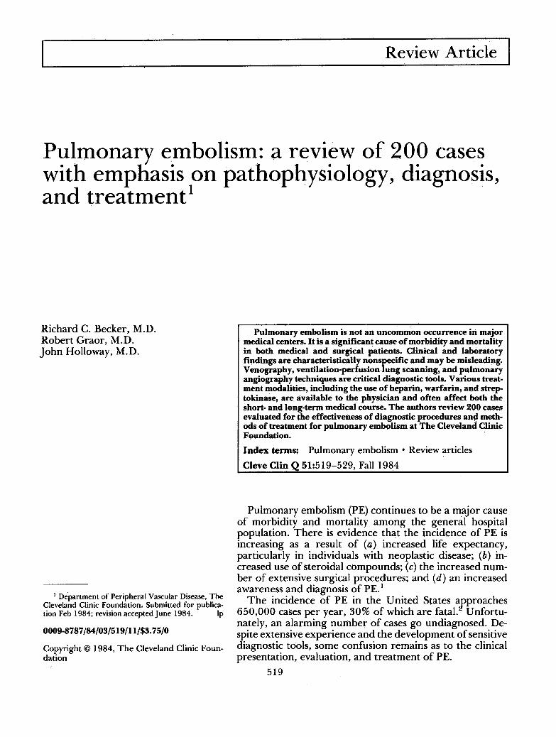
Fig. 2. A. Selective right pu lmonary a r t e ry ang iog ram, showing a large pu lmona r y embolus . B. Repea t sub t rac t ion selective ang iog ram fol lowing adminis t ra t ion of in t raar ter ia l s t rep tokinase , showing clot lysis and resumpt ion of
normal b lood flow.
mally perfused will usually be normally venti-lated. This is r e fe r r ed to as a ventilation-perfu-sion (V/Q) mismatch. Disorders such as chronic obstructive pulmonary disease, pulmonary infil-trates, tumors, granulomatous diseases, and pri-mary lung disease may have perfusion scan ab-normalities. However , they are usually accom-panied by areas of abnormal ventilation as well ( V / Q match).
T h e diagnosis of PE can be de te rmined with high probablity, based on ventilation-perfusion scan findings (Table 4).
Angiography. Selective pulmonary angiography is the definitive test by which PE can be diag-nosed. Positive signs are a sharp cutoff of the artery filled with contrast material a n d / o r an intravascular filling defect (Fig. 2, A and B). An-giography is particularly useful when: (a) there are perfusion defects that match radiographic abnormalities; (b) there are multiple perfusion defects, some normally ventilated, some not; (c) there is a history of unconf i rmed pulmonary em-
boli in a patient with an indeterminate lung scan; and (d) there is massive embolism necessitating surgical or fibrinolytic intervention. In addition, pulmonary angiography may help different ia te recur ren t embolism f rom embolus f ragmenta t ion with distal migration.
Pulmonary ar tery and pressures of the right side of the hear t can be obtained dur ing pulmo-nary angiography. These values are useful in determining the magni tude of the hemodynamic disturbance caused by the PE. In patients with preexisting cardiopulmonary disease, the inter-pretat ion of these values may be difficult.
Treatment of pulmonary embolism When PE is suspected, initial therapy should
be directed toward stabilizing the patient and initiating anticoagulation.
Heparin therapy. Hepar in is the anticoagulant of choice for the t rea tment of acute PE. Unless contraindicated, it should be given immediately to all patients suspected of having PE before

Fall 1984 Pulmonary embolism 525
diagnostic testing. Heparin is the ideal agent for initial treatment because its onset of action is immediate and it is rapidly metabolized.25
Heparin's major effect is the inhibition of the coagulation cascade, thereby preventing further thrombus formation, propagation, and emboli-zation. The body's intrinsic thrombolytic system may then dissolve the thrombus or allow it to become organized and firmly adherent to the vessel wall.
The usual initial dosage is 500 U/kg daily, given as a continuous intravenous infusion. A baseline partial thromboplastin time (PTT) is obtained before beginning therapy and the dose of heparin is adjusted to maintain the P T T 11/2-2 times the baseline value. A loading dose of 10,000 units (150 U/kg) given intravenously is used in the patient with massive PE or when there will be a delay in instituting a continuous infusion.
Hemorrhage is the major side effect of hepa-rin.27 Patients at highest risk include the elderly, those with underlying coagulopathy, those re-cently having undergone surgery, and individuals with significant hepatic dysfunction. Heparin-in-duced thrombocytopenia is an uncommon im-mune pheonomenon.28 '29
Warfarin therapy. Warfarin sodium (Coumadin) is the prototype of a class of drugs that inhibits the hepatic synthesis of the vitamin K-dependent coagulation factors II, VII, IX, and X, thereby preventing thrombin formation. It is adminis-tered one or two days after the institution of heparin therapy. The starting dose is 10 mg/day by mouth and is adjusted to maintain the pro-thrombin time at twice normal. The daily dose required varies, depending upon concomitant medical illness and other drug ingestion (Table 5). Three months is the usual length of time that warfarin is continued following PE. Beyond three months, the risk of bleeding usually outweighs the benefit.30'31
It is important that heparin therapy be contin-ued during the first four to five days of warfarin therapy to assure a state of anticoagulation while adequate depletion of all vitamin K-dependent factors is taking place.32
Thrombolytic therapy. In the patient with respi-ratory or hemodynamic compromise caused by PE, thrombolytic therapy should be considered. This is the only treatment available that promotes actual lysis of thrombus, thereby restoring nor-mal pulmonary circulation. Currently, two drugs are available: streptokinase and urokinase. Strep-
Table 5. Factors affecting coagulation response to Coumadin
Increased response Liver disease Neoplasia
Congestive heart failure Antibiotics
Decreased response Diabetes mellitus Hyperlipidemias Hypothyroid state Antacids Antihistamines
Diuretics Nonsteroidal anti-inflammatory
agents Quinidine Oral hypoglycemics
Oral contraceptives Barbiturates Vitamin K. administration Vitamin C (excessive)
tokinase acts by combining with plasminogen to form an activator substance, which indirectly con-verts plasminogen to plasmin, thereby promoting fibrin degradation and clot lysis. Urokinase acts directly on plasminogen to yield the same re-suits.33'34
Streptokinase or urokinase may be directly in-fused into the involved pulmonary artery via a catheter inserted through either a femoral or brachial vein. This approach may produce more rapid clot lysis, and further, monitoring progress with repeated pulmonary angiograms can be eas-ily accomplished. When clot lysis is confirmed, the catheter is removed and the patient is treated with conventional intravenous and oral antico-agulants. Guidelines to thrombolytic therapy are outlined (Table 6).
Inferior vena cava interruption therapy. Occa-sionally, a patient who has had a PE and has a contraindication to anticoagulation or who em-bolizes while receiving adequate anticoagulation may require inferior vena cava interruption with a caval filter or by ligation.35-39 This provides protection from further embolic episodes. How-ever, it does not treat the pulmonary embolus itself or the underlying thrombotic process. A risk (3%-5%) of thrombus extension causing in-ferior vena cava thrombosis with embolization from above the umbrella exists. Inferior vena cava thrombosis may result in severe chronic venous insufficiency. 0-44
Pulmonary embolectomy. Surgical removal of massive pulmonary emboli can be lifesaving in patients with shock who have been refractory to medical management. The surgery depends upon angiographic documentation of the em-bolus and the availability of an experienced car-diothoracic surgical team.45,46

526 Cleveland Clinic Quarterly Vol. 51, No. 3
Table 6. Guidelines for thrombolytic therapy I. Patient selection
1. Diagnosis of pulmonary embolism 2. Duration of symptoms, 7 days 3. Cardiopulmonary compromise 4. Contraindications
A. Absolute — Active internal bleeding — Recent (within 2 months) cerebrovascular accident or other active intracranial process. There is evidence that currently used
thrombolytic regimens carry the hazard of inducing cerebral hemorrhage in those with a recent history of stroke. (However, studies are currently underway using lower dosage for the treatment of stroke.)
B. Relative Major •— Recent (<10 days) major surgery, obstetrical delivery, organ biopsy, previous puncture of noncompressible vessels — Recent serious gastrointestinal bleeding — Recent serious trauma
— Severe arterial hypertension (>200 systolic or > 110 diastolic)
Minor — Recent minor trauma, including cardiopulmonary resuscitation — High likelihood of a left heart thrombus, e.g., mitral valve disease with atrial fibrillation — Bacterial endocarditis — Hemostasis defects, including those associated with severe hepatic or renal disease •— Pregnancy — Age: > 7 5 years — Diabetic hemorrhagic retinopathy
II. Drug administration Loading dose: streptokinase (250,000 units IV for 2 0 - 3 0 min)
urokinase (4,400 units/kg IV for 10 min) Maintenance: streptokinase (100,000 U/hr for 4 8 - 7 2 hr)
urokinase (4,400 U/kg/hr for 12 -24 hr)
III. Monitoring Thrombin time: 1 'A times normal Fibrinogen level: < 2 0 0 mg/100 mL If lytic state is not achieved in 4 hr, reload and continue infusion as indicated. When lytic state is achieved, obtain clotting parameters once or twice daily.
IV. Complications 1. Major bleeding requiring cessation of therapy (<5%) 2. Febrile reaction (streptokinase) (20%) 3. Allergic cutaneous reaction (streptokinase) (<1 %)
V. Treatment of complications 1. Bleeding: minimize invasive procedures
compression dressings 2. Major bleeding: discontinue infusion
fresh frozen plasma/RBC transfusion 3. Febrile/skin rash: hydrocortisone (100 mg IV)
diphenhydramine (50 mg IV)
IV = intravenous.
Prevention. Death from a PE in a patient who has otherwise undergone successful surgical or medical treatment is tragic. Prophylactic mea-sures are available that decrease the incidence of deep vein thrombosis and subsequent emboliza-
tion. Heparin (5,000 units) given subcutaneously every 12 hours inactivates factor X, thereby pre-venting thrombus formation. The efficacy of this regimen has proved effective in patients with heart disease, stroke, neoplasia, and in those >40

Fall 1984 Pulmonary embolism 527
Clinically suspected PE
CXR, ECG, ABG I
Perfusion lung scan
Abnormal I
Ventilation Scan
Normal
Low or indetermanent
probability
Venography or
Doppier/IPG
High probability
PE
PE excluded
reatment
DVT and possible PE
PE treatment
Pulmonary Angiogram
PE excluded
PE treatment
Fig. 3. Algor i thm fo r d iagnos ing PE.
years of age undergoing thoracic or abdominal surgery. Patients undergo ing major or thopedic or urologie surgery are not protected by low-dose heparin therapy.4 9 In termi t tent compres-sion stockings alone or combined with low-molec-ular weight dext ran 5 0 or low-dose warfarin seem to be effective prophylactic measures in these patients.51 When anticoagulation is contraindi-cated, such as in neurosurgical or ophthalmolog-ical procedures, in termit tent compression stock-ings may be effective.
O the r methods of prophylaxis are available and include adjusted doses of heparin,5 2 '5 3 hepa-rin and dihydroergotamine , cont inuous passive motion devices, and antiplatelet agents such as aspirin.
Conclusion T h e mortality of undiagnosed PE approaches
30%. Short- and long-term morbidity exceeds this figure. O u r retrospective review of patients diagnosed as having suffered a PE while hospi-

528 Cleveland Clinic Quarterly Vol. 51, No. 3
talized at The Cleveland Clinic Foundation em-phasize that the approach to evaluating a patient suspected of having a PE must be logical, precise, and systematic (Fig. 3).
In summary, PE is common in the hospital setting. Most emboli originate from thrombi within the deep veins of the pelvis and lower extremities. The presentation of PE may be non-specific or classic with chest pain, dyspnea, he-moptysis, and hemodynamic compromise. PE should always be considered in a patient at risk with unexplained cardiopulmonary signs and symptoms. The approach to diagnosis should be prompt and systematic, using ventilation-perfu-sion lung scanning and pulmonary angiography when indicated. Early treatment significantly af-fects morbidity and mortality.
Robert Graor, M.D. Department of Peripheral
Vascular Disease The Cleveland Clinic Foundation 9500 Euclid Ave. Cleveland OH 44106
References 1. Rosenow EC III, Osmundson PJ, Brown L. Pulmonary em-
bolism. Mayo Clin Proc 1981; 56:161-178. 2. Dalen JE, AlpertJS. Natural history of pulmonary embolism.
Prog Cardiovasc Dis 1975; 17:259-270. 3. Barritt DW, Jordan SC. Anticoagulant drugs in the treat-
ment of pulmonary embolism: a controlled trial. Lancet 1960; 1:1309-1312.
4. Satiani B. Management of acute deep vein thrombosis. Am Fam Physician 1982; 25:103-109.
5. Moser KM, LeMoine JR. Is embolic risk conditioned by location of deep venous thrombosis? Anh Intern Med 1981; 94:439-444.
6. Maurer BJ, Wray R, Shillingsford JP. Frequency of venous thrombosis after myocardial infarction. Lancet 1971; 2:1385-1387.
7. Harvey-Kemble JV. Incidence of deep vein thrombosis. Br J Hosp Med 1971; 6:721-726.
8. Virchow R. Cellular pathology as based upon physiological and pathological histology. Translated from 2nd ed. London, John Churchill, 1860.
9. Allison PR. Pulmonary embolism and thrombophlebitis. Br J Surg 1967 54:466-470.
10. Browse NL. Effect of surgery on resting calf blood flow. Br Med J 1962; 1:1714-1721.
11. Moncada S, Higgs EA, Vane JR. Human arterial and venous tissues generate prostacylcin (prostaglandin X), a potent inhib-itor of platelet aggregation. Lancet 1977; 1:18-20.
12. Moncada S, Vane JR. Pharmacology and endogenous roles of prostaglandin endoperoxides, thromboxane A2, and pros-tacyclin. Pharmacol Rev 1979; 30:293-332.
13. Du P Heyns A, Badenhorst CJ, Retief FP. A stable non-prostaglandin inhibitor of platelet aggregation in human aorta intima extracts. S Afr Med J 1979; 55:908-912.
14. Henderson ES, Rapaport SI. The thrombotic activity of activation product. J Clin Invest 1962; 41:235-240.
15. A cooperative study. Urokinase pulmonary embolism trial, phase 1 results. JAMA 1970; 214:2163-2172.
16. A cooperative study. Urokinase-streptokinase embolism trial, phase 2 results. JAMA 1974; 229:1606-1613.
17. Nelson JR, Smith JR. The pathologic physiology of pulmo-nary embolism: a physiologic discussion of the vascular reac-tions following pulmonary arterial obstruction by emboli of varying size. Am Heart J 1959; 58:916-932.
18. Widdicombe JG. Reflex mechanisms in pulmonary throm-boembolism. [In] Moser KM, Stein M, eds. Pulmonary Thromboembolism. Chicago, Year Book Medical Publishers, 1973.
19. Stein M, Hirose T , Yasutkat, Tarabeih A. Airways response to pulmonary embolism: pharmacologic aspects. [In] Moser KM, Stein M, eds. Pulmonary Thromboembolism. Chicago, Year Book Medical Publishers, 1973.
20. Jardin F, Gurdjian F, Desfonds P, Fouilladieu JL, Margairaz A. Hemodynamic factors influencing arterial hypoxemia in massive pulmonary embolism with circulatory failure. Circu-lation 1979; 59:909-912.
21. Hampton AO, Castleman B. Correlation of postmortem chest teleroentgenograms with autopsy findings, with special reference to pulmonary embolism and infarction. AJR 1940; 43:305-326.
22. Chang CH. Radiological considerations in pulmonary em-bolism. Clin Radiol 1967; 18:301-309.
23. Kerr IH, Simon G, Sutton GC. The value of the plain radiograph in acute massive pulmonary embolism. Br J Radiol 1971;44:751-757.
24. Mclntyre KM, Sasabara AA. Determinations of cardiovas-cular responses to pulmonary embolism. [In] Moser KM, Stein M, eds. Chicago, Year Book Medical Publishers, 1973.
25. Sasahara AA. Therapy for pulmonary embolism. JAMA 1974; 229:1795-1798.
26. Genton E, Hirsh J. Observations in anticoagulant and throm-bolytic therapy in pulmonary embolism. Prog Cardiovasc Dis 1975; 17:335-343.
27. Kelton JG, Hirsh J. Bleeding associated with antithrombotic therapy. Semin Hematol 1980; 17:259-291.
28. Bell WR, Royall RM. Heparin-associated thrombocytopenia: a comparison of three heparin preparations. N Engl J Med 1980; 303:902-907.
29. Powers PJ, Cuthbert D, Hirsh J. Thrombocytopenia found uncommonly during heparin therapy. JAMA 1979; 241:2396-2397.
30. Thomas DP. Heparin in the prophylaxis and treatment of venous thromboembolism. Semin Hematol 1978; 15:1-17.
31. Brozovic M. Oral anticoagulants in clinical practice. Semin Hematol 1978; 15:27-34.
32. Gurewich V. Guidelines for the management of anticoagu-lant therapy. Semin Thromb Hemostas 1976; 2: 176-196.
33. Sherry S, Lindemeyer RI, Fletcher AP, Alkjaersig N. Studies on enhanced fibrinolytic activity in man. J Clin Invest 1959; 38:810-822.
34. Sherry S, Fletcher AP, Alkjaersig N. Fibrinolysis and fibrin-olytic activity in man. Physiol Rev 1959; 39:343-382.
35. Bergan JJ, Trippel OH. Vena cava operations for prevention of pulmonary embolism. Surg Clin North Am 1966; 46:195-207.
36. Wheeler CG, Thompson JE, Austin DJ, Patman RD, Stockton RL. Interruption of the inferior vena cava for thromboem-

Fall 1984 Pulmonary embolism 529
bolism: comparison of ligation and plication. Ann Surg 1966; 163:199-204.
37. Mobin-Uddin K, Callard GM, Bolooki H, Rubinson R, Michie D, Jude JR. Transvenous caval interruption with umbrella filter. N Engl J Med 1972; 286:55-58.
38. Silver D, Sabiston DC Jr. The role of vena caval interruption in the management of pulmonary embolism. Surgery 1975; 77:1-10.
39. Greenfield LJ, ZoccoJJ. Intraluminal management of acute massive pulmonary thromboembolism. J Thorac Cardiovasc Surg 1979; 77:402-410.
40. Gurewich V, Thomas DP, Rabinov KR. Pulmonary embo-lism after ligation of the inferior vena cava. N Engl J Med 1966; 274:1350-1354.
41. Piccone VA Jr, Vidal E, Yarnoz M, Glass P, LeVeen HH. The late results of caval ligation. Surgery 1970; 68:980-998.
42. Wingerd M, Bernhard VM, Maddison F, Towne JB. Comparison of caval filters in the management of venous thromboembolism. Arch Surg 1978; 113:1264-1271.
43. Donaldson MC, Wirthlin LS, Donaldson GA. Thirty-year experience with surgical interruption of the inferior vena cava for prevention of pulmonary embolism. Ann Surg 1980; 191:367-372.
44. Stewart JR, Greenfield LJ. Transvenous vena caval filtration and pulmonary embolectomy. Surg Clin North Am 1982; 62:411-430.
45. Gifford RW, Groves LK. Limitations in the feasibility of pulmonary embolectomy: a clinicopathologic study of 101
cases of massive pulmonary embolism. Circulation 1969; 39:523-530.
46. Sabiston DC Jr, Wolfe WG. Pulmonary embolectomy. [In] Moser KM, Stein M. eds. Pulmonary Thromboembolism. Chicago, Year Book Medical Publishers, 1973.
47. McNeil BJ. A diagnostic strategy using ventilation-perfusion studies in patients suspect for pulmonary embolism. J Nucl Med 1976; 17:613-616.
48. McNeil BJ. Ventilation-perfusion studies and the diagnosis of pulmonary embolism: concise communication. J Nucl Med 1980; 21:319-323.
49. Hampson WG, Harris FC, Lucas HK, et al. Failure of low-dose heparin to prevent deep-vein thrombosis after hip-re-placement arthroplasty. Lancet 1974; 2:795-797.
50. Nicolaides AN, Fernandes e Fernandes J, Pollock AV. Intermittent sequential pneumatic compression of the legs in the prevention of venous stasis and postoperative deep venous thrombosis. Surgery 1980; 87:69-75.
51. Hull R, Delmore T, Carter C, et al. Adjusted subcutaneous heparin versus warfarin sodium in the long-term treatment of venous thrombosis. N Engl J Med 1982; 306:189-194.
52. Leyvraz PF, Richard J, Bachmann F, et al. Adjusted versus fixed-dose subcutaneous heparin in the prevention of deep-vein thrombosis after total hip replacement. N Engl J Med 1983; 309:954-958.
53. Harris WH, Salzman EW, Athanasoulis C, Waltman AC, Baum S, DeSanctis RW. Comparison of warfarin, low-mo-lecular-weight dextran, aspirin, and subcutaneous heparin in prevention of venous thromboembolism following total hip replacement. J Bone Joint Surg 1974; 56-A:l 552-1562 .