pud0916100004 physician up 09-sp-2 · michael choti and timothy paw-lik are determined to improve...
TRANSCRIPT

W hen she first came under his care in October 2005, the 63-year-old office manager told urologist E. James
Wright that she’d “had enough” with recurring pelvic floor problems. She’d recently been to two gynecologists who told her they were out of per-manent solutions. Now she felt like her “insides were falling out,” as her bladder “was more on the outside of me than the inside of me.”
It didn’t take Wright long to reach an assess-ment. About 100 times in the last five years, he’s performed a type of minimally invasive prolapse surgery to address loss of support in women’s pelvic organs. He says one in four women will suffer from pelvic floor disorders in some form during their lifetime, and up to 20 percent will experience prolapse.
The compromised support issues, he says, are re-ally a “consequence of a life well-lived.” His patients have typically given birth to multiple children and been physically active over many years. Many are smokers. Some are overweight. Most are past menopause and have had hysterectomies and other surgeries that can weaken support of pelvic structures.
Symptoms usually arrive with a general sense of pressure in the lower pelvic area, which some patients describe as an uncomfortable pulling sensation or a bulge in the vagina that worsens toward the end of a long day of activity. “Patients have told me it almost feels like they’re sitting on a tennis ball,” Wright says.
The condition can sometimes cause urinary retention or leakage, stool trapping and difficulty with intimacy as the bladder drops and/or the uterus slips lower, sometimes working its way through the birth canal.
Traditional repair techniques have relied on reshaping the fallen pelvic structures in an overlapping “leafing” pattern with traditional suturing approaches. Those methods have shown a high rate—up to 15 percent—of prolapse recurrence. Laparoscopic and open surgical techniques can also be used but require entrance into the abdomen, adding to recovery and greater risk. Wright says his less invasive method leaves fewer external scars, lasts longer, reduces pain and allows patients to leave the hospital and resume normal
activities much faster.Describing his approach as “tension-free,” Wright creates a
hammock-like structure with a polypropylene mesh that cradles the targeted pelvic structures. The mesh is self-anchoring with the patient’s native pelvic tissues, much in the way Velcro works. “It integrates with the body’s natural supporting points,” Wright says, “getting back to a situation close to the normal anatomy.” Al-though some experts argue that the mesh is capable of eroding over time, that’s happened in only six of Wright’s cases and was, he says, easily corrected.
The procedure is mostly accomplished transvaginally with a mini-mum number of external incisions. It can usually be completed in less than two hours, on an outpatient basis or followed by a single overnight stay. Patients resume normal activities in about one or two weeks.
“Dr. Wright repaired the floor, the ceiling, the back wall, and also touched up the bowel,” says the office manager patient, whose only remaining issue is a rare instance of urinary leakage when she sneezes. “I just want more people to know that this procedure is out there.”
% 410-550-7008 to refer a patient.
Non-Profit OrgU.S. Postage PAIDPermit No. 5415Baltimore, MD
Johns Hopkins MedicineReferring Physician Services901 South Bond Street / Suite 350 Baltimore, Maryland 21231-3339
This newsletter is published for the Johns Hopkins Clinical Practice Association by Johns Hopkins Medicine Marketing and Communications
Clinical Practice AssociationWilliam Baumgartner, M.D., Johns Hopkins Medicine vice dean for clinical affairs; Clinical Practice Association president
Marketing and CommunicationsDalal Haldeman, Ph.D., M.B.A., vice presidentPatrick Gilbert, director of editorial servicesMary Ann Ayd, managing editorMarjorie Centofanti, Ramsey Flynn, Lauren Manfuso, Daphne Swancutt, writers David Dilworth, designer; Keith Weller, photographer
Questions or comments about this issue?Call 410-955-2902 or e-mail [email protected]
© 2009 The Johns Hopkins University and The Johns Hopkins Health System Corporation
CHANGE SERVICE REQUESTED
UROLOGY
PhysicianUpdate
Your Vital LinksJohns Hopkins Medicine offers the following links to physicians in the surrounding community. We also urge M.D.’s to use our Physician Liaison Service to offer suggestions and comments.
Hopkins Access Line (HAL): Physician-Only Line for Consultations, Referrals and Patient Transfers 1-800-765-5447 (Continental United States)410-955-9444 (Baltimore area and international calls)
Online Referral Directoryhopkinsmedicine.org/referraldirectory
Physician Liaison Service: 1-800-759-7734 (Continental United States) 410-502-2737 (Baltimore area and international calls) [email protected]
CME Program Informationhopkinscme.eduInformation: 410-502-9634Fax registration: 410-955-0807E-mail: [email protected]
Johns Hopkins USA: For Out-of-State Patients 410-735-4872 [email protected]
Services for the Militaryhopkinsmedicine.org/usfhp/
Cancer Center Clinical Trialshopkinskimmelcancercenter.org/ clinicaltrials
Some of the research reported in this newsletter may have corporate sponsor-ship. For more information, please contact the Office of Policy Coordination at 410-516-5560.
E. James Wright: Women have an 11 percent lifetime risk of needing surgical correction for prolapse.
New Support for the Pelvic Floor
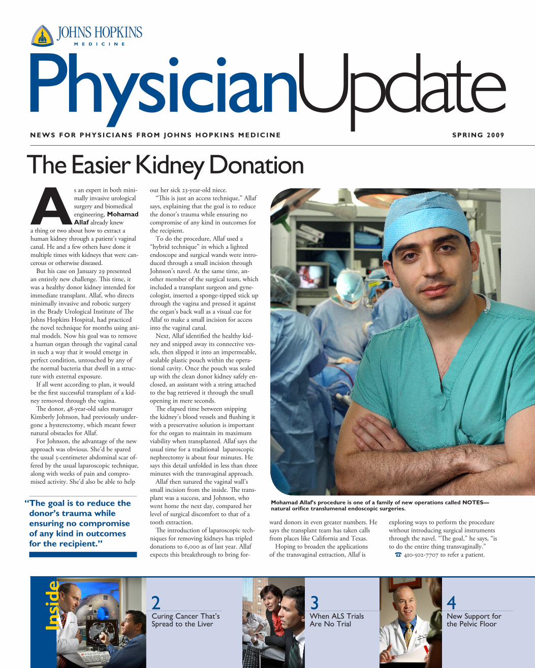
PhysicianUpdateA s an expert in both mini-
mally invasive urological surgery and biomedical engineering, Mohamad Allaf already knew
a thing or two about how to extract a human kidney through a patient’s vaginal canal. He and a few others have done it multiple times with kidneys that were can-cerous or otherwise diseased.
But his case on January 29 presented an entirely new challenge. This time, it was a healthy donor kidney intended for immediate transplant. Allaf, who directs minimally invasive and robotic surgery in the Brady Urological Institute of The Johns Hopkins Hospital, had practiced the novel technique for months using ani-mal models. Now his goal was to remove a human organ through the vaginal canal in such a way that it would emerge in perfect condition, untouched by any of the normal bacteria that dwell in a struc-ture with external exposure.
If all went according to plan, it would be the first successful transplant of a kid-ney removed through the vagina.
The donor, 48-year-old sales manager Kimberly Johnson, had previously under-gone a hysterectomy, which meant fewer natural obstacles for Allaf.
For Johnson, the advantage of the new approach was obvious. She’d be spared the usual 5-centimeter abdominal scar of-fered by the usual laparoscopic technique, along with weeks of pain and compro-mised activity. She’d also be able to help
out her sick 23-year-old niece.“This is just an access technique,” Allaf
says, explaining that the goal is to reduce the donor’s trauma while ensuring no compromise of any kind in outcomes for the recipient.
To do the procedure, Allaf used a “hybrid technique” in which a lighted endoscope and surgical wands were intro-duced through a small incision through Johnson’s navel. At the same time, an-other member of the surgical team, which included a transplant surgeon and gyne-cologist, inserted a sponge-tipped stick up through the vagina and pressed it against the organ’s back wall as a visual cue for Allaf to make a small incision for access into the vaginal canal.
Next, Allaf identified the healthy kid-ney and snipped away its connective ves-sels, then slipped it into an impermeable, sealable plastic pouch within the opera-tional cavity. Once the pouch was sealed up with the clean donor kidney safely en-closed, an assistant with a string attached to the bag retrieved it through the small opening in mere seconds.
The elapsed time between snipping the kidney’s blood vessels and flushing it with a preservative solution is important for the organ to maintain its maximum viability when transplanted. Allaf says the usual time for a traditional laparoscopic nephrectomy is about four minutes. He says this detail unfolded in less than three minutes with the transvaginal approach.
Allaf then sutured the vaginal wall’s small incision from the inside. The trans-plant was a success, and Johnson, who went home the next day, compared her level of surgical discomfort to that of a tooth extraction.
The introduction of laparoscopic tech-niques for removing kidneys has tripled donations to 6,000 as of last year. Allaf expects this breakthrough to bring for-
ward donors in even greater numbers. He says the transplant team has taken calls from places like California and Texas.
Hoping to broaden the applications of the transvaginal extraction, Allaf is
exploring ways to perform the procedure without introducing surgical instruments through the navel. “The goal,” he says, “is to do the entire thing transvaginally.”
% 410-502-7707 to refer a patient.
SPRING 2009
When ALS Trials Are No Trial
New Support for the Pelvic Floor
2 3 4Curing Cancer That’s Spread to the Liver
N E W S f O R P H Y S I C I A N S f R O M J O H N S H O P k I N S M E D I C I N E
Insi
de
The Easier Kidney Donation
Mohamad Allaf’s procedure is one of a family of new operations called NOTES—natural orifice translumenal endoscopic surgeries.
“ The goal is to reduce the donor’s trauma while ensuring no compromise of any kind in outcomes for the recipient.”

for some patients with primary liver cancer, liver transplantation has long been a treatment op-tion. But when colon or other cancers have metastasized to the
liver, patients are often told that no surgical treatment is available.
Michael Choti and Timothy Paw-lik are determined to improve the out-comes in cases that most surgeons consider inoperable. Choti, a leading liver cancer surgeon, and Pawlik, director of hepato-biliary surgery, have been exploiting the liver’s natural ability to regenerate itself with a “divide and conquer” approach.
When there are a limited number of tu-mors in the liver, surgeons can resect them, and the organ rather quickly regenerates itself. But when there are many tumors in all areas of the liver, or very large tumors, surgery becomes far more difficult because patients need a certain amount of hepatic reserve to live. “We are one of the few centers in the country,” says Pawlik, “help-ing patients who have tumors that require removal of 70 to 80 percent of the liver.”
Using images provided by liver radiolo-gist Ihab kamel, they determine how much of the liver must be removed to get all of the cancer. If it is too much, they turn to interventional radiologist Jeff Geschwind for a technique called portal vein embolization.
Relying on the fact that each of the liver’s halves has its own portal vein to carry critical growth factors that give the liver its unique rejuvenation capabilities, Geschwind blocks off blood supply to the tumor-filled side of the liver, caus-ing the nontumorous side to grow larger. When it gets big enough to represent 20 percent to 30 percent of the total liver, Chotic and Pawlik can operate and remove the tumors.
For patients deemed inopera-ble because of tumors through-out both sides of the liver, they take a two-step approach. They can remove all the cancer from one side of the liver, wait for the other side of the liver to regrow, then remove the remaining can-cer in the other half of the liver. This pro-cedure, they say, has cured some patients who’d been told they had no options.
In some instances, the same approach is used to treat carcinoid or neuroendo-crine tumors that have spread to the liver. Among the success stories is a 45-year-old man who had a pancreatic neuroendocrine tumor and multiple liver metastases. Paw-lik performed a pancreatectomy to remove the pancreatic neuroendocrine tumor and also did a two-step liver surgery to remove
all the disease in the liver. Two years later, the patient remains disease free.
To help patients with primary liver can-cers who aren’t candidates for transplanta-tion and whose cancers are too advanced to be resected, the liver team offers tran-sarterial chemo embolization. A catheter inserted via the femoral artery is threaded to the blood vessel feeding the primary liver tumor. The catheter delivers chemo-therapy directly to the tumor, after which the blood vessel is embolized to block the tumor’s vascular supply and ensure that the anticancer drugs stay in the tumor.
Choti says the combination of these techniques and new anticancer drugs is
improving outcomes. Over the last 10 to 15 years, survival rates for patients with meta-static colorectal cancer to the liver have doubled, and five years out are now more than 50 percent.
Noting the progress also being made for other cancers of the liver, Pawlik has launched the one-day, one-stop Johns Hopkins Medicine Liver Tumor Multidis-ciplinary Clinic that provides a compre-hensive approach to treat patients with a wide range of liver tumors. “As we expand the resection criteria,” he says, “a multidis-ciplinary approach is critical to ensure the best chance of success for these patients.”
% 1-877-LIVER-99 to refer a patient.
2 • Johns hopkins phys ic ian update • S PR ING 2009
Curing Cancer That’s Spread to the Liver
GASTROENTEROLOGY
ONCOLOGIC SURGERY
Michael Choti and Timothy Pawlik: “We are seeing more and more patients who’ve been told they are unresectable because of the type of cancer they have or because of the technical aspects of the surgery.”
Just a few short years ago, identify-ing esophageal cancers endoscopi-cally was a hit-or-miss mission. While endoscopy has enabled minimally invasive access to the
gastrointestinal tract, identifying and diagnosing suspicious tissues have been quantity versus quality exercises.
“We could scope out large areas for biopsy,” says gastroenterologist kerry Dunbar, “but missing has been the abil-ity to be more precise and selective about what we biopsy.”
That’s meant multiple biopsies and tissue samples being sent to pathology, and needle-in-the-haystack searches. It’s time-consuming and frustrating, especially when patients are anxious for immediate results.
That waiting game changed when Johns Hopkins recently became the first facility in the United States to use confocal endo-
microscopy to instantly diagnose patients with gastrointestinal cancer.
Tipping the end of a traditional endo-scope is a miniaturized microscope, which lets physicians view cellular, vascular and connective structures in detail. Where the devil is in those details, Dunbar and gas-troenterologist Marcia Canto are seeing a more precise view of them than ever.
“We’re seeing the mucosa magnified about a thousand times,” says Dunbar. “And, that’s letting us see in vivo differ-ences between normal and precancerous cellular changes.”
While that’s helping to improve di-agnosis of diseases of the esophagus and gastrointestinal tract, Dunbar is also using endomicroscopy specifically for Barrett’s esophagus.
The lifetime cancer risk for Barrett’s patients is about 5 percent. What’s been tricky for doctors is identifying whether
and to what degree dysplasia—that atypical change in cell nuclei or growth patterns—exists in Barrett’s.
“The idea is to be able to identify it early,” says Dunbar. Endomicroscopy may help that endeavor by not only magnify-ing the field of vision but allowing a more discerning tissue selection.
“What that means,” says Dunbar, “is that we’re sending fewer tissue samples to pathologists and only those that have been identified as suspicious via endomicros-copy.”
If Dunbar proves its effectiveness for potential cancers in Barrett’s patients, endomicroscopy could change the way physicians view and evaluate abnormal lesions during endocospies, particularly in determining the need for biopsies and potential removal of lesions.
% 410-955-9696 to refer a patient.
Endomicroscopy Magnifies the Field of Vision for GI Cancers
Gastroenterologist kerry Dunbar
I can’t stress enough how important it is to get people with ALS into the clinic early in the disease,” says neurologist/neuroscientist Nicholas Maragakis.
Given that amyotrophic lateral sclerosis is incurable, it may be tempting to ask why the hurry. There are two reasons. Johns Hopkins’ ALS Multidisciplinary Clinic has existed for years and, due in part to Director Lora Clawson’s unwavering insistence on consummate care, early-entry patients tend to keep function longer with a higher quality of life. Studies on patients in large academic centers underscore that.
And, increasingly, those with ALS benefit from closeness to clinical trials. Even though no solidly therapeutic ALS drug has surfaced, results of Hopkins’ best practices studies, for example, reach its patients’ bedsides faster. That includes a recent look at earlier use of forced venti-lation —BiPAP—showing it can prolong life roughly a year. That result has already changed clinical practice. The same holds for gastrostomy tube feeding: Earlier use improves survival.
“Even the negative studies help,” says Clawson. When pre-clinical work with the antibiotic minocycline looked interesting enough that ALS patients nationwide asked their physicians for the drug, trials at Hopkins and elsewhere showed it was a danger-
ous option.Each year, some 50 percent of ALS patients join trials at Hop-
kins. “The breadth of our human studies,” says Maragakis, “is exceptional—drug therapy, genetics, even stem cells. And the benefits are real.”
% 410-955-8511 to refer a patient to the ALS Multidisciplinary Clinic
A lthough it has long been known that both men and women can transmit human papillomavirus, the clinical and research
focus once centered almost entirely on HPV’s ability to cause cervical cancer—a disease to which men are obviously not susceptible. That thinking changed in 2000, when Johns Hopkins scientists first linked HPV to head and neck cancers.
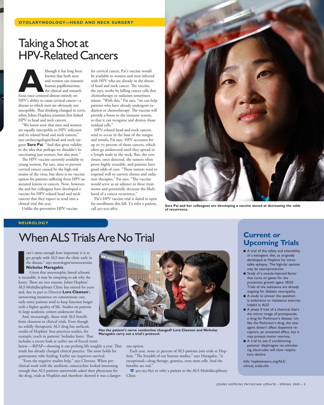
“We know now that men and women are equally susceptible to HPV infection and its related head and neck tumors,” says otolaryngologist/head and neck sur-geon Sara Pai. “And that gives validity to the idea that perhaps we shouldn’t be vaccinating just women, but also men.”
The HPV vaccine currently available to young women, Pai says, aims to prevent cervical cancer caused by the high-risk strains of the virus, but there is no vaccine option for patients suffering from HPV-as-sociated lesions or cancers. Now, however, she and her colleagues have developed a vaccine for HPV-related head and neck cancers that they expect to send into a clinical trial this year.
Unlike the preventive HPV vaccine
for cervical cancer, Pai’s vaccine would be available to women and men infected with HPV who are already in the throes of head and neck cancer. The vaccine, she says, works by killing cancer cells that chemotherapy or radiation sometimes misses. “With this,” Pai says, “we can help patients who have already undergone ra-diation or chemotherapy. The vaccine will provide a boost to the immune system, so that it can recognize and destroy those residual cells.”
HPV-related head and neck cancers tend to occur in the base of the tongue and tonsils, Pai says. HPV accounts for up to 70 percent of those cancers, which often go undetected until they spread to a lymph node in the neck. But, she con-tinues, once detected, the tumors often prove highly treatable, and patients have good odds of cure. “These tumors tend to respond well to current chemo and radia-tion therapies,” Pai says. “The vaccine would serve as an adjunct to these treat-ments and potentially decrease the likeli-hood of a cancer recurrence.”
Pai’s HPV vaccine trial is slated to open for enrollment this fall. To refer a patient, call 410-502-9825.
When ALS Trials Are No TrialNEUROLOGY
Taking a Shot at HPV-Related Cancers
Has the patient’s nerve conduction changed? Lora Clawson and Nicholas Maragakis carry out a trial’s protocol.
OTOLARYNGOLOGY—HEAD AND NECk SURGERY
Sara Pai and her colleagues are developing a vaccine aimed at decreasing the odds of recurrence.
Current or Upcoming Trialsn A trial of the safety and tolerability
of a ketogenic diet, as originally developed at Hopkins for intrac-table epilepsy. The high-fat options may be neuroprotective.
n Study of a muscle-injected factor that turns on genes for the protective growth agent VEGF. Trials of the substance are already ongoing for diabetic neuropathy.
n A study to answer the question: Is endurance or resistance exercise helpful in ALS?
n A phase II trial of a chemical that’s the mirror image of pramipexole, a drug for Parkinson’s disease. Un-like the Parkinson’s drug, the new agent doesn’t affect dopamine re-ceptors, an unwanted effect, but it may protect motor neurons.
n A trial to see if conditioning patients’ diaphragms via stimulat-ing electrodes will slow respira-tory decline.
Info: hopkinsneuro.org/ALS/ clinical_trials.cfm
Johns hopkins phys ic ian update • spR inG 2009 • 3