psma-targeted radionuclide therapy of metastatic castration
TRANSCRIPT
PSMA-targeted radionuclide therapy
of metastatic castration-resistant prostate cancer
with Lu-177 labeled PSMA-617
Clemens Kratochwil1, Frederik L. Giesel1, 2, Melsa Stefanova1, Martina
Benešová3, Marcus Bronzel4, Ali Afshar-Oromieh1, 2, Walter Mier1, Matthias
Eder3, Klaus Kopka3, and Uwe Haberkorn1, 2
1Department of Nuclear Medicine, University Hospital Heidelberg, Germany
2Clinical Cooperation Unit Nuclear Medicine, German Cancer Research Center
(dkfz), Heidelberg, Germany
3Division of Radiopharmaceutical Chemistry, German Cancer Research Center
(dkfz), Heidelberg, Germany
4ABX-CRO, Dresden, Germany
Corresponding author:
Dr. med. Clemens Kratochwil
Department of Nuclear Medicine
University of Heidelberg
Im NeuenheimerFeld 400
69120 Heidelberg
Tel. +49-6221-56-37164 (Fax. +49-6221-56-5473)
Email: [email protected]
Running Title: 177Lu-PSMA-617 targeted therapy of mCRPC
Journal of Nuclear Medicine, published on March 16, 2016 as doi:10.2967/jnumed.115.171397by on February 13, 2018. For personal use only. jnm.snmjournals.org Downloaded from
ABSTRACT
Prostate-specific membrane antigen (PSMA) is an excellent target for
radionuclide therapy of metastasized castration-resistant prostate cancer
(mCRPC). Besides high affinity and long tumor retention, the DOTA-
conjugated ligand PSMA-617 has low kidney uptake making it an excellent
choice for therapeutic application.
We retrospectively report our experience with 177Lu-PSMA-617 targeted
radionuclide therapy in a case series of mCRPC patients resistant to other
treatments.
Methods: Patients with PSMA-positive tumor phenotypes were selected by
molecular imaging. 30 patients received 1-3 cycles of 177Lu-PSMA-617. During
therapy pharmacokinetics and radiation-dosimetry were evaluated. Blood cell
count was checked every two weeks after the first and every four weeks after
succeeding cycles. Prostate specific antigen (PSA) was determined every four
weeks. Radiological restaging was performed after three cycles.
Results: 21/30 patients had a PSA response; in 13/30 the PSA decreased
>50%. After 3 cycles 8/11 patients achieved a sustained PSA response
(>50%) for over 24 weeks which also correlated with radiological response
(decreased lesion number and size). Normally, acute hematotoxicity was mild.
Diffuse bone marrow involvement was a risk factor for higher grade
myelosuppression but could be identified by PSMA-imaging in advance.
Xerostomia, nausea and fatigue occurred sporadically (<10%). Clearance of
non-tumor-bound tracer is predominantly renal and widely completed by 48h.
Safety dosimetry reveals kidney doses of approx. 0.75 Gy/GBq, red-marrow
0.03 Gy/GBq, salivary glands 1.4 Gy/GBq; irrespective of tumor burden and
consistent on subsequent cycles. Mean tumor absorbed dose ranged 6-22
Gy/GBq during cycle-1.
by on February 13, 2018. For personal use only. jnm.snmjournals.org Downloaded from
Conclusion: 177Lu-PSMA-617 is a promising new option for therapy of
mCRPC and deserves more attention in larger prospective trials.
Keywords: PSMA, Lu-177, castration-resistant prostate cancer, radionuclide
therapy, pharmacokinetics and dosimetry
by on February 13, 2018. For personal use only. jnm.snmjournals.org Downloaded from
INTRODUCTION
Despite recent approval of some novel drugs, metastatic castration-resistant
prostate cancer (mCRPC) remains a lethal disease and additional treatment
options are still needed.
PSMA is a promising target for directing new therapies. It is found in the
majority of prostate cancers (1) and its overexpression correlates with
traditional adverse prognostic factors (2). Binding of a ligand leads to
internalization via clathrin-coated pits (3) and prolonged retention in the cell.
PSMA-antibody-auristatin conjugates have been considered one option (4) but
they face the inherent resistance of mCRPC against most (excepting taxanes)
conventional chemotherapies. In contrast, prostate cancer is usually
radiosensitive. Radiotherapy is a standard treatment for localized prostate
cancer, for palliative management of mCRPC and even radiopharmaceuticals
targeting the surrounding bone matrix instead of the tumor itself can improve
survival (5). Therefore it seems more promising that a radioactive PSMA-ligand
which is directly internalized into tumor cells will be effective in delivering high
doses for systemic endo-radiotherapy. A phase-2 study using the radiolabelled
antibody 177Lu-J591 already demonstrated moderate anti-tumor effects (6), but
the slow diffusion of antibodies into solid lesions and hematotoxicity caused by
a long circulation time in blood are limitations (7,8). Due to faster kinetics, the
PSMA targeted small molecule MIP-1095, when labelled with 131I
demonstrated superior outcomes to the antibody approach with PSA
responses in 17/28 patients (9). Unfortunately, the co-emission of high energy
photons from 131I requires elaborate radiation protection. Unlike 131I, 177Lu is a
more pure Beta particle emitter and preferable for clinical routine. The DOTA-
conjugated PSMA-617 can be labelled with 177Lu-Lu3+ and was further refined
in tumor-targeting with low nanomolar affinity in the range of Ki = 0.37 nM
(NAALADase assay) and Ki = 2.34 nM (equilibrium dissociation constant on
by on February 13, 2018. For personal use only. jnm.snmjournals.org Downloaded from
LNCaP) and highly efficient internalization with approx. 75% of the total cell
associated activity internalized after 3 h of incubation on LNCaP (10,11,12).
Here we report our first clinical experience with 177Lu-PSMA-617 in patients
with advanced mCRPC resistant or with contraindications to other conventional
therapies and PSMA-positive tumor phenotypes as demonstrated by molecular
imaging using structurally related diagnostic analogues (Figure-1). All used
PSMA-ligands share the Glu-Urea-motif for binding to the proteolytic domain
and a lipophilic chelate or linker region to interact with the hydrophobic
accessory pocket proposed by Bařinka et al. (13).
MATERIALS AND METHODS
Patients
177Lu-PSMA-617 was offered as surrogate therapy in accordance with the
updated Declaration of Helsinki, paragraph-37 “Unproven Interventions in
Clinical Practice” and in accordance with German regulations for
“compassionate use” which includes priority of all approved treatments (without
contraindications) and confirmation of the indication by both a nuclear
medicine physician and an external expert in urology or oncology. In brief: All
30 patients were refractory to LHRH-analogs and anti-androgens (Table 1). 23
patients had prior treatment with abiraterone or enzalutamide, 11 of them had
received both. 14 patients were refractory to docetaxel, 4 had subsequently
also been treated with cabazitaxel and 4 with estramustine. 6 patients were
pretreated with Ra-223. In contrast to a formal clinical trial, no systematic
patient selection was performed, except all patients had to present with a
PSMA-positive tumor phenotype based on PSMA-imaging. All patients were
informed about the experimental nature of this therapy and gave written
by on February 13, 2018. For personal use only. jnm.snmjournals.org Downloaded from
informed consent. The institutional review board approved this retrospective
study.
Imaging based patient stratification
PSMA-imaging was performed <4 weeks prior to the first treatment cycle. Two
different kinds of PSMA-imaging were used prior to treatment.
Patients with a public health care provider (only reimbursement for
scintigraphy) received planar scans and dual bed position SPECT/CT (GE
Infinity) covering thorax/abdomen/pelvis 3h after i.v. injection of 500-700 MBq
99mTc-MIP1427 (50 nmol ligand). The precursor was produced in house as
previously described (14) and labeled according to the protocol described with
minor modifications; in short the deprotected precursor was radiolabeled with
the tricarbonyl method using the CRS Isolink kit (PSI, Switzerland). The
intensity of tumor uptake was scored visually.
Patients with a commercial health care provider and reimbursement for
positron emission tomography/ computed tomography (PET/CT) received
PSMA-PET/CT. This was either done in our department on a Biograph 6
PET/CT (Siemens, Erlangen) 1h post injection of 150 MBq +/-20% (2 nmol
ligand) 68Ga-PSMA-11 (15) or in outside PET centers before the patients were
scheduled to receive therapy in our department. PSMA-PET scans were
quantified by measuring SUVmax values for the hottest bone, soft tissue and
lymph node metastasis (as prospectively defined index lesions), respectively.
177Lu-Labeling of PSMA-617
by on February 13, 2018. For personal use only. jnm.snmjournals.org Downloaded from
The precursor PSMA-617 was synthesized as described previously (10) or was
obtained from ABX advanced biochemical compounds (Radeberg, Germany)
and dissolved with DMSO to obtain a 10 mM solution. 2 µl (20 nmol) of this
solution was used per 1 GBq of [177Lu]LuCl3 (Perkin Elmer, NEZ307D; 0.04M
HCl) mixed with 1.25 µl 20% ascorbic acid and 100µl 0.4M sodium acetate
buffer (pH 5; adjusted with acetic acid) and injected directly into the
[177Lu]LuCl3 delivery vial. After heating to 95°C for 10 minutes quality check per
RP-HPLC and ITLC was performed and the final product was diluted in 2 ml
0.9% NaCl.
Pharmacokinetics and Dosimetry
Thorough descriptions of the methods used for evaluation of pharmacokinetics
and dosimetry are provided online (Supplemental Methods).
Treatment regime and follow-up
According to German radiation protection laws the patients were treated as in-
patients on the nuclear medicine ward until 48h post injection. Clinical exam
was done prior and 1 day after therapy. Patients received i.v. hydration (2000
ml 0.9% NaCl, flow 333ml/h) starting 30 min prior to therapy. The therapy
solution was administered with a slow (30-60 s) freehand injection through a
0.20 µm sterile filter with low protein binding (Filtropur S 0.2, Sarstedt,
Nuembrecht, Germany). Our initial treatment regime was based on 3.7-4.0
GBq per cycle repeated every 2 months which was derived from data with I-
131-MIP1095 (9). Once first ligand specific dosimetry data became available
for Lu-177-PSMA617 the dose was increased to 6 GBq per cycle. An overview
of the administered activities is provided in (Table 1). After the first cycle blood
by on February 13, 2018. For personal use only. jnm.snmjournals.org Downloaded from
cell count was done every 2 weeks, during the succeeding cycles at least
every 4 weeks. Serum creatinine, blood-urea-nitrogen, liver enzymes and PSA
were checked every 4 weeks. Baseline and follow-up values of lab tests were
classified into toxicity gradings using the Common Terminology Criteria for
Adverse Events 3.0 (16). After 3 cycles imaging-based restaging was
performed with either 68Ga-PSMA11-PET/CT or 99mTc-MIP1427-SPECT/CT as
available baseline.
RESULTS
Pharmakokinetics
The initial volume of distribution 1h p.i. was 22 (+/- 12) liters, which
approximates extracellular body water (EBW) (17). Comparison of full-blood
samples and serum revealed that there is neither a relevant passive diffusion
of PSMA-617 into cellular blood components nor absorption at their surface.
Blood clearance could be fitted bi-exponential with half-lives of 4 h and 95 h
(Supplemental Fig. 1a); interpretable as fast clearance from EBW and a slow
clearance averaged from organs with specific uptake (including tumor tissue)
assuming equilibrium between blood and the particular compartment,
respectively. Approximately 50% of the injected activity is eliminated by urine
during the first 48h, then the cumulative clearance curve reaches a plateau
(Supplemental Fig. 1b). The intestine presented maximum contrast in the 20h
p.i. image followed by a normal colon passage speed. Approximately 1-5% of
the injected dose is eliminated by fecal excretion.
After 48 h the direct gamma emission was <2 µSv/h at 2 m distance for all
patients. Due to the observation that urine clearance of non-tumor bound
PSMA-617 is almost completed 48h p.i. and clearance from the intestine can
be stimulated with moderate laxatives administered 24h after 177Lu-PSMA-617,
by on February 13, 2018. For personal use only. jnm.snmjournals.org Downloaded from
all patients could be discharged after 48h in accordance with our currently valid
radiation protection regulations (18).
Dosimetry
The dosimetry analyses of 4 patients during their first and second treatment
cycle revealed a mean (+/- standard-deviation) kidney dose of 0.75(+/-0.19)
Gy/GBq 177Lu-PSMA-617. Red marrow (RM) dose was 0.03(+/-0.01) Gy/GBq,
parotid 1.28(+/-0.40) Gy/GBq and submandibular gland 1.48(+/-0.37) Gy/GBq.
There was no relevant difference in dosimetry for the patients with low or high
tumor-load. In addition, there was no relevant difference in the kidney and red
marrow dose between the first and second treatment cycle. Distinct values and
additional (not dose limiting) organs are presented in (Table-2). The red
marrow dose consists from approx. 45% “self-dose”, i.e. beta radiation during
perfusion and passive diffusion into the interstitial space, and 55% “spill-in”
radiation (5% from the delineable source organs, 50% from the “remainder
body” including tumor lesions).
Treatment efficacy
8 weeks after the first treatment cycle 21/30 patients demonstrated a decrease
in PSA, in 18 patients the decrease was >25%, in 13 patients even >50%.
However, 8 patients demonstrated a rising PSA and 1 patient remained stable
(Fig. 2A). After 24 weeks, i.e. nearly 6 month after initial therapy, 9/11 patients
receiving 3 treatment cycles presented with a sustained decrease in PSA in
comparison to the baseline value, the decrease was >25% for all of these 9
patients and >50% in 8 pt (Fig. 2B). Follow up between the week-8 and week-
24 PSA response (Fig. 2C) revealed that in 8/11 patients the PSA levels
by on February 13, 2018. For personal use only. jnm.snmjournals.org Downloaded from
further decreased from cycle-1 to cycle-3. One patient who already presented
with PSA progression after the 1st cycle continued therapy due to favorable
symptomatic response and had further PSA progression after the 3rd cycle.
Two patients initially responded to cycle-1 but had PSA relapse by cycle-3;
however, in one of them the PSA was still <50% in comparison to baseline. In
these patients imaging findings also demonstrated partial remission in
comparison to baseline staging.
Imaging based restaging revealed a positive response in 10 of the 11 patients;
surprisingly, a positive imaging response was even found in 1 of the 2 patients
with rising PSA. 6 patients were re-staged with PSMA-PET/CT and all
presented with a decrease of >50% (average of index lesions) in SUVmax (Fig.
3A). Three patients were assessed with 99mTc-PSMA-SPECT/CT and
presented with visual response (Fig. 3B). In patients with soft tissue or lymph
node metastases (target lesions according to RECIST) response was
additionally demonstrated with CT (Fig. 3C). Also the post-therapeutic
emission scans based on the inherent imaging capabilities of 177Lu (co-
emission of gamma radiation) seem sufficient to monitor treatment response
despite a minimal lower resolution and higher noise (Fig. 4). Due to the
multitude of lesions we did not assess the exact lesion number; as long as the
total number of delineable metastases decreased by visual estimation the
situation was considered a radiological response. Thus, in similar to the use of
bone scans in clinical trials (19), single new lesions were not considered
“progressive disease”.
Clinically, the treatment was able to stabilize the patient’s well-being. None of
the patients discontinued treatment due to worsening of their general clinical
condition. The body weight remained fairly stable (mean body weight at
baseline: 83kg, at week-24: 81 kg). None of the 24/30 patients without opioid
analgesics at baseline had to start such a medication during follow-up. The
dose of the 6/30 patients with opioid analgesics at baseline remained stable.
by on February 13, 2018. For personal use only. jnm.snmjournals.org Downloaded from
Treatment toxicity
Creatinine and urea as well as liver enzymes were not significantly changed
during the complete follow up period, which was 12 weeks for the 19 patients
receiving one treatment cycle and 24 weeks for the 11 patients receiving three
treatment cycles. Thus, follow-up is sufficient to report acute and mid-term
toxicities but not late effects.
Among 15 patients with normal baseline hemoglobin 6 developed I° anemia, 9
patients had no red cell toxicity. In 10 patients with I° anemia before therapy
only 3 patients had an decline to II°, 6 patients remained stable and one
patient improved to the normal range, this patient simultaneously presented
with striking radiological improvement of bone metastases. From 3 patients
that already had II° anemia at baseline, one worsened to III° (after only one
treatment cycle), one was stable, one improved to I°. In comparison to
baseline, 18/27 patients had no worsening of anemia (66%), 9/27 worsened by
one grade (33%); no patient had a decline of more than one grade. The only
patient with III° anemia had diffuse pattern bone marrow involvement on pre-
therapeutic imaging. 2 patients had already received substitution of
erythrocytes <6 weeks before PSMA-therapy and were omitted from evaluation
of anemia.
With regard to white blood cell (WBC) count (Fig. 5A) 22 patients never
developed CTCAE-toxicity higher than baseline. Grade I leucopenia was
observed in 6 patients mainly after the third cycle. Grade II was observed in 2
patients, both with diffuse pattern bone marrow involvement.
Platelet count (Fig. 5B) demonstrated high inter-individual variability. However,
in 23 patients the absolute platelet count never dropped below the normal
range. In 4 patients grade I thrombocytopenia was observed. One patient
by on February 13, 2018. For personal use only. jnm.snmjournals.org Downloaded from
developed grade II and one patient grade III thrombocytopenia. Both patients
had previously presented with diffuse pattern bone marrow infiltration during
imaging and were the same patients who developed the highest WBC toxicity.
In one patient grade IV thrombocytopenia was already present at baseline.
Despite the fact that the absolute platelet count stayed within the normal range
(150-300/nl) for 23/30 patients, we observed a relative decline in the mean
platelet count of -14% with nadir 4-6 weeks after the first therapy that
recovered after 8 weeks. However, in the 11 patients receiving 3 cycles we
found a chronic decrease of platelets (-20%) from baseline to week-24.
Most of the patients reported no relevant dysfunction of salivary glands.
Substitution of saliva (spray/gel) was prescribed to 2/30 patients; both
developed the xerostomia after the third cycle. After the first and second
treatment cycle only temporal xerostomia without relevant loss in quality of life
was occasionally reported. Mild fatigue over baseline was regularly reported
but only two times it was attributed to affect activities of daily living. Nausea
and loss of appetite during the first weeks after therapy were reported
infrequently.
DISCUSSION
Here we report our clinical experience with 177Lu-PSMA-617, which revealed
anti-tumor activity in the majority of patients with mild to moderate toxicities.
In contrast to conventional pharmaceuticals, the toxicity and response
probability of a radiopharmaceutical predominantly depends on the radiation
absorbed dose to healthy and tumor tissue, respectively. There are well-
defined radiation tolerance limits for normal organs. Therefore, empiric dose
escalation studies can partially be omitted and dosing of radioactive drugs can
be based on dosimetry. Our dosimetry data are well in line with two other
by on February 13, 2018. For personal use only. jnm.snmjournals.org Downloaded from
recent investigations (20,21). The highest normal organ dose was found for the
salivary glands. Thus, the sporadic incidence of reversible xerostomia which
was mainly observed after the third cycle is reasonable taking into account
published radiation tolerance limits (22). However, if mild xerostomia is
considered to be an annoying but harmless side effect, kidneys are the only
essential dose limiting organs and their tolerance limits would permit about
twice the cumulative dose, i.e. 36 GBq 177Lu-PSMA-617 (23), which vice versa
would still stay below the limits to provoke severe and irreversible xerostomia
(22). Additionally, recent attempts to reduce kidney uptake of PSMA-ligands
raise hope to further increase the therapeutic index (24). Selecting the ideal
single fraction dose is more challenging because bone-marrow reserve can be
reduced after previous chemotherapy and the published tolerance limits are
not reliable (25). Also dosimetry can underestimate red-marrow dose because
the beta-radiation arising from bone metastases cannot be sufficiently
modeled. The 497 keV beta-energy of 177Lu corresponds to a mean/max tissue
range of only 0.5mm/2mm (i.e. 10-50 cell diameters) and it is plausible to
neglect this dose contribution if only a limited number of solid bone metastases
are present. However, it might be relevant in case of diffuse bone-marrow
involvement. Therefore, we initially administered conservative 4 GBq fractions.
Once it became clear, that only diffuse-type bone-marrow involvement,
eventually in combination with previous chemotherapy, present a risk factor for
higher hematotoxicity, we escalated to 6 GBq and patients with diffuse-pattern
were subsequently stratified to receive PSMA-617 labeled with an alpha
emitter. Targeted alpha radiation therapy was already demonstrated to reduce
red-marrow toxicity in similar situations (26). However, the reliability of this
tailored approach has still to be proven. Despite moderate acute
hematotoxicity, we observed a chronic decline of platelets during 3 cycles, thus
further dose escalations of 177Lu-PSMA-617 should be conducted cautiously.
Nevertheless, there is still some room to improve the treatment regime.
by on February 13, 2018. For personal use only. jnm.snmjournals.org Downloaded from
The main limitation of this report is that the patients were not systematically
selected in a prospective manner with stringent inclusion criteria like in a
typical clinical trial. Therefore, the results of this retrospective evaluation
should only be considered explorative. Nevertheless, the findings are
noteworthy in view of the high number of prior treatments seen by our patients
prior to receiving 177Lu-PSMA-617. The novel mCRPC-agents have been
approved with hormone therapy (Cougar-302, PREVAIL) or hormone and
docetaxel (Cougar-301, AFFIRM, TROPIC) being the only pre-treatments (27).
In contrast, if the novel drugs are applied consecutively, the >50% PSA
response rate is commonly less than 40% (28). Our cohort is very high risk
with negative prognostic factors such as high Gleason score and visceral
metastases (29) making the high response rate with the absence of severe
toxicity all the more remarkable.
It has been reported that tubulin-targeting with taxanes inhibits androgen
receptor (AR) nuclear translocation (30). As abiraterone or enzalutamide also
interfere with AR-signaling, these drugs are somehow competitive in their
mechanism of action and cross resistance may occur, making optimal
sequencing of the new drugs challenging (28,30). In contrast, PSMA genes are
suppressed by androgens; and androgen independency as well as androgen-
deprivation therapy may even increase the expression of PSMA in mCRPC
(31,32). Thus, PSMA-targeting is rather complementary to the currently
approved drugs and can still be effective when targeting the AR-axis fails. This
would explain the high rate of radiological and PSA responses despite
excessive pretreatment.
On the other hand, the reported patients include some selection bias. Patients
with diffuse bone-marrow involvement were excluded, once it became
apparent that these patients have a higher probability to develop
by on February 13, 2018. For personal use only. jnm.snmjournals.org Downloaded from
hematotoxicity. Additionally, a PSMA-positive tumor phenotype based on PET
or scintigraphy was a precondition to receive therapy. However, treatment
stratification based on prognostic factors is a desired objective in modern
oncology and it is beneficial that PSMA-positive tumors can be easily identified
noninvasively with PSMA-imaging (33). In addition, a diagnostic study with
PSMA-PET/CT found PSMA-positive tumor phenotypes in 88% of prostate
cancer relapses, suggesting that the majority of mCRPC patients may be
potential candidates for PSMA-targeted therapy (34).
CONCLUSION
177Lu-PSMA-617 is a new treatment option for mCRPC that demonstrates
substantial anti-tumor activity with few side-effects. 177Lu-PSMA-617 therefore,
deserves more attention in larger prospective trials.
DISCLOSURE
Pending Patent for PSMA-617: M. Benesova, M. Eder, K. Kopka, U. Haberkorn
This research was supported by the Klaus-Tschira-Stiftung (project no.
00.198.2012)
by on February 13, 2018. For personal use only. jnm.snmjournals.org Downloaded from
REFERENCES
1. Bostwick DG, Pacelli A, Blute M, Roche P, Murphy GP. Prostate specific
membrane antigen expression in prostatic intraepithelial neoplasia and
adenocarcinoma: a study of 184 cases. Cancer. 1998;82:2256-2261.
2. Perner S, Hofer MD, Kim R, et al. Prostate-specific membrane antigen
expression as a predictor of prostate cancer progression. Hum Pathol.
2007;38:696-701.
3. Liu H, Rajasekaran AK, Moy P, et al. Constitutive and antibody-induced
internalization of prostate-specific membrane antigen. Cancer Res.
1998;58:4055-4060.
4. Ma D, Hopf CE, Malewicz AD, et al. Potent antitumor activity of an
auristatin-conjugated, fully human monoclonal antibody to prostate-specific
membrane antigen. Clin Cancer Res. 2006;12:2591-2596.
5. Rose JN, Crook JM. The role of radiation therapy in the treatment of
metastatic castrate-resistant prostate cancer. Ther Adv Urol. 2015;7:135-
145.
6. Tagawa ST, Milowsky MI, Morris M, et al. Phase II study of lutetium-177-
labeled anti-prostate-specific membrane antigen monoclonal antibody J591
for metastatic castration-resistant prostate cancer. Clin Cancer Res.
2013;19:5182–5191.
7. Vallabhajosula A, Goldsmith SJ, Hamacher KA, et al. Prediction of
myelotoxicity based on bone marrow radiation-absorbed dose:
radioimmunotherapy studies using 90Y- and 177Lu-Labeled J591 antibodies
specific for prostate-specific membrane antigen. J Nucl Med. 2005;46:850–
858
8. Vallabhajosula S, Goldsmith SJ, Kostakoglu L, Milowsky MI, Nanus DM,
Bander NH. Radioimmunotherapy of prostate cancer using 90Y- and
177Lu-labeled J591 monoclonal antibodies: effect of multiple treatments on
myelotoxicity. Clin Cancer Res. 2005;11:7195s-7200s.
by on February 13, 2018. For personal use only. jnm.snmjournals.org Downloaded from
9. Zechmann CM, Afshar-Oromieh A, Armor T, et al. Radiation dosimetry and
first therapy results with a 124I/ 131I-labeled small molecule (MIP-1095)
targeting PSMA for prostate cancer therapy. Eur J Nucl Med Mol Imaging.
2014;41:1280-1292.
10. Benešová M, Schäfer M, Bauder-Wüst U, et al. Preclinical evaluation of a
tailor-made DOTA-conjugated PSMA inhibitor with optimized linker moiety
for imaging and endoradiotherapy of prostate cancer. J Nucl Med.
2015;56:914-920.
11. Kratochwil C, Giesel FL, Eder M, et al. [177Lu]Lutetium-labelled PSMA
ligand-induced remission in a patient with metastatic prostate cancer. Eur J
Nucl Med Mol Imaging. 2015;42:987-988.
12. Gourni E, Canovas C, Goncalves V, Denat F, Meyer PT, Maecke HR. (R)-
NODAGA-PSMA: A versatile precursor for radiometal labeling and nuclear
imaging of PSMA-positive tumors. PLoS One. 2015;10:e0145755.
13. Barinka C, Byun Y, Dusich CL, et al. Interactions between human
glutamate carboxypeptidase II and urea-based inhibitors: structural
characterization. J Med Chem. 2008;51:7737-7743.
14. Lu G, Maresca KP, Hillier SM, et al. Synthesis and SAR of ⁹⁹mTc/Re-
labeled small molecule prostate specific membrane antigen inhibitors with
novel polar chelates. Bioorg Med Chem Lett. 2013;23:1557-1563.
15. Eder M, Neels O, Müller M, et al. Novel preclinical and radiopharmaceutical
aspects of [68Ga]Ga-PSMA-HBED-CC: a new PET tracer for imaging of
prostate cancer. Pharmaceuticals (Basel). 2014;7:779-796.
16. Common Terminology Criteria for Adverse Events 3.0 (NIH/NCI).
http://ctep.cancer.gov/protocolDevelopment/electronic_applications/docs/ct
caev3.pdf. Accessed 01 February 2016.
17. Leard SE, Freis ED. Changes in the volume of the plasma, interstitial and
intracellular fluid spaces during hydration and dehydration in normal and
edematous subjects. Am J Med. 1949;7:647-654.
by on February 13, 2018. For personal use only. jnm.snmjournals.org Downloaded from
18. SSK (BMUB). Notwendigkeit der stationären Durchführung der
Ganzkörperszintigraphie mit I-131 beim Schilddrüsenkarzinom.
http://www.ssk.de/SharedDocs/Beratungsergebnisse_PDF/2004/Ganzkoer
perszintigraphie_I131.html?nn=2241514. BAnz Nr. 158 24.08.2004.
Accessed 01 September 2015.
19. Scher HI, Halabi S, Tannock I, et al. Design and end points of clinical trials
for patients with progressive prostate cancer and castrate levels of
testosterone: recommendations of the Prostate Cancer Clinical Trials
Working Group. J Clin Oncol. 2008;26:1148-1159.
20. Delker A, Fendler WP, Kratochwil C, et al. Dosimetry for 177Lu-DKFZ-
PSMA-617: a new radiopharmaceutical for the treatment of metastatic
prostate cancer. Eur J Nucl Med Mol Imaging. 2015;43:42-51.
21. Kabasakal L, AbuQbeitah M, Aygün A, et al. Pre-therapeutic dosimetry of
normal organs and tissues of 177Lu-PSMA-617 prostate-specific
membrane antigen (PSMA) inhibitor in patients with castration-resistant
prostate cancer. Eur J Nucl Med Mol Imaging. 2015;42:1976-1983.
22. Hey J, Setz J, Gerlach R, et al. Parotid gland-recovery after radiotherapy in
the head and neck region--36 months follow-up of a prospective clinical
study. Radiat Oncol. 2011;6:125.
23. Cremonesi M, Ferrari M, Di Dia A, et al. Recent issues on dosimetry and
radiobiology for peptide receptor radionuclide therapy. Q J Nucl Med Mol
Imaging. 2011;55:155–167.
24. Kratochwil C, Giesel FL, Leotta K, et al. PMPA for nephroprotection in
PSMA-targeted radionuclide therapy of prostate cancer. J Nucl Med.
2015;56:293-298.
25. Siegel JA, Yeldell D, Goldenberg DM, et al. Red marrow radiation dose
adjustment using plasma FLT3-L cytokine levels: improved correlations
between hematologic toxicity and bone marrow dose for
radioimmunotherapy patients. J Nucl Med. 2003;44:67–76.
by on February 13, 2018. For personal use only. jnm.snmjournals.org Downloaded from

26. Kratochwil C, Giesel FL, Bruchertseifer F, et al. ²¹³Bi-DOTATOC receptor-
targeted alpha-radionuclide therapy induces remission in neuroendocrine
tumours refractory to beta radiation: a first-in-human experience. Eur J
Nucl Med Mol Imaging. 2014;41:2106-2119.
27. Crawford ED, Higano CS, Shore ND, Hussain M, Petrylak DP. Treating
patients with metastatic castration resistant prostate cancer: A
comprehensive review of available therapies. J Urol. 2015;194:1537-1547.
28. Chi K, Hotte SJ, Joshua AM, et al. Treatment of mCRPC in the AR-axis-
targeted therapy-resistant state. Ann Oncol. 2015;26:2044-2056.
29. Pond GR, Sonpavde G, de Wit R, Eisenberger MA, Tannock IF, Armstrong
AJ. The prognostic importance of metastatic site in men with metastatic
castration-resistant prostate cancer. Eur Urol. 2014;65:3-6.
30. van Soest RJ, van Royen ME, de Morrée ES, et al. Cross-resistance
between taxanes and new hormonal agents abiraterone and enzalutamide
may affect drug sequence choices in metastatic castration-resistant
prostate cancer. Eur J Cancer. 2013;49:3821-3830.
31. Evans MJ, Smith-Jones PM, Wongvipat J, et al. Noninvasive measurement
of androgen receptor signaling with a positron-emitting radiopharmaceutical
that targets prostate-specific membrane antigen. Proc Natl Acad Sci.
2011;108:9578-9582.
32. Wright GL Jr, Grob BM, Haley C, et al. Upregulation of prostate-specific
membrane antigen after androgen-deprivation therapy. Urology.
1996;48:326-334.
33. Lee DY, Li KC. Molecular theranostics: a primer for the imaging
professional. AJR Am J Roentgenol. 2011;197:318-324.
34. Afshar-Oromieh A, Avtzi E, Giesel FL, et al. The diagnostic value of
PET/CT imaging with the (68)Ga-labelled PSMA ligand HBED-CC in the
diagnosis of recurrent prostate cancer. Eur J Nucl Med Mol Imaging.
2015;42:197-209.
by on February 13, 2018. For personal use only. jnm.snmjournals.org Downloaded from
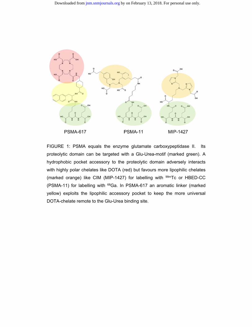
FIGURE 1: PSMA equals the enzyme glutamate carboxypeptidase II. Its
proteolytic domain can be targeted with a Glu-Urea-motif (marked green). A
hydrophobic pocket accessory to the proteolytic domain adversely interacts
with highly polar chelates like DOTA (red) but favours more lipophilic chelates
(marked orange) like CIM (MIP-1427) for labelling with 99mTc or HBED-CC
(PSMA-11) for labelling with 68Ga. In PSMA-617 an aromatic linker (marked
yellow) exploits the lipophilic accessory pocket to keep the more universal
DOTA-chelate remote to the Glu-Urea binding site.
by on February 13, 2018. For personal use only. jnm.snmjournals.org Downloaded from
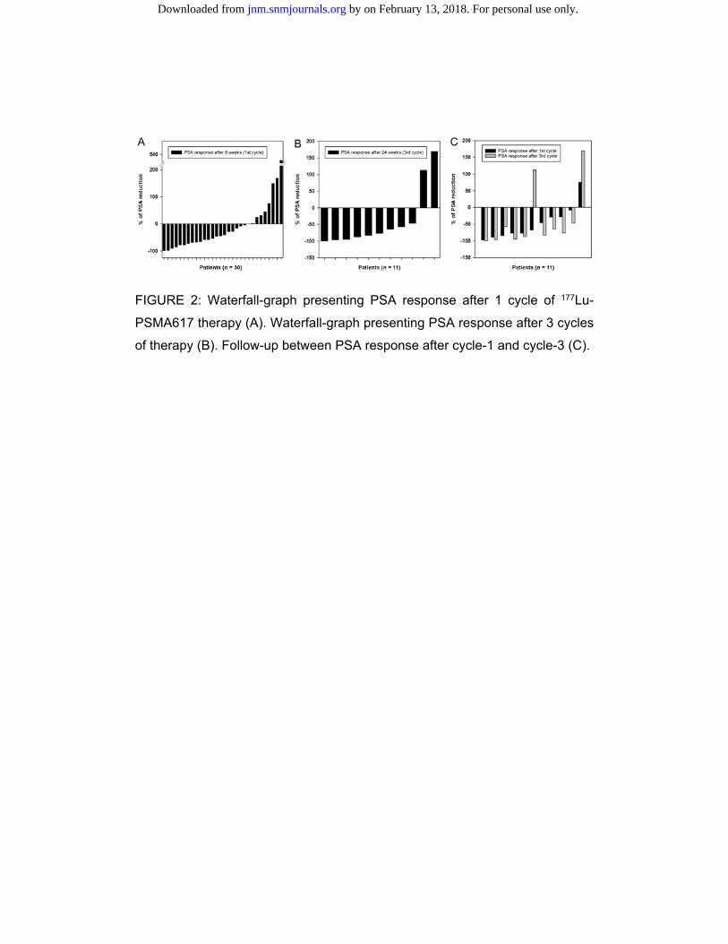
FIGURE 2: Waterfall-graph presenting PSA response after 1 cycle of 177Lu-
PSMA617 therapy (A). Waterfall-graph presenting PSA response after 3 cycles
of therapy (B). Follow-up between PSA response after cycle-1 and cycle-3 (C).
by on February 13, 2018. For personal use only. jnm.snmjournals.org Downloaded from
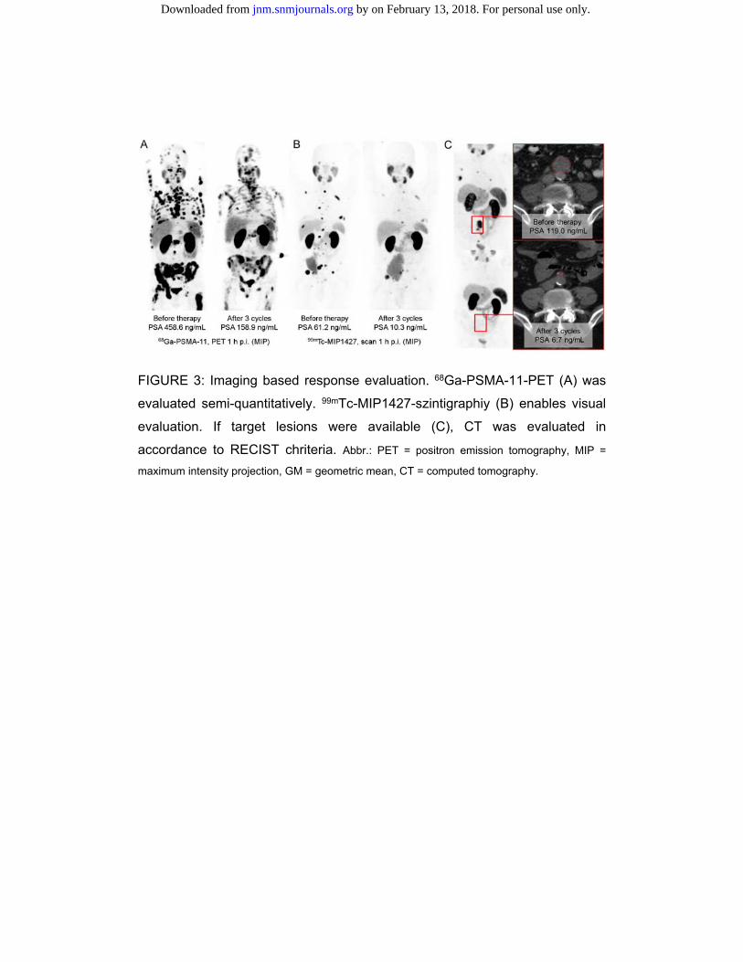
FIGURE 3: Imaging based response evaluation. 68Ga-PSMA-11-PET (A) was
evaluated semi-quantitatively. 99mTc-MIP1427-szintigraphiy (B) enables visual
evaluation. If target lesions were available (C), CT was evaluated in
accordance to RECIST chriteria. Abbr.: PET = positron emission tomography, MIP =
maximum intensity projection, GM = geometric mean, CT = computed tomography.
by on February 13, 2018. For personal use only. jnm.snmjournals.org Downloaded from
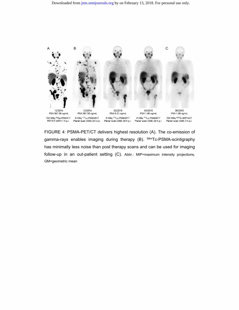
FIGURE 4: PSMA-PET/CT delivers highest resolution (A). The co-emission of
gamma-rays enables imaging during therapy (B). 99mTc-PSMA-scintigraphy
has minimally less noise than post therapy scans and can be used for imaging
follow-up in an out-patient setting (C). Abbr.: MIP=maximum intensity projections,
GM=geometric mean
by on February 13, 2018. For personal use only. jnm.snmjournals.org Downloaded from
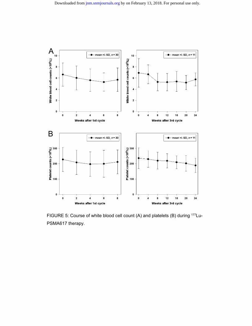
FIGURE 5: Course of white blood cell count (A) and platelets (B) during 177Lu-
PSMA617 therapy.
by on February 13, 2018. For personal use only. jnm.snmjournals.org Downloaded from
Tables
Table-1: Patient characteristics
No Age GS OP RTx CRPC Abirat Enza Ra-223 CTx Cycles [GBq] Visceral Metastases
1 68 7 1 B 1 0 0 0 D 6 / 6 / 6 Lung
2 71 4 0 L/B 1 1 1 0 D/ C 4 / 4 /4 Liver
3 75 9 1 B 1 0 0 0 0 4 / 4 / 6 0
4 61 8 1 L/B 1 1 0 0 D/ Sorafenib 6 / 6 / 6 Liver
5 67 9 0 L/B 1 0 0 0 0 6 / 6 / 6 0
6 78 8 1 L/B 1 0 0 0 0 6 / 6 / 6 0
7 71 9 0 0 1 0 1 0 D/ C/ EMP/ HU 4 / PD Liver
8 78 7b 1 B 1 1 1 1 D/ EMP 6 0
9 68 9 1 0 1 0 0 0 D 6 / 6/ 6 Brain
10 74 9 1 0 1 1 1 0 0 4/ 6/ 6 Liver
11 66 9 1 L 1 1 0 0 0 6 / 6 / 6 0
12 78 8 1 0 1 0 0 0 0 6 / 6 0
13 79 7b 1 0 1 0 0 1 0 3 / Tox Lung, Adrenal
14 73 9 1 B 1 1 1 0 0 4 / 6/ 6 Liver, Adrenal
15 71 7 0 L 1 1 0 0 0 4/ 6 Liver
16 68 na 0 0 1 1 0 1 D/ EMP 6 0
17 73 na 1 L/B 1 1 0 1 0 4 / 4 0
18 78 8 1 L 1 1 0 1 0 4/ 6/ 6 0
19 73 na 1 L/B 1 1 0 0 D 4 / Tox Lung
20 68 7 1 B 1 1 1 0 D 6 0
21 85 7a 1 B 1 1 1 0 D 6/ 6/ 0
22 71 7 0 L 1 1 0 0 0 4 / PD Rectum
23 66 9 1 L/B 1 1 1 0 0 6/ 6 0
24 75 8 1 B 1 1 1 0 D 6 0
25 80 7 1 B 1 1 1 0 D/ C 6 Liver, Lung
26 64 9 0 B 1 1 0 1 0 6 0
27 61 9 1 L/B 1 1 1 0 D/ C 6 Liver
28 69 8 1 L/B 1 1 0 0 0 6/ 6/ Lung
29 73 9 0 L 1 1 1 0 D 6/ 6 0
30 75 na 1 L 1 0 1 0 0 6/ Tox 0
Abbr.: 0 = patient did not receive that therapy, 1 = patient had history of that treatment, GS = gleason score, OP = prostatectomy, RTx = radiation therapy to prostate bed (=L) or bone (=B), CRPC = hormone therapy with both an LHRH-Analogue/Antagonist and an anti-androgen, Abirat = Abiraterone, Enza = Enzalutamide, CTx = chemotherapy with docetaxel (=D), cabazitacel (=C), estramustin monophosphate (=EMP) or hydroxyurea (=HU). Cycles = therapy with 177Lu-PSMA-617 with the given activities [GBq] in bi-monthly fractions. Fractionated therapy had to be discontinued due to toxicity (=Tox) or progressive disease (=PD)
by on February 13, 2018. For personal use only. jnm.snmjournals.org Downloaded from
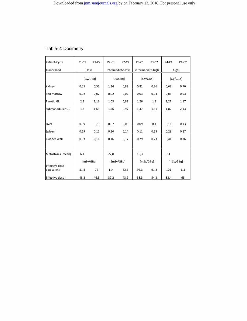
Table-2: Dosimetry
Patient-Cycle P1-C1 P1-C2 P2-C1 P2-C2 P3-C1 P3-C2 P4-C1 P4-C2
Tumor load low Intermediate-low intermediate-high high
[Gy/GBq] [Gy/GBq] [Gy/GBq] [Gy/GBq]
Kidney 0,55 0,56 1,14 0,82 0,81 0,76 0,62 0,76
Red Marrow 0,02 0,02 0,02 0,02 0,03 0,03 0,05 0,03
Parotid Gl. 2,2 1,16 1,03 0,82 1,26 1,3 1,27 1,17
Submandibular Gl. 1,3 1,69 1,26 0,97 1,37 1,31 1,82 2,13
Liver 0,09 0,1 0,07 0,06 0,09 0,1 0,16 0,13
Spleen 0,19 0,15 0,26 0,14 0,11 0,13 0,28 0,27
Bladder Wall 0,03 0,16 0,16 0,17 0,29 0,23 0,41 0,36
Metastases (mean) 6,1 22,8 15,3 14
[mSv/GBq] [mSv/GBq] [mSv/GBq] [mSv/GBq] Effective dose equivalent 81,8 77 114 82,5 96,3 91,2 126 111
Effective dose 48,2 46,5 37,2 43,9 58,3 54,3 83,4 65
by on February 13, 2018. For personal use only. jnm.snmjournals.org Downloaded from
Doi: 10.2967/jnumed.115.171397Published online: March 16, 2016.J Nucl Med. Mier, Matthias Eder, Klaus Kopka and Uwe HaberkornClemens Kratochwil, Frederik L. Giesel, Melsa Stefanova, Martina Benesová, Marcus Bronzel, Ali Afshar-Oromieh, Walter with Lu-177 labeled PSMA-617PSMA-targeted radionuclide therapy of metastatic castration-resistant prostate cancer
http://jnm.snmjournals.org/content/early/2016/03/16/jnumed.115.171397This article and updated information are available at:
http://jnm.snmjournals.org/site/subscriptions/online.xhtml
Information about subscriptions to JNM can be found at:
http://jnm.snmjournals.org/site/misc/permission.xhtmlInformation about reproducing figures, tables, or other portions of this article can be found online at:
and the final, published version.proofreading, and author review. This process may lead to differences between the accepted version of the manuscript
ahead of print area, they will be prepared for print and online publication, which includes copyediting, typesetting,JNMcopyedited, nor have they appeared in a print or online issue of the journal. Once the accepted manuscripts appear in the
. They have not beenJNM ahead of print articles have been peer reviewed and accepted for publication in JNM
(Print ISSN: 0161-5505, Online ISSN: 2159-662X)1850 Samuel Morse Drive, Reston, VA 20190.SNMMI | Society of Nuclear Medicine and Molecular Imaging
is published monthly.The Journal of Nuclear Medicine
© Copyright 2016 SNMMI; all rights reserved.
by on February 13, 2018. For personal use only. jnm.snmjournals.org Downloaded from