radionuclide imaging of osteomyelitis - sociedad uruguaya … · 2016-02-10 · radionuclide...
TRANSCRIPT

32
*DepartHem
†DivisioIslan
AddressMed270
Radionuclide Imaging of OsteomyelitisChristopher J. Palestro, MD*,†
http://dx.doi.or0001-2998/& 2
ment of Radipstead, NY.n of Nucleard Jewish Healreprint requesicine and Mo-05 76th Ave,
Radionuclide procedures frequently are performed as part of the diagnostic workup ofosteomyelitis. Bone scintigraphy accurately diagnoses osteomyelitis in bones not affectedby underlying conditions. Degenerative joint disease, fracture, and orthopedic hardwaredecrease the specificity of the bone scan, making it less useful in these situations. Gallium-67scintigraphy was often used as an adjunct to bone scintigraphy for diagnosing osteomyelitis.However, now it is used primarily for spinal infections when 18F-FDG imaging cannot beperformed. Except for the spine, in vitro–labeled leukocyte imaging is the nuclearmedicine testof choice for diagnosing complicating osteomyelitis. Leukocytes accumulate in bone marrowas well as in infection. Performing complementary bone marrow imaging with 99mTc-sulfurcolloid facilitates the differentiation between osteomyelitis and normal marrow and improvestest overall accuracy. Antigranulocyte antibodies and antibody fragments, such as 99mTc-besilesomab and 99mTc-sulesomab, were developed to eliminate the disadvantages associ-ated with in vitro–labeled leukocytes. These agents, however, have their own shortcomingsandare notwidely available. As biotin is used as agrowth factor by certain bacteria, 111In-biotinis useful to diagnose spinal infections. Radiolabeled synthetic fragments of ubiquicidin, anaturally occurring human antimicrobial peptide that targets bacteria, can differentiateinfection from sterile inflammation and may be useful to monitor response to treatment.18F-FDG is extremely useful in the diagnostic workup of osteomyelitis. Sensitivity in excess of95% and specificity ranging from 75%-99% have been reported. 18F-FDG is the radionuclidetest of choice for spinal infection. The test is sensitive, with a high negative predictive value,and reliably differentiates degenerative from infectious vertebral body end-plate abnormalities.Data on the accuracy of 18F-FDG for diagnosingdiabetic pedal osteomyelitis are contradictory,and its role for this indication remains to be determined. Initial investigations suggested that18F-FDG accurately diagnoses prosthetic joint infection; more recent data indicate that itcannot differentiate infection from other causes of prosthetic failure. Preliminary data on thePET agents gallium-68 and iodine-124 fialuridine indicate that these agents may have a role indiagnosing osteomyelitis.Semin Nucl Med 45:32-46 C 2015 Elsevier Inc. All rights reserved.
Osteomyelitis is an infection of the bone and may belocalized or involve periosteum, cortex, marrow, and
cancellous tissue. Acute osteomyelitis can arise hematogenouslyor through inoculation from direct trauma, a contiguous focusof infection, or sepsis following surgery.1 The diagnosis of oste-omyelitis is not always obvious, and radionuclide proceduresfrequently are performed as part of the diagnostic workup.
g/10.1053/j.semnuclmed.2014.07.005015 Elsevier Inc. All rights reserved.
ology Hofstra, NorthShore-LIJ School of Medicine,
Medicine and Molecular Imaging, North Shore Longth System, Manhasset & New Hyde Park, NY.ts to Christopher J. Palestro, MD, Division of Nuclearlecular Imaging, Long Island Jewish Medical Center,New Hyde Park, NY 11040. E-mail: [email protected]
RadiopharmaceuticalsSingle-Photon–Emitting Agents99mTc-DiphosphonatesBone scintigraphy usually is performed with technetium-99m-methylene diphosphonate (99mTc-MDP). Uptake of this radio-pharmaceutical, which binds to the hydroxyapatite crystal,depends on blood flow and rate of new bone formation.Whenosteomyelitis is the indication, a 3-phase bone scan usually isperformed. Three-phase bone scintigraphy consists of a dyn-amic imaging sequence, the flow or perfusion phase, followedimmediately by static images of the region of interest, the bloodpool or soft tissue phase. The third, or bone, phase consists ofimages of the area of interest, acquired 2-4 hours after injection.Focal hyperperfusion, focal hyperemia, and focally increased

Figure 1 Right ulnar osteomyelitis. There is focal hyperperfusion, focal hyperemia, and focally increased bone uptake ofradiopharmaceutical in the right ulna.
Radionuclide imaging of osteomyelitis 33
bony uptake are the classic presentation of osteomyelitis on a3-phase bone scan (Fig. 1). The test is both sensitive andspecific for diagnosing osteomyelitis in bones not affected byunderlying conditions. Abnormalities on bone scintigraphyreflect the rate of new bone formation in general andconsequently in the setting of preexisting conditions such asdegenerative joint disease, fracture, and orthopedic hardware,the test, because of decreased specificity, is less useful (Fig. 2).2
Gallium-67Several factors contribute to gallium-67 (67Ga) uptake ininfection. Approximately 90% of circulating 67Ga is transferrinbound in the plasma. Increased blood flow and vascularmembrane permeability result in increased 67Ga delivery andaccumulation at infectious foci. 67Ga binds to lactoferrin,which is present in high concentrations in sites of infection.Direct bacterial uptake, complexing with siderophores, andleukocyte transport also may contribute to 67Ga uptake ininfection. Imaging generally is performed 18-72 hours afterinjection.2 Presently, the role of 67Ga imaging in musculoske-letal infection is limited almost exclusively to the spine (Fig. 3).
In Vitro–Labeled LeukocytesIn vitro leukocyte (white blood cell [WBC]) labelingusually is performed with 111In oxyquinolone (In) or
Figure 2 Left knee osteoarthritis. The findings on the 3-phase billustrating the limitations of bone scintigraphy in individualsperformed to evaluate a painful right knee arthroplasty; the left
99mTc-exametazime (Tc). Uptake depends on intact chemo-taxis, number and types of cells labeled, and cellular responsein a particular condition. A circulating WBC count of at least2000 per microliter is needed for satisfactory image quality.Most WBCs labeled usually are neutrophils, and the test ismost sensitive for detecting neutrophil-mediated infections.3
111In-WBC advantages include label stability; a normaldistribution limited to liver, spleen, and bone marrow; andthe ability to perform delayed imaging. Complementary bonemarrow imaging can be performed during cell labeling, as asimultaneous dual-isotope acquisition, or after 111In-WBCimaging. Disadvantages include low-resolution images andthe interval of 16-30 hours between injection and imaging.3
The normal distribution of 99mTc-WBCs is more variablethan that of 111In-WBCs. In addition to the reticuloendothelialsystem, activity normally is present in the urinary tract, largebowel (within 4 hours after injection), and occasionally gallbladder. 99mTc-WBC advantages include high-resolutionimages, and the ability to detect abnormalities within a fewhours after injection. Disadvantages include label instabilityand the short half-life of 99mTc, which limits delayed imaging.When performing bone marrow imaging, there must be aninterval of 2-3 days between the 2 procedures.3
Leukocytes accumulate in both infection and bone marrow.The normal distribution of hematopoietically active bone
one scan in this case mimic those seen in osteomyelitis,with preexisting skeletal abnormalities. This study wasknee was asymptomatic.

Figure 3 Cervical spine osteomyelitis. The sagittal CT image (left) demonstrates destructive changes at the C6-C7 level(arrows), which corresponds to an area of intense radiopharmaceutical uptake on the sagittal 67Ga SPECT image (right).
C.J. Palestro34
marrow is variable and is affected by the patient 's age, systemicconditions such as sickle cell disease and tumor, and localconditions such as fractures and orthopedic hardware. Con-sequently, it may not be possible to determine if an area ofactivity on a WBC image represents infection or marrow.Performing 99mTc-sulfur colloid bone marrow (marrow)imaging overcomes this difficulty. Both WBCs and 99mTc-sulfur colloid accumulate inmarrow;WBCs also accumulate ininfection, but sulfur colloid does not. The combined studyresult is positive for infection when activity is present on theWBC image without corresponding activity on the marrowimage. Any other pattern is negative for infection(Figs. 4 and 5).4
In Vivo–Labeled LeukocytesBesilesomab, a 150-kDa murine monoclonal antibody of theIgG1 kappa isotype, binds to nonspecific cross-reactingantigen-95, an epitope expressed on cell membranes ofgranulocytes and granulocyte precursors. Approximately10% of the 99mTc-besilesomab injected is neutrophil boundby 45 minutes. Another 20% circulates freely, presumablylocalizing in infection through nonspecific mechanisms. Theincidence of human antimurine antibody response ranges from
Figure 4 Right sternoclavicular osteomyelitis. Noncontrast CT scawall mass, swelling, and tenderness, was interpreted as consisteclavicle and in the right retrosternal region. On the 111In-WBC imslightly larger than the left. The intensity of the uptake, howevconclusions. On the marrow image (right), there is a well-definedclavicle extending into the right half of the manubrium. The co
less than 5% in patients receiving a single dose of 125 μg ofantibody to more than 30% in patients receiving repeatedinjections. Tominimize potential problems, patients should beprescreened for human antimurine antibody, injected with nomore than 250 μg of antibody, and should avoid repeatadministration.5
Sulesomab, a 50-kDa fragment antigen-binding (Fab0)portion of an IgG1 class murine monoclonal antibody, bindsto normal cross-reactive antigen-90 present on leukocytes.Approximately 3%-6% of the 99mTc-sulesomab injected isassociated with circulating neutrophils; at 24 hours afterinjection, approximately 35% of the remaining activity is inthe bone marrow. Initial investigations suggested that suleso-mab binds to circulating neutrophils that migrate to foci ofinfection and to leukocytes already present at the site ofinfection. Subsequent data, however, suggest that accumula-tion in infection is nonspecific.5
111In-BiotinBiotin is necessary for cell growth, fatty acid production, andmetabolism of fats and amino acids. It also is used as a growthfactor by certain bacteria. 111In-biotin has been used primarilyfor diagnosing spinal infections.5
n finding (not shown), in a patient with right anterior chestnt with myositis adjacent to the medial head of the rightage (left), the right sternoclavicular region (arrow) appearser, is virtually the same, making it difficult to draw anyphotopenic defect (arrow) in the medial head of the right
mbined study result is positive for osteomyelitis.

Figure 5 Marrow expansion. Interpreted in isolation, the intense activity in the distal right femur (arrow) on the 111In-WBCimage (left) could easily bemistaken for osteomyelitis. On themarrow image (right), the distribution of activity in the distalright femur (arrow) is virtually identical to that on the 111In-WBC image, and the combined study finding is negative forosteomyelitis. The intensity of uptake on labeled leukocyte images is not useful for determining the presence or absence ofosteomyelitis. (Compare with Fig. 4).
Radionuclide imaging of osteomyelitis 35
Radiolabeled Antimicrobial PeptidesAntimicrobial peptides are an integral component of thebiological defenses of multicellular organisms. Radiolabeledsynthetic fragments of ubiquicidin (UBI), a human antimicro-bial peptide that targets bacteria, possess the ability to differ-entiate infection from sterile inflammation and may be usefulfor monitoring the efficacy of antibacterial agents in certaininfections.5
Positron-Emitting Agents18F-FDG18F-FDG is transported into cells via glucose transporters andphosphorylated by hexokinase to 18F-20-18F-FDG-6 phos-phate but is not metabolized further. 18F-FDG accumulates
Figure 6 Lumbar spine compression fracture. A 3-month-old combe appreciated on the CT component of the study. There is unifoincluding the third lumbar vertebra. (Reproduced with permiss
in virtually all leukocytes, and its uptake in these cells is relatedto their metabolic rate and the number of glucose transporters.Increased 18F-FDG accumulation in infection presumably isdue to several factors. There is an increased number of glucosetransporters and an increased expression of these glucosetransporters by activated inflammatory cells. There is anincreased affinity of these transporters for 18F-FDG in inflam-mation, probably owing to the effects of circulating cytokinesand growth factors.6
18F-FDG PET offers several advantages over single-photon–emitting tracers. PET is a high-resolution tomographic techni-que that enables precise localization of radiopharmaceuticalaccumulation. The small 18F-FDG molecule enters poorlyperfused areas rapidly. The procedure is completed in 1-2hours and has a relatively low radiation dose. Uptake usually
pression fracture of the third lumbar vertebra (arrow) canrm distribution of 18F-FDG throughout the lumbar spineion from Palestro.6)

C.J. Palestro36
normalizes within 3-4 months after trauma or surgery.Degenerative bone changes ordinarily show only mildlyincreased 18F-FDG uptake (Figs. 6 and 7).6
18F-FDG-Labeled LeukocytesIn an effort to develop a more specific PET tracer for infectionimaging, leukocytes have been labeled in vitro with 18F-FDG.Despite satisfactory results, it is unlikely that 18F-FDG-WBCimaging ever will enter clinical practice. The 110 minutes half-life of 18Fmakes it impractical for labeling to be performed off-site, which means that the test would be limited to those sitescapable of performing labeling. In some situations, imaging atlater time points (eg, 24 hours after injection) may be needed.The short half-life of 18F precludes imaging much later than4-5 hours after injection. The labeling efficiency is significantlylower andmore variable thanwhat can be achievedwith 111In-oxine, and, perhaps most importantly, a large fraction of18F-FDG rapidly elutes from the leukocytes. In vitro dataindicate that, by 4 hours after labeling, approximately 40% ofthe activity is eluted from the cells.7-11
Gallium-68This PET tracer is generator produced. The imaging character-istics of gallium-68 (68Ga) are superior to those of 67Ga. 68Gahas a high positron yield and a half-life of 68 minutes. 68Ga-citrate is produced with high radiochemical yield and purityand has been used to detect inflammation and infection.5,12
124I-fialuridneBacteria possess a thymidine kinase whose substrate specificityis different from that of the major human thymidine kinase.This difference was used to develop a molecular imagingtest for detecting viable bacteria. The potential of 124I-fialuridne
Figure 7 Degenerative spinal arthritis. Although there are extensivthe examination, there is uniform distribution of 18F-FDG thro
(FIAU) PET/CT for diagnosing musculoskeletal infection wasstudied in 8 subjects with suspected musculoskeletal infectionand 1 control. All patients with musculoskeletal infectiondemonstrated 124I-FIAU accumulation at the site of infectionwithin 2 hours after injection. There was no abnormal radio-pharmaceutical uptake in the 1 control.5,13
IndicationsIt is important to recognize that no single agent is equallyefficacious in all regions of the skeleton. The selection of anappropriate study is governed by the clinical question posed.In adults, it is useful to divide musculoskeletal infectionsinto 3 broad locations: spine, orthopedic hardware, anddiabetic foot.
Spinal InfectionSpinal osteomyelitis-discitis, which has a predilection for theelderly, accounts for 2%-7% of all cases of osteomyelitis.Infection usually is confined to the vertebral body andintervertebral disc, but the posterior elements are involved inup to 20% of cases. Soft tissue abscesses often accompanyspinal infection.14
MRI is the best imaging available for spinal infection.Radionuclide imaging is a useful adjunct to MRI. Althoughbone scintigraphy frequently is used as a screening test, falsenegative results have been reported in the elderly, possiblysecondary to arteriosclerosis-induced ischemia. The test is notuseful for detecting soft tissue infections that often accompany,or mimic, spinal osteomyelitis. Abnormalities may persist evenafter the infection has resolved, owing to ongoing bonyremodeling during healing. Consequently, if used at all, bone
e degenerative changes (arrows) on the CT component ofughout the lumbar spine.

Radionuclide imaging of osteomyelitis 37
scintigraphy should not be the only radionuclide test per-formed.14-16
Complementary 67Ga imaging improves the specificity ofthe bone scan. 67Gamay detect infection sooner than the bonescan and can identify accompanying soft tissue infection. Loveet al14 reported that 67Ga SPECT is as accurate as combinedbone-gallium imaging for diagnosing spinal osteomyelitis andconcluded that when 67Ga SPECT was performed the bonescan was unnecessary (Fig. 3).There are few data on the role of 67Ga SPECT-CT in the
diagnosis of spinal infection. Lievano et al17 reported that67Ga SPECT-CT precisely localized focal radiopharma-ceutical uptake seen on planar images, thereby avoiding afalse-positive diagnosis of spinal osteomyelitis. Domínguezet al18 reported that the combination of functional andanatomical images improves disease detection. Fuster etal19 reported that 67Ga SPECT-CT helped identify softtissue involvement in 10 of 18 patients with spinalosteomyelitis.
67Ga imaging, regardless of how it is performed, haslimitations. The physical characteristics and normal biodistri-bution of the agent are impediments to image analysis.Although the test result may become positive within a fewhours, imaging typically is performed 18-72 hours afterinjection, necessitating multiple visits to nuclear medicine.There is nothing specific about 67Ga uptake in infection; itaccumulates in many other conditions, including tumor andtrauma.With all of the limitations inherent in radionuclide bone and
67Ga imaging, it is not surprising that investigators have soughtalternative tracers. Lazzeri et al20 reported that 111In-biotin,alone and in combination with strepavidin, accurately diag-noses spinal infections. 111In-biotin does not accumulate innormal bone or bone marrow and there are few anatomiclandmarks on the images. Including SPECT-CT as part of theprocedure affects patient management by accurately differ-entiating bone from soft tissue infection and guiding theselection of therapy.21
Published data strongly support the value of 18F-FDG fordiagnosing spinal infection.22-28 Guhlmann et al22 reportedthat 18F-FDG-PET correctly diagnosed all 3 cases of infectionandwas true negative in 1 patient without infection. In anotherseries, Guhlmann et al23 reported that 18F-FDG-PET wassignificantly more accurate than 99mTc-besilesomab for diag-nosing spinal osteomyelitis. Schiesser et al24 reported that in 6patients with possible spinal implant infection, 18F-FDG-PETfinding was true negative for infection in all. In another series,18F-FDG-PET yielded true-positive results in all 7 patientswithspinal osteomyelitis.25 Schmitz et al26 reported that the testresult was true positive in all 12 patients with spinal infectionand true negative in 3 of 4 patients without infection. In aprospective study of 8 patients (9 sites), suspected of havingspinal osteomyelitis, Meller et al27 reported that FDG-PET was100% accurate.Hartmann et al,28 as part of a larger investigation, reported
that 18F-FDG-PET-CT finding was true positive in all 7patients with spinal osteomyelitis and true negative in bothpatients without spinal osteomyelitis. The precise anatomical
localization provided by PET-CT was useful for planningsurgical intervention and for differentiating soft tissue frombone involvement, thereby guiding treatment.Gratz et al29 observed that 18F-FDG-PET was superior to
67Ga for detecting paraspinal soft tissue infection and wassuperior to bone scintigraphy for differentiating advanceddegenerative arthritis from infection. Fuster et al19 studied 34patients, including 18 with spinal osteomyelitis. The sensitiv-ity, specificity, and accuracy of 18F-FDG-PET-CT were 89%,88%, and 88%, respectively. The sensitivity, specificity, andaccuracy of combined bone-gallium imaging were 78%, 81%,and 79%, respectively. 18F-FDG-PET-CT identified soft tissueinfection in 12 patients; 67Ga SPECT-CT identified soft tissueinvolvement in 10 patients.Gratz et al29 reported that 18F-FDG-PET was superior to
MRI for detecting low-grade spondylitis or discitis. Ohtoriet al30 studied 18 patients with Modic type 1 changes on MRI,including 11 with spinal osteomyelitis who underwent18F-FDG-PET. The authors reported that there was a 100%concordance between the results of 18F-FDG-PET and the finaldiagnosis.Stumpe et al31 compared 18F-FDG-PET with MRI in 30
patients (38 sites) with lumbar spine vertebral end-plateabnormalities. 18F-FDG-PET finding was true positive in all5 infected sites and true negative in all 33 uninfected sites(100% sensitivity and 100% specificity). The sensitivity andspecificity of MRI were 50% and 96%, respectively (Fig. 8).Seifen et al32 analyzed 38 consecutive cases of suspected
spondylodiscitis in patients with inconclusive results on MRIor other conventional modalities. A total of 22 patients werediagnosedwith spinal osteomyelitis. The sensitivity, specificity,and accuracy of 18F-FDG-PET-CTwere 81.8%, 100%, 89.5%,respectively; positive predictive value and negative predictivevaluewere 100%and 80%, respectively. Sensitivity, specificity,and accuracy of MRI, which was performed in 27 cases, were75%, 71.4%, 74.1%, respectively; the positive predictive valueandnegative predictive valuewere 88.2%and50%, respectively.There are potential limitations to the test. It is likely that
18FDG-PET and PET-CT will be less reliable for differentiatinginfection from tumor and infection superimposed on tumor.18F-FDG accumulation in degenerative changes usually isrelatively low, but significant focal 18F-FDG uptake in degen-erative spine disease has been reported.33
The presence of a foreign body can incite an intense immuneresponse, and increased 18F-FDG uptake around spinalimplants, in the absence of infection, has been observed. deWinter et al34 reported that the specificity of 18F-FDG-PETwasconsiderably lower in patients with than in patients withoutspinal implants (65% vs 92%).Regardless of its potential limitations, the data accumu-
lated over the past several years demonstrate convincinglythat 18F-FDG imaging accurately diagnoses spinal osteomye-litis and support its use as an adjunct to MRI. The test issensitive, completed in a single session, and image quality issuperior to that obtained with single-photon–emitting trac-ers, even when SPECT-CT is performed. Finally, in com-parative investigations, 18F-FDGhas outperformed bone and67Ga imaging.
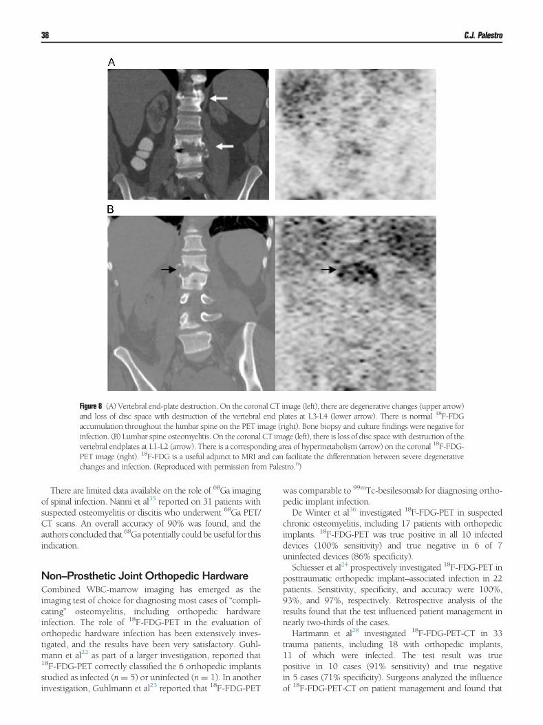
Figure 8 (A) Vertebral end-plate destruction. On the coronal CT image (left), there are degenerative changes (upper arrow)and loss of disc space with destruction of the vertebral end plates at L3-L4 (lower arrow). There is normal 18F-FDGaccumulation throughout the lumbar spine on the PET image (right). Bone biopsy and culture findings were negative forinfection. (B) Lumbar spine osteomyelitis. On the coronal CT image (left), there is loss of disc space with destruction of thevertebral endplates at L1-L2 (arrow). There is a corresponding area of hypermetabolism (arrow) on the coronal 18F-FDG-PET image (right). 18F-FDG is a useful adjunct to MRI and can facilitate the differentiation between severe degenerativechanges and infection. (Reproduced with permission from Palestro.6)
C.J. Palestro38
There are limited data available on the role of 68Ga imagingof spinal infection. Nanni et al35 reported on 31 patients withsuspected osteomyelitis or discitis who underwent 68Ga PET/CT scans. An overall accuracy of 90% was found, and theauthors concluded that 68Ga potentially could be useful for thisindication.
Non–Prosthetic Joint Orthopedic HardwareCombined WBC-marrow imaging has emerged as theimaging test of choice for diagnosing most cases of “compli-cating” osteomyelitis, including orthopedic hardwareinfection. The role of 18F-FDG-PET in the evaluation oforthopedic hardware infection has been extensively inves-tigated, and the results have been very satisfactory. Guhl-mann et al22 as part of a larger investigation, reported that18F-FDG-PET correctly classified the 6 orthopedic implantsstudied as infected (n¼ 5) or uninfected (n¼ 1). In anotherinvestigation, Guhlmann et al23 reported that 18F-FDG-PET
was comparable to 99mTc-besilesomab for diagnosing ortho-pedic implant infection.De Winter et al36 investigated 18F-FDG-PET in suspected
chronic osteomyelitis, including 17 patients with orthopedicimplants. 18F-FDG-PET was true positive in all 10 infecteddevices (100% sensitivity) and true negative in 6 of 7uninfected devices (86% specificity).Schiesser et al24 prospectively investigated 18F-FDG-PET in
posttraumatic orthopedic implant–associated infection in 22patients. Sensitivity, specificity, and accuracy were 100%,93%, and 97%, respectively. Retrospective analysis of theresults found that the test influenced patient management innearly two-thirds of the cases.Hartmann et al28 investigated 18F-FDG-PET-CT in 33
trauma patients, including 18 with orthopedic implants,11 of which were infected. The test result was truepositive in 10 cases (91% sensitivity) and true negativein 5 cases (71% specificity). Surgeons analyzed the influenceof 18F-FDG-PET-CT on patient management and found that

Figure 9 (A) Infected right hip arthroplasty. There is mild hyperperfusion and hyperemia, with irregularly increasedradiopharmaceutical accumulation on the bone phase, around the 3-year-old right hip arthroplasty. (B) Asepticallyloosened right hip arthroplasty. The findings on this 3-phase bone scan are very similar to those in (A). Most investigatorsagree that the 3-phase bone scan does not improve accuracy of the test for differentiating infection fromother causes of jointreplacement failure.
Radionuclide imaging of osteomyelitis 39
the test contributed relevant information in more than halfof the cases.
Prosthetic Joint InfectionNearly 1 million lower extremity arthroplasties are performedannually in the United States. Aseptic loosening, the mostcommon cause of prosthetic failure, usually is caused by aninflammatory reaction to 1 or more of the prosthetic compo-nents. A synovial-like pseudomembrane develops, consistingof histiocytes (95% of specimens), giant cells (80%), andoccasionally, lymphocytes and plasma cells (25%). Neutro-phils rarely are present (10%).37 Aseptic loosening usually ismanaged by a single-stage exchange arthroplasty completed ina single hospital admission with 1 surgical intervention.Infection accounts for approximately 2%of primary implant
failures and approximately 5% of revision implant failures. Theinflammatory reaction accompanying the infected prosthesiscan be similar to that in aseptic loosening, except thatneutrophils, which rarely are present in aseptic loosening,invariably are present, and usually in large numbers.37 Thetreatment of the infected joint replacement usually involvesmore than 1 hospital admission. An excisional arthroplasty isperformed followed by weeks to months of antimicrobialtherapy, followed eventually by a revision arthroplasty.
Because their management is so very different, distinguish-ing aseptic loosening from infection of a prosthetic joint isextremely important. A sensitive but nonspecific test can resultin multiple unnecessary, and costly, operations when a singleintervention would have sufficed. The specific, but insensitive,test also will result in additional surgical interventions, becauseundiagnosed infection will cause any revision implant to failwith potentially serious consequences.37
Joint aspiration with culture is the definitive preoperativediagnostic procedure. Though specific, the sensitivity of thistest is variable. Tomas et al38 reported that joint aspirationwithculture was 100% specific but only 70% sensitive fordiagnosing prosthetic hip infection.Among imaging studies, radiographs are not specific and
hardware-induced artifacts limit to some degree, CT and MRI.Radionuclide studies are extremely useful in the evaluation ofjoint replacements, especially when infection is suspected. Themost widely and often the initial radionuclide test performed isbone scintigraphy. Although sensitive bone scintigraphy is notspecific, and it is most useful for screening purposes. A normalstudy result makes it unlikely that the patient 's symptoms arerelated to the prosthesis. An abnormal study result requiresfurther investigation. The accuracy of bone scintigraphy isbetween 50% and 70%. Performing the test as a 3-phase studydoes not improve accuracy (Fig. 9).37,39-42
C.J. Palestro40
67Ga imaging has been used to improve the specificity ofbone scintigraphy. 67Ga, either alone or in combination withbone scintigraphy, has accuracy between 60% and 80% andoffers only a modest improvement over bone scintigraphyalone and has fallen into disuse.37
Presently, the best available imaging test for diagnosingprosthetic joint infection is WBC marrow imaging with anaccuracy of approximately 90% (Fig. 10). All of the studiespublished over the past 3 decades confirm that this test ishighly specific for diagnosing joint replacement infection. Innearly all of the investigations, the test has proved to besensitive as well.39,43-48 Its value notwithstanding, as alreadynoted, there are significant disadvantages to the WBCmarrowprocedure, and investigators continue to search for suitablealternatives.Boubaker et al49 reported that 99mTc-besilesomab was 67%
sensitive and 75% specific for diagnosing prosthetic hipinfection. When interpreted together with bone scintigraphy,specificity improved to 84%. Gratz et al50 reported thataccuracy improved from 80% for 99mTc-besilesomab aloneto 89% when interpreted in conjunction with bone scintig-raphy. Semiquantitative analysis has also been suggested as away to improve the sensitivity and specificity of 99mTc-besilesomab for diagnosing lower extremity prosthetic jointinfection.51,52
Von Rothenburg et al53 reported a sensitivity of 93% and aspecificity of 65% for diagnosing lower extremity prostheticinfection with 99mTc-sulesomab. Iyengar and Vinjamuri54
reported similar results. Pakos et al55 reported that 99mTc-sulesomab was 75% sensitive, 86% specific, and 79% accuratefor diagnosing prosthetic joint infection. Rubello et al56,57
reported that specificity is improved by imaging at 4 and 20-24hours after injection. Gratz et al58 reported that analysis of timeactivity curves significantly improves the accuracy of 99mTc-sulesomab for diagnosing moderate and mild prosthetic jointinfections.Although in vivo–labeled leukocytes accumulate in the bone
marrow, scant attention has been paid to combining thesestudies with bone marrow imaging. In one of the fewinvestigations in which bone marrow imaging was performed,Sousa et al59 reported that, by performing complementary
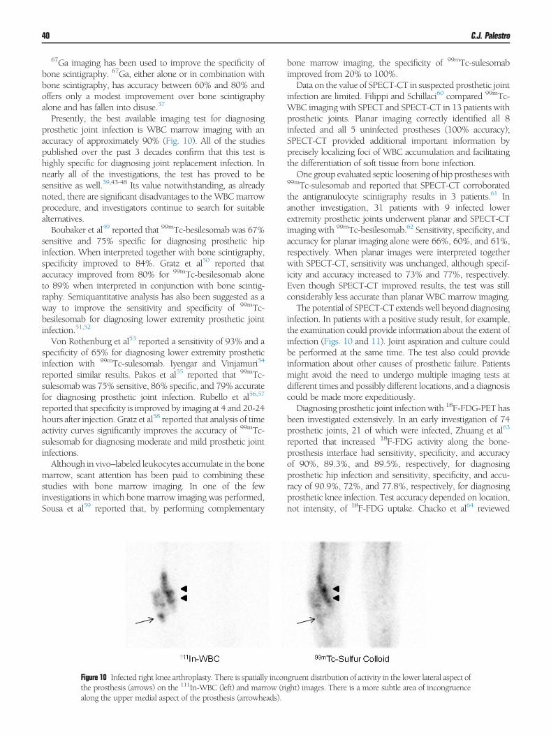
Figure 10 Infected right knee arthroplasty. There is spatially inconthe prosthesis (arrows) on the 111In-WBC (left) and marrow (ralong the upper medial aspect of the prosthesis (arrowheads).
bone marrow imaging, the specificity of 99mTc-sulesomabimproved from 20% to 100%.Data on the value of SPECT-CT in suspected prosthetic joint
infection are limited. Filippi and Schillaci60 compared 99mTc-WBC imaging with SPECT and SPECT-CT in 13 patients withprosthetic joints. Planar imaging correctly identified all 8infected and all 5 uninfected prostheses (100% accuracy);SPECT-CT provided additional important information byprecisely localizing foci of WBC accumulation and facilitatingthe differentiation of soft tissue from bone infection.One group evaluated septic loosening of hip prostheses with
99mTc-sulesomab and reported that SPECT-CT corroboratedthe antigranulocyte scintigraphy results in 3 patients.61 Inanother investigation, 31 patients with 9 infected lowerextremity prosthetic joints underwent planar and SPECT-CTimaging with 99mTc-besilesomab.62 Sensitivity, specificity, andaccuracy for planar imaging alone were 66%, 60%, and 61%,respectively. When planar images were interpreted togetherwith SPECT-CT, sensitivity was unchanged, although specif-icity and accuracy increased to 73% and 77%, respectively.Even though SPECT-CT improved results, the test was stillconsiderably less accurate than planar WBC marrow imaging.The potential of SPECT-CT extendswell beyond diagnosing
infection. In patients with a positive study result, for example,the examination could provide information about the extent ofinfection (Figs. 10 and 11). Joint aspiration and culture couldbe performed at the same time. The test also could provideinformation about other causes of prosthetic failure. Patientsmight avoid the need to undergo multiple imaging tests atdifferent times and possibly different locations, and a diagnosiscould be made more expeditiously.Diagnosing prosthetic joint infectionwith 18F-FDG-PET has
been investigated extensively. In an early investigation of 74prosthetic joints, 21 of which were infected, Zhuang et al63
reported that increased 18F-FDG activity along the bone-prosthesis interface had sensitivity, specificity, and accuracyof 90%, 89.3%, and 89.5%, respectively, for diagnosingprosthetic hip infection and sensitivity, specificity, and accu-racy of 90.9%, 72%, and 77.8%, respectively, for diagnosingprosthetic knee infection. Test accuracy depended on location,not intensity, of 18F-FDG uptake. Chacko et al64 reviewed
gruent distribution of activity in the lower lateral aspect ofight) images. There is a more subtle area of incongruence

Figure 11 Infected right knee arthroplasty. On the sagittal images from the simultaneously acquired dual-isotope SPECT-CT, spatially incongruent distribution of activity on 111In-WBC (top) andmarrow (bottom) images can be identified clearlyanterior and posterior to the femoral component (arrows) and posterior to the tibial component (arrowheads). (Samepatient as illustrated in Fig. 10.)
Radionuclide imaging of osteomyelitis 41
18F-FDG-PET scans performed on 89 lower extremity jointreplacements. The sensitivity, specificity, and accuracy of thetest were 91%, 98%, and 96% respectively, for diagnosingprosthetic hip infection and 92%, 75%, and 81%, respectively,for diagnosing prosthetic knee infection. Test accuracydepended on location and not on intensity of 18F-FDG uptake.Chacko et al65 in an investigation of 41 painful hip arthro-plasties, reported that bone-prosthesis interface activity alongthe shaft of the femoral component was 92% sensitive and97% specific for infection and that accuracy depended onlocation, not intensity, of 18F-FDG uptake.Reinartz et al42 studied 92 hip prostheses with 3-phase
bone scintigraphy and 18F-FDG-PET. Sensitivity, specific-ity, and accuracy of 3-phase bone scintigraphy were 68%,76%, and 74% vs 94%, 95%, and 95%, respectively, for 18F-FDG-PET. They observed that, on 18F-FDG-PET images,activity around the acetabular component and proximalaspect of the femoral component was not associated withinfection and that periprosthetic uptake patterns wereuseful for differentiating infection from aseptic looseningbut intensity of uptake was not. Cremerius et al66 studied 18patients with painful hip replacements and reported that18F-FDG-PET was 89% accurate for diagnosing infection.Gravius et al67 studied 20 patients with painful kneeprostheses. The sensitivity and specificity of 18F-FDG-PET
for diagnosing infection were 89% and 82%, respectively.Pill et al48 studied 92 painful hip prostheses, 21 of whichwere infected, and reported that 18F-FDG-PET was 95%sensitive and 93% specific for diagnosing infection. A totalof 51 prostheses, including 10 infected devices, also werestudied with WBC-marrow imaging. The sensitivity andspecificity of WBC-marrow imaging in this subgroup were50% and 95.1%, respectively.Manthey et al68 studied 28 lower extremity prostheses and
reported that 18F-FDG-PET was 96% accurate for diagnosingprosthetic joint infection. They also reported that by analyzingboth intensity and patterns of periprosthetic uptake, it waspossible to accurately differentiate aseptic loosening, synovitis,and infection and that activity around the femoral head andneck indicated synovitis plus infection, observations thatcontradict those of other investigations.63,65
Stumpe et al69 compared bone-prosthesis interface activityto urinary bladder activity in 35 painful hip prostheses. Studiesin which periprosthetic activity was intense were classified aspositive for infection. 18F-FDG-PET was reasonably specific(81% for reader 1 and 85% for reader 2) but not sensitive (33%for reader 1 and 56% for reader 2) for diagnosing infection(33% for reader 1 and 56% for reader 2). The accuracy of thetest for both readers was 69%. Bone scintigraphy was moreaccurate than 18F-FDG-PET (80% vs 69%).

C.J. Palestro42
Van Acker et al70 studied 21 patients with suspectedprosthetic knee infection. 18F-FDG-PET was 100% sensitivebut only 73% specific for diagnosing infection. In the samepopulation, 99mTcWBC-bone imaging was 100% sensitiveand 93% specific. Vanquickenborne et al71 reported that18F-FDG-PET and 99mTc-WBC-bone imaging both were88% sensitive for diagnosing the infected hip replacement.Specificity of 18F-FDG-PET was 78% vs 100% for 99mTc-WBC-bone imaging.García-Barrecheguren et al72 studied 24 hip replace-
ments and reported that 18F-FDG-PET was neithersensitive (64%) nor specific (67%) for infection. Delanket al73 studied 27 patients with failed lower extremityjoint replacements and reported that the test couldnot reliably differentiate infection from asepticinflammation.Love et al46 evaluated 59 failed lower extremity joint
prostheses with 18F-FDG-PET and 111InWBC-marrow imag-ing. Among several different criteria used for image interpre-tation, the presence of bone-prosthesis interface activity, with atarget to background ratio greater than 3.6 for hip replace-ments and 3.1 for knee replacements, was the most accurate(71%) for diagnosing infection. The accuracy of 111In-WBCmarrow imaging was 95%. Presently, 18F-FDG imaging doesnot appear to have a role in the diagnosis of the infected jointreplacement.In view of the similarities in presentation between the
inflamed, aseptically loosened prosthesis, and the infectedprosthesis and the dramatic differences in their manage-ment, the development of an infection-specific imagingagent would be a welcome improvement over currentprocedures.
99mTc-UBI 29-41, a radiolabeled synthetic fragment of thenaturally occurring human antimicrobial peptide UBI, appearsto be able to differentiate between infection and sterileinflammation.5 In an animal model of prosthetic joint infec-tion, all 6 infected devices studied were positive on day 9.74
Aryana et al75 studied 34 painful hip prostheses, 10 of whichwere infected. The authors interpreted images obtained
Figure 12 Osteomyelitic right great toe. There is focally increasedthe dorsal and plantar images (arrows). There is a second focus oseen only on the planar image (arrowhead). Imaging was perfo
30 minutes after injection and reported that the test was100% accurate.
Diabetic Foot InfectionDiabetic patients can have a significant foot infection with-out pain and not mount a systemic inflammatory response,and the diagnosis of osteomyelitis often is overlooked.Imaging studies are therefore an essential part of thediagnostic evaluation of these individuals. WBC imaging isconsidered the radionuclide “gold standard” for diagnosingpedal osteomyelitis in diabetic patients. The sensitivity ofplanar imaging, using 111In-WBC, has ranged from 72%-100% and the specificity from 67%-100%. The sensitivityand specificity of 99mTc-WBC planar imaging for diagnosingdiabetic pedal osteomyelitis have ranged from 86%-93%and from 80%-98%, respectively.76 The accuracy of WBCimaging is limited by poor image resolution and the smallsize of the structure being evaluated, and several investi-gators have used SPECT-CT in an effort to improve theresults.Heiba et al77 investigated dual-isotope SPECT-CT using
111In-WBC, bone scintigraphy, and when necessary, bonemarrow imaging for diagnosing pedal osteomyelitis in diabeticpatients. A total of 213 patients, including 38 with osteomye-litis, were included in their investigation. Simultaneousdual-isotope (111In-WBC þ 99mTc-MDP) SPECT-CT wassignificantly more accurate than both planar imaging andsingle-isotope (bone or 111In-WBC) SPECT-CT. Because of thepoor resolution inherent in 111In-WBC imaging and the smallstructures being evaluated, it was not always possible, evenwith the CT component of the examination, to distinguishbetween soft tissue and bone infection. The addition of boneSPECT-CT permitted precise localization of WBC accumula-tion, improving both accuracy and confidence of diagnosis.In another investigation, dual-isotope SPECT-CT wasmore accurate than conventional imaging for diagnosing andlocalizing infection in diabetic patients. This technique pro-vided guidance on patient treatment and was associated
99mTc-labeled leukocyte activity in the right great toe onf increased activity, medial and proximal to this, which isrmed about 6 hours after 99mTc-WBC injection.
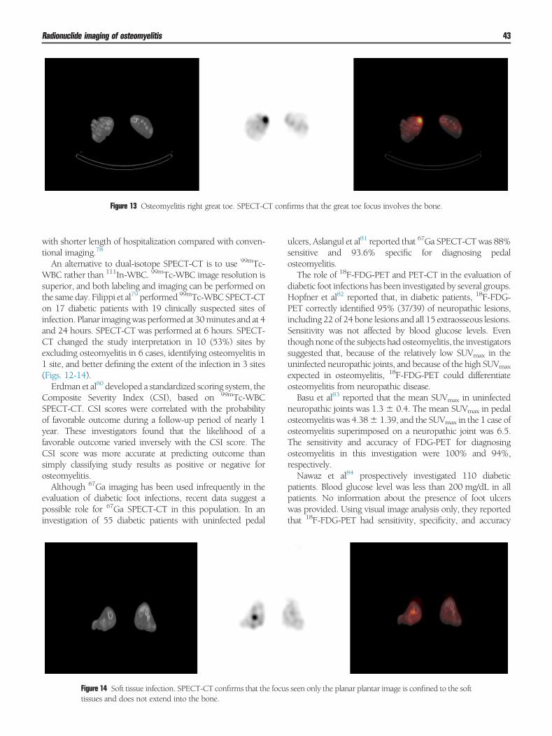
Figure 13 Osteomyelitis right great toe. SPECT-CT confirms that the great toe focus involves the bone.
Radionuclide imaging of osteomyelitis 43
with shorter length of hospitalization compared with conven-tional imaging.78
An alternative to dual-isotope SPECT-CT is to use 99mTc-WBC rather than 111In-WBC. 99mTc-WBC image resolution issuperior, and both labeling and imaging can be performed onthe same day. Filippi et al79 performed 99mTc-WBCSPECT-CTon 17 diabetic patients with 19 clinically suspected sites ofinfection. Planar imagingwas performed at 30minutes and at 4and 24 hours. SPECT-CT was performed at 6 hours. SPECT-CT changed the study interpretation in 10 (53%) sites byexcluding osteomyelitis in 6 cases, identifying osteomyelitis in1 site, and better defining the extent of the infection in 3 sites(Figs. 12-14).Erdman et al80 developed a standardized scoring system, the
Composite Severity Index (CSI), based on 99mTc-WBCSPECT-CT. CSI scores were correlated with the probabilityof favorable outcome during a follow-up period of nearly 1year. These investigators found that the likelihood of afavorable outcome varied inversely with the CSI score. TheCSI score was more accurate at predicting outcome thansimply classifying study results as positive or negative forosteomyelitis.Although 67Ga imaging has been used infrequently in the
evaluation of diabetic foot infections, recent data suggest apossible role for 67Ga SPECT-CT in this population. In aninvestigation of 55 diabetic patients with uninfected pedal
Figure 14 Soft tissue infection. SPECT-CT confirms that the focutissues and does not extend into the bone.
ulcers, Aslangul et al81 reported that 67Ga SPECT-CTwas 88%sensitive and 93.6% specific for diagnosing pedalosteomyelitis.The role of 18F-FDG-PET and PET-CT in the evaluation of
diabetic foot infections has been investigated by several groups.Hopfner et al82 reported that, in diabetic patients, 18F-FDG-PET correctly identified 95% (37/39) of neuropathic lesions,including 22 of 24 bone lesions and all 15 extraosseous lesions.Sensitivity was not affected by blood glucose levels. Eventhoughnone of the subjects hadosteomyelitis, the investigatorssuggested that, because of the relatively low SUVmax in theuninfected neuropathic joints, and because of the high SUVmax
expected in osteomyelitis, 18F-FDG-PET could differentiateosteomyelitis from neuropathic disease.Basu et al83 reported that the mean SUVmax in uninfected
neuropathic joints was 1.3� 0.4. The mean SUVmax in pedalosteomyelitis was 4.38� 1.39, and the SUVmax in the 1 case ofosteomyelitis superimposed on a neuropathic joint was 6.5.The sensitivity and accuracy of FDG-PET for diagnosingosteomyelitis in this investigation were 100% and 94%,respectively.Nawaz et al84 prospectively investigated 110 diabetic
patients. Blood glucose level was less than 200 mg/dL in allpatients. No information about the presence of foot ulcerswas provided. Using visual image analysis only, they reportedthat 18F-FDG-PET had sensitivity, specificity, and accuracy
s seen only the planar plantar image is confined to the soft

C.J. Palestro44
of 81%, 93%, and 90% respectively, for diagnosing pedalosteomyelitis.Schwegler et al85 prospectively evaluated 18F-FDG-PET for
diagnosing clinically unsuspected osteomyelitis in 20 diabeticpatients with pedal ulcers. Information on blood glucose levelsat the time of imaging was not provided. Only visual imageanalysis was performed. 18F-FDG-PET detected only 2 (29%sensitivity) of 7 cases of osteomyelitis.Keidar et al86 compared 18F-FDG-PET and PET-CT in 18
clinically suspected sites of infection. The accuracy of 18F-FDG-PET-CT for diagnosing pedal osteomyelitis was approx-imately 94%. The mean SUVmax in infection was 5.7 andranged from 1.7-11.1 for both osseous and soft tissue foci ofinfection. There was no relationship between the patients 'glycemic state and degree of 18F-FDG uptake.Kagna et al87 investigated 18F-FDG-PET-CT in 39 diabetic
patients (46 sites), 14 of whom had been included in thepublication of Keidar et al.86 Sensitivity, specificity, andaccuracy for diagnosing osteomyelitis was 100%, 93%, and96%, respectively.Familiari et al88 compared 18F-FDG-PET-CT with planar
Tc-WBC imaging in 13 diabetic patients with a high pretestlikelihood of pedal osteomyelitis. All patients had a bloodglucose level of less than 160 mg/dL. 18F-FDG-PET-CTimaging was performed at 10 minutes and 1 and 2 hours afterinjection. The highest accuracy for 18F-FDG-PET-CT at 54%was achieved when the SUVmax was Z2.0 at 1 and 2 hoursafter injection and increased over time. Accuracy improved to62%when CT findings were taken into account. The accuracyof planar 99mTc-WBC imaging, in contrast, was 92%.Presently, the role of 18F-FDG imaging in the workup of
diabetic foot requires further investigation.
References1. Osman DR: Diagnosis and management of musculoskeletal infection. In:
Fitzgerald RH, Haufer H, Malkani RL, (eds): Orthopedics. St. Louis:Mosby; 2002. pp. 695-707
2. Palestro CJ, Love C: Radionuclide imaging of musculoskeletal infection:Conventional agents. Semin Musculoskelet Radiol 2007;11:335-352
3. Palestro CJ, Love C, Bhargava KK: Labeled leukocyte imaging: Currentstatus and future directions.Q JNuclMedMol Imaging 2009;53:105-123
4. Palestro CJ, Love C, Tronco GG, et al: Combined labeled leukocyte andtechnetium-99m sulfur colloid marrow imaging for diagnosing muscu-loskeletal infection: Principles, technique, interpretation, indications andlimitations. Radiographics 2006;26:859-870
5. Palestro CJ, Glaudemans AWJM, R.A.J.O. Dierckx: Multiagent imaging ofinflammation and infection. Clin Transl Imaging 2013;1:385-396
6. Palestro CJ: FDG-PET in musculoskeletal infections. Semin Nucl Med2013;43:367-376
7. Forstrom LA, Dunn WL, Mullan BP: Biodistribution and dosimetry of[F-18] fluorodeoxyglucose labeled leukocytes in normal human subjects.Nucl Med Commun 2002;23:721-725
8. Dumarey N, Egrise D, Blocklet D, et al: Imaging infection with 18F-FDGlabeled leukocyte PET/CT: Initial experience in 21 patients. J Nucl Med2006;47:625-632
9. Rini JN, BhargavaKK, TroncoGG, et al: PETwith FDG-labeled leukocytesversus scintigraphy with 111In-oxine-labeled leukocytes for detection ofinfection. Radiology 2006;238:978-987
10. Aksoy SY, Asa S, Ozhan M, et al: FDG and FDG-labelled leucocyte PET/CT in the imaging of prosthetic joint infection. Eur J Nucl Med MolImaging 2014;41:556-564
11. Bhargava KK, Gupta RK, Nichols KJ, et al: In-vitro human leukocytelabeling with 64Cu: An intraindividual comparison with 111In-oxine and18F-FDG. Nucl Med Biol 2009;36:545-549
12. Kumar V, Boddeti DK, Evans SG, et al: Potential use of 68Ga-apo-transferrin as a PET imaging agent for detecting Staphylococcus aureusinfection. Nucl Med Biol 2011;38:393-398
13. Diaz LA, Foss CA, Thornton K, et al: Imaging of musculoskeletal bacterialinfections by [124I]FIAU-PET/CT. PLoS One 2007;10:e1007
14. Love C, Patel M, Lonner BS, et al: Diagnosing spinal osteomyelitis:A comparison of bone and gallium scintigraphy and magnetic resonanceimaging. Clin Nucl Med 2002;25:963-977
15. Palestro CJ, Love C,Miller TT: Imaging ofmusculoskeletal infections. BestPract Res Clin Rheumatol 2006;20:1197-1218
16. Gemmel F, Dumarey N, Palestro CJ: Radionuclide imaging of spinalinfections. Eur J Nucl Med Mol Imaging 2006;33:1226-1237
17. Lievano P, De la Cueva L, Navarro P, et al: 67Ga SPECT/low-dose CT.A case report of spondylodiscitis and Schmorl 's node. Rev Esp Med Nucl2009;28:288-290
18. Domínguez ML, Lorente R, Rayo JI, et al: SPECT-CT with (67)Ga-citratein the management of spondylodiscitis. Rev Esp Med Nucl Imagen Mol2012;31:34-39
19. Fuster D, Solà O, Soriano A, et al: A prospective study comparingwhole-body FDG PET/CT to combined planar bone scan with 67GaSPECT/CT in the diagnosis of spondylodiskitis. Clin Nucl Med 2012;37:827-832
20. Lazzeri E, Pauwels EKJ, Erba P, et al: Clinical feasibility of two-stepstreptavidin/111In-biotin scintigraphy in patients with suspected vertebralosteomyelitis. Eur J Nucl Med Mol Imaging 2004;31:1505-1511
21. Lazzeri E, Erba P, Perri M, et al: Clinical impact of SPECT/CTwith In-111biotin on themanagement of patients with suspected spine infection. ClinNucl Med 2010;35:12-17
22. Guhlmann A, Brecht-Krauss D, Suger G, et al: Chronic osteomyelitis:Detection with FDG PET and correlation with histopathologic findings.Radiology 1998;206:749-754
23. Guhlmann A, Brecht-Krauss D, Suger G, et al: Fluorine-18-FDG PET andtechnetium-99m antigranulocyte antibody scintigraphy in chronic osteo-myelitis. J Nucl Med 1998;39:2145-2152
24. Schiesser M, Stumpe KD, Trentz O, et al: Detection of metallic implant-associated infections with FDG PET in patients with trauma: Correlationwith microbiologic results. Radiology 2003;226:391-398
25. Kalicke T, Schmitz A, Risse JH, et al: Fluorine-18 fluorodeoxyglucose PETin infectious bone diseases: Results of histologically confirmed cases. EurJ Nucl Med Mol Imaging 2000;27:524-528
26. Schmitz A, Risse JH, Grunwald F, et al: Fluorine-18 fluorodeoxyglucosepositron emission tomography findings in spondylodiscitis: Preliminaryresults. Eur Spine J 2001;10:534-539
27. Meller J, Koster G, Liersch T, et al: Chronic bacterial osteomyelitis:Prospective comparison of F-18-FDG imaging with a dual-head coinci-dence camera and In-111-labelled autologous leucocyte scintigraphy. EurJ Nucl Med Mol Imaging 2002;29:53-60
28. Hartmann A, Eid K, Dora C, et al: Diagnostic value of 18F-FDGPET/CT intraumapatientswith suspected chronic osteomyelitis. Eur JNuclMedMolImaging 2007;34:704-714
29. Gratz S, Dorner J, Fischer U, et al: F-18-FDG hybrid PET in patientswith suspected spondylitis. Eur J Nucl Med Mol Imaging 2002;29:516-524
30. Ohtori S, Suzuki M, Koshi T, et al: 18F-fluorodeoxyglucose-PET forpatients with suspected spondylitis showing Modic change. Spine2010;15(35):E1599-E1603
31. Stumpe KD, Zanetti M, Weishaupt D, et al: FDG positron emissiontomography for differentiation of degenerative and infectious endplateabnormalities in the lumbar spine detected on MR imaging. AmJ Roentgenol 2002;179:1151-1157
32. Seifen T, Rettenbacher L, Thaler C, et al: Prolonged back pain attributed tosuspected spondylodiscitis. The value of 18F-FDG PET/CT imaging in thediagnostic work-up of patients. Nuklearmedizin 2012;51:194-200
33. Rosen RS, Fayad L, Wahl RL: Increased 18F-FDG uptake in degenerativedisease of the spine: Characterization with 18FFDG PET/CT. J Nucl Med2006;47:1274-1280

Radionuclide imaging of osteomyelitis 45
34. de Winter F, Gemmel F, Van de Wiele C, et al: 18-Fluorine fluorodeox-yglucose positron emission tomography for the diagnosis of infection inthe postoperative spine. Spine 2003;28:1314-1319
35. Nanni C, Errani C, Boriani L, et al: 68Ga-Citrate PET/CT for evaluatingpatients with infections of the bone: Preliminary results. J Nucl Med2010;51:1932-1936
36. De Winter F, van de Wiele C, Vogelaers D, et al: Fluorine-18fluorodeoxyglucose positron emission tomography: A highly accurateimaging modality for the diagnosis of chronic musculoskeletal infections.J Bone Joint Surg Am 2001;83-A:651-660
37. Love C, Marwin SE, Palestro CJ: Nuclear medicine and the infected jointreplacement. Semin Nucl Med 2009;39:66-78
38. Tomas X, Bori G, Garcia S, et al: Accuracy of CT-guided joint aspiration inpatients with suspected infection status post-total hip arthroplasty.Skeletal Radiol 2011;40:57-64
39. Palestro CJ, Swyer AJ, Kim CK, et al: Infected knee prostheses: Diagnosiswith In-111 leukocyte, Tc-99m sulfur colloid, and Tc-99mMDP imaging.Radiology 1991;179:645-648
40. Magnuson JE, Brown ML, Hauser MF, et al: In-111 labeled leukocytescintigraphy in suspected orthopedic prosthesis infection: Comparisonwith other imaging modalities. Radiology 1988;168:235-239
41. Levitsky KA, Hozack WJ, Balderston RA, et al: Evaluation of the painfulprosthetic joint. Relative value of bone scan, sedimentation rate, and jointaspiration. J Arthroplasty 1991;6:237-244
42. Reinartz P, Mumme T, Hermanns B, et al: Radionuclide imaging of thepainful hip arthroplasty: Positron-emission tomography versus triple-phase bone scanning. J Bone Joint Surg Br 2005;87-B:465-470
43. MulambaL, FerrantA, LenersN, et al: Indium-111 leucocyte scanning in theevaluation of painful hip arthroplasty. Acta Orthop Scand 1983;54:695-697
44. Palestro CJ, KimCK, Swyer AJ, et al: Total hip arthroplasty: Periprostheticindium-111-labeled leukocyte activity and complementary technetium-99m-sulfur colloid imaging in suspected infection. J Nucl Med1990;31:1950-1955
45. Joseph TN,MujitabaM, Chen AL, et al: Efficacy of combined technetium-99m sulfur colloid/indium-111 leukocyte scans to detect infected total hipand knee arthroplasties. J Arthroplasty 2001;16:753-758
46. Love C, Marwin SE, Tomas MB, et al: Diagnosing infection in the failedjoint replacement: A comparison of coincidence detection fluorine-18FDG and indium-111-labeled leukocyte/technetium-99m-sulfur colloidmarrow imaging. J Nucl Med 2004;45:1864-1871
47. El Espera I, Blondet C, Moullart V, et al: The usefulness of 99mTc sulfurcolloid bone marrow scintigraphy combined with 111In leucocytescintigraphy in prosthetic joint infection. Nucl Med Commun2004;25:171-175
48. Pill SG, Parvizi J, Tang PH, et al: Comparison of fluorodeoxyglucosepositron emission tomography and (111)indium-white blood cell imagingin the diagnosis of periprosthetic infection of the hip. J Arthroplasty2006;21:91-97
49. Boubaker A, Delaloye AB, Blanc CH, et al: Immunoscintigraphy withantigranulocyte monoclonal antibodies for the diagnosis of septic loosen-ing of hip prostheses. Eur J Nucl Med 1995;22:139-147
50. Gratz S, HöffkenH, Kaiser JW:Nuclearmedical imaging in case of painfulknee arthroplasty. Radiologe 2009;49:59-67
51. Klett R, Steiner D, Puille M, et al: Antigranulocyte scintigraphy of septicloosening of hip endoprosthesis: Effect of different methods of analysis.Nuklearmedizin 2001;40:75-79
52. Klett R, Kordelle J, Stahl U, et al: Immunoscintigraphy of septic looseningof knee endoprosthesis: A retrospective evaluation of the antigranulocyteantibody BW250/183. Eur J NuclMedMol Imaging 2003;30:1463-1466
53. Von Rothenburg T, Schoellhammer M, Schaffstein J, et al: Imaging ofinfected total arthroplasty with Tc-99m-labeled antigranulocyte antibodyFab0 fragments. Clin Nucl Med 2004;29:548-551
54. Iyengar KP, Vinjamuri S: Role of 99mTc Sulesomab in the diagnosisof prosthetic joint infections. Nucl Med Commun 2005;26:489-496
55. Pakos EE, Fotopoulos AD, Stafilas KS, et al: Use of (99m)Tc-sulesomab forthe diagnosis of prosthesis infection after total joint arthroplasty. J IntMedRes 2007;35:474-481
56. Rubello D, Casara D, Maran A, et al: Role of anti-granulocyte Fab0
fragment antibody scintigraphy (LeukoScan) in evaluating bone infection:Acquisition protocol, interpretation criteria and clinical results. Nucl MedCommun 2004;25:39-47
57. Rubello D, Rampin L, Banti E, et al: Diagnosis of infected total kneearthroplasty with anti-granulocyte scintigraphy: The importance of adual-time acquisition protocol. Nucl Med Commun 2008;29:331-335
58. Gratz S, Behr TM, Reize P, et al: (99m)Tc-Fab0 fragments (sulesomab) forimaging septically loosened total knee arthroplasty. J Int Med Res2009;37:54-67
59. Sousa R, Massada M, Pereira A, et al: Diagnostic accuracy of combined99mTc-sulesomab and 99mTc-nanocolloid bone marrow imaging indetecting prosthetic joint infection. Nucl Med Commun 2011;32:834-839
60. Filippi L, Schillaci O: Tc-99mHMPAO-labeled leukocyte scintigraphy forbone and joint infections. J Nucl Med 2006;47:1908-1913
61. Kaisidis A, Megas P, Apostolopoulos D, et al: SPECT scan with 99mTc-labeled monoclonal antibodies. Orthopade 2005;34:462-469
62. Graute V, Feist M, Lehner S, et al: Detection of low-grade prosthetic jointinfections using 99mTc-antigranulocyte SPECT/CT: Initial clinical results.Eur J Nucl Med Mol Imaging 2010;37:1751-1759
63. Zhuang H, Duarte PS, Pourdehnad M, et al: The promising role of 18F-FDGPET in detecting infected lower limbprosthesis implants. JNuclMed2001;42:44-48
64. Chacko TK, Zhuang H, Nakhoda KZ, et al: Applications of fluorodeox-yglucose positron emission tomography in the diagnosis of infection.NuclMed Commun 2003;24:615-624
65. Chacko TK, ZhuangH, Stevenson K, et al: The importance of the locationof fluorodeoxyglucose uptake in periprosthetic infection in painful hipprostheses. Nucl Med Commun 2002;23:851-855
66. Cremerius U, Mumme T, Reinartz P, et al: Analysis of (18)F-FDG uptakepatterns in PET for diagnosis of septic and aseptic loosening after total hiparthroplasty. Nuklearmedizin 2003;42:234-239
67. Gravius S, Gebhard M, Ackermann D, et al: Analysis of 18F-FDG uptakepattern in PET for diagnosis of aseptic loosening versus prosthesisinfection after total knee arthroplasty. A prospective pilot study. Nuklear-medizin 2010;49:115-123
68. Manthey N, Reinhard P, Moog F, et al: The use of [18F] fluorodeox-yglucose positron emission tomography to differentiate between synovitis,loosening and infection of hip and knee prostheses. Nucl Med Commun2002;23:645-653
69. Stumpe KD, Notzli HP, Zanetti M, et al: FDG PET for differentiation ofinfection and aseptic loosening in total hip replacements: Comparisonwith conventional radiography and three-phase bone scintigraphy.Radiology 2004;231:333-341
70. Van Acker F, Nuyts J, Maes A, et al: FDG-PET, 99mTc-HMPAO whiteblood cell SPET and bone scintigraphy in the evaluation of painful totalknee arthroplasties. Eur J Nucl Med 2001;28:1496-1504
71. Vanquickenborne B,Maes A, Nuyts J, et al: The value of (18)FDG-PET forthe detection of infected hip prosthesis. Eur J Nucl Med Mol Imaging2003;30:705-715
72. García-Barrecheguren E, Rodríguez Fraile M, Toledo Santana G, et al:FDG-PET: A new diagnostic approach in hip prosthetic replacement. RevEsp Med Nucl 2007;26:208-220
73. Delank KS, Schmidt M, Michael JW, et al: The implications of 18F-FDGPET for the diagnosis of endoprosthetic loosening and infection in hip andknee arthroplasty: Results from a prospective, blinded study. BMCMusculoskelet Disord 2006;7:20
74. Sarda-Mantel L, Saleh-Mghir A, Welling MM, et al: Evaluation of 99mTc-UBI 29-41 scintigraphy for specific detection of experimental Staph-ylococcus aureus prosthetic joint infections. Eur J Nucl MedMol Imaging2007;34:1302-1309
75. Aryana K, Hootkani A, Sadeghi R, et al: (99m)Tc-labeled ubiquicidinscintigraphy: A promising method in hip prosthesis infection diagnosis.Nuklearmedizin 2012;51:133-139
76. Palestro CJ, Love C: Nuclear medicine and diabetic foot infections. SeminNucl Med 2009;39:52-65

C.J. Palestro46
77. Heiba SI, Kolker D,Mocherla B, et al: The optimized evaluation of diabeticfoot infection by dual isotope SPECT/CT imaging protocol. J. Foot AnkleSurg 2010;49:529-536
78. Heiba S, Kolker D, Ong L, et al: Dual-isotope SPECT/CT impact onhospitalized patients with suspected diabetic foot infection: Saving limbs,lives, and resources. Nucl Med Commun 2013;34:877-884
79. Filippi L, Uccioli L, Giurato L, et al: Diabetic foot infection: Usefulness ofSPECT/CT for 99mTc-HMPAO-labeled leukocyte imaging. J Nucl Med2009;50:1042-1046
80. Erdman WA, Buethe J, Bhore R, et al: Indexing severity of diabetic footinfection with 99mTc-WBC SPECT/CT hybrid imaging. Diabetes Care2012;35:1826-1831
81. Aslangul E,M 'bemba J, Caillat-VigneronN, et al: Diagnosing diabetic footosteomyelitis in patients without signs of soft tissue infection by couplinghybrid 67Ga SPECT/CT with bedside percutaneous bone puncture.Diabetes Care 2013;36:2203-2210
82. Hopfner S, Krolak C, Kessler S, et al: Preoperative imaging of Charcotneuroarthropathy in diabetic patients: Comparison of ring PET, hybridPET, and magnetic resonance imaging. Foot Ankle Int 2004;25:890-895
83. Basu S, Chryssikos T, Houseni M, et al: Potential role of FDG-PET in thesetting of diabetic neuro-osteoarthropathy: Can it differentiate uncompli-cated Charcot’s neuropathy from osteomyelitis and soft tissue infection?Nucl Med Commun 2007;28:465-472
84. Nawaz A, Torigian DA, Siegelman ES, et al: Diagnostic performance ofFDG-PET, MRI, and plain film radiography (PFR) for the diagnosis ofosteomyelitis in the diabetic foot. Mol Imaging Biol 2010;12:335-342
85. Schwegler B, Stumpe KD, Weishaupt D, et al: Unsuspected osteomyelitisis frequent in persistent diabetic foot ulcer and better diagnosed byMRI than by 18F-FDG PET or 99mTc-MOAB. J Intern Med 2008;263:99-106
86. Keidar Z, Militianu D, Melamed E, et al: The diabetic foot: Initialexperience with 18F-FDG-PET/CT. J Nucl Med 2005;46:444-449
87. Kagna O, Srour S, Melamed E, et al: FDG PET/CT imaging in thediagnosis of osteomyelitis in the diabetic foot. Eur J Nucl Med MolImaging 2012;39:1545-1550
88. Familiari D, Glaudemans AWJM, Vitale V, et al: Can sequential 18F-FDG-PET/CT imaging replace WBC imaging in the diabetic foot? J Nucl Med2011;52:1012-1019