proton therapy – process support with anesthesia · d-23775-2009 in october 2009, dräger talked...
TRANSCRIPT

D-2
3775
-200
9
In October 2009, Dräger talked to Dr. Morten Eckermann, head/director of the anesthesia department for the Munich Rinecker Proton Therapy Center (RPTC, Fig. 1), about his experience with a new type of therapy and process support with anesthesia.
PREFACE A radiation unit for proton therapy has a technical specialty of a radiation head (nozzle) with a magnetic field of approxi-mately one Tesla, which deflects the proton beam over the tumor surface to be radiated (Fig. 2). This brings up the question of whether a MRT capable anesthetic device must be used or if a conventional standard device is sufficient.
The Dräger Fabius Tiro with the Delta patient monitor and the Scio gas mod-ule was selected as the conventional anesthetic work place for introduction and on-site radiation.
Preliminary tests performed at the Hahn-Meitner Institute in Berlin monitored the magnetic field strength and magnetic field influences with the desired Fabius Tiro configuration and concluded that a standard anesthesia workplace is suffi-cient.
In close teamwork between Dräger and the RPTC, the Fabius Tiro was separately measured and verified directly at its working position in the gantry.
The first radiation unit started operation in March 2009. After six months of evaluation, we are now able to expand on the previous knowledge as regards the anesthesia related workflows with patients.
Proton therapy – Process support with anesthesia
Fig. 1. Rinecker Proton Therapy Center Radiation center
02 |
D-2
3776
-200
9
PROTON THERAPY – PROCESS SUPPORT WITH ANESTHESIA.
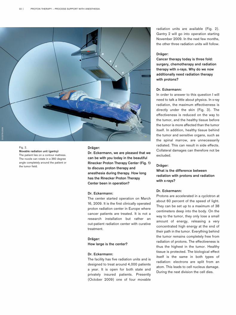
Fig. 2. Movable radiation unit (gantry) The patient lies on a contour mattress. The nozzle can rotate in a 360 degree angle completely around the patient or the tumor field.
radiation units are available (Fig. 2). Gantry 2 will go into operation starting November 2009. In the next few months, the other three radiation units will follow.
Dräger: Cancer therapy today is three fold: surgery, chemotherapy and radiation therapy with x-rays. Why do we now additionally need radiation therapy with protons?
Dr. Eckermann: In order to answer to this question I will need to talk a little about physics. In x-ray radiation, the maximum effectiveness is directly under the skin (Fig. 3). The effectiveness is reduced on the way to the tumor, and the healthy tissue before the tumor is more affected than the tumor itself. In addition, healthy tissue behind the tumor and sensitive organs, such as the spinal marrow, are unnecessarily radiated. This can result in side effects. Collateral damages can therefore not be excluded.
Dräger: What is the difference between radiation with protons and radiation with x-rays?
Dr. Eckermann: Protons are accelerated in a cyclotron at about 60 percent of the speed of light. They can be set up to a maximum of 38 centimeters deep into the body. On the way to the tumor, they only lose a small amount of energy, releasing a very concentrated high energy at the end of their path in the tumor. Everything behind the tumor remains completely free from radiation of protons. The effectiveness is thus the highest in the tumor. Healthy tissue is protected. The biological effect itself is the same in both types of radiation: electrons are split from an atom. This leads to cell nucleus damage. During the next division the cell dies.
Dräger: Dr. Eckermann, we are pleased that we can be with you today in the beautiful Rinecker Proton Therapy Center (Fig. 1) to discuss proton therapy and anesthesia during therapy. How long has the Rinecker Proton Therapy Center been in operation?
Dr. Eckermann: The center started operation on March 16, 2009. It is the first clinically operated proton radiation center in Europe where cancer patients are treated. It is not a research installation but rather an out-patient radiation center with curative treatment.
Dräger: How large is the center?
Dr. Eckermann: The facility has five radiation units and is designed to treat around 4,000 patients a year. It is open for both state and privately insured patients. Presently (October 2009) one of four movable

|03
D-1
8278
-201
0
PROTON THERAPY – PROCESS SUPPORT WITH ANESTHESIA
Fig. 3: Comparison of proton radiation (green) and x-ray radiation (orange): The effectiveness is the highest in the tumor with proton therapy. Healthy tissue is protected.
Dräger: Which tumors can be handled with proton therapy?Dr. Eckermann: Based on the physical and biological properties of the protons, basically all tumors that could previously be treated with x-ray radiation therapy can also be treated with proton radiation. Including but not limited to organs such as lungs, liver, pancreas, gallbladder and prostate. But we can also undergo proton therapy on tumors in the brain area, neck and in the nose area. Here, this is much less aggressive for the tissue surrounding the tumor. In addition, with proton therapy, we can also address cancer localization, which cannot be treated with x-ray radiation because of excess side effects. We can then even start proton therapy if the patient has already reached the tolerance limits of side effects according to conventional x-ray therapy. Due to the reduced chance of secondary tumor triggering, proton therapy is absolutely preferred for children over x-ray radiation.
Dräger: Approximately how many radiation treatments are necessary for proton therapy at RPTC?
Dr. Eckermann: The number of radiation treatments depends on the type and size of the tumor. On average, patients must count on approximately 18 sessions. At RPTC we radiate Monday through Saturday. On Sunday the units are serviced.
Dräger: Who is bearing the treatment costs?
Dr. Eckermann: The first state health insurance to provide a provision contract for proton radiation at RPTC for their insured patients was AOK Bayern. AOK Bayern also represents the AOKs of other German states as a clearing house. Also the Landesverband
Bayern of the Betriebskrankenkassen (BKK), which covers all of Germany, and the Landwirtschaftlichen Krankenkassen Bayern have signed corresponding contracts with the RPTC. More insurance companies will follow. Proton therapy in Munich at the RPTC is thus a service of the state health insurance.
Dräger: Generally, how long does one radia-tion session last?
Dr. Eckermann: The actual proton radiation lasts from about one to five minutes per field, depending on the size of the tumor. It is completely painless. The radiation process with anesthesia at this time is around a 30 to 45 minutes stay in the gantry. The first control examination is in about eight weeks. Then we compare, using MRT and CT, whether the original size and characteristics of the tumor have changed or not.
Dräger: Is any anesthesia necessary during proton therapy?
Dr. Eckermann: According to our experience, we do not require any anesthetist for around 80 to 90 percent of the patients. Cooperating children and adults are treated and cared for on an outpatient basis by the department for radiation therapy. An anesthetist is used only for the remaining 10 to 20 percent of the patients. How the patients will develop in future remains to be seen.
Dräger: What tasks are performed by the anesthetist during proton therapy? Dr. Eckermann: The anesthetist performs two tasks. He/she sedates smaller children and fearful or excited patients. This ensures their immobilization.
He/she also relaxes patients with general anesthesia if the targeted organs move with breathing. This includes, for example, lungs, liver or pancreas.
Dräger: This means a patient with a liver tumor, for example, must always have general anesthesia during proton therapy?
Dr. Eckermann: Yes, because if the patient breathes spontaneously in this case the liver would move by a few centimeters in the longitudinal direction. Then we could not radiate the liver tumor in a single pass. In the gantry of the proton unit we precisely set the exact beam position within one millimeter. In this way radiation takes place in defined angles and layers. This means at the same time: the tumor must remain „stationary“ so that we can hit it effectively. To keep organs still by generating an apnea phase in controlled general anesthesia, was the critical idea from Dr. Rinecker.

04 |
D-2
3778
-200
9
PROTON THERAPY – PROCESS SUPPORT WITH ANESTHESIA
folded out. The x-ray images on the proton radiation unit are compared to the CT based images. Then we can accom-modate the actual position to the target position. In the CT we use the same equipment as with the initiation of the diagnostics.
Dräger: The proton therapy is done a few days after the diagnostics. What does the process for sedating children look like with you?
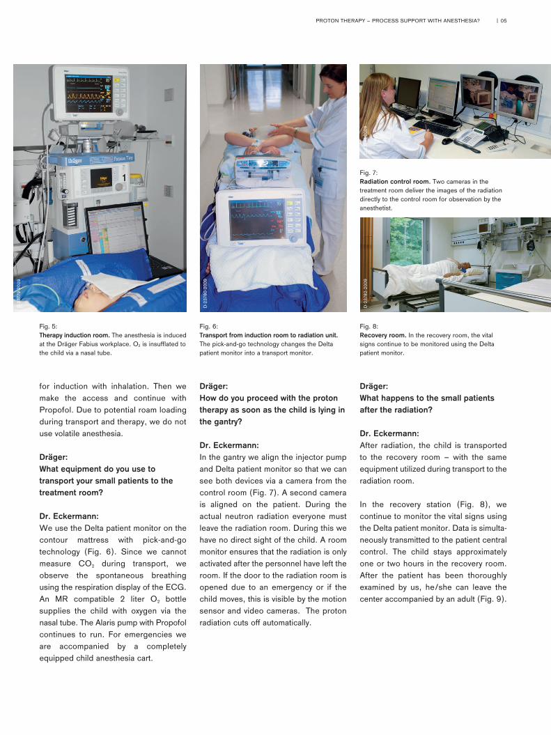
Dr. Eckermann: In the therapy induction room (Fig. 5), we induce anesthesia at the Fabius work-place. Monitoring is performed with ECG, SpO2, NiBP and rectal temperature measurement and insufflating O2 via nasal tube and monitoring the etCO2. Sedation is done with Propofol via Perfusor. In exceptional cases, if there is no intravenous access available and the child is quite excited, we use Sevoflurane
pump outside of the MRT. After oral or rectal preliminary medication with Midazolam, sedation is done during the examination – in general with Propofol. We monitor children with EKG, SpO2, NiBP and rectal temperature measure-ment. We apply O2 via a nasal tube. Spontaneous breathing is monitored in the MRT. Occasionally, we use Sevoflurane in order to avoid the long lines of the Perfusor.
Dräger: How do you determine the position of the tumor?
Dr. Eckermann: We basically utilize a CT for precise goal planning. This way we can later verify the positioning of the child at the radiation site. Two x-ray machines are installed in diagonal positions in the gantry for this purpose. The x-ray machines affect two x-ray sensitive flat panels. They are integrated on the side of the nozzle to be
Dräger: How do you prepare the patients for proton radiation?
Dr. Eckermann: During proton therapy the beam must hit with a precision of less than one millimeter. For this the patient must take the same position for each radiation treatment. We place the patient in a contour mattress, which is a large mattress with Styrofoam balls (Fig. 2). At the beginning of the radiation planning it is evacuated. Each patient gets his/her own contour mattress, which is assigned to the patient using a barcode. For radiation treatment on the head, we additionally use a face mask or a bite profile from the upper jaw. This can be imagined as a fixed tooth brace as is familiar in dentistry. In this way our patients are immobilized. We check if the patient is lying properly using digital x-rays. These x-rays are supplied by flat panels which are installed on the side of the nozzle, the location where the radi-ation exits.
Dräger: How do you conduct the diagnostics of your patients?
Dr. Eckermann: In general, each of our patients under-goes a total body diagnosis before the radiation. We plan the intervention using CT or MRT (Fig. 4). First we scan the entire body to record all tumor manifesta-tions. We work with our two Philips Achieva MR systems with 1.5 Tesla MRT. For children, a sedative is oftentimes necessary for this diagnostics.
In the RT we use a Titus MRI with Ventilog C, Cosy 1, PM8050 MRI and four meter long breathing hoses. It is supplied by two MRT compatible 10-liter O2 bottles fastened on the device. Patient monitoring is done by an Invivo MR monitor. The diagnostics are initiated on a Fabius Tiro workplace with Delta patient monitor, Scio gas module and Alaris injection
Fig. 4: The entire body of the patient is scanned in the MRT for diagnostics. This way all tumor manifestations are recorded.
| 05D
-237
79-2
009
D-2
3780
-200
9
D-2
3782
-200
9D
-237
81-2
009
PROTON THERAPY – PROCESS SUPPORT WITH ANESTHESIA?
for induction with inhalation. Then we make the access and continue with Propofol. Due to potential roam loading during transport and therapy, we do not use volatile anesthesia.
Dräger: What equipment do you use to transport your small patients to the treatment room?
Dr. Eckermann: We use the Delta patient monitor on the contour mattress with pick-and-go technology (Fig. 6). Since we cannot measure CO2 during transport, we observe the spontaneous breathing using the respiration display of the ECG. An MR compatible 2 liter O2 bottle supplies the child with oxygen via the nasal tube. The Alaris pump with Propofol continues to run. For emergencies we are accompanied by a completely equipped child anesthesia cart.
Dräger: How do you proceed with the proton therapy as soon as the child is lying in the gantry?
Dr. Eckermann: In the gantry we align the injector pump and Delta patient monitor so that we can see both devices via a camera from the control room (Fig. 7). A second camera is aligned on the patient. During the actual neutron radiation everyone must leave the radiation room. During this we have no direct sight of the child. A room monitor ensures that the radiation is only activated after the personnel have left the room. If the door to the radiation room is opened due to an emergency or if the child moves, this is visible by the motion sensor and video cameras. The proton radiation cuts off automatically.
Dräger: What happens to the small patients after the radiation?
Dr. Eckermann: After radiation, the child is transported to the recovery room – with the same equipment utilized during transport to the radiation room.
In the recovery station (Fig. 8), we continue to monitor the vital signs using the Delta patient monitor. Data is simulta-neously transmitted to the patient central control. The child stays approximately one or two hours in the recovery room. After the patient has been thoroughly examined by us, he/she can leave the center accompanied by an adult (Fig. 9).
Fig. 7: Radiation control room. Two cameras in the treatment room deliver the images of the radiation directly to the control room for observation by the anesthetist.
Fig. 8: Recovery room. In the recovery room, the vital signs continue to be monitored using the Delta patient monitor.
Fig. 6: Transport from induction room to radiation unit. The pick-and-go technology changes the Delta patient monitor into a transport monitor.
Fig. 5: Therapy induction room. The anesthesia is induced at the Dräger Fabius workplace. O2 is insufflated to the child via a nasal tube.
06 |
D-2
3783
-200
9D
-237
84-2
009
PROTON THERAPY – PROCESS SUPPORT WITH ANESTHESIA
in the induction room. We monitor ECG, SpO2 and NiBP, place a venous access and preoxygenate (Fig. 10).
Dräger: Which anesthesia do you use for your adult patients?
Dr. Eckermann: For preliminary medication we use Dormicum and Propofol as hypnotic. In order to protect the respiratory tract dur-ing general anesthesia from around one to two hours and to create an exact reproducible condition, we use Atracuri-um as muscle relaxant and intubate our adult patients. Since the radiation is not painful, we do not use large doses of opiates.
Dräger: Do you transport your adult patients with the same equipment as you use for children?
Dr. Eckermann: Yes. An adult patient is transported to the radiation unit under the same conditions as a child.
Dräger: How is the adult patient prepared in the radiation room for the proton therapy?
Dr. Eckermann: After arriving in the radiation room, a long breathing hose is connected to the Fabius Tiro anesthesia unit (Fig. 11). The adult patient is ventilated with 100 percent O2, volume controlled, standard practice with a tidal volume corresponding to the ideal body weight. The frequency is between 10 and 14 rpm, the PEEP between 0 and 5 millibar and the live gas flow at approximately one liter per minute. We then continue the anesthesia as pure intravenous anesthesia administering continuous Propofol and, if necessary, post relaxant. Just as with our small patients, we establish the exact position of the target area with x-rays via the flat
Dräger: At what age can a child be successfully treated with proton therapy?
Dr. Eckermann: Our youngest patients were aged one year and/or two years and six months.
Dräger: Now we get to the adult patients: How does the proton therapy of an adult differ from that of a child?
Dr. Eckermann: As already mentioned, we perform radiation of organs, which move with breathing, in general anesthesia via controlled apnea phase. In order to better control the position of a movable tumor with an adult, we place (different than with a child) small gold threads, for example, at one to three locations in the surrounding tissue outside of the planed direction of radiation. They serve as x-ray positive marks. Alternatively, the same function can be done with already existing stints or metal clips from previous interventions. For the final planning or targeting CT, we use the same form of anesthesia as with the radiation itself; that is to say also with the necessary apnea phase. In cases of doubt, here it is finally decided whether the patient is suitable from an anesthesia point of view for repeated narcoses and apnea situations during the radiation or not.
Dräger: Is the narcosis induction for proton therapy for adults exactly the same as with other operations?
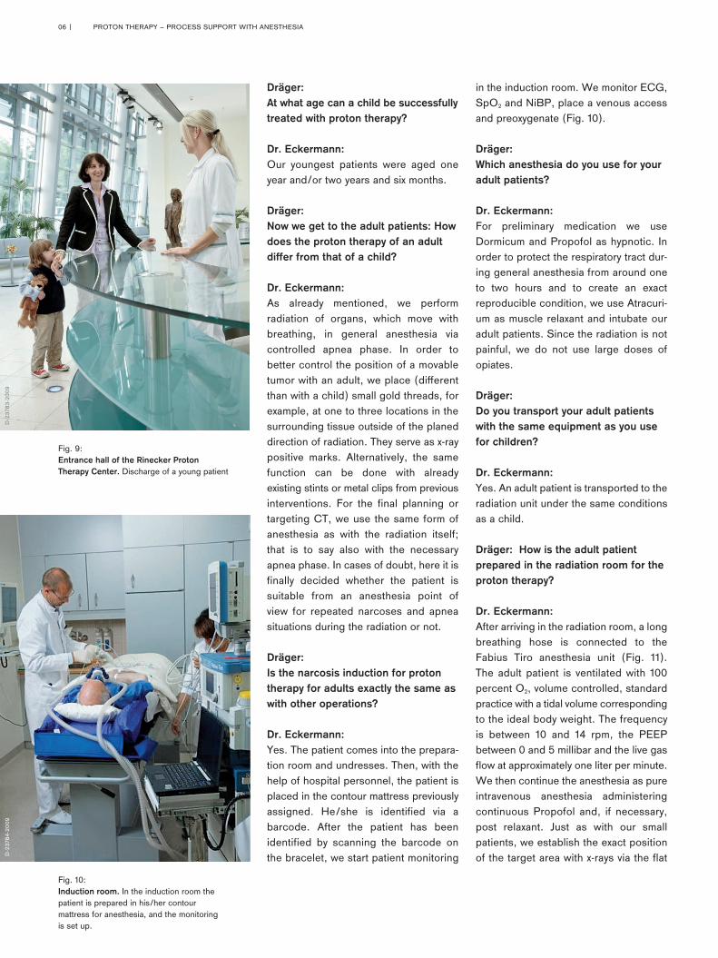
Dr. Eckermann: Yes. The patient comes into the prepara-tion room and undresses. Then, with the help of hospital personnel, the patient is placed in the contour mattress previously assigned. He/she is identified via a barcode. After the patient has been identified by scanning the barcode on the bracelet, we start patient monitoring
Fig. 9: Entrance hall of the Rinecker Proton Therapy Center. Discharge of a young patient
Fig. 10: Induction room. In the induction room the patient is prepared in his/her contour mattress for anesthesia, and the monitoring is set up.

| 07
D-2
3785
-200
9
PROTON THERAPY – PROCESS SUPPORT WITH ANESTHESIA.
panel on the nozzle. We compare the images with the data from the targeting CT.
Dräger: How does radiation exactly progress with an adult?
Dr. Eckermann: To prepare for the apnea phase, we hyperventilate the patients with approx. 30 mmHg etCO2. During the functional apnea we set the anesthesia device at 100 percent O2 at the smallest tidal vol-ume (40 ml) and low frequency. It has been shown that a pressure consistency in the long respiratory tracts is not guaranteed with purely manual or sponta-neous mode. No measured respiratory tract parameters are transferred to the anesthesia protocol system.
Dräger: How long does the functional apnea phase last with an adult patient?
Dr. Eckermann: It takes about three minutes. The time window is composed as follows: 30 sec-onds for the request for the proton beam and leaving the room, 40 seconds for the room switchover of the motion sensor, and 50 seconds to two minutes for the radiation. We supply the patients with a constant O2 flow of one liter per minute. This replaces the O2 uptake of the patient. Studies show that with such an arrangement apnea times can be performed theoretically far above the therapeutically required time without hypoxemia. The duration of the apnea is limited for by the CO2 rise in the blood, since no exhalation occurs.
Dräger: How do you keep a safe view of your adult patients in the gantry from the control room?
Dr. Eckermann: Exactly like our children. During radiation
we are in the control room, about ten meters from the patient. Here there are two information sources available to us: We have in the control room information on ECG, SpO2, NiBP, etCO2 via the slave screen as well as ventilation parameters of the Dräger Infinity Delta patient monitor from the radiation room in view. We also observe the patient and the anesthesia workplace via several cameras.
Dräger: Assuming that you have to radiate several fields or radiate from various angles. How does this work?
Dr. Eckermann: Then we go between the position chang-es to the patient in the gantry, ventilate the patient to reduce the CO2 and then again leave the room.
Dräger: What do you do, however, if the patient moves?
Dr. Eckermann: In case of an emergency (for example the door is opened or the patient moves), the motion sensor and video cameras detect this and the proton beam cuts off auto-matically and direct patient contact is quickly possible again.
Dräger: What happens after cutting off the beam with your adult patients?
Dr. Eckermann: After the therapy has ended, the patient is ventilated with normal tidal volume and a slightly increased frequency. This normalizes the existing CO2 value. Depending on the duration of the apnea
Fig. 11: Radiation room. The patient is connected via long breathing hoses in the radiation room to the Fabius Tiro anesthesia device. The doctors can determine with x-ray images via the flat panels on the nozzle whether the tumor is positioned precisely on target or not.

08 |
D-2
3787
-200
9
D-2
3786
-200
9
90 6
6 46
9 |
15.0
6-3
| C
omm
unic
atio
ns &
Sal
es M
arke
ting
| P
P |
PR
| L
E |
Prin
ted
in G
erm
any
| C
hlor
ine-
free
– e
nviro
nmen
tally
com
patib
le |
Sub
ject
to m
odifi
catio
ns |
© 2
015
Drä
gerw
erk
AG &
Co.
KG
aA
REGION EUROPE CENTRAL AND EUROPE NORTHDrägerwerk AG & Co. KGaA Moislinger Allee 53–5523558 Lübeck, GermanyTel +49 451 882 0Fax +49 451 882 [email protected]
REGION EUROPE SOUTHDräger Médical S.A.S. Parc de Haute Technologie d’Antony 225, rue Georges Besse92182 Antony Cedex, FranceTel +33 1 46 11 56 00Fax +33 1 40 96 97 [email protected]
REGION MIDDLE EAST, AFRICADrägerwerk AG & Co. KGaABranch OfficeP.O. Box 505108Dubai, United Arab EmiratesTel +971 4 4294 600Fax +971 4 4294 [email protected]
REGION ASIA / PACIFICDraeger Medical South East Asia Pte Ltd.25 International Business Park#04-27/29 German CentreSingapore 609916, SingaporeTel +65 6572 4388Fax +65 6572 4399 [email protected]
REGION NORTH AMERICADraeger Medical, Inc.3135 Quarry Road Telford, PA 18969-1042, USATel +1 215 721 5400Toll-free +1 800 437 2437Fax +1 215 723 [email protected]
REGION CENTRAL AND SOUTH AMERICADräger Panama Comercial S. de R.L.Complejo Business Park, V tower, 10th floorPanama CityTel +507 377 9100Fax +507 377 [email protected]
CORPORATE HEADQUARTERSDrägerwerk AG & Co. KGaAMoislinger Allee 53–5523558 Lübeck, Germany
www.draeger.com
Manufacturer:Drägerwerk AG & Co. KGaAMoislinger Allee 53–5523558 Lübeck, Germany
Locate your Regional Sales Representative at: www.draeger.com/contact
PROTON THERAPY – PROCESS SUPPORT WITH ANESTHESIA
phase, it can increase up to 60 mmHg. The patient is then transported to the recovery room. There we extubate the patient and monitor him/her for about 1- 1 1/2 hours. As is common in out-patient facilities, we only discharge the patient if the Aldrete score criteria are met. A companion person then takes the patient home.
Dräger: After six months of operation in this magnetic environment, how did the Dräger anesthesia device/ monitor combination perform in your opinion?
Dr. Eckermann: Six months of practical experience shows that the Fabius Tiro anesthesia workplace
with the Delta patient monitor and the Scio gas module (Fig. 12) are working without any problems.
Dräger: Dr. Eckermann, thank you for your time – and of course for your detailed explanations.
ReferenceDr. H. Rinecker: Protonentherapie. (Proton therapy) Neue Chance bei Krebs.(New change with cancer). Herbig Verlagsbuchhandlung GmbH, Munich; ISBN 3-7766-2422-1. Please also refer to the Internet at www.rptc.de
Fig. 12: Anesthesia workplace. Fabius Tiro with Delta patient monitor and Scio gas module.
PERSONAL INFORMATIONDr. Morten Eckermann studied medicine in Regensburg and at the TU Munich in Germany. After several years of continuing education in surgery, he trained in several large Munich clinics as an anesthetist. He had worked as chief physician in the surgery clinic of Dr. Rinecker for ten years before he became the director of the anesthesia depart-ment at RPTC in March 2009.