proteomics characterization of the cytotoxicity mechanism of
TRANSCRIPT
Proteomics Characterization of the CytotoxicityMechanism of Ganoderic Acid D andComputer-automated Estimation of thePossible Drug Target Network*□S
Qing-Xi Yue‡§, Zhi-Wei Cao§¶, Shu-Hong Guan‡, Xiao-Hui Liu�, Lin Tao¶,Wan-Ying Wu‡, Yi-Xue Li¶, Peng-Yuan Yang�, Xuan Liu‡**, and De-An Guo‡ ‡‡
Triterpenes isolated from Ganoderma lucidum could inhibitthe growth of numerous cancer cell lines and were thoughtto be the basis of the anticancer effects of G. lucidum.Ganoderic acid D (GAD) is one of the major components inGanoderma triterpenes. GAD treatment for 48 h inhibitedthe proliferation of HeLa human cervical carcinoma cellswith an IC50 value of 17.3 � 0.3 �M. Flow cytometric analysisand DNA fragmentation analysis indicated that GAD in-duced G2/M cell cycle arrest and apoptosis. To identify thecellular targets of GAD, two-dimensional gel electrophore-sis was performed, and proteins altered in expressionallevel after GAD exposure of cells were identified by MALDI-TOF MS/MS. The regulation of proteins was also confirmedby Western blotting. The cytotoxic effect of GAD was asso-ciated with regulated expression of 21 proteins. Further-more these possible GAD target-related proteins were eval-uated by an in silico drug target searching program,INVDOCK. The INVDOCK analysis results suggested thatGAD could bind six isoforms of 14-3-3 protein family, an-nexin A5, and aminopeptidase B. The direct binding affinityof GAD toward 14-3-3 � was confirmed in vitro using surfaceplasmon resonance biosensor analysis. In addition, the in-tensive study of functional association among these 21 pro-teins revealed that 14 of them were closely related in theprotein-protein interaction network. They had been found toeither interact with each other directly or associate witheach other via only one intermediate protein from previousprotein-protein interaction experimental results. When thenetwork was expanded to a further interaction outward, all21 proteins could be included into one network. In this way,the possible network associated with GAD target-relatedproteins was constructed, and the possible contribution ofthese proteins to the cytotoxicity of GAD is discussed in thisreport. Molecular & Cellular Proteomics 7:949–961, 2008.
Ganoderma lucidum is a medicinal mushroom known to theChinese as “Lingzhi.” It has been used as a home remedy intraditional Chinese medicine (TCM)1 for over 2000 years (1). InTCM, it was believed to preserve the human vitality and topromote longevity. More recently, it has been used for theprevention or treatment of a variety of diseases includingcancer. And in Western countries, the dried powder of G.lucidum is also popularly used as a dietary supplement (2).
Among the reported biological/pharmacological propertiesof G. lucidum, their antitumor activities are of particular inter-est. Investigations into the anticancer activity of G. lucidumhave been performed in both in vitro and in vivo studies,supporting its application for cancer treatment and prevention(for reviews, see Refs. 3 and 4). Polysaccharides and triter-penes are two major categories of the bioactive ingredientsfrom G. lucidum, and it has been found previously that po-lysaccharides exert their anticancer effect mainly via an im-mune-modulatory mechanism, whereas triterpenes directlysuppress growth and invasive behavior of cancer cells (5).Triterpenes were reported to be able to inhibit growth, induceapoptosis, and cause cell cycle arrest of cancer cells (6–9).However, the cytotoxicity mechanism of Ganoderma triterpe-nes is still far from clear. In the present study, ganoderic acidD (GAD), a main component of Ganoderma triterpenes, withpurity greater than 99% was used. We checked the GAD-mediated response on the proliferation of HeLa human cervi-cal carcinoma cells. Then for a comprehensive analysis of themolecular targets of GAD, a proteomics approach was usedfor identifying proteins altered in steady-state levels afterexposure of HeLa cells to GAD for 48 h. 2-DE was conducted,and then differentially expressed proteins were identified by
From the ‡Shanghai Research Center for Modernization of Tradi-tional Chinese Medicine, Shanghai Institute of Materia Medica, Chi-nese Academy of Sciences, Shanghai 201203, China, ¶ShanghaiCenter for Bioinformation Technology, Shanghai 200235, China, and�Institutes of Biomedical Sciences, Fudan University, Shanghai200032, China
Received, June 1, 2007, and in revised form, December 17, 2007Published, MCP Papers in Press, December 31, 2007, DOI
10.1074/mcp.M700259-MCP200
1 The abbreviations used are: TCM, traditional Chinese medicine;GAD, ganoderic acid D; SPR, surface plasmon resonance; RU, re-sponse unit(s); eIF5A, eukaryotic translation initiation factor 5A;PRDX3, thioredoxin-dependent peroxide reductase mitochondrialprecursor; 14-3-3E, 14-3-3 �; EB1, microtubule-associated proteinRP/EB family member 1; AHA1, activator of heat shock 90-kDa pro-tein ATPase homolog 1; PDI, protein-disulfide isomerase; 2-DE, two-dimensional gel electrophoresis; 3-D, three-dimensional; MTT, 3-(4,5-dimethylthiazol-2-yl)-2,5-diphenyltetrazolium bromide; PMF, peptidemass fingerprint; PPI, protein-protein interaction.
Research
© 2008 by The American Society for Biochemistry and Molecular Biology, Inc. Molecular & Cellular Proteomics 7.5 949This paper is available on line at http://www.mcponline.org
at UN
IVE
RS
ITE
T I B
ER
GE
N on July 11, 2008
ww
w.m
cponline.orgD
ownloaded from
/DC1
http://www.mcponline.org/cgi/content/full/M700259-MCP200Supplemental Material can be found at:

MALDI-TOF MS/MS and further confirmed by Western blotanalysis. Moreover a computational program, INVDOCK, wasapplied to verify the possible direct targets of GAD. Thepredicted binding between GAD and 14-3-3 � was then con-firmed by using surface plasmon resonance (SPR) biosensoranalysis. And finally a comprehensive network analysis wasconducted to mine the functional association between theexperimentally defined proteins.
EXPERIMENTAL PROCEDURES
Chemicals
GAD was isolated and purified from G. lucidum by the laboratory ofTCM chemistry, Shanghai Research Center for Modernization of Tra-ditional Chinese Medicine, Shanghai Institute of Materia Medica, Chi-nese Academy of Sciences as reported before (10). The structure ofGAD (including the chemical structure and 3-D structure) is shown inFig. 1. GAD was identified by spectral analyses, primarily NMR andMS, and comparison with previous literature (11). After identification,it was further purified by HPLC to yield authorized compound with apurity of at least 99%. The result of spectral analyses and HPLCanalysis of GAD is shown in supplemental Figs. 1–4. All reagents usedin 2-DE were purchased from Bio-Rad. Other chemicals, exceptwhere specially noted, were purchased from Sigma-Aldrich.
Cell Culture
The HeLa human cervical carcinoma cell line (CCL-2) was obtainedfrom the American Type Culture Collection (Manassas, VA), and cellswere cultured in minimum essential medium (Invitrogen) with 2 mM
L-glutamine, 1.5 g/liter sodium bicarbonate, 0.1 mM non-essentialamino acids, 1.0 mM sodium pyruvate, and 10% fetal bovine serum.Antibiotics added were 100 units/ml penicillin and 100 �g/ml strep-tomycin (Invitrogen).
Cytotoxicity Assay
The cytotoxicity of GAD was determined by a calorimetric tetrazo-lium (3-(4,5-dimethylthiazol-2-yl)-2,5-diphenyltetrazolium bromide(MTT)) assay as reported before (12). Briefly cells were plated in96-well flat bottomed plates at a density of 1 � 103 cells/well incomplete medium and incubated overnight. Then the media werechanged into fresh media containing various amounts of GAD for 24,
48, or 72 h. At the end of the incubation, 20 �l of the dye MTT (5mg/ml) was added to each well, and the plates were incubated for 3 hat 37 °C. Then 100 �l of lysis buffer (20% SDS in 50% N,N-dimeth-ylformamide containing 0.5% (v/v) 80% acetic acid and 0.4% (v/v) 1N HCl) was added to each well and incubated overnight (16 h). Cellviability was evaluated by measuring the mitochondria-dependentconversion of the yellow tetrazolium salt MTT to purple formazancrystals by metabolic active cells. The optical density (proportional tothe number of live cells) was assessed with a Bio-Rad 550 microplatereader at 570 nm. Each experiment was performed in triplicate. Re-sults of three independent experiments were used for statistical anal-ysis. IC50 value (half-maximal inhibitory concentration) was calculatedby the Logit method.
Flow Cytometric Analysis of Cell Cycle
Flow cytometric analysis of cell cycle was conducted as reportedbefore (13). Briefly adherent and detached cells were harvested withtrypsin, washed with PBS three times, and then fixed in ice-cold 70%ethanol at 4 °C for 2 h. After centrifugation at 100 � g for 2 min, cellswere resuspended in propidium iodide stain buffer (0.1% TritonX-100, 10 �g/ml DNase-free RNase A, and 50 �g/ml propidium iodidein PBS) for 30 min in the dark. Flow cytometric analysis was con-ducted using a BD Biosciences FACStar Plus flow cytometer.
Imaging of Morphological Changes of GAD-treated Cells
To detect morphological changes in the apoptotic process, nuclearstaining was performed as reported before (13). Briefly after treatmentwith GAD (10 or 50 �M) for 48 h, cells were washed with PBS, andthen fixed with 4% paraformaldehyde (pH 7.4) for 30 min at roomtemperature. After PBS washes, cells were stained with a 0.5 mg/mlsolution of 4,6-diamido-2-phenylindole hydrochloride in PBS for 10min at room temperature. The cells were washed twice with PBS andphotographed using an Olympus UV light fluorescence microscope.
DNA Fragmentation Assay (DNA Ladder)
The integrity of the genomic DNA of the cells was assessed byagarose gel electrophoresis. Briefly after treatment with GAD (10 or 50�M) for 48 h, cells were washed with PBS and then collected byscraping. The cell genomic DNA was extracted using DNAzol (Invitro-gen) and then loaded on 2% agarose gels for electrophoresis. Thegels were stained with ethidium bromide (0.5 mg/l) and photographedunder UV illumination.
2-DE Analysis
Sample Preparation—For sample preparation, cells were culturedin 75-cm2 flasks at a density of 2 � 105 cells/flask. Cells at 70%confluency were incubated for 48 h with medium containing 0.1%DMSO (solvent control) or in addition with 10 �M GAD. Subsequentlycells were washed three times with ice-cold PBS and then scraped offwith a cell scraper. Cells of two flasks were combined and subse-quently centrifuged for 10 min at 2500 � g. The supernatant wasdiscarded, and cell pellets were dissolved in 200 �l of lysis buffercontaining 7 M urea, 2 M thiourea, 2% CHAPS, 1% DTT, 0.8% Phar-malyte, and protease inhibitor (all from Bio-Rad). Homogenization ofthe cells was achieved by ultrasonication (10 strokes, low amplitude)on ice. The lysed cells were centrifuged at 15,000 � g for 30 min at4 °C, and the supernatant containing the solubilized proteins wasused directly or stored at �80 °C. Protein samples from at least threeindependent experiments were collected for 2-DE assay.
2-DE—2-DE was carried out similarly to that described by Robertset al. (14) using a Bio-Rad 2-DE system following the Bio-Rad hand-book (15). Briefly a 150-�g protein sample was applied for IEF using
FIG. 1. Structure of GAD. A, The chemical structure (A) and 3-Dstructure (B) of GAD are shown.
Cytotoxicity Mechanism of Ganoderic Acid D
950 Molecular & Cellular Proteomics 7.5
at UN
IVE
RS
ITE
T I B
ER
GE
N on July 11, 2008
ww
w.m
cponline.orgD
ownloaded from
the ReadyStrip IPG strips (17 cm, pH 4–7; Bio-Rad). The strips wereplaced into a Protean IEF cell (Bio-Rad) and were rehydrated at 50 Vfor 12 h, and then the proteins were separated based on their pIaccording to the following protocol: 250 V with linear climb for 30 min,1000 V with rapid climb for 60 min, 10,000 V with linear climb for 5 h,and 10,000 V with rapid climb until 60,000 V-h was reached. After IEF,the IPG strips were equilibrated for 15 min in a buffer containing 50mM Tris-HCl, pH 8.8, 30% glycerol, 7 M urea, 2% SDS, and 1% DTTfollowed by further treatment in a similar buffer (but containing 4%iodoacetamide instead of DTT) for 15 min and then directly appliedonto 12% homogeneous SDS-PAGE gels for electrophoresis using aProtean II xi cell system (Bio-Rad). Furthermore two kinds of electro-phoresis conditions, which were suitable for the separation of pro-teins with higher molecular weight (10 mA/gel for 30 min followed by30 mA/gel for 5.5 h) and for the separation of proteins with lowermolecular weight (10 mA/gel for 30 min followed by 20 mA/gel for 8 h),respectively, were both used. The gels were then silver-stained usingBio-Rad Silver Stain Plus kit reagents (Bio-Rad) according to themanufacturer’s instructions.
Image Analysis and MALDI-TOF MS/MS
The silver-stained gels were scanned using a GS-800 densitometer(Bio-Rad) and then analyzed using PDQuest software (Bio-Rad).Paired (control and GAD-treated) protein samples from three inde-pendent experiments were analyzed by 2-DE. And for each pair ofprotein samples, triplicate electrophoreses were performed to ensurereproducibility. Comparisons were made between gel images of pro-tein profiles obtained from the GAD-treated group and control group.The individual protein spot quantity was normalized as follows: theraw quantity of each spot in a member gel was divided by the totalquantity of the valid spots in the gel, and normalized spot intensitieswere expressed in ppm. Quantitative analysis was performed usingthe Student’s t test between protein gels from the control and GAD-treated group. The significantly differentially expressed protein spots(p � 0.05) with 2-fold or more increased or decreased intensitybetween the control and GAD-treated group were selected and sub-jected to further identification by MALDI-TOF MS/MS.
Proteins of interest were excised from the gels with an EXQuestspot cutter (Bio-Rad) and placed into a 96-well microtiter plate. MSanalysis was performed at the Institutes of Biomedical Sciences,Fudan University, Shanghai, China (16). Briefly gel pieces weredestained with a solution of 15 mM potassium ferricyanide and 50 mM
sodium thiosulfate (1:1) for 2 min at room temperature. Then the gelpieces were washed twice with deionized water and shrunk by de-hydration in ACN. The samples were then swollen in a digestion buffercontaining 25 mM ammonium bicarbonate and 12.5 ng/�l trypsin at4 °C. After 30-min incubation, the gels were digested for more than12 h at 37 °C. Peptides were then extracted twice using 0.1% TFA in50% ACN. The extracts were dried under the protection of N2. ForMALDI-TOF MS/MS, peptides were mixed with 0.7 �l of MALDImatrix (5 mg/ml �-cyano-4-hydroxycinnamic acid diluted in 0.1% TFAand 50% ACN) and spotted onto the 192-well stainless steel MALDItarget plates. MS measurements were carried out on an ABI 4700Proteomics Analyzer with delayed ion extraction (Applied Biosys-tems). PMFs and peptide sequence spectra were obtained using thesettings presented in supplemental Tables 1 and 2. The first fiveprecursor ions with highest intensity were selected for fragmentation.The accelerated voltage was operated at 20 kV, and the positive ionmass spectra were recorded. MS accuracy was internally calibratedwith trypsin-digested peptides of horse myoglobin. Using the individ-ual PMF spectra, peptides exceeding a signal-to-noise ratio of 20 thatpassed through a mass exclusion filter (supplemental Table 3) weresubmitted to fragmentation analysis. MS/MS accuracy was calibratedagainst the MS/MS fragments of m/z 1606.85, which is one of the
peaks generated in myoglobin PMF. The parameters for peak match-ing were: minimum signal-to-noise ratio was 20, mass tolerance was0.2 Da, minimum peaks to match reference masses was 4, andmaximum outlier error was set to 100 ppm. The number of total shotsfor each PMF spectrum was 2000, whereas for MS/MS the totalnumber of shots was 3000. All PMF and MS/MS peak list data weregenerated by GPS Explorer software 3.6 with parameter settings assummarized in supplemental Table 4. Data search files were gener-ated according to the settings presented in supplemental Table 5 andsubmitted for protein homology identification by using the MASCOT2.1 search engine (Matrix Science) against the Homo sapiens (human,138,060 sequences) subset of the sequences in the National Centerfor Biotechnology non-redundant (NCBInr) database (updated onMarch 17, 2007 with 4,736,044 sequences; 1,634,373,987 residues).Peptide differential modifications allowed during the search werecarbamidomethylation of cysteines and oxidation of methionines. Themaximum number of missed cleavages was set to 1 with trypsin asthe protease. Protein homology identifications of the top hit (first rank)with a relative score exceeding 95% probability and additional hits(second rank or more) with a relative score exceeding 98% probabilitythreshold were retained. The probability-based score, assuming thatthe observed match is significant (p � 0.05), had to be more than 64when submitting PMF data to the database and be more than 30 forindividual peptide ions when submitting peptide sequence spectra.Proteins belonging to a protein family with multiple members weresingled out based on the identification of unique and diagnosticpeptides.
Western Blotting Analysis
As reported before (12), cells were washed three times with coldTBS, harvested using a cell scraper, and lysed in 10 volume of coldlysis buffer (50 mM Tris-HCl, pH 7.2, 250 mM NaCl, 0.1% NonidetP-40, 2 mM EDTA, 10% glycerol, 1 mM PMSF, 5 �g/ml aprotinin, and5 �g/ml leupeptin) on ice. Lysates were centrifuged, and then thesupernatant protein was denatured by mixing with an equal volume of2� sample loading buffer and then boiling at 100 °C for 5 min. Analiquot (containing 50 �g protein) of the supernatant was loaded onto a12% SDS gel, separated electrophoretically, and transferred to a PVDFmembrane (Bio-Rad). After the PVDF membrane was incubated with 10mM TBS with 1.0% Tween 20 and 10% dehydrated skim milk to blocknonspecific protein binding, the membrane was incubated with primaryantibodies overnight at 4 °C. The primary antibodies used were mouseanti-eIF-5A monoclonal antibody (1:1000; BD Biosciences), rabbit anti-14-3-3E polyclonal antibody (1:1000; Abgent, San Diego, CA), rabbitanti-PRDX3 polyclonal antibody (1:1000; Proteintech Group, Chicago,IL), mouse anti-EB1 monoclonal antibody (1:500; BD Biosciences), andmouse anti-actin monoclonal antibody (1:2000; Sigma). Blots were thenincubated with horseradish peroxidase-conjugated goat anti-mouseIgG (Sigma) or horseradish peroxidase-conjugated goat anti-rabbit IgG(Sigma) for 1 h at room temperature at a 1:5000 dilution and thenvisualized using chemiluminescence (Pierce).
Statistical Analysis
Significances of difference between groups were determined by anon-paired Student’s t test. For each variable three independentexperiments were carried out. Data are given as the mean � S.D.
Identification of Potential Protein Targets for GAD
To verify the proteins related to possible GAD targets derived fromthe experimental results, a flexible ligand-protein inverse dockingprogram, INVDOCK, was adopted that can predict proteins directlybinding with a small molecule through an automatic search of everyentry in a protein cavity database (17). To save the computing time, a
Cytotoxicity Mechanism of Ganoderic Acid D
Molecular & Cellular Proteomics 7.5 951
at UN
IVE
RS
ITE
T I B
ER
GE
N on July 11, 2008
ww
w.m
cponline.orgD
ownloaded from

subset of the cavity database was derived from the 3-D structures ofall the experimentally derived proteins beforehand. And this smalldataset, instead of the huge cavity database derived from all ProteinData Bank entries, was used to run INVDOCK. Those proteins con-taining the cavities hit by the GAD molecule were predicted as pos-sible protein targets of GAD.
SPR Biosensor Analysis
The binding affinity of GAD to 14-3-3 � in vitro was assayed by theDrug Discovery and Design Center, Shanghai Institute of MateriaMedica, Chinese Academy of Sciences using an SPR-based Biacore3000 instrument (Biacore AB, Uppsala, Sweden) as reported before(18, 19). Human recombinant GST-14-3-3 � protein expressed inEscherichia coli (molecular mass, 55 kDa; pI 5.36 in PBS) with a purityof more than 90% was bought from Calbiochem. The manufacturerindicated that it could be used in in vitro binding assays. Humanrecombinant GST expressed in E. coli (molecular mass, 27 kDa; pI8.91 in PBS) was a gift from Prof. Jia Li (Shanghai Institute of MateriaMedica, Chinese Academy of Sciences) and used as control in theSPR analysis. Both the GST-14-3-3 � and GST protein were dissolvedin coupling buffer (15 �g/ml, in 10 mM sodium acetate, pH 4.36) andimmobilized onto the same sensor chip but on different flow cells. TheGST-14-3-3 � and GST protein were immobilized on a CM5 sensorchip as ligand in 8000 response units (RU) with N-ethyl-N�-(3-dimeth-ylaminopropyl) carbodiimide and N-hydroxysuccinimide according tothe standard primary amine-coupling procedures, and HBS-EP (10mM HEPES, 150 mM NaCl, 3 mM EDTA, and 0.005% (v/v) surfactantP20, pH 7.4) was used as the running buffer. Equilibration of the baseline was performed by a continuous flow of HBS-EP through the chipsurface for 1–2 h. Biacore data were collected at 25 °C with HBS-EPas the running buffer at a constant flow of 30 �l/min. GAD was seriallydiluted into the running buffer to a final DMSO concentration of 0.5%.The samples were injected into the channels at a flow rate of 30�l/min followed by washing with the running buffer. The bindingresponses were recorded continuously in RU at a frequency of 1 Hzas sensorgrams and presented as a function of time. The association(kon) and dissociation (koff) rate constants and the equilibrium disso-ciation constant (KD � koff/kon) were determined by analysis of thesensorgram curves obtained at different concentrations of GAD byuse of BIA evaluation software version 3.1 (Biacore) and the 1:1Langmuir binding fitting model. The curve fitting efficiency was eval-uated by statistical parameter �2.
Network Construction and Simplification for Protein Association
Various on-line databases containing experimental information ofprotein interactions and associations have been set up with thedevelopment of high throughput proteomics technology (20). A PPInetwork was mapped among those experimentally derived proteinsbased on the collective information retrieved through exhaustivesearch from these resources. The direct partners interacting with ourexperimental proteins were further used as a new query seed to fishout another round of partner proteins. Through this way, the networkwas expanded step by step until the proteins of interest could beincluded into the network. Then for better clarification, the networkwas simplified to a minimum network containing experimentally de-rived proteins through the Steiner minimal tree algorithm (21).
RESULTS
Cytotoxic Effects by GAD Treatment in CarcinomaCells—As shown in Fig. 2A, after treatment of the cells withincreasing concentrations (1, 5, 10, 20, and 50 �M) of GAD for24, 48, and 72 h, the cell survival rate of cells was reduced in
a dose- and time-dependent manner. The IC50 value of GADwas 17.3 �. 0.3 �M for 48-h treatment. Furthermore as shownin Fig. 2B, the representative DNA histograms of HeLa cellsexposed to GAD showed that GAD at 10 and 50 �M bothinduced G2/M phase arrest and apoptosis. For example, cellstreated with 10 �M GAD displayed a cell cycle profile with anelevated G2/M cell population after 24-h treatment (16.7 and28.8% for control and GAD-treated, respectively). At this time,the apoptosis rates of cells were 0.7 and 7.9% for control and10 �M GAD-treated, respectively. This indicated that somecells were arrested in G2/M phase with no significant changein cell viability (about 90%, as shown in Fig. 2A). After 48 htreatment, the G2/M cell population was 0.30% in the 10 �M
GAD-treated group and 18.2% in the control group. At thesame time, the apoptosis rate was 17.7% in the 10 �M GAD-treated group and 3.8% in the control group. The possiblereason for this is that the cells blocked in G2/M phase under-went apoptosis and eventually died after extended culturewithout progressing to mitosis. And the total cell viability alsomarkedly decreased (about 69%, as shown in Fig. 2A) at thetime point of 48 h. Apoptosis of cells induced by GAD also
FIG. 2. Effect of GAD on HeLa cell viability, cell cycle arrest, andapoptosis. A, HeLa cells were treated with 1, 5, 10, 20, and 50 �M
GAD for 24, 48, and 72 h, and cell viability was determined by MTTassay. B, DNA histograms of HeLa cells obtained by flow cytometryanalysis. Accumulation in G2/M phase was observed in 10 and 50 �M
GAD-treated cells after 24-h treatment. An increase in the percentageof apoptotic cells was observed in GAD-treated cells after 48-h treat-ment. C, morphological change induced by 10 and 50 �M GAD inHeLa cells after 48-h treatment (�600 magnification). Typical apopto-tic morphological change in GAD-treated cells was observed. D, DNAfragmentation induced by 10 and 50 �M GAD in HeLa cells after 48-htreatment. Typical apoptotic DNA fragmentation (DNA ladder) wasobserved in HeLa cells treated with 50 �M GAD. Shown are repre-sentative results of three independent experiments.
Cytotoxicity Mechanism of Ganoderic Acid D
952 Molecular & Cellular Proteomics 7.5
at UN
IVE
RS
ITE
T I B
ER
GE
N on July 11, 2008
ww
w.m
cponline.orgD
ownloaded from

could be characterized by nuclear fragmentation and chro-matin condensation. As shown in Fig. 2C, treatment with 10�M GAD or 50 �M GAD for 48 h induced a morphologicalchange typical of apoptosis in HeLa cells. The result of theDNA ladder assay (Fig. 2D) indicated that treatment with 50�M GAD for 48 h induced typical apoptosis-related DNA frag-mentation (ladder) in HeLa cell genomic DNA.
2-DE of Control and GAD-treated HeLa Cells—To furtherinvestigate the mechanism of cell toxicity induced by GAD,protein profiles of control and GAD-treated cells were studiedby comparative proteomics analysis. Representative two-di-mensional gel images of control and GAD-treated cells areshown in Fig. 3A. To identify more protein spots, two kinds ofelectrophoresis conditions were used. Panels a and b are thegel images with better separation of higher molecular weightproteins, whereas panels c and d are the gel images withbetter separation of lower molecular weight proteins. Each gelresolved up to 700 protein spots. The proteome maps ofcontrol and GAD-treated cells were compared with PDQuestsoftware to identify the protein spot variations. After GADtreatment, significantly differentially expressed protein spots(p � 0.05) with 2-fold or more increased or decreased inten-sity as observed in all nine replicate gels were scored. Sevendown-regulated protein spots and 14 up-regulated proteinspots were found as indicated by the spots marked witharrows in Fig. 3A and by the expanded plots in Fig. 3B. TableI shows the average intensity values and their standard devi-ations of the spots, the statistical assay results, and the -folddifferences between the control and GAD-treated group. The-fold difference is represented by the ratio of the intensityvalue of the GAD-treated group to the value of the controlgroup.
Identification of the Differentially Expressed Proteins—Afteranalyzing the two-dimensional gels, peptides were extractedfrom each differentially expressed protein spot by in-gel tryp-tic digestion, and proteins were identified using MS/MS. Theresults of MS/MS analysis are summarized in Table II. Theprotein score, coverage, number of identified peptides, andbest ion score of each spot are also shown in Table II. Theresult of MALDI-TOF MS/MS analysis of spot 9 is shown inFig. 4 as an example.
Confirmation of Differentially Expressed Proteins by West-ern Blotting—Western blotting was used to assess the ex-pression of eukaryotic translation initiation factor 5A (eIF5A),14-3-3E, thioredoxin-dependent peroxide reductase mito-chondrial precursor (PRDX3), and microtubule-associatedprotein RP/EB family member 1 (EB1) in control and GAD-treated HeLa cells. Consistent with the proteomics results,eIF5A and EB1 were found to be down-regulated whereas14-3-3E and PRDX3 were found to be up-regulated in GAD-treated HeLa cells (Fig. 5).
Identification of Potential Protein Targets for GAD—Amongthe 21 proteins derived from the experiments, 19 of them(except 26 S proteasome subunit p40.5 and mitofilin) have
Protein Data Bank structures. Because the premise of search-ing for GAD targets through the INVDOCK program is the 3-Dstructure of the protein, we are not able to compute whetherthey can bind to GAD or not if the proteins do not have 3-Dstructures. Currently INVDOCK results suggest that eight ofthe 19 proteins with Protein Data Bank structures can bindwith the GAD molecule directly. The ligand-protein interaction
FIG. 3. The proteome maps (2-DE images) of GAD-treated HeLacells. A, panels a and b, are 2-DE images with better separation ofhigher molecular weight proteins of control and GAD-treated HeLacells, respectively. Panels c and d are 2-DE images with better sep-aration of lower molecular weight proteins of control and GAD-treatedHeLa cells, respectively. GAD-treated HeLa cells were treated with 10�M GAD for 48 h. The gel pair is the representative gel of nine replicategels collected from three independent experiments. Differentially ex-pressed spots are shown by the arrows. B, the expanded region ofdifferentially expressed protein spots. The proteins within the circlesare the differentially expressed proteins.
Cytotoxicity Mechanism of Ganoderic Acid D
Molecular & Cellular Proteomics 7.5 953
at UN
IVE
RS
ITE
T I B
ER
GE
N on July 11, 2008
ww
w.m
cponline.orgD
ownloaded from

energy values of binding between GAD and the eight proteinsare listed in Table III. Interestingly six of them (14-3-3 �/�,14-3-3 �/�, 14-3-3 �, 14-3-3 , 14-3-3 , and 14-3-3 �) belongto the same 14-3-3 family. The other two binding proteins are
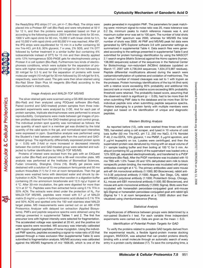
annexin A5 and aminopeptidase B. The conformation of theGAD molecule binding with 14-3-3 � (Protein Data Bank code1qja) is shown in Fig. 6 as an illustration. In fact, GAD waspredicted to accept three cavities in 14-3-3 proteins theoret-
TABLE ISummary of differentially expressed proteins in GAD-treated HeLa cells
Spot Pairs of gels (n)Spot volume
-Fold difference p valueControl(mean � S.D.)
GAD-treated(mean � S.D.)
ppm
1 9 2749.6 � 184.8 1266.3 � 260.0 0.46 �0.052 9 465.7 � 115.3 4885.3 � 552.6 10.49 �0.053 9 2614.6 � 735.4 8326.3 � 1727.1 3.18 �0.054 9 1517.3 � 491.1 4684.3 � 1367.8 3.09 �0.055 9 1478.0 � 483.6 5152.1 � 1598.4 3.49 �0.056 9 1629.2 � 642.3 5153.0 � 1586.8 3.16 �0.057 9 3418.4 � 481.1 1453.7 � 343.4 0.43 �0.058 9 963.7 � 75.3 2913.7 � 511.4 3.02 �0.059 9 3774.1 � 1725.8 9645.6 � 2282.7 2.56 �0.05
10 9 1308.2 � 291.9 519.3 � 162.7 0.40 �0.0511 9 1842.9 � 660.9 5574.3 � 1569.6 3.02 �0.0512 9 1189.2 � 153.3 522.3 � 160.3 0.44 �0.0513 9 423.7 � 29.5 1722.3 � 160.1 4.06 �0.0514 9 475.7 � 159.6 1529.7 � 217.1 3.22 �0.0515 9 3262.0 � 303.4 1456.1 � 384.8 0.45 �0.0516 9 2307.7 � 339.7 1010.3 � 219.8 0.44 �0.0517 9 2263.7 � 300.6 4950.3 � 646.7 2.19 �0.0518 9 2726.3 � 275.6 1267.3 � 171.8 0.46 �0.0519 9 697.7 � 33.6 1488.7 � 135.7 2.13 �0.0520 9 1531.7 � 114.4 3520.6 � 317.4 2.30 �0.0521 9 192.7 � 64.4 711.0 � 215.3 3.69 �0.05
TABLE IIThe results of protein identifications of differentially expressed proteins using MALDI-TOF MS/MS
Spot Target proteinNCBI
accessionno.
Theoreticalmolecular
mass(kDa)/pI
Proteinscore
Sequencecoverage
Number ofpeptidesmatched/
unmatched
Uniquepeptides
Bestion
score
%
1 Translation initiation factor 5A (eIF5A) 54037409 16.7/5.08 334 35 19/11 3 1532 Ephrin receptor EphA7 (EphA7) 4758282 31.8/5.14 82 56 4/11 2 1023 PRDX3 2507171 27.7/7.67 171 19 18/15 2 1134 14-3-3 �/� 1345590 28.1/4.76 137 62 8/7 3 905 14-3-3 �/� 68085578 27.7/4.73 252 46 27/23 2 846 14-3-3 � 54696890 29.1/5.17 126 27 17/12 2 397 14-3-3 16306737 24.3/4.77 78 37 6/12 2 408 14-3-3 5726310 28.3/4.66 73 29 9/31 2 339 14-3-3 � (14-3-3E) 51702210 29.2/4.63 296 40 26/10 3 88
10 EB1 20138589 29.8/5.02 93 39 15/23 3 6711 Annexin A5 113960 35.8/4.94 648 61 28/9 4 20412 Spermidine synthase 134811 33.8/5.30 139 30 21/11 3 4613 26 S proteasome subunit p40.5 3618343 42.9/5.53 183 27 16/16 3 4314 AHA1 6912280 38.2/5.41 137 28 10/7 2 5915 Cytokeratin 19 24234699 44.1/5.04 96 34 19/24 4 6416 Cytokeratin 1 1346343 66.0/8.16 94 49 9/31 2 3117 Calumenin 2809324 37.0/4.47 196 34 9/11 3 3618 Ubiquinol-cytochrome c reductase core I
protein (Core I protein)515634 52.6/5.94 109 15 11/26 2 43
19 PDI 860986 56.6/6.10 98 22 12/13 2 3020 Aminopeptidase B 40316915 72.5/5.51 80 15 17/38 2 4221 Mitofilin 8131894 83.6/6.08 124 41 16/2 2 33
Cytotoxicity Mechanism of Ganoderic Acid D
954 Molecular & Cellular Proteomics 7.5
at UN
IVE
RS
ITE
T I B
ER
GE
N on July 11, 2008
ww
w.m
cponline.orgD
ownloaded from

ically, including the cavity between the homodimers and thetwo symmetrical phosphopeptide binding sites in the twomonomers. But INVDOCK does not compare and choosewhich cavity is the best to accommodate a chemical moleculeamong several putative cavities in a protein structure. In re-ality, there is the possibility for the GAD molecule to chooseonly one type of cavity to attach. According to a previousreport (22), the phosphopeptide binding sites of binding sitesof proteins belong to 14-3-3 family are the common sites thatcould be occupied by client proteins. So in Fig. 6, only the
docking model of GAD with the phosphopeptide binding sitesin the two monomers is shown.
The Binding Affinity of GAD Toward 14-3-3 � Estimated bySPR Biosensor Analysis—To verify the prediction from IN-VDOCK analysis that GAD could bind directly to 14-3-3 pro-teins, the binding affinity of GAD toward 14-3-3 � was deter-mined by using SPR biosensor technology. The binding abilityof GAD toward 14-3-3 � was reflected by RU values recordeddirectly by the Biacore 3000 instrument. As shown in Fig. 7A,RU increased with increasing GAD concentration, indicatingthat GAD was able to bind to 14-3-3 � in a dose-dependentmanner. The association (kon), dissociation (koff), and equilib-rium dissociation (KD) constants of GAD binding to the immo-bilized GST-14-3-3 � were (5.02 � 0.24) � 103 M�1 s�1,(2.03 � 0.17) � 10�2 s�1, and (4.04 � 0.32) � 10�6 M,respectively. The curve fitting efficiency was evaluated bystatistical parameter �2, a statistical parameter in the SPRassay. The �2 value was calculated to be 0.32. In a controlstudy, GAD was injected over the immobilization GST surface,and the result exhibited weak nonspecific binding affinity asshown in Fig. 7B. The kon, koff, KD, and �2 values of GADbinding to the GST were (3.62 � 0.15) � 103 M�1 s�1, (2.92 �
0.21) � 10�2 s�1, (8.06 � 0.37) � 10�6 M, and 0.15, respec-tively. The nonspecific interaction between GAD and GSTprotein might be partly caused by the attraction between
FIG. 4. The result of the MALDI-TOF MS/MS analysis of proteinthat marked as spot 9 in Fig. 1. The protein was identified to behuman 14-3-3E by protein database search. A, peptide mass finger-print of the tryptic digest of spot 9. * indicates unique peptides furtheridentified by MS/MS. B, MS/MS profile of the peptide with a mass of1819.95 Da. C, MS/MS profile of the peptide with a mass of 1384.70Da. D, MS/MS profile of the peptide with a mass of 1256.61 Da. y-ionsresulting from fragmentation of the peptides and amino acids theyrepresent are indicated.
FIG. 5. Western blotting of eIF5A, 14-3-3E, PRDX3, andEB1.“Control” and “GAD-treated” above the panel represent the con-trol cells and HeLa cells treated with 10 �M GAD for 48 h, respectively.Each blot is the representative result of three independentexperiments.
TABLE IIIThe INVDOCK-predicted binding between GAD and proteins
Number Protein
Protein DataBank
identificationnumber
Ligand-proteininteraction
energy value
1 14-3-3 � 1qja �48.62 14-3-3 �/� 2c23 �37.43 14-3-3 � 2btp �48.54 14-3-3 1ywt �56.15 14-3-3 2b05 �41.56 14-3-3 � 2br9 �45.27 Aminopeptidase B 1hs6 �49.38 Annexin A5 1sav �39.2
Cytotoxicity Mechanism of Ganoderic Acid D
Molecular & Cellular Proteomics 7.5 955
at UN
IVE
RS
ITE
T I B
ER
GE
N on July 11, 2008
ww
w.m
cponline.orgD
ownloaded from
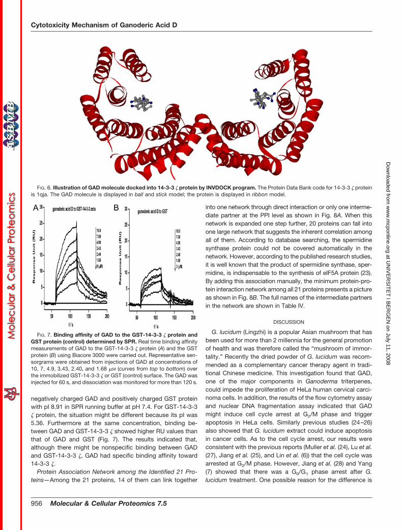
negatively charged GAD and positively charged GST proteinwith pI 8.91 in SPR running buffer at pH 7.4. For GST-14-3-3� protein, the situation might be different because its pI was5.36. Furthermore at the same concentration, binding be-tween GAD and GST-14-3-3 � showed higher RU values thanthat of GAD and GST (Fig. 7). The results indicated that,although there might be nonspecific binding between GADand GST-14-3-3 �, GAD had specific binding affinity toward14-3-3 �.
Protein Association Network among the Identified 21 Pro-teins—Among the 21 proteins, 14 of them can link together
into one network through direct interaction or only one interme-diate partner at the PPI level as shown in Fig. 8A. When thisnetwork is expanded one step further, 20 proteins can fall intoone large network that suggests the inherent correlation amongall of them. According to database searching, the spermidinesynthase protein could not be covered automatically in thenetwork. However, according to the published research studies,it is well known that the product of spermidine synthase, sper-midine, is indispensable to the synthesis of eIF5A protein (23).By adding this association manually, the minimum protein-pro-tein interaction network among all 21 proteins presents a pictureas shown in Fig. 8B. The full names of the intermediate partnersin the network are shown in Table IV.
DISCUSSION
G. lucidum (Lingzhi) is a popular Asian mushroom that hasbeen used for more than 2 millennia for the general promotionof health and was therefore called the “mushroom of immor-tality.” Recently the dried powder of G. lucidum was recom-mended as a complementary cancer therapy agent in tradi-tional Chinese medicine. This investigation found that GAD,one of the major components in Ganoderma triterpenes,could impede the proliferation of HeLa human cervical carci-noma cells. In addition, the results of the flow cytometry assayand nuclear DNA fragmentation assay indicated that GADmight induce cell cycle arrest at G2/M phase and triggerapoptosis in HeLa cells. Similarly previous studies (24–26)also showed that G. lucidum extract could induce apoptosisin cancer cells. As to the cell cycle arrest, our results wereconsistent with the previous reports (Muller et al. (24), Lu et al.(27), Jiang et al. (25), and Lin et al. (6)) that the cell cycle wasarrested at G2/M phase. However, Jiang et al. (28) and Yang(7) showed that there was a G0/G1 phase arrest after G.lucidum treatment. One possible reason for the difference is
FIG. 6. Illustration of GAD molecule docked into 14-3-3 � protein by INVDOCK program. The Protein Data Bank code for 14-3-3 � proteinis 1qja. The GAD molecule is displayed in ball and stick model; the protein is displayed in ribbon model.
FIG. 7. Binding affinity of GAD to the GST-14-3-3 � protein andGST protein (control) determined by SPR. Real time binding affinitymeasurements of GAD to the GST-14-3-3 � protein (A) and the GSTprotein (B) using Biacore 3000 were carried out. Representative sen-sorgrams were obtained from injections of GAD at concentrations of10, 7, 4.9, 3.43, 2.40, and 1.68 �M (curves from top to bottom) overthe immobilized GST-14-3-3 � or GST (control) surface. The GAD wasinjected for 60 s, and dissociation was monitored for more than 120 s.
Cytotoxicity Mechanism of Ganoderic Acid D
956 Molecular & Cellular Proteomics 7.5
at UN
IVE
RS
ITE
T I B
ER
GE
N on July 11, 2008
ww
w.m
cponline.orgD
ownloaded from

the use of different cell lines. For example, Jiang et al. (28)found that G. lucidum induced G0/G1 phase arrest in MDA-MB-231 breast cancer cells but induced G2/M phase arrest inPC-3 prostate cancer cells (25).
Mechanistically the cytotoxic effects of G. lucidum have beenimplicated in the (i) down-regulation of Akt/NF-�B signaling andthus the expression of NF-�B-regulated cyclin D1 (28), (ii) up-regulation of expression of p21 and Bax (25, 26), and (iii) sup-pression of protein kinase C and activation of mitogen-activatedprotein kinases (6). However, the molecular mechanism in whichG. lucidum, especially Ganoderma triterpenes, induces prolifer-ation inhibition, apoptosis, and cell cycle arrest is still not clear.
This study implemented the proteomics scheme to search glo-bally for the differentially expressed proteins in HeLa cells af-fected by GAD, a purified Ganoderma triterpene.
In the present study, 21 proteins whose expressions weresignificantly changed under GAD treatment were identified.Among the 19 proteins with three-dimensional structures,eight of them were predicted by INVDOCK analysis to pos-sess the ability of binding to GAD. The proteins identified inthe proteomics study might include both direct targets anddownstream regulated proteins. Proteins that can directlybind to GAD might be considered as possible direct targets ofGAD. Among the eight proteins that were predicted by IN-VDOCK to be able to bind directly to GAD, the most interest-ing proteins were the six members of the 14-3-3 family, i.e.14-3-3 �/�, 14-3-3 �/�, 14-3-3 �, 14-3-3 , 14-3-3 , and14-3-3 �. The 14-3-3 protein family is a family of highly con-served dimeric phosphoserine-binding proteins. In mammals,there are nine homologous members including 14-3-3 �/�,14-3-3 , 14-3-3 �, 14-3-3 , 14-3-3 , 14-3-3 �, and 14-3-3�/�. The 14-3-3 � and � are the phosphorylated forms of14-3-3 � and �, respectively. The binding affinity of GADtoward 14-3-3 � was confirmed in the present study usingSPR biosensor analysis. Interestingly the results of networkconstruction also suggested the central role of 14-3-3 pro-teins in all proteins identified in the proteomics study. Theresults suggested that 14-3-3 proteins might play importantroles in the cytotoxicity mechanism of GAD. The predictionthat 14-3-3 proteins are possible direct targets of GAD alsosupports the previous study results (6, 25, 26, 28) about thecytotoxicity mechanism of G. lucidum. It is well known that14-3-3 proteins are involved in many different cellular pro-cesses, including mitogenesis, cell cycle control, and apopto-sis. Several models for how 14-3-3 proteins function havebeen recently proposed: 1) 14-3-3 proteins can modulate thebiochemical activity of certain ligands like Raf-1, protein ki-nase C, and tryptophan hydroxylase; 2) 14-3-3 can affect theactivity of ligands such as BAD, FKHRL1, and Cdc25 byaltering their intracellular localization; and 3) 14-3-3 proteinsmight function as novel adapters or scaffold molecules (29–31). So by regulating 14-3-3 proteins, GAD might suppressprotein kinase C (14-3-3 proteins were previously called “pro-tein kinase C inhibitor protein 1”), consistent with the reportedsuppression of protein kinase C by G. lucidum (6). And byregulating 14-3-3 proteins, GAD might adjust some apoptosis-related proteins and some cell cycle-related proteins, consistentwith the report of Hu et al. (26) and Jiang et al. (28), respectively.
In the present study, GAD treatment also caused the reg-ulation of 15 other proteins besides 14-3-3 proteins. Brieflybased on their biological functions, these 15 proteins could begenerally classified into one of the following four categories: 1)cell survival and proliferation, 2) cell death and protein deg-radation, 3) metabolism, and 4) cytoskeleton structure. Notethat some proteins may have multiple functions and play rolesin more than one pathway.
FIG. 8. The constructed minimum protein-protein interactionnetwork. The red dots illustrate 14-3-3 proteins; the blue dots areother proteins identified from experiments. Proteins in the network areinteracting with each other via intermediate partners (shown in gray)from known PPI information. A, the network constructed by 14 iden-tified proteins. The 14 proteins can link together into one networkthrough direct interaction or only one intermediate partner. B, theexpanded network constructed by 21 identified proteins. 20 of theproteins can link together into one network through no more than twointermediate partners. Compared with A, this network was expandedone step further. The yellow dot is the protein (spermidine synthase)manually added according to the reported relation with protein eIF5A.Symbols and full names of the intermediate partners in the networkare shown in Table IV.
Cytotoxicity Mechanism of Ganoderic Acid D
Molecular & Cellular Proteomics 7.5 957
at UN
IVE
RS
ITE
T I B
ER
GE
N on July 11, 2008
ww
w.m
cponline.orgD
ownloaded from

Proteins including eIF5A and spermidine synthase play im-portant roles in cell growth and proliferation. eIF5A is a small(16–18-kDa) abundant protein that is highly conserved ineukaryotes, and it is fundamental to cell survival and prolifera-tion (32, 33). Spermidine synthase, also known as putrescineaminopropyltransferase, is the enzyme responsible for the syn-thesis of spermidine. Protein eIF5A is the only known cellularprotein that undergoes an unusual post-translational modifica-tion on a specific lysine residue to form hypusine. The uniquehypusine modification in mammalian cells occurs by a two-steppathway that involves the attachment of an aminobutyl groupfrom spermidine to the �-amine group of lysine 50 followed byhydroxylation on carbon 2 of the butyl group to form hypusine.Thus, the essential nature of spermidine for hypusine modifica-tion of eIF5A is well established. And in addition to the indis-pensable role of spermidine for hypusine modification in eIF5A,polyamines are also required for optimal growth of mammaliancells (34). Many studies have indicated that down-regulation orinhibition of hypusine synthesis impedes cancer cell growth,and eIF-5A can be considered as a target of anticancer strate-gies (35–37). Thus, the decreased expression of eIF5A andspermidine synthase after GAD treatment may contribute to thecell growth inhibition induced by GAD in HeLa cells.
Proteins including annexin A5 and 26 S proteasome subunitp40.5 play important roles in cell death and protein degrada-tion. Annexin A5 is a calcium-binding protein that belongs tothe annexin family, a superfamily of ubiquitous proteins char-acterized by their calcium-dependent ability to bind to biolog-ical membranes. The involvement of annexins in several phys-iological processes, such as membrane trafficking, calciumsignaling, cell motility, proliferation and differentiation, and apo-ptosis has been proposed. Importantly annexins have beenimplicated in the pathogenesis of benign and malignant neo-plasms of different origins (38–40). As to annexin A5, its ex-pression exhibits different regulation tendency in carcinoma
development of different organs. For example, the loss of an-nexin A5 was identified as a marker for cutaneous squamouscell carcinoma (41), whereas annexin A5 protein expression wasaugmented in growth hormone-secreting carcinoma (42). Im-portantly the expression of annexin A5 was markedly sup-pressed in both cervical and endometrial carcinoma cells whencompared with their normal counterparts (43). Because theHeLa cell line used in the present study is a type of humancervical carcinoma cell line, the increased expression of annexinA5 in GAD-treated HeLa cells may contribute to growth inhibi-tion induced by GAD. Similarly an increase of annexin A5 levelswas also observed in butyrate-treated colon adenocarcinomacell lines, whereas butyrate induced cell differentiation andgrowth arrest in these cells (44). Note that GAD could directlybind to annexin A5 according to the INVDOCK analysis. The roleof annexin A5 in the cytotoxicity of GAD deserves further study.26 S proteasome subunit p40.5 is an important subunit ofproteasomes, which are eukaryotic ring-shaped or cylindricalparticles with multicatalytic protease activities. The increase of26 S proteasome subunit p40.5 found in the present study maycontribute to the possible protein degradation of HeLa cellsinduced by GAD treatment.
Proteins including ephrin receptor EphA7, thioredoxin-de-pendent peroxide reductase mitochondrial precursor, activa-tor of heat shock 90-kDa protein ATPase homolog 1, ubiqui-nol-cytochrome c reductase core I protein, protein-disulfideisomerase, aminopeptidase B, and mitofilin are enzymes orregulators of enzymes that play important roles in cell metab-olism. The regulation of these proteins by GAD might cause achange of metabolism in HeLa cells. Moreover some proteinswere also involved in pathways like cell proliferation/cell deathand play important roles in carcinogenesis. Ephrin receptorEphA7 (receptor protein-tyrosine kinase, EC 2.7.10.1) is areceptor for members of the ephrin-A family and can catalyzethe reaction of “ATP � a L-tyrosine in protein � ADP � a
TABLE IVSymbols and full names of the intermediate partners in the network shown in Fig. 8
Number Symbol Full name
1 ACTG1 Actin, 12 APCS Amyloid P component, serum3 CLASP1 Cytoplasmic linker-associated protein 14 COL10A1 Collagen, type X, �1 (Schmid metaphyseal chondrodysplasia)5 CYCS Cytochrome c, somatic6 FEZ1 Fasciculation and elongation protein �1 (zygin I)7 HSPA8 Heat shock 70-kDa protein 88 HSP90AA1 Heat shock protein 90-kDa �, class A member 19 IKBKG Inhibitor of � light polypeptide gene enhancer in B-cells, kinase
10 MLLT4 Myeloid/lymphoid or mixed lineage leukemia (trithorax homolog, Drosophila);translocated to 4
11 PPP1CC Protein phosphatase 1, catalytic subunit, isoform12 SUMO4 SMT3 suppressor of mif two 3 homolog 4 (Saccharomyces cerevisiae)13 TG Thyroglobulin14 UBQLN4 Ubiquilin 415 XPO1 Exportin 1 (CRM1 homolog, yeast)16 ZFYVE9 Zinc finger, FYVE domain-containing 9
Cytotoxicity Mechanism of Ganoderic Acid D
958 Molecular & Cellular Proteomics 7.5
at UN
IVE
RS
ITE
T I B
ER
GE
N on July 11, 2008
ww
w.m
cponline.orgD
ownloaded from

L-tyrosine phosphate in protein.” A previous report showedthat a significant reduction of EphA7 expression is found inhuman colorectal cancers (45). In the present study, GADtreatment increased the expression of EphA7 in HeLa cells.PRDX3, a type of peroxiredoxin (EC 1.11.1.15), was found tobe up-regulated in HeLa cells by GAD. Peroxiredoxins are afamily of peroxidases that reduce hydrogen peroxide (H2O2)and alkyl hydroperoxides to water and alcohol, respectively.The major role of peroxiredoxins is to control the constitutivelevel of H2O2 in the cell and thus protect cell against reactiveoxygen species-induced damage. PRDX3 expression isthought to play a role in the antioxidant defense system andhomeostasis within the mitochondria. It was reported thatPRDX3 overexpression led to decreased cell growth (46). Sothe increase in PRDX3 expression may be related to thegrowth inhibition caused by GAD treatment in HeLa cells.Activator of heat shock 90-kDa protein ATPase homolog 1(AHA1) could act as cochaperone that stimulates HSP90ATPase activity by influencing the conformational state of the“ATP lid” and consequent N-terminal dimerization (47).Ubiquinol-cytochrome c reductase core I protein (EC 1.10.2.2)is a component of the ubiquinol-cytochrome c reductasecomplex (complex III or cytochrome bc1 complex), which ispart of the mitochondrial respiratory chain. Protein-disulfideisomerase (PDI; EC 5.3.4.1) catalyzes the rearrangement of-S–S- bonds in proteins. It was reported that resistance to theapoptosis-inducing agent Aplidin in HeLa cells was related tothe down-regulation of PDI expression (48). In GAD-treatedHeLa cells, the expression of PDI was increased; this maycontribute to sensitivity of HeLa cells to apoptosis. Amin-opeptidase B (EC 3.4.11.6) is an exopeptidase that selectivelyremoves arginine and/or lysine residues from the N terminusof several peptide substrates including Arg-Leu-enkephalin,Arg-Met-enkephalin, and Arg-Lys-somatostatin-14. Note thataminopeptidase B was predicted to be able to bind directlywith GAD in the INVDOCK analysis of present study. And itwas reported that the aminopeptidase B activity was de-creased in human renal cell carcinoma samples comparedwith non-tumor tissues (49). The increase of aminopeptidaseB protein expression in GAD-treated HeLa cells may play animportant role in the cytotoxicity of GAD. Mitofilin is a trans-membrane protein of the inner mitochondrial membrane andmay be involved in catabolic pathways (50, 51). The contri-bution of mitofilin to the cytotoxicity of GAD is unknown.
Proteins including microtubule-associated protein RP/EBfamily member 1, cytokeratin 19, cytokeratin 1, and calumeninare generally cytoskeleton-related proteins. And these pro-teins were also reported to participate in pathways such ascell cycle control and apoptosis. For example, the plus endsof microtubules are important binding sites for proteins thatregulate microtubules. EB1 is one of the best characterized“plus end-binding proteins.” Properly regulating the dynamicproperties of microtubules is critical for ensuring the accuratesegregation of chromosomes in mitosis. The function of EB1
as an “antipausing” factor is well conserved, and inhibition ofEB1 in a number of systems results in nondynamic microtu-bules that spend the majority of time in a paused state (52–54). Thus, it can be anticipated that the decrease of EB1expression in GAD-treated HeLa cells will contribute to thecell cycle arrest induced by GAD. Cytokeratin 19 and cytok-eratin 1 are intermediate filament proteins associated with theintegrity of cell structure. According to previous reports, ker-atin expression may be related to carcinogenesis (55). Inter-estingly in cervical cancer cells like HeLa cells, the functionalrole of cytokeratin 19 was shown to be associated with theapoptosis prevention and drug resistance of cells. Cytokeratin19 expression was found to be higher in cervical carcinomacell lines compared with control cell lines, and the elevation ofthe cytokeratin 19 level was associated with clinical cervicalcancer staging. The reduction of the cytokeratin 19 level byspecific antibody caused apoptosis in a cervical carcinomaHeLa cell line (56, 57). So the decrease of cytokeratin 19expression in GAD-treated HeLa cells may contribute to apo-ptosis induced by GAD. Calumenin is a multiple EF-hand Ca2�-binding protein located in endo/sarcoplasmic reticulum ofmammalian tissues (58). It was suggested in the previous re-ports to be related to the organization of cytoskeleton andcarcinoma metastasis. The expression of calumenin was foundto be decreased in carcinoma cell strains with higher metastasispotential (59, 60). The increase of calumenin in GAD-treatedHeLa cells may also play a role in the cytotoxicity of GAD.
To date, this study is the first to use a proteomics techniqueto search globally for the proteins influenced in cancer cells bya purified G. lucidum component. We found 21 proteins thatmight be target-related proteins of GAD. By using computer-automated analysis, we tried to predict the possible targetsand network of GAD. Most interestingly, our results suggestedthe important role of 14-3-3 proteins in the cytotoxicity mech-anism of GAD. The results of the present study shed light onthe anticancer mechanism of G. lucidum from a molecularperspective. Furthermore we are continuing to find new trit-erpenes from G. lucidum (61) and other herbs. It is possiblethat we can obtain promising triterpenes for cancer therapyeither by isolating them from herbs or by modifying the struc-ture of natural triterpenes. Understanding of the cytotoxicitymechanism of GAD will be helpful to the study and the use oflikely promising triterpenes.
* This work was supported in part by grants from the Ministry ofScience and Technology of China (2006AA02Z317, 2004CB720103,2003CB715901, and 2006AA02312), National Natural Science Foun-dation of China (30500107 and 30670953), Science and TechnologyCommission of Shanghai Municipality (06DZ19731 and 06PJ14072),and Shanghai Pudong Science and Technology Committee(PKJ2006-L07). The costs of publication of this article were defrayedin part by the payment of page charges. This article must therefore behereby marked “advertisement” in accordance with 18 U.S.C. Section1734 solely to indicate this fact.
Cytotoxicity Mechanism of Ganoderic Acid D
Molecular & Cellular Proteomics 7.5 959
at UN
IVE
RS
ITE
T I B
ER
GE
N on July 11, 2008
ww
w.m
cponline.orgD
ownloaded from

□S The on-line version of this article (available at http://www.mcponline.org) contains supplemental material.
§ Both authors contributed equally to this work.** To whom correspondence may be addressed. Tel./Fax: 86-21-
50272223; E-mail: [email protected].‡‡ To whom correspondence may be addressed. Tel./Fax: 86-21-
50272223; E-mail: [email protected].
REFERENCES
1. Yun, T. K. (1999) Update from Asia. Asian studies on cancer chemopre-vention. Ann. N. Y. Acad. Sci. 889, 157–192
2. Sliva, D. (2004) Cellular and physiological effects of Ganoderma lucidum(Reishi). Mini-Rev. Med. Chem. 4, 873–879
3. Yuen, J. W., and Gohel, M. D. (2005) Anticancer effects of Ganodermalucidum: a review of scientific evidence. Nutr. Cancer 53, 11–17
4. Sliva, D. (2006) Ganoderma lucidum in cancer research. Leuk. Res. 30,767–768
5. Yeung, W. H., Lu, Q. L., Zhang, Q., and Go, V. L. W. (2004) Chemical andbiochemical basis of the potential anti-tumor properties of Ganodermalucidum. Curr. Top. Nutraceutical Res. 2, 67–77
6. Lin, S. B., Li, C. H., Lee, S. S., and Kan, L. S. (2003) Triterpene-enrichedextracts from Ganoderma lucidum inhibit growth of hepatoma cells viasuppressing protein kinase C, activating mitogen-activated protein ki-nases and G2-phase cell cycle arrest. Life Sci. 72, 2381–2390
7. Yang, H. L. (2005) Ganoderic acid produced from submerged culture ofGanoderma lucidum induces cell cycle arrest and cytotoxicity in humanhepatoma cell line BEL7402. Biotechnol. Lett. 27, 835–838
8. Kimura, Y., Taniguchi, M., and Baba, K. (2002) Antitumor and antimetastaticeffects on liver triterpenoid fractions of Ganoderma lucidum: mechanismof action and isolation of active substance. Anticancer Res. 22, 3309–3318
9. Min, B. S., Gao, J. J., Nakamura, N., and Hattori, M. (2000) Triterpenes fromthe spores of Ganoderma lucidum and their cytotoxicity against meth-Aand LLC tumor cells. Chem. Pharm. Bull. 48, 1026–1033
10. Wang, X. M., Yang, M., Guan, S. H., Liu, R. X., Xia, J. M., Bi, K. S., and Guo,D. A. (2006) Quantitative determination of six major triterpenoids inGanoderma lucidum and related species by high performance liquidchromatography. J. Pharm. Biomed. Anal. 41, 838–844
11. Komoda, Y., Nakamura, H., Ishihara, S., Uchida, M., Kohda, H., and Ya-masaki, K. (1985) Structures of new terpenoid constituents of Ganodermalucidum (Fr.) Karst (Polyporaceae). Chem. Pharm. Bull. 33, 4829–4835
12. Liu, X., Fan, X. L., Zhao, Y., Luo, G. R., Li, X. P., Li. R., and Le, W. D. (2005)Estrogen provides neuroprotection against activated microglia-induceddopaminergic neuronal injury through both estrogen receptor-� andestrogen receptor-� in microglia. J. Neurosci. Res. 81, 653–665
13. Liu, X., and Zhu, X. Z. (1999) Roles of p53, c-Myc, Bcl-2, Bax and caspasesin glutamate-induced neuronal apoptosis and the possible neuroprotec-tive mechanism of basic fibroblast growth factor. Brain Res. Mol. BrainRes. 71, 210–216
14. Roberts, K., Bhatia, K., Stanton, P., and Lord, R. (2004) Proteomic analysisof selected prognostic factors of breast cancer. Proteomics 4, 784–792
15. Garfin, D., and Heerdt, L. (2001) 2-D Electrophoresis for Proteomics: aMethods and Product Manual, pp. 25–27, Bio-Rad Laboratories, Rich-mond, CA
16. Shen, H., Cheng, G., Fan, H., Zhang, J., Zhang, X., Lu, H., Liu, C., Sun, F.,Jin, H., Xu, X., Xu, G., Wang, S., Fang, C., Bao, H., Wang, Y., Wang, J.,Zhong, H., Yu, Z., Liu, Y., Tang, Z., and Yang, P. (2006) Expressedproteome analysis of human hepatocellular carcinoma in nude mice(LCI-D20) with high metastasis potential. Proteomics 6, 528–537
17. Chen, Y. Z, and Zhi, D. G. (2001) Ligand-protein inverse docking and itspotential use in the computer search of protein targets of a small mole-cule. Proteins 43, 217–226
18. Ye, F., Zhang, Z. S., Luo, H. B., Shen, J. H., Chen, K. X., Shen, X., and Jiang,H. L. (2006) The dipeptide H-Trp-Glu-OH shows highly antagonisticactivity against PPAR: bioassay with molecular modeling simulation.Chembiochem 7, 74–82
19. Chen, S., Chen, L. L., Tan, J. Z., Chen, J., Du, L., Sun, T., Shen, J. H., Chen,K. X., Jiang, H. L., and Shen, X. (2005) Severe acute respiratory syn-drome coronavirus 3C-like proteinase N terminus is indispensable forproteolytic activity but not for enzyme dimerization. J. Biol. Chem. 280,164–173
20. Chaurasia, G., Iqbal, Y., Hanig, C., Herzel, H., Wanker, E. E., and Futschik,M. E. (2007) UniHI: an entry gate to the human protein interactome.Nucleic Acids Res. 35, D590–DD594
21. Klein, P., and Ravi, R. A (1995) Nearly best-possible approximation algo-rithm for node-weighted Steiner trees. J. Algorithms 19, 104–115
22. Gardino, A. K., Smerdonb, S. J., and Yaffe, M. B. (2006) Structural deter-minants of 14-3-3 binding specificities and regulation of subcellularlocalization of 14-3-3-ligand complexes: A comparison of the X-raycrystal structures of all human 14-3-3 isoforms. Semin. Cancer Biol. 16,173–182
23. Park, M. H. (2006) The post-translational synthesis of a polyamine-derivedamino acid, hypusine, in the eukaryotic translation initiation factor 5A(eIF5A). J. Biochem. (Tokyo) 139, 161–169
24. Muller, C. I., Kumagai, T., O’Kelly, J., Seeram, N. P., Heber, D., and Koeffler,H. P. (2006) Ganoderma lucidum causes apoptosis in leukemia, lym-phoma and multiple myeloma cells. Leuk. Res. 30, 841–848
25. Jiang, J., Slivova, V., Valachovicova, T., Harvey, K., and Sliva, D. (2004)Ganoderma lucidum inhibits proliferation and induces apoptosis in hu-man prostate cancer cells PC-3. Int. J. Oncol. 24, 1093–1100
26. Hu, H., Ahn, N. S., Yang, X., Lee, Y. S., and Kang, K. S. (2002) Ganodermalucidum extract induces cell cycle arrest and apoptosis in MCF-7 humanbreast cancer cell. Int. J. Cancer 102, 250–253
27. Lu, Q. Y., Jin, Y. S., Zhang, Q., Zhang, Z., Heber, D., Go, V. L., Li, F. P., andRao, J. Y. (2004) Ganoderma lucidum extracts inhibit growth and induceactin polymerization in bladder cancer cells in vitro. Cancer Lett. 216, 9–20
28. Jiang, J., Slivova, V., Harvey, K., Valachovicova, T., and Sliva, D. (2004)Ganoderma lucidum suppresses growth of breast cancer cells throughthe inhibition of Akt/NF-�B signaling. Nutr. Cancer 49, 209–216
29. Tzivion, G., Gupta, V. S., Kaplun, L., and Balan, V. (2006) 14-3-3 proteins aspotential oncogenes. Semin. Cancer Biol. 16, 203–213
30. Hermeking, H., and Benzinger, A. (2006) 14-3-3 proteins in cell cycleregulation. Semin. Cancer Biol. 16, 183–192
31. Masters, S. C., and Fu, H. (2001) 14-3-3 proteins mediate an essentialanti-apoptotic signal. J. Biol. Chem. 276, 45193–45200
32. Jao, D. L., and Chen, K. Y. (2002) Subcellular localization of the hypusine-containing eukaryotic initiation factor 5A by immunofluorescent stainingand green fluorescent protein tagging. J. Cell. Biochem. 86, 590–600
33. Park, M. H., Lee, Y. B., and Joe, Y. A. (1997) Hypusine is essential foreukaryotic cell proliferation. Biol. Signals 6, 115–123
34. Nishimura, K., Murozumi, K., Shirahata, K., Park, M. H., Kashiwagi, K., andIgarashi, K. (2005) Independent roles of eIF5A and polyamines in cellproliferation. Biochem. J. 385, 779–785
35. Caraglia, M., Budillon, A., Vitale, G., Lupoli, G., Tagliaferri, P., and Abbruzz-ese, A. (2000) Modulation of molecular mechanisms involved in proteinsynthesis machinery as a new tool for the control of cell proliferation. Eur.J. Biochem. 267, 3919–3936
36. Caraglia, M., Marra, M., Giuberti, G., D’Alessandro, A. M., Budillon, A., delPrete, S., Lentini, A., Beninati, S., and Abbruzzese, A. (2001) The role ofeukaryotic initiation factor 5A in the control of cell proliferation andapoptosis. Amino Acids 20, 91–104
37. Kang, H. A., and Hershey, J. W. B. (1994) Effect of initiation factor eIF-5Adepletion on protein synthesis and proliferation of Saccharomyces cer-evisiae. J. Biol. Chem. 269, 3934–3940
38. Kenis, H., van Genderen, H., Bennaghmouch, A., Rinia, H.A., Frederik, P.,Narula, J., Hofstra, L., and Reutelingsperger, C. P. (2004) Cell surface-expressed phosphatidylserine and annexin A5 open a novel portal of cellentry. J. Biol. Chem. 279, 52623–52629
39. Hayes, M. J., and Moss, S. E. (2004) Annexins and disease. Biochem.Biophys. Res. Commun. 322, 1166–1170
40. Bastian, B. C. (1997) Annexins in cancer and autoimmune diseases. CMLSCell. Mol. Life Sci. 53, 554–556
41. Dooley, T. P., Reddy, S. P., Wilborn, T. W., and Davis, R. L. (2003) Biomar-kers of human cutaneous squamous cell carcinoma from tissues and celllines identified by DNA microarrays and qRT-PCR. Biochem. Biophys.Res. Commun. 306, 1026–1036
42. Mulla, A., Christian, H. C., Solito, E., Mendoza, N., Morris, J. F., andBuckingham, J. C. (2004) Expression, subcellular localization and phos-phorylation status of annexins 1 and 5 in human pituitary adenomas anda growth hormone-secreting carcinoma. Clin. Endocrinol. (Oxf.) 60,107–119
Cytotoxicity Mechanism of Ganoderic Acid D
960 Molecular & Cellular Proteomics 7.5
at UN
IVE
RS
ITE
T I B
ER
GE
N on July 11, 2008
ww
w.m
cponline.orgD
ownloaded from

43. Karube, A., Shidara, Y., Hayasaka, K., Maki, M., and Tanaka, T. (1995)Suppression of calphobindin I (CPB I) production in carcinoma of uterinecervix and endometrium. Gynecol. Oncol. 58, 295–300
44. Guzman-Aranguez, A., Olma, N., Turnay, J., Lecona, E., Perez-Ramos, P.,Lopez de Silanes, I., and Lizarbe, M. A. (2005) Differentiation of humancolon adenocarcinoma cells alters the expression and intracellular local-ization of annexins A1, A2, and A5. J. Cell. Biochem. 94, 178–193
45. Wang, J., Kataoka, H., Suzuki, M., Sato, N., Nakumura, R., Tao, H., Ma-ruyama, K., Isogaki, J., Kanaoka, S., Ihara, M., Tanaka, M., Kanamori, M.,Nakamura, I., Shinmura, K., and Sugimura, H. (2005) Downregulation ofEphA7 by hypermethylation in colorectal cancer. Oncogene 24,5637–5647
46. Nonn, L., Berggren, M., and Powis, G. (2003) Increased expression ofmitochondrial peroxiredoxin-3 (thioredoxin peroxidase-2) protects can-cer cells against hypoxia and drug-induced hydrogen peroxide-depend-ent apoptosis. Mol. Cancer Res. 1, 682–689
47. Siligardi, G., Hu, B., Panaretou, B., Piper, P. W., Piper, P. W., Pearl, L. H.,and Prodromou, C. (2004) Co-chaperone regulation of conformationalswitching in the Hsp90 ATPase cycle. J. Biol. Chem. 279, 51989–51998
48. Gonzalez-Santiago, L., Alfonso, P., Suarez, Y., Nunez, A., Garcia-Fernan-dez, L. F., Alvarez, E., Munoz, A., and Casal, J. I. (2007) Proteomicanalysis of the resistance to Aplidin in human cancer cells. J. ProteomeRes. 6, 1286–1294
49. Varona, A., Blanco, L., Lopez, J. I., Gil, J., Agirregoitia, E., Irazusta, J., andLarrinaga, G. (2007) Altered levels of acid, basic, and neutral peptidaseactivity and expression in human clear cell renal cell carcinoma. Am. J.Physiol. 292, F780–F788
50. Odgren, P. R., Toukatly, G., Bangs, P. L., Gilmore, R., and Fey, E. G. (1996)Molecular characterization of mitofilin (HMP), a mitochondria-associatedprotein with predicted coiled coil and intermembrane space targetingdomains. J. Cell Sci. 109, 2253–2264
51. Navet, R., Mathy, G., Douette, P., Dobson, R. L., Leprince, P., De Pauw, E.,Sluse-Goffart, C., and Sluse, F. E. (2007) Mitoproteome plasticity of ratbrown adipocytes in response to cold acclimation. J. Proteome Res. 6,25–33
52. Green, R. A., Wollman, R., and Kaplan, K. B. (2005) APC and EB1 functiontogether in mitosis to regulate spindle dynamics and chromosome align-ment. Mol. Biol. Cell 16, 4609–4622
53. Kline-Smith, S. L., and Walczak, C. E. (2004) Mitotic spindle assembly andchromosome segregation: refocusing on microtubule dynamics. Mol.Cell 15, 317–327
54. Tirnauer, J. S., Grego, S., Salmon, E. D., and Mitchison, T. J. (2002)EB1-microtubule interactions in Xenopus egg extracts: role of EB1 inmicrotubule stabilization and mechanisms of targeting to microtubules.Mol. Biol. Cell 13, 3614–3626
55. Chu, P. G., and Weiss, L. M. (2002) Keratin expression in human tissuesand neoplasms. Histopathology 41, 365–366
56. Yuan, C. C., Huang, T. S., Ng, H. T., Liu, R. S., Hung, M. W., and Tsai, L. C.(1998) Elevated cytokeratin-19 expression associated with apoptoticresistance and malignant progression of human cervical carcinoma.Apoptosis 3, 161–169
57. Yuan, C. C., Huang, H. C., Tsai, L. C., Ng, H. T., and Huang, T. S. (1997)Cytokeratin-19 associated with apoptosis and chemosensitivity in hu-man cervical cancer cells. Apoptosis 2, 101–105
58. Jung, D. H., Mo, S. H., and Kim, D. H. (2006) Calumenin, a multipleEF-hands Ca2�-binding protein, interacts with ryanodine receptor-1 inrabbit skeletal sarcoplasmic reticulum. Biochem. Biophys. Res. Com-mun. 343, 34–42
59. Ding, S. J., Li, Y., Shao, X. X., Zhou, H., Zeng, R., Tang, Z. Y., and Xia, Q. C.(2004) Proteome analysis of hepatocellular carcinoma cell strains,MHCC97-H and MHCC97-L, with different metastasis potentials. Pro-teomics 4, 982–994
60. Wu, W., Tang, X., Hu, W., Lotan, R., Hong, W. K., and Mao, L. (2002)Identification and validation of metastasis-associated proteins in headand neck cancer cell lines by two-dimensional electrophoresis and massspectrometry. Clin. Exp. Metastasis 19, 319–326
61. Guan, S. H., Yang, M., Liu, X., Xia, J. M., Wang, X. M., Jin, H., and Guo,D. A. (2006) Two new lanostanoid triterpenes from the fruit body ofGanoderma lucidum—the major component of SunRecome�. Nat. Prod.Commun. 1, 177–181
Cytotoxicity Mechanism of Ganoderic Acid D
Molecular & Cellular Proteomics 7.5 961
at UN
IVE
RS
ITE
T I B
ER
GE
N on July 11, 2008
ww
w.m
cponline.orgD
ownloaded from