proteomic analysis of the fish pathogen flavobacterium
TRANSCRIPT

Dumpala et al. Proteome Science 2010, 8:26http://www.proteomesci.com/content/8/1/26
Open AccessR E S E A R C H
ResearchProteomic analysis of the fish pathogen Flavobacterium columnarePradeep R Dumpala1, Nagihan Gülsoy2, Mark L Lawrence1 and Attila Karsi*1
AbstractBackground: Flavobacterium columnare causes columnaris disease in cultured and wild fish populations worldwide. Columnaris is the second most prevalent bacterial disease of commercial channel catfish industry in the United States. Despite its economic importance, little is known about the expressed proteins and virulence mechanisms of F. columnare. Here, we report the first high throughput proteomic analysis of F. columnare using 2-D LC ESI MS/MS and 2-DE MALDI TOF/TOF MS.
Results: Proteins identified in this study and predicted from the draft F. columnare genome were clustered into functional groups using clusters of orthologous groups (COGs), and their subcellular locations were predicted. Possible functional relations among the identified proteins were determined using pathway analysis. The total number of unique F. columnare proteins identified using both 2-D LC and 2-DE approaches was 621, of which 10.95% (68) were identified by both methods, while 77.29% (480) and 11.76% (73) were unique in 2-D LC and 2-DE, respectively. COG groupings and subcellular localizations were similar between our data set and proteins predicted from the whole genome. Twenty eight pathways were significantly represented in our dataset (P < 0.05).
Conclusion: Results from this study provide experimental evidence for many proteins that were predicted from the F. columnare genome annotation, and they should accelerate functional and comparative studies aimed at understanding virulence mechanisms of this important pathogen.
BackgroundFlavobacterium columnare is a long Gram-negative rodin the family Flavobacteriaceae, one of the main phyleticlines within the Bacteroidetes group from the domainBacteria [1]. Several species in Flavobacteriaceae causedisease in fish. F. columnare is the causative agent ofcolumnaris disease [2], which exists both in fresh andbrackish water throughout the world [3]. Outbreaks mayresult in high mortality, especially during spring andautumn, and are most likely associated with poor envi-ronmental conditions causing stress [4,5]. Stressful con-ditions are common in commercial aquaculture whereproduction is kept at maximum levels.
Columnaris disease generally begins as an externalinfection on the skin, fins, gills, or oral cavity [6]. On theskin and fins, lesions are characterized by dull, grayish-white or yellow erosive lesions that can progress to deep
ulcers in the underlying muscle. External infection oftenis concurrent with systemic infection and subacute mor-talities [3]. In some cases, systemic infection with little orno visible external or internal pathological signs mayoccur [7]. F. columnare infections can be chronic, butmore often, the disease appears suddenly and causesmortalities within a few days [6].
A substantial amount of work has been done on F.columnare phylogeny [1,8], isolation, identification, anddetection of F. columnare [9-18], and characterization ofF. columnare strains [19-25]. In addition, genetic tools forthe manipulation of F. columnare have recently beenreported [26]. Efforts have been made to understand themechanisms of virulence employed by the organism [27-30], but much remains poorly understood. F. columnareproduces several extracellular proteases that are believedto be important virulence factors contributing to thebranchial and cutaneous necrosis [28,31-33], but the roleof the proteases has not been definitively elucidated. Asurface capsular material that may be involved in adhe-sion has been described [34]. Lipopolysaccharide (LPS)
* Correspondence: [email protected] Department of Basic Sciences, College of Veterinary Medicine, Mississippi State University, Mississippi State, MS 39762-6100, USAFull list of author information is available at the end of the article
© 2010 Dumpala et al; licensee BioMed Central Ltd. This is an Open Access article distributed under the terms of the Creative CommonsAttribution License (http://creativecommons.org/licenses/by/2.0), which permits unrestricted use, distribution, and reproduction inany medium, provided the original work is properly cited.

Dumpala et al. Proteome Science 2010, 8:26http://www.proteomesci.com/content/8/1/26
Page 2 of 11
may also play an important role in columnaris pathogene-sis [35]. Chondroitin lyase activity was found to be signif-icantly related to strain virulence in a temperaturedependent manner [30].
Although, 14 F. columnare outer membrane relatedproteins were reported recently [36], little is known aboutthe expressed F. columnare proteins. In the present study,we report the first protein expression analysis from F.columnare using the complementary technologies of 2-DLC ESI MS/MS and 2-DE MALDI TOF/TOF MS. Resultsof this study provide experimental evidence for the pre-dicted F. columnare proteins and should accelerate func-tional and comparative studies to delineate pathogenicmechanisms of F. columnare.
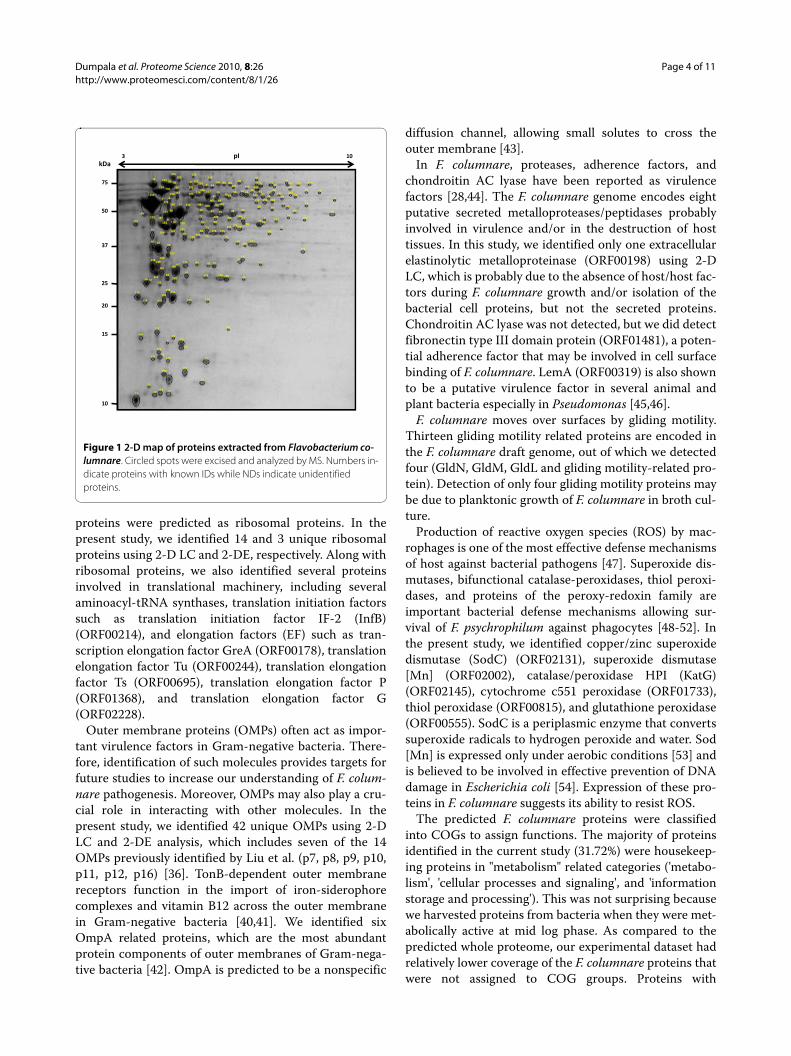
ResultsProtein identification using 2-D LC and 2-DE2-D LC analysis yielded total 548 F. columnare proteins(Additional File 1: Table S1) representing 19.01% of thepredicted F. columnare proteome (2,882 ORFs). 2-DEanalysis of four CBB stained gels showed approximately600 spots. Among the common spots, 192 were excisedand analyzed from one of these gels. We were able toidentify 182 (94.79%) of the excised spots (Additional File2: Table S2), which represented 141 unique F. columnareproteins. In 2-DE analysis, 41 (22.53%) proteins wereidentified in multiple spots, probably due to post-transla-tional modifications and processing. Together, 2-D LCand 2-DE analyses resulted in the identification of 621 F.columnare proteins, 480 (77.29%) of which were identi-fied only by 2-D LC, while 73 (11.76%) were identifiedonly by 2-DE (Figure 1), and 68 (10.95%) were detected byboth approaches. Analysis of the functional role catego-ries assigned by the J. Craig Venter Institute's AnnotationEngine indicated some important categories under cellenvelope and cellular processes. Sub categories underthese main groups had potential to include proteins thatmay be involved in F. columnare pathogenesis (Table 1).
Functional classification of identified proteinsFunctional classifications of the 621 identified uniqueproteins and the 2,882 predicted F. columnare proteinsare summarized in Figure 2. A total of 88 (14.17%, 88/621) proteins from our dataset and 372 (12.91%, 372/2,882) proteins from the predicted whole proteome wereclassified as information storage and processing relatedcategories (J, K, and L). Cellular processes and signalingrelated categories (D, M, N, O, and T) included 112(18.04%) and 370 (12.84%) proteins from our proteindataset and the whole proteome database, respectively. Atotal of 197 (31.72%) and 658 (22.83%) proteins from ourprotein dataset and the predicted whole proteome,respectively, were represented in metabolism related cat-
egories (C, E, F, G, H, I, P, and Q). Poorly characterizedCOG groups (R and S) contained 70 (11.27%) and 318(11.03%) proteins in our protein dataset and the pre-dicted whole proteome, respectively. "No COGs" (pro-teins not belong to any of the currently-defined COGs)was the top represented category in our protein dataset(23.51%) and in the whole proteome (36.33%). Eight(1.29%) proteins from our protein dataset and 117(4.06%) proteins from whole proteome were assigned to"No hit" (non-significant short or low-complexitysequences) category. A summary of Pfam domain analysison proteins detected by 2-D LC and 2-DE, and predictedfrom the F. columnare draft genome are given in Addi-tional Files 3, 4, and 5: Tables S3, S4 and S5, respectively.
Subcellular localization of identified proteinsSubcellular locations of F. columnare proteins identifiedin this study and predicted from the draft genome weredetermined (Figure 3). 2-DE and 2-D LC generallyresulted in similar coverage of subcellular compartmentscompared to the predicted proteome, except 2-DE gavebetter coverage of cytoplasmic proteins and poorer cov-erage of cytoplasmic membrane proteins. 2-D LC hadbetter representation of proteins from both membranecompartments and extracellular proteins. Both 2-D LCand 2-DE had lower representation of the "unknown"subcellular location proteins than the percentage fromthe predicted proteome. In this study, 42 proteins werepredicted to locate in the F. columnare outer membraneby PSORTb (Table 2).
Pathway analysisWe used Pathway Studio analysis to gain insight into vari-ous pathways represented in F. columnare proteins identi-fied by 2-D LC and 2-DE analysis. Twenty seven pathwaysbelonging to metabolism and one pathway in geneticinformation processing were significantly represented (P< 0.05, Additional File 6: Table S6). Out of 28 pathways,eight related to amino acid metabolism (Figure 4), fivecarbohydrate metabolism, four cofactor and vitaminmetabolism, four xenobiotics biodegradation and metab-olism, two glycan biosynthesis and metabolism, twonucleotide metabolism, one energy metabolism, one bio-synthesis of secondary metabolites, and one related totranslational process. 277 proteins from our dataset wereintegral to these pathways. Of the significantly repre-sented pathways, purine metabolism contained the high-est number of proteins (24 proteins), while biotinmetabolism pathway had the fewest (4 proteins). Of allthe pathways, folate biosynthesis pathway was the mostsignificant pathway in our dataset (P = 2.25E-09), whichwas followed by purine metabolism pathway (P = 1.44E-07).

Dumpala et al. Proteome Science 2010, 8:26http://www.proteomesci.com/content/8/1/26
Page 3 of 11
DiscussionThis research analyzed the soluble cell proteome of Fla-vobacterium columnare using complementary technolo-gies of 2-D LC and 2-DE. Proteomic data from this studyhas provided experimental evidence for the expression ofF. columnare proteins that were previously only predictedfrom the draft genome sequence. Data from this studyprovides a picture of the expressed F. columnare pro-teome during log-phase growth in FCGM along with a 2-D reference map, which will provide an important basisfor comparison under other environmental conditions.
Currently, the most popular methods employed toexplore protein expression utilize protein separation by
2-D LC (gel-free) or 2-DE (gel-based) followed by MSanalysis [37]. Use of more than one methodology in pro-teomic investigations improves proteome coveragebecause each method identifies a set of unique proteins[38,39]. Therefore, in the present study, we employed thecomplementary techniques of 2-DE and 2-D LC to iden-tify 621 unique proteins from F. columnare (480 unique to2-D LC, 73 unique to 2-DE, and 68 common to both),which provided experimental evidence for more than onefifth (21.55%) of the predicted 2,882 F. columnare ORFs.
Ribosomal proteins, one of the most abundantlyexpressed protein types in cells, were detected in highnumbers. In the draft F. columnare genome sequence, 60
Table 1: Flavobacterium columnare potential pathogenesis-related proteins.
ORF # Protein name Approach/SNa) Role categoryb)
ORF00560 3-deoxy-D-manno-octulosonate 8-phosphate phosphatase, YrbI family 2-D LC Cell envelope (A)
ORF01244 3-deoxy-D-manno-octulosonate cytidylyltransferase 2-D LC Cell envelope (A)
ORF02664 Amidinotransferase family protein 2-D LC Cellular processes (E)
ORF01484 CHU large protein; gliding motility-related protein; putative adhesin AidA-related 2-D LC Cellular processes (C)
ORF01824 DTDP-4-dehydrorhamnose 3,5-epimerase 2-DE/120 Cell envelope (A)
ORF00198 Extracellular elastinolytic metalloproteinase 2-D LC Cellular processes (D)
ORF01481 Fibronectin type III domain protein 2-D LC Cellular processes (D)
ORF02336 GldL Both/152 Cellular processes (C)
ORF02335 GldM 2-D LC Cellular processes (C)
ORF02334 GldN 2-DE/151 Cellular processes (C)
ORF02678 Glucose-1-phosphate thymidylyltransferase 2-D LC Cell envelope (A)
ORF00396 Glycosyl transferase, group 1 2-D LC Cell envelope (A)
ORF02332 Glycosyltransferase, putative 2-D LC Cell envelope (A)
ORF00532 GTP-binding protein TypA/BipA 2-DE/36, 37 Cellular processes (F)
ORF01035 Hemagglutinin 2-D LC Cellular processes (E)
ORF02801 Lipid-A-disaccharide synthase 2-D LC Cell envelope (A)
ORF01136 LPS biosynthesis protein , RfbU family 2-D LC Cell envelope (A)
ORF02060 Mannosyltransferase 2-D LC Cell envelope (A)
ORF00475 Membrane protein, putative 2-D LC Cellular processes (E)
ORF01989 Monofunctional biosynthetic peptidoglycan transglycosylase 2-D LC Cell envelope (B)
ORF02568 PepSY-associated TM helix family 2-D LC Cellular processes (C)
ORF02672 Polysaccharide export outer membrane protein 2-D LC Cell envelope (A)
ORF01163 Tetraacyldisaccharide 4'-kinase 2-D LC Cell envelope (A)
ORF01370 UDP-3-0-acyl N-acetylglucosamine deacetylase 2-D LC Cell envelope (A)
ORF01367 UDP-3-O-[3-hydroxymyristoyl] glucosamine N-acyltransferase 2-D LC Cell envelope (A)
ORF02676 UDP-glucose 6-dehydrogenase Both/169 Cell envelope (A)
ORF02536 UDP-N-acetylglucosamine 2-epimerase 2-D LC Cell envelope (A)
ORF00211 Universal stress protein family protein 2-DE/10 Cellular processes (F)
a) SN indicates spot number marked on the 2-DE image (Figure 1).b) A, Biosynthesis and degradation of surface polysaccharides and lipopolysaccharides; B, Biosynthesis and degradation of murein sacculus and peptidoglycan; C, Chemotaxis and motility; D, Pathogenesis; E, Toxin production and resistance; F, Adaptations to atypical conditions.
Dumpala et al. Proteome Science 2010, 8:26http://www.proteomesci.com/content/8/1/26
Page 4 of 11
proteins were predicted as ribosomal proteins. In thepresent study, we identified 14 and 3 unique ribosomalproteins using 2-D LC and 2-DE, respectively. Along withribosomal proteins, we also identified several proteinsinvolved in translational machinery, including severalaminoacyl-tRNA synthases, translation initiation factorssuch as translation initiation factor IF-2 (InfB)(ORF00214), and elongation factors (EF) such as tran-scription elongation factor GreA (ORF00178), translationelongation factor Tu (ORF00244), translation elongationfactor Ts (ORF00695), translation elongation factor P(ORF01368), and translation elongation factor G(ORF02228).
Outer membrane proteins (OMPs) often act as impor-tant virulence factors in Gram-negative bacteria. There-fore, identification of such molecules provides targets forfuture studies to increase our understanding of F. colum-nare pathogenesis. Moreover, OMPs may also play a cru-cial role in interacting with other molecules. In thepresent study, we identified 42 unique OMPs using 2-DLC and 2-DE analysis, which includes seven of the 14OMPs previously identified by Liu et al. (p7, p8, p9, p10,p11, p12, p16) [36]. TonB-dependent outer membranereceptors function in the import of iron-siderophorecomplexes and vitamin B12 across the outer membranein Gram-negative bacteria [40,41]. We identified sixOmpA related proteins, which are the most abundantprotein components of outer membranes of Gram-nega-tive bacteria [42]. OmpA is predicted to be a nonspecific
diffusion channel, allowing small solutes to cross theouter membrane [43].
In F. columnare, proteases, adherence factors, andchondroitin AC lyase have been reported as virulencefactors [28,44]. The F. columnare genome encodes eightputative secreted metalloproteases/peptidases probablyinvolved in virulence and/or in the destruction of hosttissues. In this study, we identified only one extracellularelastinolytic metalloproteinase (ORF00198) using 2-DLC, which is probably due to the absence of host/host fac-tors during F. columnare growth and/or isolation of thebacterial cell proteins, but not the secreted proteins.Chondroitin AC lyase was not detected, but we did detectfibronectin type III domain protein (ORF01481), a poten-tial adherence factor that may be involved in cell surfacebinding of F. columnare. LemA (ORF00319) is also shownto be a putative virulence factor in several animal andplant bacteria especially in Pseudomonas [45,46].
F. columnare moves over surfaces by gliding motility.Thirteen gliding motility related proteins are encoded inthe F. columnare draft genome, out of which we detectedfour (GldN, GldM, GldL and gliding motility-related pro-tein). Detection of only four gliding motility proteins maybe due to planktonic growth of F. columnare in broth cul-ture.
Production of reactive oxygen species (ROS) by mac-rophages is one of the most effective defense mechanismsof host against bacterial pathogens [47]. Superoxide dis-mutases, bifunctional catalase-peroxidases, thiol peroxi-dases, and proteins of the peroxy-redoxin family areimportant bacterial defense mechanisms allowing sur-vival of F. psychrophilum against phagocytes [48-52]. Inthe present study, we identified copper/zinc superoxidedismutase (SodC) (ORF02131), superoxide dismutase[Mn] (ORF02002), catalase/peroxidase HPI (KatG)(ORF02145), cytochrome c551 peroxidase (ORF01733),thiol peroxidase (ORF00815), and glutathione peroxidase(ORF00555). SodC is a periplasmic enzyme that convertssuperoxide radicals to hydrogen peroxide and water. Sod[Mn] is expressed only under aerobic conditions [53] andis believed to be involved in effective prevention of DNAdamage in Escherichia coli [54]. Expression of these pro-teins in F. columnare suggests its ability to resist ROS.
The predicted F. columnare proteins were classifiedinto COGs to assign functions. The majority of proteinsidentified in the current study (31.72%) were housekeep-ing proteins in "metabolism" related categories ('metabo-lism', 'cellular processes and signaling', and 'informationstorage and processing'). This was not surprising becausewe harvested proteins from bacteria when they were met-abolically active at mid log phase. As compared to thepredicted whole proteome, our experimental dataset hadrelatively lower coverage of the F. columnare proteins thatwere not assigned to COG groups. Proteins with
Figure 1 2-D map of proteins extracted from Flavobacterium co-lumnare. Circled spots were excised and analyzed by MS. Numbers in-dicate proteins with known IDs while NDs indicate unidentified proteins.
10
15
20
25
75
37
50
kDa3 10pI
13 47153 34
3150
31
40
112
35170
14
15
75
125
141
6
16
128
120152
41
173
86
10
134
70
64
102
179 178 180
84 83 8230
65
160162
16195
155
116182
131146
147
555756
68139
85
13662
174
149148
107
29
108
4546
79
111159
164
104 105
151 110
165
109
60126
15642
73
43
176 175
21
69
137
48
91
10090
98172
25
101
117103 118
106
181 168 6378
27144
44
99
19
5239
171
37
138
101
33
77 76
282
113518
97 967
7487
58
71
133
59167
53
2312 72 11
22
177
122
16688
11489
6751
129
169
26
115
4994509238
ND127
124
549
17
66143
8142
140
4119
1132
157
20
158
32
154 130
8013581
145163
ND ND
ND
61
123
NDND
ND
193123
24
ND
ND
ND

Dumpala et al. Proteome Science 2010, 8:26http://www.proteomesci.com/content/8/1/26
Page 5 of 11
unknown function may be expressed only under special-ized conditions like host environment or stressed condi-tions and may not be expressed under laboratory cultureconditions. Analysis of metabolism-related proteins inour list implies that amino acids, instead of carbohy-drates, could be the major sources of carbon or energy.The FCGM medium we used for bacterial growth is agood source of amino acids, nitrogen, and certain saltslike MgSO4 and CaCl2 but it lacks readily available carbo-hydrate sources. Most of the metabolic proteins indenti-fied in our study were amino acid kinases, including fivehistidine kinases and five aminotransferases. Only threecarbohydrate kinases involved in core carbohydratemetabolisms were identified. We found 18 peptidases,which may be useful in utilizing available amino acids as amajor source of carbon. Previous studies on F. psychro-
philum also suggest that this bacterium was unable to usereadily available carbohydrates as a major energy source[55].
We used the PSORTb algorithm to predict the subcel-lular locations of F. columnare proteins. Many proteinswere mapped to the cytoplasm, which is expectedbecause these proteins are not hydrophobic, and thus sol-ubility does not hinder protein isolation and separation.2-DE was particularly effective at detection of cytoplas-mic proteins. By contrast, cytoplasmic and outer mem-brane proteins were identified in higher percentagesusing 2-D LC when compared to 2-DE. The same buffersolution containing 7M urea and 2% CHAPSO was usedto solubilize proteins during isolations for both 2D-LCand 2-DE, so it appears that the difference is not due tothe isolation procedure, but rather the separation tech-
Figure 2 Comparison of COG categories. Proteins identified in this study (621 proteins in both 2-D LC and 2-DE analysis) and predicted from the Flavobacterium columnare draft genome (2,882 ORFs) were organized into COG functional groups. Percentages were calculated by dividing the num-ber of proteins in the particular COG category by the number of unique proteins in each analysis. COG categories are as follows: J, translation, ribo-somal structure and biogenesis; K, transcription; L, replication, recombination, and repair; D, cell cycle control, cell division, chromosome partitioning; M, cell wall/membrane/envelop biogenesis; N, cell motility; O, post-translational modification and protein turnover, chaperones; T, signal transduction mechanisms; U, intracellular trafficking, secretion, and vesicular transport; V, defense mechanisms; C, energy production and conversion; E, amino acid transport and metabolism; F, nucleotide transport and metabolism; G, carbohydrate transport and metabolism; H, coenzyme transport and metabo-lism; I, lipid transport and metabolism; P, inorganic ion transport and metabolism; Q, secondary metabolites biosynthesis, transport and catabolism; R, general function prediction only; S, function unknown. "No hit" group indicates non-significant short or low-complexity sequences. Query proteins not belong to any of the currently-defined COGs (23.51% in 2-D LC and 2-DE analysis and 36.33% in predicted ORFs) were not included in the figure for clarity.
0.00%
1.00%
2.00%
3.00%
4.00%
5.00%
6.00%
7.00%
8.00%
9.00%
10.00%
Perc
ent o
f tot
al p
rote
ins
COG categories
2-D LC and 2-DE ORFs

Dumpala et al. Proteome Science 2010, 8:26http://www.proteomesci.com/content/8/1/26
Page 6 of 11
Table 2: Outer membrane proteins of Flavobacterium columnare predicted by PSORTb.
Protein name ORF # COGa) Approach/SNb)
Conserved hypothetical protein ORF02881 - 2-D LC
Conserved hypothetical protein ORF00226 - 2-D LC
Conserved hypothetical protein ORF01938 D 2-D LC
Conserved hypothetical protein ORF00224 - 2-D LC
Conserved hypothetical protein ORF02143 - 2-D LC
Conserved hypothetical protein ORF02328 R 2-D LC
Extracellular elastinolytic metalloproteinase ORF00198 - 2-D LC
Fjo24 ORF02628 - 2-D LC
Hypothetical protein ORF02366 - 2-D LC
Hypothetical protein ORF01273 - 2-D LC
Hypothetical protein ORF01272 - 2-D LC
Hypothetical protein ORF01485 - 2-D LC
Hypothetical protein ORF02042 P 2-D LC
Lipoprotein, putative ORF01143 - 2-D LC
Lipoprotein, putative ORF02619 - Both/166, 167
Omp assembly complex, YaeT protein ORF02858 M 2-D LC
OmpA ORF02005 M Both/132
OmpA ORF00341 M 2-D LC
OmpA family protein ORF00085 M 2-D LC
OmpA/MotB family ORF00937 M 2-D LC
OmpA/MotB family ORF01639 N 2-D LC
OmpA/MotB family ORF00225 M 2-DE/11, 12
Outer membrane efflux protein ORF00073 MN 2-D LC
Outer membrane insertion C- signal domain protein ORF00187 - 2-D LC
Peptidyl-prolyl cis-trans isomerase C ORF02829 O 2-D LC
Putative TonB-dependent receptor ORF00266 P 2-D LC
RagA protein, putative ORF00258 P 2-D LC
Secreted peptidase, family M23 ORF01434 M 2-D LC
Secreted protein ORF02176 - 2-DE/141
Snf2 family helicase ORF00742 KL 2-D LC
Tetratricopeptide repeat domain protein ORF01855 R 2-D LC
TonB-dependent outer membrane receptor ORF00779 P 2-D LC
TonB-dependent outer membrane receptor ORF02179 P 2-D LC
TonB-dependent outer membrane receptor ORF00147 P 2-D LC
TonB-dependent outer membrane receptor ORF02172 P 2-D LC
TonB-dependent outer membrane receptor ORF02232 P 2-D LC
TonB-dependent outer membrane receptor ORF01690 P 2-D LC
TonB-dependent outer membrane receptor, putative ORF00704 P 2-D LC
TonB-dependent outer membrane receptor, putative ORF02424 P 2-D LC
TonB-dependent receptor plug domain protein ORF01433 P 2-D LC
TonB-dependent receptor, putative ORF01511 P 2-D LC
Transcriptional regulator, AraC family protein ORF01879 R 2-D LC
a) COG categories are as follows: M, cell wall/membrane/envelop biogenesis; D, cell cycle control, cell division, chromosome partitioning; P, inorganic ion transport and metabolism; S, function unknown; N, cell motility; R, general function prediction only; O, post-translational modification and protein turnover, chaperones; K, transcription; L, replication, recombination and repair; -, "No COGs".b) SN indicates spot number marked on the 2-DE image (Figure 1).

Dumpala et al. Proteome Science 2010, 8:26http://www.proteomesci.com/content/8/1/26
Page 7 of 11
niques. Other studies have reported that hydrophobicmembrane proteins are typically difficult to identify using2-DE. Hydrophobic proteins were found to be under-rep-resented in 2-DE based proteome analyses of membraneprotein fractions from E. coli [56-58] and Bacillus subtilis[39,59,60]. It is important to keep in mind that we choseto analyze only 192 common spots (30.81%) out ofapproximately 600 spots identified on our gels, so the biastoward cytoplasmic proteins in our 2-DE results may bebecause these proteins were more abundant or consis-tently present in gels. However, for future functionalstudies of membrane proteins, specialized isolation tech-niques may be needed. The ease and higher sensitivity of2-D LC for detection of hydrophobic membrane proteinsmay make this the preferred method for studying mem-brane proteins.
From the Pathway Studio analysis, we identified path-ways that were significantly represented in our proteindataset. Folate biosynthesis was the most significantlyrepresented metabolic pathway, which was followed bypurine metabolism pathway. Higher representation ofmetabolic and translational process related pathways isexpected because proteins were isolated from F. colum-nare during a metabolically active state. Amino acidmetabolism related pathways (glutamate; histidine; sele-noamino acid; alanine and aspartate; glycine, serine, andthreonine metabolism; lysine biosynthesis; lysine degra-dation; and valine, leucine, and isoleucine degradation)
are highly represented in our dataset compared to carbo-hydrate metabolism. Moreover, we have identified 18peptidases in our protein list suggesting breakdown ofhost proteins and usage of resultant amino acids as amajor source of energy, carbon, and nitrogen by F. colum-nare. Similarly, Glycosylphosphatidylinositol (GPI)-anchor biosynthesis and LPS biosynthesis pathways weresignificantly represented in our dataset, which may playan important role in cell wall synthesis. Moreover, LPSbiosynthesis may be one of the important virulence-related systems in F. columnare pathogenesis. LPS is animportant component of the cell envelope in Gram-nega-tive bacteria and is an immunodominant antigen.
ConclusionsOur main objective was to identify the expressed proteinsof F. columnare during normal growth. We showed forthe first time the expression of 621 proteins from F.columnare, which provides experimental evidence formany proteins that were predicted from the F. columnaregenome annotation. We expect this information couldaccelerate functional and comparative studies aimed atunderstanding virulence mechanisms of this importantpathogen. For example, comparative protein expressionanalysis between low and high virulence F. columnarestrains could help determine proteins involved in viru-lence. Furthermore, orthologous protein mapping ofidentified F. columnare proteins to well studied bacteria
Figure 3 Subcellular locations of Flavobacterium columnare proteins determined by PSORTb prediction. Percentages were calculated by di-viding the number of proteins in the particular subcellular location by the number of unique proteins in each analysis (548 proteins in 2D-LC, 141 proteins in 2-DE, and 2,882 ORFs predicted from the draft genome). 2-D LC and 2-DE indicates proteins identified in this study, while ORFs indicates proteins predicted from the whole genome. Unknown category includes proteins with multiple subcellular locations or unknown location.
0.00%
10.00%
20.00%
30.00%
40.00%
50.00%
60.00%
Cytoplasmic Cytoplasmic Membrane Extracellular Outer Membrane Periplasmic Unknown
Perc
ent o
f tot
al p
rote
ins
Subcellular compartment
2-D LC 2-DE ORFs

Dumpala et al. Proteome Science 2010, 8:26http://www.proteomesci.com/content/8/1/26
Page 8 of 11
would reveal protein targets for mutational analysis tounderstand gene function as well as to develop live atten-uated vaccines.
MethodsProtein extractionF. columnare growth medium (FCGM) [tryptone (8.00 g),yeast extract (0.80 g), MgSO4 7 H2O (1.00 g), CaCl2 2 H2O(0.74 g), NaCl (5.00 g), and sodium citrate (1.50 g) perliter] was used to grow F. columnare. Isolate ATCC 49512was streaked on FCGM agar plates (FCGM medium plus8.00 g agar per liter) and incubated at 30°C for 48 h. Fourcolonies were grown in 12 ml FCGM broth separately at
30°C under continuous shaking at 200 rpm. Growth ofbacteria was monitored by measuring optical density at600 nm (OD600), and bacteria were harvested at mid-exponential phase (OD600 0.6) by centrifugation at 3,750rpm for 15 min at 30°C.
The four bacterial pellets were separately resuspendedin cold urea-CHAPS buffer (7 M urea, 50 mM tris-HCl,2% CHAPS, 8 mM PMSF pH 8.0), and cells were lysedimmediately on ice by applying ten intermittent pulses of10 s with a sonicator. Bacterial homogenates were centri-fuged at 14,000 rpm for 5 min at 4°C to remove cell debrisand unbroken cells. Proteins from supernatant were pre-cipitated by trichloroacetic acid/Acetone, and the resul-
Figure 4 Amino acid metabolism related pathways in Flavobacterium columnare. Amino acid metabolism related pathways include glutamate; histidine; selenoamino acid; alanine and aspartate; glycine, serine, and threonine metabolism; lysine biosynthesis; lysine degradation; and valine, leu-cine, and isoleucine degradation pathways. Entities shown in oval represent genes involved in this pathway. Filled ovals represent proteins detected in this study. Unconnected genes were removed from the figure to reduce complexity.

Dumpala et al. Proteome Science 2010, 8:26http://www.proteomesci.com/content/8/1/26
Page 9 of 11
tant protein pellets were resuspended in rehydrationbuffer (7 M urea, 20 mM tris-HCl, 5 mM EDTA, 5 mMMgCl2, 4% CHAPS, and 5 mM PMSF pH 8.0). Proteinconcentrations were estimated using a 2-D Quant Kit (GEHealthcare, Piscataway, NJ).
2-D LC ESI MS/MS analysis100 μg of protein was digested with trypsin (1:50 w/w) at37°C for 16 h. The resultant peptides were desalted, driedin a vacuum centrifuge, and resuspended in 20 μL of 5%acetonitrile and 0.1% formic acid. Mass spectrometricanalysis was accomplished using a ProteomeX Worksta-tion (Thermo Scientific, Waltham, MA) as described pre-viously [61]. The F. columnare draft genome wasannotated using Annotation Engine at the J. Craig VenterInstitute, which uses the Glimmer algorithm for gene pre-diction [62,63]. The F. columnare protein database with2,882 open reading frames (ORFs) is available on ourwebsite http://www.miangel.msstate.edu, while the F.columnare genome project is hosted at The OklahomaUniversity Health Sciences Center http://micro-gen.ouhsc.edu. To identify proteins, mass spectra andtandem mass spectra were searched against the in silicotrypsin digested F. columnare protein database as previ-ously reported [64]. Protein identifications and associatedMS data were submitted to the PRIDE database [Acces-sion number: 9749].
2-DE and MALDI MS analysisProteins were extracted from four mid-exponential phasebacterial cultures as described in section 2.1. Protein pel-lets were resuspended in 350 μL of freshly prepared rehy-dration buffer (7 M urea, 2 M thio urea, 2% CHAPS, 1:50carrier ampholytes, 0.3% DTT) and quantified using 2-DQuant Kit (GE Healthcare). For in-gel rehydration, 800-1000 μg of solubilized proteins were loaded onto eachIPG strip (17 cm pH 3-10 NL). IEF, in-gel trypsin diges-tion, and MALDI MS analysis were performed as previ-ously described [64,65].
Identification of COGs, domains, protein locations, and pathwaysAll identified proteins from our dataset and the predictedwhole F. columnare proteome were organized into COGfunctional groups using COGnitor tool [66-68]. Similarly,protein domain analysis was conducted by using batchPfam analysis [69]. The subcellular location of proteinsfrom our dataset as well as from the whole F. columnareproteome were predicted using PSORTb v2.0.4 [70,71].Pathways with significant protein representation in ourexperimentally derived protein dataset were identifiedusing Pathway Studio (Ariadne Genomics, Rockville,MD). Due to lack of a molecular interaction database forF. columnare in Pathway Studio, all F. columnare proteins
were mapped to their orthologs in E. coli, F. psychrophi-lum, and F. johnsoniae by identifying reciprocal-best-BLAST hits. The resulting ortholog map file was used topredict pathways in F. columnare proteins. We used P ≤0.05 to select pathways with significant protein coverage.
Additional material
Competing interestsThe authors declare that they have no competing interests.
Authors' contributionsPRD carried out the experiments, analysis and interpretation of the data, andpreparation of the manuscript. NG participated in the experiments and prepa-ration of the manuscript. MLL contributed to preparation and critical review ofthe manuscript. AK contributed to the overall conception and design of theproject as well as preparation and critical review of the manuscript. All authorsread and approved the final manuscript.
AcknowledgementsThe authors would like to thank The J. Craig Venter Institute for providing auto-mated annotation. We thank Edward Zaitshik, Dr. Allison Gillaspy, and Dr. David Dyer at the Laboratory for Genomics and Bioinformatics at the University of Oklahoma Health Sciences Center for maintaining the annotation in Manatee. We also thank Tibor Pechan and Dr. Erdogan Memili for technical assistance, and Divya Swetha Peddinti for support with data analysis. Mass spectrometry was conducted at the Life Sciences and Biotechnology Institute, Mississippi State University. This project was partially supported by competitive grants from the National Research Initiative of the United States Department of Agri-culture Cooperative State Research, Education and Extension Service (2006-35600-16571 and 2007-35204-18404). Approved for publication as Journal Article No J-11703 of the Mississippi Agricultural and Forestry Experiment Sta-tion, Mississippi State University.
Author Details1Department of Basic Sciences, College of Veterinary Medicine, Mississippi State University, Mississippi State, MS 39762-6100, USA and 2Department of Biology, Faculty of Art and Science, Marmara University, Göztepe, İstanbul 34722, Turkey
References1. Bernardet J, Segers P, Cancanneyt M, Berthe M, Kersters K, Vandamme P:
Cutting a Gordian knot: emended classification and description of the genus Flavobacterium, emended description of the family Flavobacteriaceae, and proposal of Flavobacterium hydatis nom. nov. (Basonym, Cytophaga aquatilis Strohl and Tait 1978). Int J Syst Bacteriol 1996, 46:128-148.
2. Bernardet JF: Immunization with bacterial antigens: Flavobacterium and Flexibacter infections. Dev Biol Stand 1997, 90:179-188.
3. Plumb J: Health maintenance and microbial diseases of cultured fishes. Iowa State University Press, Ames, IA 1999.
Additional file 1 Table S1 - Flavobacterium columnare proteins identi-fied by 2-D LC analysis.Additional file 2 Table S2 - Flavobacterium columnare proteins identi-fied by 2-DE analysis.Additional file 3 Table S3 - Pfam analysis of proteins identified by 2-D LC analysis.Additional file 4 Table S4 - Pfam analysis of proteins identified by 2-DE analysis.Additional file 5 Table S5 - Pfam analysis of proteins predicted from the Flavobacterium columnare genome.Additional file 6 Table S6 - Pathways significantly represented in Fla-vobacterium columnare protein dataset.
Received: 4 February 2010 Accepted: 4 June 2010 Published: 4 June 2010This article is available from: http://www.proteomesci.com/content/8/1/26© 2010 Dumpala et al; licensee BioMed Central Ltd. This is an Open Access article distributed under the terms of the Creative Commons Attribution License (http://creativecommons.org/licenses/by/2.0), which permits unrestricted use, distribution, and reproduction in any medium, provided the original work is properly cited.Proteome Science 2010, 8:26
Dumpala et al. Proteome Science 2010, 8:26http://www.proteomesci.com/content/8/1/26
Page 10 of 11
4. Moore A, Eimers ME, Cardella NA: Attempts to control Flexibacter columnaris epizootics in pond-reared channel catfish by vaccination. J Aquat Anim Health 1990, 2:109-111.
5. Wakabayashi H: Effect of environmental conditions on the infectivity of Flexibacter columnaris to fish. J Fish Dis 1991, 14:279-290.
6. Austin B, Austin DA: Bacterial Fish Pathogens: Disease of Farmed and Wild Fish Edinburgh, UK: Heriot-Watt University; 1999.
7. Hawke JP, Thune RL: Systemic isolation and antimicrobial susceptibility of Cytophaga columnaris from commercially reared channel catfish. J Aquat Anim Health 1992, 4:109-113.
8. Bernardet JF, Grimont PAD: Deoxyribonucleic acid relatedness and phenotypic characterization of Flexibacter columnaris sp. nov., nom. rev., Flexibacter psychrophilus sp. nov., nom. rev., and Flexibacter maritimus Wakabayashi, Hikida, and Masumura 1986. Int J Syst Bacteriol 1989, 39:346-354.
9. Ordal EJ, Rucker RR: Pathogenic myxobacteria. Proc Soc Exp Biol Med 1944, 56:15-18.
10. Fijan FJ: Antibiotic additives for the isolation of Chondrococcus columnaris from fish. Appl Microbiol 1969, 17:333-334.
11. Decostere A, Haesebrouck F, Devriese LA: Shieh medium supplemented with tobramycin for selective isolation of Flavobacterium columnare (Flexibacter columnaris) from diseased fish. J Clin Microbiol 1997, 35:322-324.
12. Griffin BR: A Simple Procedure for Identification of Cytophaga columnaris. J Aquat Anim Health 1992, 4:63-66.
13. Bader JA, Shoemaker CA, Klesius PH: Rapid detection of columnaris disease in channel catfish (Ictalurus punctatus) with a new species-specific 16-S rRNA gene-based PCR primer for Flavobacterium columnare. J Microbiol Methods 2003, 52:209-220.
14. Darwish AM, Ismaiel AA, Newton JC, Tang J: Identification of Flavobacterium columnare by a species-specific polymerase chain reaction and renaming of ATCC43622 strain to Flavobacterium johnsoniae. Mol Cell Probes 2004, 18:421-427.
15. Shoemaker CA, Arias CR, Klesius PH, Welker TL: Technique for identifying Flavobacterium columnare using whole-cell fatty acid profiles. J Aquat Anim Health 2005, 17:267-274.
16. Panangala VS, Shelby RA, Shoemaker CA, Klesius PH, Mitra A, Morrison EE: Immunofluorescent test for simultaneous detection of Edwardsiella ictaluri and Flavobacterium columnare. Dis Aquat Org 2006, 68:197-207.
17. Panangala VS, Shoemaker CA, Klesius PH: TaqMan real-time polymerase chain reaction assay for rapid detection of Flavobacterium columnare. Aquac Res 2007, 38:508-517.
18. Yeh HY, Shoemaker CA, Klesius PH: Sensitive and rapid detection of Flavobacterium columnare in channel catfish Ictalurus punctatus by a loop-mediated isothermal amplification method. J Appl Microbiol 2006, 100:919-925.
19. Shamsudin MN, Plumb JA: Morphological, biochemical and physiological characterization of Flexibacter columnaris isolates from four fish species of fish. J Aquat Anim Health 1996, 8:335-382.
20. Decostere A, Haesebrouck F, Devriese LA: Characterization of four Flavobacterium columnare (Flexibacter columnaris) strains isolated from tropical fish. Vet Microbiol 1998, 62:35-45.
21. Darwish AM, Ismaiel AA: Genetic diversity of Flavobacterium columnare examined by restriction fragment length polymorphism RNA gene and the and sequencing of the 16S ribosomal 16S-23S rDNA spacer. Mol Cell Probes 2005, 19:267-274.
22. Thomas-Jinu S, Goodwin AE: Morphological and genetic characteristics of Flavobacterium columnare isolates: correlations with virulence in fish. J Fish Dis 2004, 27:29-35.
23. Schneck JL, Caslake LF: Genetic diversity of Flavobacterium columnare isolated from fish collected from warm and cold water. J Fish Dis 2006, 29:245-248.
24. Olivares-Fuster O, Shoemaker CA, Klesius PH, Arias CR: Molecular typing of isolates of the fish pathogen, Flavobacterium columnare, by single-strand conformation polymorphism analysis. FEMS Microbiol Lett 2007, 269:63-69.
25. Soto E, Mauel MJ, Karsi A, Lawrence ML: Genetic and virulence characterization of Flavobacterium columnare from channel catfish (Ictalurus punctatus). J Appl Microbiol 2008, 104(5):1302-10.
26. Staroscik AM, Hunnicutt DW, Archibald KE, Nelson DR: Development of methods for the genetic manipulation of Flavobacterium columnare. BMC Microbiol 2008, 8:115.
27. Bader JA, Shoemaker CA, Klesius PH: Production, characterization and evaluation of virulence of an adhesion defective mutant of Flavobacterium columnare produced by beta-lactam selection. Letters in applied microbiology 2005, 40:123-127.
28. Newton J, Wood TM, Hartley MM: Isolation and partial characterization of extracellular proteases produced by isolates of Flavobacterium columnare derived from channel catfish. J Aquat Anim Health 1997, 9:75-85.
29. Stringer-Roth KM, Yunghans W, Caslake LF: Differences in chondroitin AC lyase activity of Flavobacterium columnare isolates. J Fish Dis 2002, 25:687-691.
30. Suomalainen LR, Tiirola M, Valtonen ET: Chondroitin AC lyase activity is related to virulence of fish pathogenic Flavobacterium columnare. J Fish Dis 2006, 29:757-763.
31. Bertolini JM, Rohovec JS: Electrophoretic detection of proteases from different Flavobacterium columnare strains and assessment of their variability. Dis Aquat Org 1992, 12:121-128.
32. Christison J, Martin SM: Isolation and preliminary characterization of an extracellular protease of Cytophaga sp. Can J Microbiol 1971, 17:1207-1216.
33. Griffin BR: Characteristics of a chondrotin AC lyase produced by Cytophaga columnais. Trans Am Fish Soc 1991, 120:391-395.
34. Pate JL, Ordal EJ: The fine structure of Chondrococcus columnaris. 3. The surface layers of Chondrococcus columnaris. J Cell Biol 1967, 35:37-51.
35. Zhang Y, Arias CR, Shoemaker CA, Klesius PH: Comparison of lipopolysaccharide and protein profiles between Flavobacterium columnare strains from different genomovars. J Fish Dis 2006, 29:657-663.
36. Liu GY, Nie P, Zhang J, Li N: Proteomic analysis of the sarcosine-insoluble outer membrane fraction of Flavobacterium columnare. J Fish Dis 2008, 31:269-276.
37. Gorg A, Weiss W, Dunn MJ: Current two-dimensional electrophoresis technology for proteomics. Proteomics 2004, 4:3665-3685.
38. Salzano AM, Arena S, Renzone G, D'Ambrosio C, Rullo R, Bruschi M, Ledda L, Maglione G, Candiano G, Ferrara L, Scaloni A: A widespread picture of the Streptococcus thermophilus proteome by cell lysate fractionation and gel-based/gel-free approaches. Proteomics 2007, 7:1420-1433.
39. Wolff S, Otto A, Albrecht D, Zeng JS, Buttner K, Gluckmann M, Hecker M, Becher D: Gel-free and gel-based proteomics in Bacillus subtilis: a comparative study. Mol Cell Proteomics 2006, 5:1183-1192.
40. Braun V, Hantke K, Koster W, (Ed): Bacterial iron transport: mechanisms, genetics, and regulation. In: Metal Ions in Biological Systems: Iron Transport and Storage in Microorganisms, Plants and Animals. New York: Marcel Dekker Inc; 1998.
41. Letain TE, Postle K: TonB protein appears to transduce energy by shuttling between the cytoplasmic membrane and the outer membrane in Escherichia coli. Mol Microbiol 1997, 24:271-283.
42. Molloy MP, Phadke ND, Maddock JR, Andrews PC: Two-dimensional electrophoresis and peptide mass fingerprinting of bacterial outer membrane proteins. Electrophoresis 2001, 22:1686-1696.
43. Sugawara E, Nikaido H: Pore-forming activity of OmpA protein of Escherichia coli. J Biol Chem 1992, 267:2507-2511.
44. Decostere A, Haesebrouck F, Van Driessche E, Charlier G, Ducatelle R: Characterization of the adhesion of Flavobacterium columnare (Flexibacter columnaris) to gill tissue. J Fish Dis 1999, 22:465-474.
45. Tan MW, Rahme LG, Sternberg JA, Tompkins RG, Ausubel FM: Pseudomonas aeruginosa killing of Caenorhabditis elegans used to identify P. aeruginosa virulence factors. Proc Natl Acad Sci USA 1999, 96:2408-2413.
46. Hrabak EM, Willis DK: The lemA gene required for pathogenicity of Pseudomonas syringae on bean is a member of a family of two-component regulators. J Bacteriol 1992, 174:3011-3020.
47. Secombes CJ: The nonspecific immune system: cellular defences. in The Fish Immune System: Organism, Pathogen and Environment San Diego: Academic Press; 1996.
48. Kawai Y, Okawarab AI, Okuyama H, Kura F, Suzuki K: Modulation of chemotaxis, O(2)(-) production and myeloperoxidase release from human polymorphonuclear leukocytes by the ornithine-containing

Dumpala et al. Proteome Science 2010, 8:26http://www.proteomesci.com/content/8/1/26
Page 11 of 11
lipid and the serineglycine-containing lipid of Flavobacterium. FEMS Immunol Med Microbiol 2000, 28:205-209.
49. Nematollahi A, Decostere A, Pasmans F, Haesebrouck F: Flavobacterium psychrophilum infections in salmonid fish. J Fish Dis 2003, 26:563-574.
50. Nematollahi A, Pasmans F, Haesebrouck F, Decostere A: Early interactions of Flavobacterium psychrophilum with macrophages of rainbow trout Oncorhynchus mykiss. Dis Aquat Org 2005, 64:23-28.
51. Sanchez-Moreno M, Monteoliva-Sanchez M, Ortega F, Ramos-Cormenzana A, Monteoliva M: Superoxide dismutase in strains of the genus Flavobacterium: isolation and characterization. Arch Microbiol 1989, 152:407-410.
52. Duchaud E, Boussaha M, Loux V, Bernardet JF, Michel C, Kerouault B, Mondot S, Nicolas P, Bossy R, Caron C, Bessières P, Gibrat JF, Claverol S, Dumetz F, Le Hénaff M, Benmansour A: Complete genome sequence of the fish pathogen Flavobacterium psychrophilum. Nat Biotechnol 2007, 25:763-769.
53. Touati D: Transcriptional and posttranscriptional regulation of manganese superoxide dismutase biosynthesis in Escherichia coli, studied with operon and protein fusions. J Bacteriol 1988, 170:2511-2520.
54. Hopkin KA, Papazian MA, Steinman HM: Functional differences between manganese and iron superoxide dismutases in Escherichia coli K-12. J Biol Chem 1992, 267:24253-24258.
55. Bernardet JF, Kerouault B: Phenotypic and genomic studies of "Cytophaga psychrophila" isolated from diseased rainbow trout (Oncorhynchus mykiss) in France. Appl Environ Microbiol 1989, 55:1796-1800.
56. Fountoulakis M, Gasser R: Proteomic analysis of the cell envelope fraction of Escherichia coli. Amino Acids 2003, 24:19-41.
57. Lai EM, Nair U, Phadke ND, Maddock JR: Proteomic screening and identification of differentially distributed membrane proteins in Escherichia coli. Mol Microbiol 2004, 52:1029-1044.
58. Molloy MP, Herbert BR, Slade MB, Rabilloud T, Nouwens AS, Williams KL, Gooley AA: Proteomic analysis of the Escherichia coli outer membrane. Eur J Biochem 2000, 267:2871-2881.
59. Bunai K, Yamane K: Effectiveness and limitation of two-dimensional gel electrophoresis in bacterial membrane protein proteomics and perspectives. J Chromatogr B Analyt Technol Biomed Life Sci 2005, 815:227-236.
60. Eymann C, Dreisbach A, Albrecht D, Bernhardt J, Becher D, Gentner S, Tam le T, Buttner K, Buurman G, Scharf C, Venz S, Völker U, Hecker M: A comprehensive proteome map of growing Bacillus subtilis cells. Proteomics 2004, 4:2849-2876.
61. McCarthy FM, Burgess SC, van den Berg BH, Koter MD, Pharr GT: Differential detergent fractionation for non-electrophoretic eukaryote cell proteomics. J Proteome Res 2005, 4:316-324.
62. Salzberg SL, Delcher AL, Kasif S, White O: Microbial gene identification using interpolated Markov models. Nucleic acids research 1998, 26:544-548.
63. Delcher AL, Harmon D, Kasif S, White O, Salzberg SL: Improved microbial gene identification with GLIMMER. Nucleic acids research 1999, 27:4636-4641.
64. Dumpala PR, Lawrence ML, Karsi A: Proteome analysis of Edwardsiella ictaluri. Proteomics 2009, 9:1353-1363.
65. Chaudhary A, Pechan T, Willett KL: Differential protein expression of peroxiredoxin I and II by benzo(a)pyrene and quercetin treatment in 22Rv1 and PrEC prostate cell lines. Toxicol Appl Pharmacol 2007, 220:197-210.
66. Tatusov RL, Galperin MY, Natale DA, Koonin EV: The COG database: a tool for genome-scale analysis of protein functions and evolution. Nucleic Acids Res 2000, 28:33-36.
67. Tatusov RL, Koonin EV, Lipman DJ: A genomic perspective on protein families. Science 1997, 278:631-637.
68. Tatusov RL, Natale DA, Garkavtsev IV, Tatusova TA, Shankavaram UT, Rao BS, Kiryutin B, Galperin MY, Fedorova ND, Koonin EV: The COG database: new developments in phylogenetic classification of proteins from complete genomes. Nucleic Acids Res 2001, 29:22-28.
69. Finn RD, Mistry J, Tate J, Coggill P, Heger A, Pollington JE, Gavin OL, Gunasekaran P, Ceric G, Forslund K, et al.: The Pfam protein families database. Nucleic acids research 2010, 38:D211-222.
70. Gardy JL, Laird MR, Chen F, Rey S, Walsh CJ, Ester M, Brinkman FS: PSORTb v.2.0: expanded prediction of bacterial protein subcellular localization and insights gained from comparative proteome analysis. Bioinformatics 2005, 21:617-623.
71. Gardy JL, Spencer C, Wang K, Ester M, Tusnady GE, Simon I, Hua S, deFays K, Lambert C, Nakai K, Brinkman FS: PSORT-B: Improving protein subcellular localization prediction for Gram-negative bacteria. Nucleic Acids Res 2003, 31:3613-3617.
doi: 10.1186/1477-5956-8-26Cite this article as: Dumpala et al., Proteomic analysis of the fish pathogen Flavobacterium columnare Proteome Science 2010, 8:26