comparative proteomic analysis of mycobacterium
TRANSCRIPT
ORIGINAL RESEARCHpublished: 09 May 2017
doi: 10.3389/fmicb.2017.00795
Frontiers in Microbiology | www.frontiersin.org 1 May 2017 | Volume 8 | Article 795
Edited by:
Biswarup Mukhopadhyay,
Virginia Tech, USA
Reviewed by:
Thomas Dick,
Rutgers University, USA
Nomakorinte Gcebe,
Agricultural Research Council,
South Africa
Jonathan M. Blackburn,
University of Cape Town, South Africa
*Correspondence:
Solomon A. Yimer
Specialty section:
This article was submitted to
Microbial Physiology and Metabolism,
a section of the journal
Frontiers in Microbiology
Received: 28 December 2016
Accepted: 18 April 2017
Published: 09 May 2017
Citation:
Yimer SA, Birhanu AG, Kalayou S,
Riaz T, Zegeye ED, Beyene GT,
Holm-Hansen C, Norheim G,
Abebe M, Aseffa A and Tønjum T
(2017) Comparative Proteomic
Analysis of Mycobacterium
tuberculosis Lineage 7 and Lineage 4
Strains Reveals Differentially Abundant
Proteins Linked to Slow Growth and
Virulence. Front. Microbiol. 8:795.
doi: 10.3389/fmicb.2017.00795
Comparative Proteomic Analysis ofMycobacterium tuberculosis Lineage7 and Lineage 4 Strains RevealsDifferentially Abundant ProteinsLinked to Slow Growth and VirulenceSolomon A. Yimer 1, 2*, Alemayehu G. Birhanu 2, 3, Shewit Kalayou 1, Tahira Riaz 2,
Ephrem D. Zegeye 4, Getachew T. Beyene 1, Carol Holm-Hansen 5, Gunnstein Norheim 5,
Markos Abebe 6, Abraham Aseffa 6 and Tone Tønjum 1, 2
1Department of Microbiology, Oslo University Hospital, Oslo, Norway, 2Department of Microbiology, University of Oslo, Oslo,
Norway, 3Department of Medical Biotechnology, Institute of Biotechnology, Addis Ababa University, Addis Ababa, Ethiopia,4Centre for Applied Biotechnology, Uni Research Environment, Bergen, Norway, 5 Infection Control and Environmental
Health, Norwegian Institute of Public Health, Oslo, Norway, 6Department of Research and Innovation, Armauer Hansen
Research Institute, Addis Ababa, Ethiopia
In order to decipher the nature of the slowly growing Mycobacterium tuberculosis
(M.tuberculosis) lineage 7, the differentially abundant proteins in strains ofM. tuberculosis
lineage 7 and lineage 4 were defined. Comparative proteomic analysis by mass
spectrometry was employed to identify, quantitate and compare the protein profiles of
strains from the two M. tuberculosis lineages. Label-free peptide quantification of whole
cells from M. tuberculosis lineage 7 and 4 yielded the identification of 2825 and 2541
proteins, respectively. A combined total of 2867 protein groups covering 71% of the
predicted M. tuberculosis proteome were identified. The abundance of 125 proteins in
M. tuberculosis lineage 7 and 4 strains was significantly altered. Notably, the analysis
showed that a number ofM. tuberculosis proteins involved in growth and virulence were
less abundant in lineage 7 strains compared to lineage 4. Five ABC transporter proteins,
three phosphate binding proteins essential for inorganic phosphate uptake, and six
components of the type 7 secretion system ESX-3 involved in iron acquisition were less
abundant in M. tuberculosis lineage 7. This proteogenomic analysis provided an insight
into the lineage 7-specific protein profile which may provide clues to understanding the
differential properties of lineage 7 strains in terms of slow growth, survival fitness, and
pathogenesis.
Keywords:Mycobacterium tuberculosis, tuberculosis, lineage 7, proteomics, Ethiopia, mass spectrometry, type 7
secretion
INTRODUCTION
Tuberculosis (TB) has claimed an uncountable number of lives over centuries. One third of theglobal population is infected with the causative agentMycobacterium tuberculosis (M. tuberculosis),which is themain cause of TB. Each year,∼10.4million people contract TB and 1.8million die fromthe disease (WHO, 2016). In line with the World Health Organization (WHO) vision, the world
Yimer et al. Mycobacterium tuberculosis Lineage 7 Proteomics
is united in the quest to eliminate TB by 2050 (WHO, 2016).A holistic approach to understand host, environmental andbacterial factors to TB susceptibility and disease is crucial toachieve the global TB elimination target. There is a large bodyof evidence on host and environmental attributes for TB (Rieder,1999). However, given the potential implications for severity ofillness and transmission, there is limited knowledge regarding thebasic mechanisms underlying the physiology and pathogenesis ofthe differentM. tuberculosis lineages.
Large sequence polymorphisms classify M. tuberculosis into7 main lineages. These are lineage 1 (Indo-Oceanic), lineage 2(East Asian including “Beijing”), lineage 3 (CAS/Delhi), lineage4 (Euro-American including Latin American Mediterranean(LAM), Haarlem, X type and T families), lineage 5 and lineage 6(West African 1 and 2, respectively), and lineage 7 (Comas et al.,2013). Lineage 7 was recently detected in Ethiopia and amongEthiopian immigrants in Djibouti (Blouin et al., 2012; Firdessaet al., 2013; Yimer et al., 2013, 2015). We have shown that cells oftheM. tuberculosis lineage 7 grow slowly in vitro and that lineage7 infections are associated with prolonged delay in seekinghealth care among patients compared to other lineages (Yimeret al., 2015). Whole genome sequencing (WGS) demonstratedthat M. tuberculosis lineage 7 cells host a high number ofmutations in genes involved in carbohydrate transport andmetabolism, transcription, energy production and conversion,all of which contribute to the slow-growth phenotype (Yimeret al., 2016). Former WGS studies have described characteristicsand attributes of the other M. tuberculosis lineages that mayplay important roles in the pathogenesis of TB (Coscolla andGagneux, 2014). For example, the Beijing genotype (lineage2) is associated with high bacillary load in acid-fast bacillus(AFB) smears and frequently acquires drug resistance (Coscollaand Gagneux, 2014). Lineages 2, 3, and 4 exhibited a lowerearly inflammatory response compared to lineage 1 and lineage6 (Chacon-Salinas et al., 2005). Lineage 3 showed a higheranti-inflammatory phenotype compared to Lineage 4 (Portevinet al., 2011). Mycobacterium africanum (lineage 6) acquires drugresistance at a lower rate than the Euro-American (lineage 4) inGhana (Albanna et al., 2011). This shows that even though theyare genetically closely related, different strains of M. tuberculosispresent very diverse clinical phenotypes in terms of virulence.We therefore hypothesized that there may be a mechanismunderlying the variation in pathogenicity observed betweenstrains of M. tuberculosis detectable at the proteomic level.Global proteomic characterization by use of mass spectrometryrepresents a powerful tool and is an important supplement togenomics in defining the protein expression patterns (indicatingwhich genes are expressed and down- or upregulated; Peirs et al.,2005; Kelkar et al., 2011; Schubert et al., 2015; Bespyatykh et al.,2016; Jhingan et al., 2016; Peters et al., 2016). To date, no studieshave addressed the relationship between the M. tuberculosislineage 7 slow-growth phenotype and its proteomic signatures.
The objective of this study was to characterize the differentiallyabundant protein profile of M. tuberculosis lineage 7 (L7-35 andL7-28) and lineage 4 H37Rv strains and define the proteomicprofiles relevant for growth and pathogenicity. This proteomicstudy generated novel insight into the differentially abundant
(DA) proteins in M. tuberculosis lineage 7 vs. lineage 4 strains.A total of 2,867 proteins covering 71% of the predicted M.tuberculosis proteome were identified. The analysis of DAproteins by pathway clustering provided an overview on thelineage-specific protein interaction profile. This information maybe used to explain the differential behavior of lineage 7 strains interms of slow growth, survival fitness, and pathogenesis.
MATERIALS AND METHODS
Mycobacterial Strains and GrowthConditionsWhole genome sequenced M. tuberculosis lineage 7 strains (L7-35 and L7-28) collected form the Amhara Region of Ethiopiaand lineage 4 strain H37Rv were streaked onto Middlebrook7H10 plates in triplicates from freezer stocks, and incubated ina humidified 37◦C, 5% CO2 incubator. After 32 days the cellswere carefully scrapped off the agar plates and put into 50mLFalcon R© tubes. The cell pellets were gently resuspended in 30 mLPBS, pH 7.4, and centrifuged at 3,900 rpm for 20min at 4◦C.The cell pellets were subsequently transferred into 2mL screwcapped tubes (Sarstedt, Nümbrecht, Germany) and resuspendedin 1 mL PBS, and heat inactivated at 80◦C for 90min. Culturingand processing of the M. tuberculosis samples up until the heatinactivation step were conducted in a biosafety level 3 facilityat Oslo University Hospital, Norway. The heat-inactivated M.tuberculosis samples were stored at −80◦C until lysed for massspectrometry analysis.
Proteomic AnalysisCell lysisThe heat-inactivated cell pellets were resuspended in lysis buffercontaining 2% SDS, 10 mM Tris-HCl (pH 7.5), 1 tablet per50mL EDTA-free Protease Inhibitor Cocktail (Sigma-Aldrich,Cleveland, US) and 1 tablet per 10mL PhosSTOP PhosphataseInhibitor Cocktail (Roche). The samples were subsequentlytransferred into Lysing Matrix B tubes (Roche) and disruptedmechanically by bead beating using MagNa Lyser (RocheDiognostics, GmbH, Mannheim, Germany) for 90 s, speed 6.0.The lysis procedure followed by 1min cooling on ice wasrepeated six times. The lysate was clarified by centrifugation(15,000 × g for 15min) at 21◦C, and the supernatant containingthe whole cell lysate proteins was transferred in to new 2mLscrew cap micro tubes (Sarstedt, Nümbrecht, Germany).
In-Gel Trypsin Digestion of Cellular ProteinsHundred µg of protein sample dissolved in NuPAGE LDSsample buffer (4x) and NuPAGE Sample Reducing Agent (10X)(Life Technologies, USA) were incubated for 10min at 70◦C andpre-fractionated by 1.0mm, 4–12% NuPAGE Novex Bis-TrisSDS-PAGE gel (Life Technologies), at 80 V for 5min followedby 20min at 200 V. Gels were Coomassie-stained using aColloidal Blue Staining kit for NuPAGE as per manufacturer’sinstructions. After staining, each gel lane was divided into 6fractions, and each fraction was subjected to in-gel reduction,alkylation, and tryptic digestion (Shevchenko et al., 2006).In brief, proteins were reduced using 10mM DTT for 1 h at
Frontiers in Microbiology | www.frontiersin.org 2 May 2017 | Volume 8 | Article 795
Yimer et al. Mycobacterium tuberculosis Lineage 7 Proteomics
56◦C and alkylated with 55mM iodoacetamide for 1 h at roomtemperature (Sigma-Aldrich, Cleveland, US). The reduced andalkylated peptides were digested with sequencing-grade trypsin(Promega, WI, USA, 1:100; w/w) for 16 h at 37◦C in 50mMNH4HCO3. The trypsin-digested protein samples were extractedfrom the gel using sequential (50 and 100%) acetonitrile (ACN),dried by SpeedVac concentrator (Eppendorf, concentrator 5301)and re-suspended using 0.05% trifluoroaceticacid (TFA). Fordesalting, the peptide samples were loaded on to C18 stage tipsactivated and equilibrated with 95% ACN/0.1% FA and 0.1%formic acid (FA), respectively. The loaded samples were washedwith 0.05% TFA and eluted with 95% ACN/0.1% FA. The eluentwas dried using a SpeedVac concentrator, re-suspended in 0.1%FA, transferred to auto-sampler nano-liquid chromatography(LC) vials and stored at −20◦C. The proteomic work flow isdepicted in Supplementary file 1.
Mass spectrometry (MS) analysisPeptide identification and quantitation were performed by label-free quantification (LFQ) LC-MS/MS using a Q Exactive hybridquadropole-orbitrap instrument interfaced with an EASY 1000-nano-LC electrospray ion source (Thermo-Fisher Scientific,Biberach, Germany). Peptides were injected in triplicates intoa pre-analytic column (Acclaim PepMap 100, 75 µm × 2 cm,nanoviper, C18, 3 µm, 100 Å, Thermo Fisher Scientific) andseparated on an analytical column (PepMap RSLC, C18, 2 µm,100 Å, 50 µm × 15 cm, Thermo Fisher Scientific) with a 75minsolvent gradient and flow rate of 0.3 µL/min at 60◦C. Thegradient used was from 2 to 30% solvent B for 30min followedby 30–75% solvent B from 30 to 35min and 75 to 90% solventB from 35 to 70min. Thereafter the gradient was kept at 90%solvent B from 70 to 75min, using 0.1% formic acid (FA) in 3%acetonitrile (ACN) as solvent A and 0.1% FA in 97% ACN assolvent B (FA: LC-MS grade, Fluka; ACN: LC-MS grade, MerckLaboratories). The MS instrument was operated in the data-dependent acquisition mode with automatic switching betweenMS and MS/MS scans. The full MS scans were acquired at 70Kresolution, with automatic gain control target of 1 × 10⁶ ions,maximum injection time of 200 ms and MS scan range 300–1,800m/z. Higher energy collision dissociation (HCD) was usedfor peptide fragmentation with normalized collision energy setto 28. The MS/MS scans were performed using a data-dependenttop10 method at a resolution of 17.5 K with an automatic gaincontrol target of 5 × 10⁴ ions at maximum injection time of 100ms and isolation window of 2.0m/z units. An under fill ratioof 10% and dynamic exclusion duration of 30 s were applied.The mass spectrometry proteomics data have been depositedto the ProteomeXchange Consortium (http://proteomecentral.proteomexchange.org) via the PRIDE partner repository [1] withthe dataset identifier PXD006117.
Database searchThe MS/MS data analysis was performed using the MaxQuant(MQ) software package (version 1.4.0.5; Cox andMatthias, 2008)for analyzing large MS/MS data sets, employing its integratedAndromeda search algorithms (Cox et al., 2011). The rawspectral data were searched against the M. tuberculosis H37Rv
reference proteome UP000001584 (UniProt-proteome) usingreverse decoy databases and a selection of known contaminantsprovided by MQ. The following parameters were applied for thedatabase search: Enzyme specificity was set as Trypsin/P, anda maximum of two missed cleavages and a mass tolerance of0.5 Da for fragment ion were applied. The “re-quantify” and“match between runs” options were utilized with a retention timealignment window of 3min. Carbamidomethylation of cysteinewas set as a fixed modification and acetylation of the protein N-terminus, conversion of N-terminal glutamine and glutamic acidto pyroglutamic acid and oxidation of methionine were set asvariable modifications for database searches. The first search forprecursor ions was performed with a mass tolerance of 20 ppmfor calibration, while 6 ppm was applied for the main search. Forprotein identification, at least 1 unique peptide was required perprotein group (Cox and Matthias, 2008; Zhao and Lin, 2010).Minimum peptide length of 7 amino acids was required foridentification. The maximum false discovery rate (FDR) cutoffof 0.01 (1%) was set at both the peptide spectra matches and theprotein group levels. For all other parameters, the default settingwas applied. Following protein identification by a databasesearch, validation for multiple comparisons was corrected usingBenjamini-Hochberg correction (Benjamini et al., 2001). To aidin the control of false positives, the database was supplementedwith additional sequences for common contaminants andreversed sequences of each entry.
Bioinformatics analysisBioinformatics analysis was performed using the Perseussoftware (version 1.5.1.6) as previously described (Tyanova et al.,2016). The protein groups output from MQ was used as thebasis for all the subsequent statistical and ontology enrichmentanalysis. LFQ intensities were used to assess differences in theabundance of proteins between the two M. tuberculosis lineages.Abundance estimation of the proteins identified was performedusing intensity-based absolute quantification (iBAQ) values.Briefly, the protein groups output was filtered by removingmatches to the reverse database, matches only identified bysite, and common contaminants. Subsequently, LFQ intensitieswere transformed to log2. For quantitative comparisons, threetechnical replicates of each biological experiment (n = 3) wereaveraged based on their median values. An LFQ intensitycategory was then created that consisted of the intensities fromtwo clinical isolates (M. tuberculosis L728 and L735) of lineage7. This merger served as a core proteome for lineage 7, whichwas later used to compare against the H37Rv lineage 4 referencestrain. For statistical analysis, at least two valid LFQ intensitiesout of the three biological experiments were required. Signals thatoriginally were zero (missing values) were imputed with randomnumbers from a normal distribution. The mean and standarddeviation were chosen to best simulate low abundance valuesbelow the noise level (width = 0.3; shift = 1.8; Hubner et al.,2010). A two-tailed unpaired t-test with an FDR value of 0.05 andS0 = 2 was applied to identify proteins for which the abundancewas significantly changed between the two M. tuberculosislineages (Tusher et al., 2001). The resulting t-test -significantproteins for each M. tuberculosis lineage were analyzed for
Frontiers in Microbiology | www.frontiersin.org 3 May 2017 | Volume 8 | Article 795
Yimer et al. Mycobacterium tuberculosis Lineage 7 Proteomics
annotation enrichments. A two-tailed Fisher’s Exact Test wasused to assess the significance of enrichment terms. Proteinsassigned to enriched term categories (p < 0.05) were groupedaccording to the Kyoto Encyclopedia of Genes and Genomes(KEGG) classification. The GO and KEGG categories of proteinsidentified were added using the Uniprot annotation for the M.tuberculosis reference proteome database (Gene ontology1).
Protein Interaction and Network AnalysisThe Search Tool for the Retrieval of Interacting Genes version10.0 (Franceschini et al., 2013; STRING, http://string-db.org/)was used to interpret the biological significance of DA proteinsin terms of predicted protein-protein interaction networks. Therequired minimum interaction score of at least 0.4 was used asthe cut-off point criterion. The Cytoscape software (Shannonet al., 2003) was used to visualize the interaction networkpredicted. A list of DA proteins, the official gene identifier,and the corresponding relative abundance value were separatelyuploaded to Cytoscape (http://www.cytoscape.org/). Propertiesof the network including node degree and edge attributes werethen analyzed. Nodes represent proteins and edges representthe interactions/connections between the proteins. The degreerepresents the number of interactions associated with the protein.Proteins with a large degree are known as hub proteins (Azuajeet al., 2010) and are considered to be the essential or key proteinsin the network (Ideker and Sharan, 2008). The Network Analyzeroption in Cytoscape 3.4.1 was used to compute the degree andbetween-ness centrality of the network (Assenov et al., 2008). TheMCODE program was used to identify the most inter-connectednodes (Bader and Hogue, 2003).
Ethics ApprovalThe study obtained ethics approval from the Regional Committeefor Medical Research Ethics in Eastern Norway (REK Øst) andthe Ethiopian Science and Technology Ministry in Addis Ababa,Ethiopia. Written informed consent was obtained from the studyparticipants before the study was conducted.
RESULTS
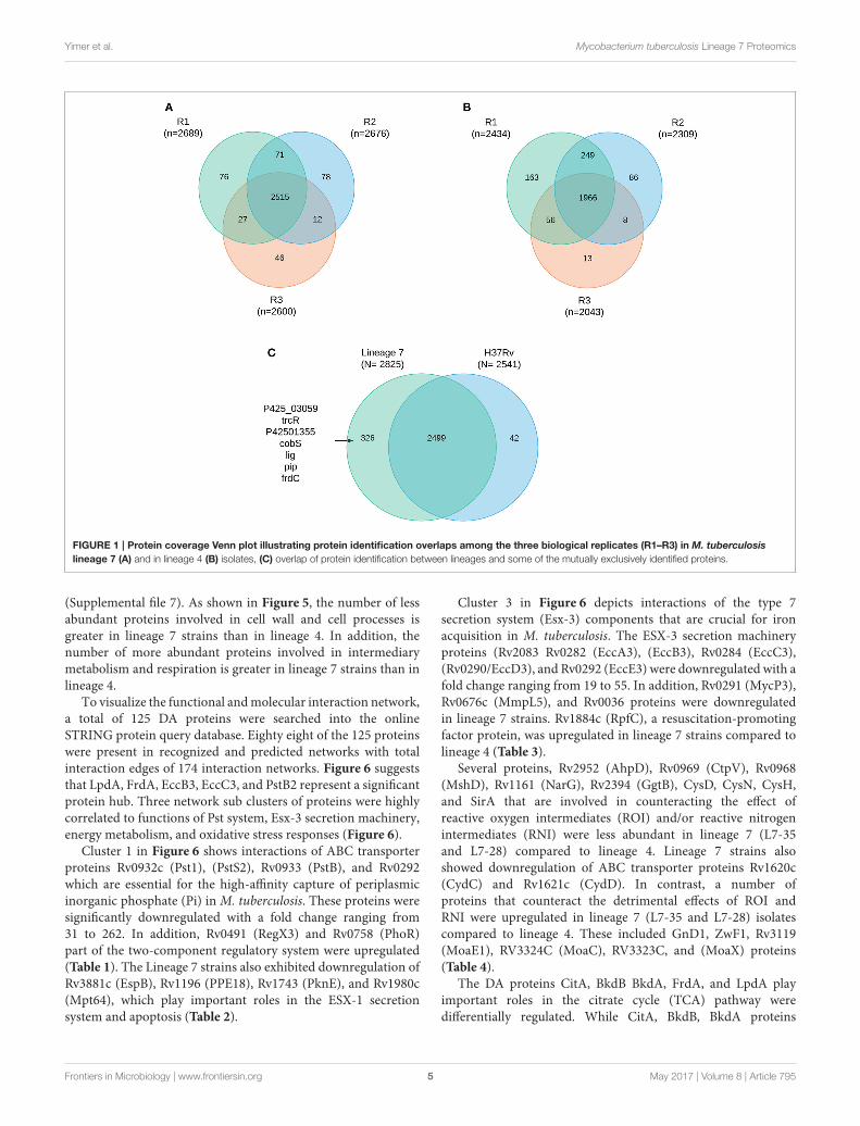
Comprehensive Proteome Analysis ofM. tuberculosis Lineage 7 and Lineage 4StrainsA total of 2867 M. tuberculosis proteins were identified with99% confidence at the peptide and protein levels (Supplementalfile 2) representing 71% protein coverage of the predicted M.tuberculosis proteome. The total number of proteins identifiedin M. tuberculosis lineage 7 (L7-35 and L7-28) and in lineage 4were 2,825 and 2,541, respectively. Among the 2,825 identifiedproteins in lineage 7 strains, 2499 (87%) proteins were shared inall the biological experiments (Figures 1A–C). Mutual exclusivityanalysis revealed 326 and 42 strain-specific protein groups inlineage 7 and lineage 4, respectively (Supplemental file 2), and themain DA component pathways are addressed below. The overlapin protein identification in the different biological replicates
1www.uniprot.org/help/gene_ontology
and M. tuberculosis lineages is shown in the Venn diagram(Figures 1A–C). Of the annotated components, 1,783 (62%) havean assigned molecular function, 1,110 (38.7%) are involved inknown biological processes, 948 (33%) are assigned by cellularcompartment, and 829 (28.9%) have an assigned KEGG function.The complete list of protein groups is presented in Supplementalfile 3.
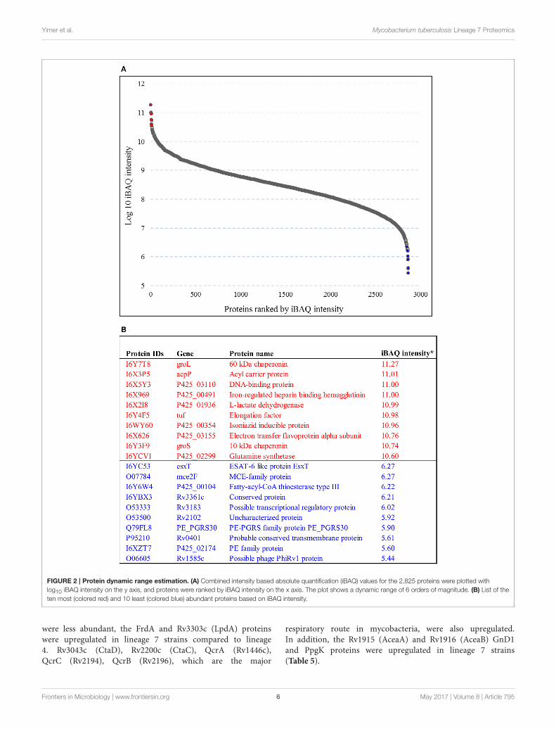
Protein Profile of M. tuberculosis Lineage 7(L7-35 and L7-28) and Lineage 4 StrainsThe abundance of the M. tuberculosis proteins identified wasquantified by iBAQ. This technique takes into account thenormalization and summation of MS/MS signals in relationto peptide size, length, and number of theoretical peptidesconsidered acceptable for all the proteins that are definedin a specific proteome run. The 10 most abundant proteinclasses identified in lineage 7 strain were chaperones, hydrolaseisomerase, ligase, oxidoreductase, transfer carrier protein, andnucleic acid-binding proteins. The least abundant proteinsidentified include the ESAT-6-like protein (EsxT), MCE-familyprotein (Mce2F), PE-PGRS family protein and PE family protein.The iBAQ intensity in the aggregate proteome covered a dynamicrange of six orders of magnitude between the most abundant andleast abundant proteins (Figures 2A,B; Supplemental file 3).
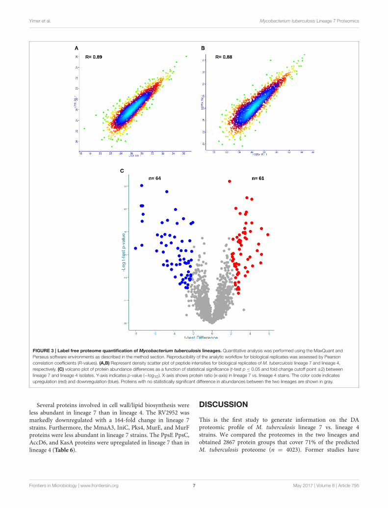
After identifying the 2,867 proteins in the composite M.tuberculosis proteome, the reproducibility of our label-freequantification workflow was assessed (Supplemental file 1). ThePearson correlations (R-values) of biological replicates usingnormalized protein LFQ intensities were computed. The analysisshowed that the R-value between normalized intensities was high(Figures 3A,B, Supplemental file 4) and thus was suitable foraccurate comparisons of protein abundance differences.
For DA protein comparisons, criteria were set that fulfill twovalid LFQ intensity values from each biological triplicate. Thisresulted in a total of 1,946 proteins. Using a set of statisticalcriteria, T-test p-value 0.05, S0 = 2 and fold change cutoff point±2, we found the abundances of 125 proteins to be significantlychanged (Figure 3C, Supplemental file 5).
The Proteomes of the M. tuberculosis
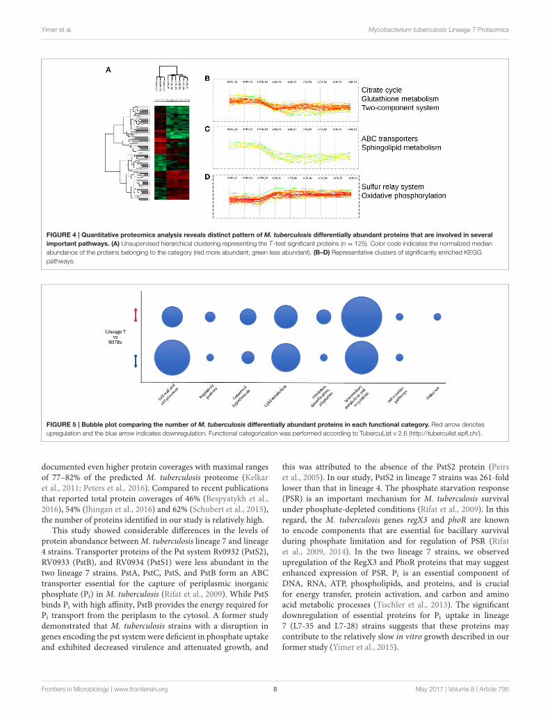
Lineage 7 Strains Are SignificantlyDifferent from Lineage 4Among the 125 differentially regulated proteins, 64 weredownregulated and 61 proteins were upregulated (Figure 3C,Supplemental file 5). The 125 DA protein groups were furthersubjected to unsupervised hierarchical cluster analysis. Theresulting cluster-gram is shown in Figure 4A. KEGG analysiswas then conducted to investigate whether these proteinswere enriched for any particular pathway. The pathwaysub-clusters that were significantly enriched are shown inFigures 4B–D (Supplementary file 6). The DA proteins werealso categorized into their functional categories as defined byTubercuList. A majority of the proteins detected belong tocategories of intermediary metabolism and respiration (39.2%),lipid metabolism (20%), cell wall- and cell processes-related(24.8%), conserved hypotheticals (2.4%) and unknowns (0.8%)
Frontiers in Microbiology | www.frontiersin.org 4 May 2017 | Volume 8 | Article 795
Yimer et al. Mycobacterium tuberculosis Lineage 7 Proteomics
FIGURE 1 | Protein coverage Venn plot illustrating protein identification overlaps among the three biological replicates (R1–R3) in M. tuberculosis
lineage 7 (A) and in lineage 4 (B) isolates, (C) overlap of protein identification between lineages and some of the mutually exclusively identified proteins.
(Supplemental file 7). As shown in Figure 5, the number of lessabundant proteins involved in cell wall and cell processes isgreater in lineage 7 strains than in lineage 4. In addition, thenumber of more abundant proteins involved in intermediarymetabolism and respiration is greater in lineage 7 strains than inlineage 4.
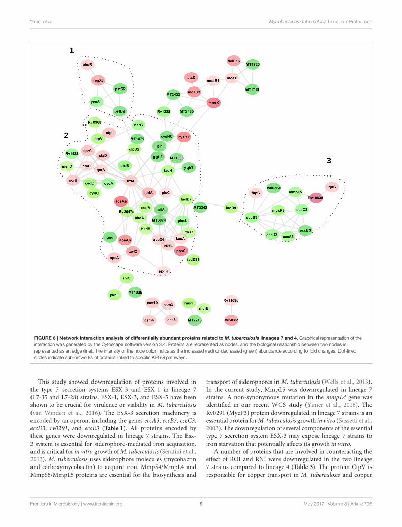
To visualize the functional andmolecular interaction network,a total of 125 DA proteins were searched into the onlineSTRING protein query database. Eighty eight of the 125 proteinswere present in recognized and predicted networks with totalinteraction edges of 174 interaction networks. Figure 6 suggeststhat LpdA, FrdA, EccB3, EccC3, and PstB2 represent a significantprotein hub. Three network sub clusters of proteins were highlycorrelated to functions of Pst system, Esx-3 secretion machinery,energy metabolism, and oxidative stress responses (Figure 6).
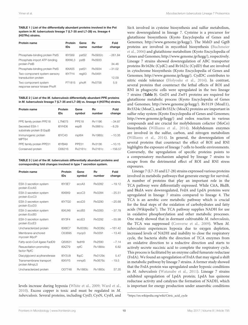
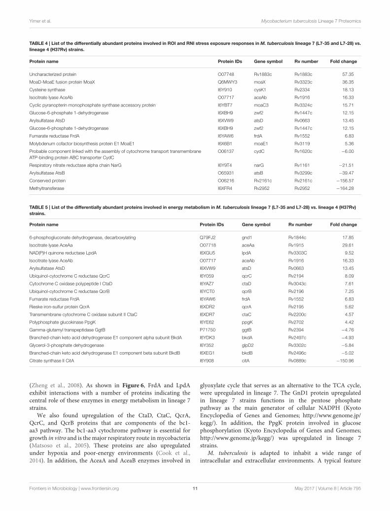
Cluster 1 in Figure 6 shows interactions of ABC transporterproteins Rv0932c (Pst1), (PstS2), Rv0933 (PstB), and Rv0292which are essential for the high-affinity capture of periplasmicinorganic phosphate (Pi) in M. tuberculosis. These proteins weresignificantly downregulated with a fold change ranging from31 to 262. In addition, Rv0491 (RegX3) and Rv0758 (PhoR)part of the two-component regulatory system were upregulated(Table 1). The Lineage 7 strains also exhibited downregulation ofRv3881c (EspB), Rv1196 (PPE18), Rv1743 (PknE), and Rv1980c(Mpt64), which play important roles in the ESX-1 secretionsystem and apoptosis (Table 2).
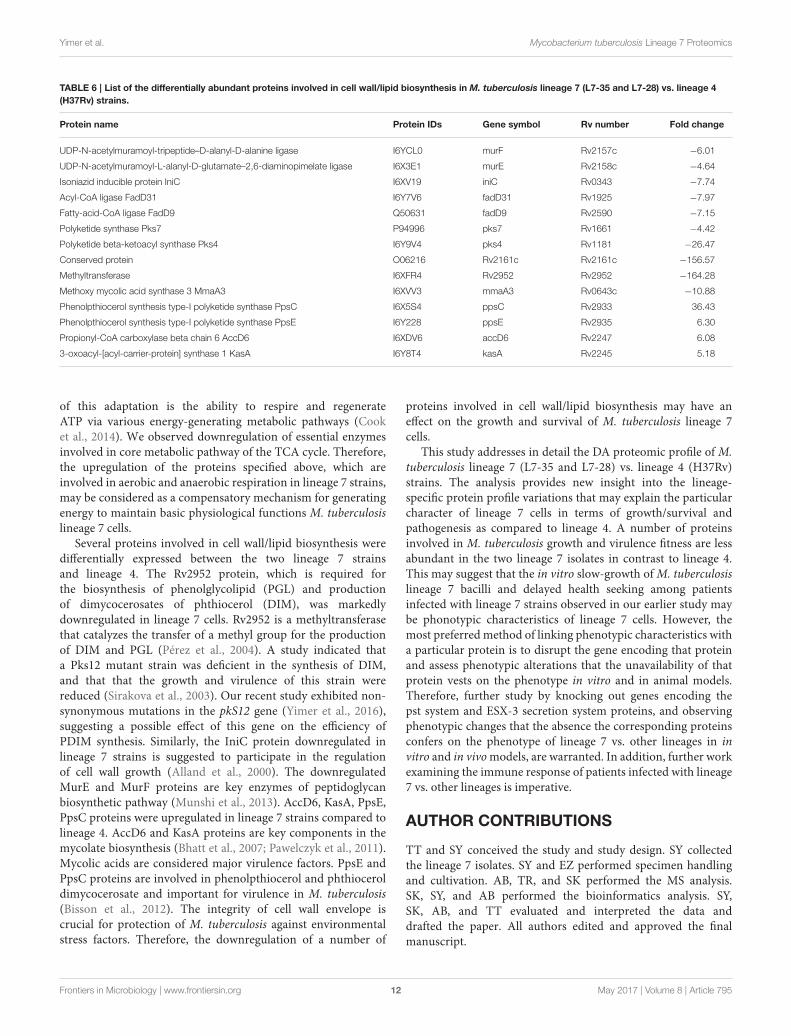
Cluster 3 in Figure 6 depicts interactions of the type 7secretion system (Esx-3) components that are crucial for ironacquisition in M. tuberculosis. The ESX-3 secretion machineryproteins (Rv2083 Rv0282 (EccA3), (EccB3), Rv0284 (EccC3),(Rv0290/EccD3), and Rv0292 (EccE3) were downregulated with afold change ranging from 19 to 55. In addition, Rv0291 (MycP3),Rv0676c (MmpL5), and Rv0036 proteins were downregulatedin lineage 7 strains. Rv1884c (RpfC), a resuscitation-promotingfactor protein, was upregulated in lineage 7 strains compared tolineage 4 (Table 3).
Several proteins, Rv2952 (AhpD), Rv0969 (CtpV), Rv0968(MshD), Rv1161 (NarG), Rv2394 (GgtB), CysD, CysN, CysH,and SirA that are involved in counteracting the effect ofreactive oxygen intermediates (ROI) and/or reactive nitrogenintermediates (RNI) were less abundant in lineage 7 (L7-35and L7-28) compared to lineage 4. Lineage 7 strains alsoshowed downregulation of ABC transporter proteins Rv1620c(CydC) and Rv1621c (CydD). In contrast, a number ofproteins that counteract the detrimental effects of ROI andRNI were upregulated in lineage 7 (L7-35 and L7-28) isolatescompared to lineage 4. These included GnD1, ZwF1, Rv3119(MoaE1), RV3324C (MoaC), RV3323C, and (MoaX) proteins(Table 4).
The DA proteins CitA, BkdB BkdA, FrdA, and LpdA playimportant roles in the citrate cycle (TCA) pathway weredifferentially regulated. While CitA, BkdB, BkdA proteins
Frontiers in Microbiology | www.frontiersin.org 5 May 2017 | Volume 8 | Article 795
Yimer et al. Mycobacterium tuberculosis Lineage 7 Proteomics
FIGURE 2 | Protein dynamic range estimation. (A) Combined intensity based absolute quantification (iBAQ) values for the 2,825 proteins were plotted with
log10 iBAQ intensity on the y axis, and proteins were ranked by iBAQ intensity on the x axis. The plot shows a dynamic range of 6 orders of magnitude. (B) List of the
ten most (colored red) and 10 least (colored blue) abundant proteins based on iBAQ intensity.
were less abundant, the FrdA and Rv3303c (LpdA) proteinswere upregulated in lineage 7 strains compared to lineage4. Rv3043c (CtaD), Rv2200c (CtaC), QcrA (Rv1446c),QcrC (Rv2194), QcrB (Rv2196), which are the major
respiratory route in mycobacteria, were also upregulated.In addition, the Rv1915 (AceaA) and Rv1916 (AceaB) GnD1and PpgK proteins were upregulated in lineage 7 strains(Table 5).
Frontiers in Microbiology | www.frontiersin.org 6 May 2017 | Volume 8 | Article 795
Yimer et al. Mycobacterium tuberculosis Lineage 7 Proteomics
FIGURE 3 | Label free proteome quantification of Mycobacterium tuberculosis lineages. Quantitative analysis was performed using the MaxQuant and
Perseus software environments as described in the method section. Reproducibility of the analytic workflow for biological replicates was assessed by Pearson
correlation coefficients (R-values). (A,B) Represent density scatter plot of peptide intensities for biological replicates of M. tuberculosis lineage 7 and lineage 4,
respectively. (C) volcano plot of protein abundance differences as a function of statistical significance (t-test p ≤ 0.05 and fold change cutoff point ±2) between
lineage 7 and lineage 4 isolates. Y-axis indicates p-value (−log10). X-axis shows protein ratio (x-axis) in lineage 7 vs. lineage 4 stains. The color code indicates
upregulation (red) and downregulation (blue). Proteins with no statistically significant difference in abundances between the two lineages are shown in gray.
Several proteins involved in cell wall/lipid biosynthesis wereless abundant in lineage 7 than in lineage 4. The RV2952 wasmarkedly downregulated with a 164-fold change in lineage 7strains. Furthermore, the MmaA3, IniC, Pks4, MurE, and MurFproteins were less abundant in lineage 7 strains. The PpsE PpsC,AccD6, and KasA proteins were upregulated in lineage 7 than inlineage 4 (Table 6).
DISCUSSION
This is the first study to generate information on the DAproteomic profile of M. tuberculosis lineage 7 vs. lineage 4strains. We compared the proteomes in the two lineages andobtained 2867 protein groups that cover 71% of the predictedM. tuberculosis proteome (n = 4023). Former studies have
Frontiers in Microbiology | www.frontiersin.org 7 May 2017 | Volume 8 | Article 795
Yimer et al. Mycobacterium tuberculosis Lineage 7 Proteomics
FIGURE 4 | Quantitative proteomics analysis reveals distinct pattern of M. tuberculosis differentially abundant proteins that are involved in several
important pathways. (A) Unsupervised hierarchical clustering representing the T-test significant proteins (n = 125). Color code indicates the normalized median
abundance of the proteins belonging to the category (red more abundant; green less abundant). (B–D) Representative clusters of significantly enriched KEGG
pathways.
FIGURE 5 | Bubble plot comparing the number of M. tuberculosis differentially abundant proteins in each functional category. Red arrow denotes
upregulation and the blue arrow indicates downregulation. Functional categorization was performed according to TubercuList v 2.6 (http://tuberculist.epfl.ch/).
documented even higher protein coverages with maximal rangesof 77–82% of the predicted M. tuberculosis proteome (Kelkaret al., 2011; Peters et al., 2016). Compared to recent publicationsthat reported total protein coverages of 46% (Bespyatykh et al.,2016), 54% (Jhingan et al., 2016) and 62% (Schubert et al., 2015),the number of proteins identified in our study is relatively high.
This study showed considerable differences in the levels ofprotein abundance betweenM. tuberculosis lineage 7 and lineage4 strains. Transporter proteins of the Pst system Rv0932 (PstS2),RV0933 (PstB), and RV0934 (PstS1) were less abundant in thetwo lineage 7 strains. PstA, PstC, PstS, and PstB form an ABCtransporter essential for the capture of periplasmic inorganicphosphate (Pi) in M. tuberculosis (Rifat et al., 2009). While PstSbinds Pi with high affinity, PstB provides the energy required forPi transport from the periplasm to the cytosol. A former studydemonstrated that M. tuberculosis strains with a disruption ingenes encoding the pst systemwere deficient in phosphate uptakeand exhibited decreased virulence and attenuated growth, and
this was attributed to the absence of the PstS2 protein (Peirset al., 2005). In our study, PstS2 in lineage 7 strains was 261-foldlower than that in lineage 4. The phosphate starvation response(PSR) is an important mechanism for M. tuberculosis survivalunder phosphate-depleted conditions (Rifat et al., 2009). In thisregard, the M. tuberculosis genes regX3 and phoR are knownto encode components that are essential for bacillary survivalduring phosphate limitation and for regulation of PSR (Rifatet al., 2009, 2014). In the two lineage 7 strains, we observedupregulation of the RegX3 and PhoR proteins that may suggestenhanced expression of PSR. Pi is an essential component ofDNA, RNA, ATP, phospholipids, and proteins, and is crucialfor energy transfer, protein activation, and carbon and aminoacid metabolic processes (Tischler et al., 2013). The significantdownregulation of essential proteins for Pi uptake in lineage7 (L7-35 and L7-28) strains suggests that these proteins maycontribute to the relatively slow in vitro growth described in ourformer study (Yimer et al., 2015).
Frontiers in Microbiology | www.frontiersin.org 8 May 2017 | Volume 8 | Article 795
Yimer et al. Mycobacterium tuberculosis Lineage 7 Proteomics
FIGURE 6 | Network interaction analysis of differentially abundant proteins related to M. tuberculosis lineages 7 and 4. Graphical representation of the
interaction was generated by the Cytoscape software version 3.4. Proteins are represented as nodes, and the biological relationship between two nodes is
represented as an edge (line). The intensity of the node color indicates the increased (red) or decreased (green) abundance according to fold changes. Dot-lined
circles indicate sub-networks of proteins linked to specific KEGG pathways.
This study showed downregulation of proteins involved inthe type 7 secretion systems ESX-3 and ESX-1 in lineage 7(L7-35 and L7-28) strains. ESX-1, ESX-3, and ESX-5 have beenshown to be crucial for virulence or viability in M. tuberculosis(van Winden et al., 2016). The ESX-3 secretion machinery isencoded by an operon, including the genes eccA3, eccB3, eccC3,eccD3, rv0291, and eccE3 (Table 1). All proteins encoded bythese genes were downregulated in lineage 7 strains. The Esx-3 system is essential for siderophore-mediated iron acquisition,and is critical for in vitro growth ofM. tuberculosis (Serafini et al.,2013). M. tuberculosis uses siderophore molecules (mycobactinand carboxymycobactin) to acquire iron. MmpS4/MmpL4 andMmpS5/MmpL5 proteins are essential for the biosynthesis and
transport of siderophores in M. tuberculosis (Wells et al., 2013).In the current study, MmpL5 was downregulated in lineage 7strains. A non-synonymous mutation in the mmpL4 gene wasidentified in our recent WGS study (Yimer et al., 2016). TheRv0291 (MycP3) protein downregulated in lineage 7 strains is anessential protein forM. tuberculosis growth in vitro (Sassetti et al.,2003). The downregulation of several components of the essentialtype 7 secretion system ESX-3 may expose lineage 7 strains toiron starvation that potentially affects its growth in vitro.
A number of proteins that are involved in counteracting theeffect of ROI and RNI were downregulated in the two lineage7 strains compared to lineage 4 (Table 3). The protein CtpV isresponsible for copper transport in M. tuberculosis and copper
Frontiers in Microbiology | www.frontiersin.org 9 May 2017 | Volume 8 | Article 795
Yimer et al. Mycobacterium tuberculosis Lineage 7 Proteomics
TABLE 1 | List of the differentially abundant proteins involved in the Pst
system in M. tuberculosis lineage 7 (L7-35 and L7-28) vs. lineage 4
(H37Rv) strains.
Protein name Protein
IDs
Gene
name
Rv
number
Fold
change
Phosphate-binding protein PstS I6Y569 pstS2 Rv0932c −261.84
Phosphate import ATP-binding
protein PstB
I6XWL3 pstB Rv0933
34.46
Phosphate-binding protein PstS I6XA55 pstS1 Rv0934 −31.02
Two-component system sensory
transduction protein
I6Y7Y4 regX3 Rv0491
12.09
Two component system
response sensor kinase PhoR
P71815 phoR Rv0758 5.9
TABLE 2 | List of the M. tuberculosis differentially abundant PPE proteins
inM. tuberculosis lineage 7 (L7-35 and L7-28) vs. lineage 4 (H37Rv) strains.
Protein name Protein
IDs
Gene
symbol
Rv
number
Fold
change
PPE family protein PPE18 L7N675 PPE18 Rv1196 −34.87
Secreted ESX-1
substrate protein B EspB
I6YHD6 espB Rv3881c −8.29
Immunogenic protein
MPT64
I6YC43 mpt64 Rv1980c −10.35
PPE family protein PPE51 I6YBA9 PPE51 Rv3136 −10.15
Conserved protein O06216 Rv2161c Rv2161c −156.57
TABLE 3 | List of the M. tuberculosis differentially abundant proteins and
corresponding fold changes involved in type 7 secretion system.
Protein name Protein
IDs
Gene
symbol
Rv
number
Fold
change
ESX-3 secretion system
protein EccA3
I6Y3E7 eccA3 Rv0282 −19.12
ESX-3 secretion system
protein EccC3
I6X8X9 eccC3 Rv0284 −25.51
ESX-3 secretion system
protein EccD3
I6Y7G0 eccD3 Rv0290 −25.68
ESX-3 secretion system
protein EccB3
I6XUX6 eccB3 Rv0283 −37.78
ESX-3 secretion system
protein EccE3
I6Y3F4 eccE3 Rv0292 −55.98
Uncharacterized protein I6X8C7 Rv0036c Rv0036c −161.42
Membrane-anchored
mycosin MycP
O53695 mycp3 Rv0291 −13.40
Fatty-acid–CoA ligase FadD9 Q50631 fadH9 Rv2590 −7.14
Resuscitation-promoting
factor RpfC
I6XZ79 rpfC Rv1884c 6.82
Diacylglycerol acyltransferase I6Y2U9 fbpC Rv0129c 5.47
Transmembrane transport
protein MmpL5
I6XVY5 mmpl5 Rv0676c −19.5
Uncharacterized protein O07748 Rv1883c Rv1883c 57.35
levels increase during hypoxia (White et al., 2009; Ward et al.,2010). Excess copper is toxic and must be regulated in M.tuberculosis. Several proteins, including CysD, CysN, CysH, and
SirA involved in cysteine biosynthesis and sulfur metabolism,were downregulated in lineage 7. Cysteine is a precursor forglutathione biosynthesis (Kyoto Encyclopedia of Genes andGenomes; http://www.genome.jp/kegg/). The MshD and GgtBproteins are involved in mycothiol biosynthesis (Buchmeieret al., 2006) and glutathione metabolism (Kyoto Encyclopedia ofGenes and Genomes; http://www.genome.jp/kegg/), respectively.Lineage 7 strains showed downregulation of ABC transporterproteins Rv1620c (CydC) and Rv1621c (CydD) that are involvedin cytochrome biosynthesis (Kyoto Encyclopedia of Genes andGenomes; http://www.genome.jp/kegg/). CydDC contributes tonitric oxide tolerance (Holyoake et al., 2016). In contrast,several proteins that counteract the lethal effects of ROI andRNI in phagocytic cells were upregulated in the two lineage7 strains (Table 3). GnD1 and ZwF1 proteins are required forglutathione metabolic process (Kyoto Encyclopedia of Genesand Genomes; http://www.genome.jp/kegg/). Rv3119 (MoaE1),Rv3324c (MoaC), and Rv3323c (MoaX) proteins are important insulfur relay system (Kyoto Encyclopedia of Genes and Genomes;http://www.genome.jp/kegg/) and redox reaction in variouscompounds and are crucial for molybdenum cofactor (MoCo)biosynthesis (Williams et al., 2014). Molybdenum enzymesare involved in the sulfur, carbon, and nitrogen metabolism(Williams et al., 2014). In general, the downregulation ofseveral proteins that counteract the effect of ROI and RNIhighlights the exposure of lineage 7 cells to hostile environments.Conversely, the upregulation of specific proteins points toa compensatory mechanism adapted by lineage 7 strains toescape from the detrimental effect of ROI and RNI stressexposures.
Lineage 7 (L7-35 and L7-28) strains expressed various proteinsinvolved in metabolic pathways that generate energy for survival.A number of proteins that play an important role in theTCA pathway were differentially expressed. While CitA, BkdB,and BkdA were downregulated, FrdA and LpdA proteins wereupregulated in lineage 7 strains compared to lineage 4. TheTCA is an aerobic core metabolic pathway which is crucialfor the final steps of the oxidation of carbohydrates and fattyacids (Wikipedia2). The TCA pathway supplies NADH for usein oxidative phosphorylation and other metabolic processes.One study showed that in dormant cultivatable M. tuberculosis,the TCA was suppressed (Converse et al., 2009). When M.tuberculosis experiences hypoxia due to oxygen depletion,increased levels of NADH and inability to close the respiratorycycle, the bacteria shifts the direction of TCA enzymes froman oxidative direction to a reductive direction and starts toactively secrete succinic acid to complete the respiratory cycle.This process is facilitated by an enzyme called fumarate reductase(FrdA).We found an upregulation of FrdA that may signal a shiftin metabolic pathway by lineage 7 strains. A former study showedthat the FrdA protein was upregulated under hypoxic conditionsin M. tuberculosis (Watanabe et al., 2011). Lineage 7 strainsexhibited upregulation of LpdA protein; LpdA has quinonereductase activity and catalyzes the formation of NADH, whichis important for energy production under anaerobic conditions
2https://en.wikipedia.org/wiki/Citric_acid_cycle
Frontiers in Microbiology | www.frontiersin.org 10 May 2017 | Volume 8 | Article 795
Yimer et al. Mycobacterium tuberculosis Lineage 7 Proteomics
TABLE 4 | List of the differentially abundant proteins involved in ROI and RNI stress exposure responses inM. tuberculosis lineage 7 (L7-35 and L7-28) vs.
lineage 4 (H37Rv) strains.
Protein name Protein IDs Gene symbol Rv number Fold change
Uncharacterized protein O07748 Rv1883c Rv1883c 57.35
MoaD-MoaE fusion protein MoaX Q6MWY3 moaX Rv3323c 36.35
Cysteine synthase I6Y910 cysK1 Rv2334 18.13
Isocitrate lyase AceAb O07717 aceAb Rv1916 16.33
Cyclic pyranopterin monophosphate synthase accessory protein I6YBT7 moaC3 Rv3324c 15.71
Glucose-6-phosphate 1-dehydrogenase I6XBH9 zwf2 Rv1447c 12.15
Arylsulfatase AtsD I6XVW9 atsD Rv0663 13.45
Glucose-6-phosphate 1-dehydrogenase I6XBH9 zwf2 Rv1447c 12.15
Fumarate reductase FrdA I6YAW6 frdA Rv1552 6.83
Molybdenum cofactor biosynthesis protein E1 MoaE1 I6X6B1 moaE1 Rv3119 5.36
Probable component linked with the assembly of cytochrome transport transmembrane
ATP-binding protein ABC transporter CydC
O06137 cydC Rv1620c −6.00
Respiratory nitrate reductase alpha chain NarG I6Y9T4 narG Rv1161 −21.51
Arylsulfatase AtsB O65931 atsB Rv3299c −39.47
Conserved protein O06216 Rv2161c Rv2161c −156.57
Methyltransferase I6XFR4 Rv2952 Rv2952 −164.28
TABLE 5 | List of the differentially abundant proteins involved in energy metabolism in M. tuberculosis lineage 7 (L7-35 and L7-28) vs. lineage 4 (H37Rv)
strains.
Protein name Protein IDs Gene symbol Rv number Fold change
6-phosphogluconate dehydrogenase, decarboxylating Q79FJ2 gnd1 Rv1844c 17.85
Isocitrate lyase AceAa O07718 aceAa Rv1915 29.61
NAD(P)H quinone reductase LpdA I6XGU5 lpdA Rv3303C 9.52
Isocitrate lyase AceAb O07717 aceAb Rv1916 16.33
Arylsulfatase AtsD I6XVW9 atsD Rv0663 13.45
Ubiquinol-cytochrome C reductase QcrC I6Y059 qcrC Rv2194 8.09
Cytochrome C oxidase polypeptide I CtaD I6YAZ7 ctaD Rv3043c 7.61
Ubiquinol-cytochrome C reductase QcrB I6YCT0 qcrB Rv2196 7.25
Fumarate reductase FrdA I6YAW6 frdA Rv1552 6.83
Rieske iron-sulfur protein QcrA I6XDR2 qcrA Rv2195 5.62
Transmembrane cytochrome C oxidase subunit II CtaC I6XDR7 ctaC Rv2200c 4.57
Polyphosphate glucokinase PpgK I6YE62 ppgK Rv2702 4.42
Gamma-glutamyl transpeptidase GgtB P71750 ggtB Rv2394 −4.76
Branched-chain keto acid dehydrogenase E1 component alpha subunit BkdA I6YDK3 bkdA Rv2497c −4.93
Glycerol-3-phosphate dehydrogenase I6Y352 glpD2 Rv3302c −5.84
Branched-chain keto acid dehydrogenase E1 component beta subunit BkdB I6XEG1 bkdB Rv2496c −5.02
Citrate synthase II CitA I6Y908 citA Rv0889c −150.96
(Zheng et al., 2008). As shown in Figure 6, FrdA and LpdAexhibit interactions with a number of proteins indicating thecentral role of these enzymes in energy metabolism in lineage 7strains.
We also found upregulation of the CtaD, CtaC, QcrA,QcrC, and QcrB proteins that are components of the bc1-aa3 pathway. The bc1-aa3 cytochrome pathway is essential forgrowth in vitro and is themajor respiratory route inmycobacteria(Matsoso et al., 2005). These proteins are also upregulatedunder hypoxia and poor-energy environments (Cook et al.,2014). In addition, the AceaA and AceaB enzymes involved in
glyoxylate cycle that serves as an alternative to the TCA cycle,were upregulated in lineage 7. The GnD1 protein upregulatedin lineage 7 strains functions in the pentose phosphatepathway as the main generator of cellular NADPH (KyotoEncyclopedia of Genes and Genomes; http://www.genome.jp/kegg/). In addition, the PpgK protein involved in glucosephosphorylation (Kyoto Encyclopedia of Genes and Genomes;http://www.genome.jp/kegg/) was upregulated in lineage 7strains.
M. tuberculosis is adapted to inhabit a wide range ofintracellular and extracellular environments. A typical feature
Frontiers in Microbiology | www.frontiersin.org 11 May 2017 | Volume 8 | Article 795
Yimer et al. Mycobacterium tuberculosis Lineage 7 Proteomics
TABLE 6 | List of the differentially abundant proteins involved in cell wall/lipid biosynthesis in M. tuberculosis lineage 7 (L7-35 and L7-28) vs. lineage 4
(H37Rv) strains.
Protein name Protein IDs Gene symbol Rv number Fold change
UDP-N-acetylmuramoyl-tripeptide–D-alanyl-D-alanine ligase I6YCL0 murF Rv2157c −6.01
UDP-N-acetylmuramoyl-L-alanyl-D-glutamate–2,6-diaminopimelate ligase I6X3E1 murE Rv2158c −4.64
Isoniazid inducible protein IniC I6XV19 iniC Rv0343 −7.74
Acyl-CoA ligase FadD31 I6Y7V6 fadD31 Rv1925 −7.97
Fatty-acid-CoA ligase FadD9 Q50631 fadD9 Rv2590 −7.15
Polyketide synthase Pks7 P94996 pks7 Rv1661 −4.42
Polyketide beta-ketoacyl synthase Pks4 I6Y9V4 pks4 Rv1181 −26.47
Conserved protein O06216 Rv2161c Rv2161c −156.57
Methyltransferase I6XFR4 Rv2952 Rv2952 −164.28
Methoxy mycolic acid synthase 3 MmaA3 I6XVV3 mmaA3 Rv0643c −10.88
Phenolpthiocerol synthesis type-I polyketide synthase PpsC I6X5S4 ppsC Rv2933 36.43
Phenolpthiocerol synthesis type-I polyketide synthase PpsE I6Y228 ppsE Rv2935 6.30
Propionyl-CoA carboxylase beta chain 6 AccD6 I6XDV6 accD6 Rv2247 6.08
3-oxoacyl-[acyl-carrier-protein] synthase 1 KasA I6Y8T4 kasA Rv2245 5.18
of this adaptation is the ability to respire and regenerateATP via various energy-generating metabolic pathways (Cooket al., 2014). We observed downregulation of essential enzymesinvolved in core metabolic pathway of the TCA cycle. Therefore,the upregulation of the proteins specified above, which areinvolved in aerobic and anaerobic respiration in lineage 7 strains,may be considered as a compensatory mechanism for generatingenergy to maintain basic physiological functions M. tuberculosislineage 7 cells.
Several proteins involved in cell wall/lipid biosynthesis weredifferentially expressed between the two lineage 7 strainsand lineage 4. The Rv2952 protein, which is required forthe biosynthesis of phenolglycolipid (PGL) and productionof dimycocerosates of phthiocerol (DIM), was markedlydownregulated in lineage 7 cells. Rv2952 is a methyltransferasethat catalyzes the transfer of a methyl group for the productionof DIM and PGL (Pérez et al., 2004). A study indicated thata Pks12 mutant strain was deficient in the synthesis of DIM,and that that the growth and virulence of this strain werereduced (Sirakova et al., 2003). Our recent study exhibited non-synonymous mutations in the pkS12 gene (Yimer et al., 2016),suggesting a possible effect of this gene on the efficiency ofPDIM synthesis. Similarly, the IniC protein downregulated inlineage 7 strains is suggested to participate in the regulationof cell wall growth (Alland et al., 2000). The downregulatedMurE and MurF proteins are key enzymes of peptidoglycanbiosynthetic pathway (Munshi et al., 2013). AccD6, KasA, PpsE,PpsC proteins were upregulated in lineage 7 strains compared tolineage 4. AccD6 and KasA proteins are key components in themycolate biosynthesis (Bhatt et al., 2007; Pawelczyk et al., 2011).Mycolic acids are considered major virulence factors. PpsE andPpsC proteins are involved in phenolpthiocerol and phthioceroldimycocerosate and important for virulence in M. tuberculosis(Bisson et al., 2012). The integrity of cell wall envelope iscrucial for protection of M. tuberculosis against environmentalstress factors. Therefore, the downregulation of a number of
proteins involved in cell wall/lipid biosynthesis may have aneffect on the growth and survival of M. tuberculosis lineage 7cells.
This study addresses in detail the DA proteomic profile of M.tuberculosis lineage 7 (L7-35 and L7-28) vs. lineage 4 (H37Rv)strains. The analysis provides new insight into the lineage-specific protein profile variations that may explain the particularcharacter of lineage 7 cells in terms of growth/survival andpathogenesis as compared to lineage 4. A number of proteinsinvolved in M. tuberculosis growth and virulence fitness are lessabundant in the two lineage 7 isolates in contrast to lineage 4.This may suggest that the in vitro slow-growth ofM. tuberculosislineage 7 bacilli and delayed health seeking among patientsinfected with lineage 7 strains observed in our earlier study maybe phonotypic characteristics of lineage 7 cells. However, themost preferred method of linking phenotypic characteristics witha particular protein is to disrupt the gene encoding that proteinand assess phenotypic alterations that the unavailability of thatprotein vests on the phenotype in vitro and in animal models.Therefore, further study by knocking out genes encoding thepst system and ESX-3 secretion system proteins, and observingphenotypic changes that the absence the corresponding proteinsconfers on the phenotype of lineage 7 vs. other lineages in invitro and in vivomodels, are warranted. In addition, further workexamining the immune response of patients infected with lineage7 vs. other lineages is imperative.
AUTHOR CONTRIBUTIONS
TT and SY conceived the study and study design. SY collectedthe lineage 7 isolates. SY and EZ performed specimen handlingand cultivation. AB, TR, and SK performed the MS analysis.SK, SY, and AB performed the bioinformatics analysis. SY,SK, AB, and TT evaluated and interpreted the data anddrafted the paper. All authors edited and approved the finalmanuscript.
Frontiers in Microbiology | www.frontiersin.org 12 May 2017 | Volume 8 | Article 795
Yimer et al. Mycobacterium tuberculosis Lineage 7 Proteomics
FUNDING
Funding was received from the Research Council of Norway(RCN) FRIMEDBIO project 204747 and RCN GLOBVACprojects 234506 to TT and 192468 to CH, and Norwegian South-Eastern Health Authority project 2013080 to SY and TT.
ACKNOWLEDGMENTS
The authors thank the patients for consenting to participatein the study. We also thank the selected health care facilities
in the Amhara Region, Ethiopia, for facilitating the study. Weare grateful to the Armauer Hansen Research Institute (AHRI),Addis Ababa, Ethiopia, and the Norwegian Institute of PublicHealth for facilitating the transfer of lineage 7 strains for WGSat Oslo University Hospital.
SUPPLEMENTARY MATERIAL
The Supplementary Material for this article can be foundonline at: http://journal.frontiersin.org/article/10.3389/fmicb.2017.00795/full#supplementary-material
REFERENCES
Albanna, A. S., Reed, M. B., Kotar, K. V., Fallow, A., McIntosh, F. A., Behr, M. A.,et al. (2011). Reduced transmissibility of East African Indian strains of Mtb.PLoS ONE 6:e25075. doi: 10.1371/journal.pone.0025075
Alland, D., Steyn, A. J., Weisbrod, T., Aldrich, K., and Jacobs, W. R. (2000).Characterization of the Mycobacterium tuberculosis iniBAC promoter, apromoter that responds to cell wall biosynthesis inhibition. J. Bacteriol. 182,1802–1811. doi: 10.1128/JB.182.7.1802-1811.2000
Assenov, Y., Ramírez, F., Schelhorn, S. E., Lengauer, T., and Albrecht, M. (2008).Computing topological parameters of biological networks. Bioinformatics 24,282–284. doi: 10.1093/bioinformatics/btm554
Azuaje, F., Devaux, Y., and Wagner, D. R. (2010). Coordinated modularfunctionality and prognostic potential of a heart failure biomarker-driveninteraction network. BMC Syst. Biol. 4:60. doi: 10.1186/1752-0509-4-60.
Bader, G. D., and Hogue, C. W. (2003). An automated method for findingmolecular complexes in large protein interaction networks. BMC Bioinform.
4:2. doi: 10.1186/1471-2105-4-2Benjamini, Y., Drai, D., Elmer, G., Kafkafi, N., and Golani, I. (2001). Controlling
the false discovery rate in behavior genetics research. Behav. Brain Res. 125,279–284. doi: 10.1016/S0166-4328(01)00297-2
Bespyatykh, J., Shitikov, E., Butenko, I., Altukhov, I., Alexeev, D., andMokrousov, I.(2016). Proteome analysis of the Mycobacterium tuberculosis Beijing B0/W148cluster. Sci. Rep. 6:28985. doi: 10.1038/srep28985
Bhatt, A., Molle, V., Besra, G. S., Jacobs, W. R. Jr., and Kremer, L. (2007). TheMycobacterium tuberculosis FAS-II condensing enzymes: their role in mycolicacid biosynthesis, acid-fastness, pathogenesis and in future drug development.Mol. Microbiol. 64, 1442–1454. doi: 10.1111/j.1365-2958.2007.05761.x
Bisson, G. P., Mehaffy, C., Broeckling, C., Prenni, J., Rifat, D., and Lun, D.(2012). Upregulation of the phthiocerol dimycocerosate biosynthetic pathwayby rifampin-resistant, rpoB mutant Mycobacterium tuberculosis. J. Bacteriol.
194, 6441–6452. doi: 10.1128/JB.01013-12Blouin, Y., Hauck, Y., Soler, C., Fabre, M., Vong, R., and Dehan, C.
(2012). Significance of the identification in the Horn of Africa of anexceptionally deep branching Mycobacterium tuberculosis clade. PLoS ONE
7:12. doi: 10.1371/journal.pone.0052841Buchmeier, N. A., Newton, G. L., and Fahey, R. C. (2006). A mycothiol
synthase mutant of Mycobacterium tuberculosis has an altered thiol-disulfidecontent and limited tolerance to stress. J. Bacteriol. 188, 6245–6252.doi: 10.1128/JB.00393-06
Chacon-Salinas, R., Serafín-López, J., Ramos-Payán, R., Méndez-Aragón, P.,Hernández-Pando, R., Van Soolingen, D., et al. (2005). Differential patternof cytokine expression by macrophages infected in vitro with differentMycobacterium tuberculosis genotypes. Clin. Exp. Immunol. 140, 443–449.doi: 10.1111/j.1365-2249.2005.02797.x
Comas, I., Coscolla, M., Luo, T., Borrell, S., Holt, K. E., Kato-Maeda,M., et al. (2013). Out-of-Africa migration and Neolithic coexpansion ofMycobacterium tuberculosis with modern humans. Nat. Genet. 45, 1176–1182.doi: 10.1038/ng.2744
Converse, P. J., Karakousis, P. C., Klinkenberg, L. G., Kesavan, A. K., Ly, L. H., andAllen, S. S. (2009). Role of the dosR-dosS two-component regulatory system in
Mycobacterium tuberculosis virulence in three animal models. Infect. Immun.
77, 1230–1237. doi: 10.1128/IAI.01117-08Cook, G. M., Hards, K., Vilchèze, C., Hartman, T., and Berney, M. (2014).
Energetics of respiration and oxidative phosphorylation in mycobacteria.Microbiol. Spectr. 2:3. doi: 10.1128/microbiolspec.MGM2-0015-2013
Coscolla, M., and Gagneux, S. (2014). Consequences of genomicdiversity in Mycobacterium tuberculosis. Semin. Immunol. 26, 441–444.doi: 10.1016/j.smim.2014.09.012
Cox, J., and Matthias, M. (2008). MaxQuant enables high peptide identificationrates, individualized ppb-range mass accuracies and proteome-wide proteinquantification. Nat. Biotechnol. 26, 1367–1372. doi: 10.1038/nbt.1511
Cox, J., Neuhauser, N., Michalski, A., Scheltema, R. A., Olsen, J. V., and Mann,M. (2011). Andromeda: a peptide search engine integrated into the MaxQuantenvironment. J. Proteome Res. 10, 1794–1805. doi: 10.1021/pr101065j
Firdessa, R., Berg, S., Hailu, E., Schelling, E., Gumi, B., Erenso, G., et al. (2013).Mycobacterial lineages causing pulmonary and extrapulmonary tuberculosis,Ethiopia. Emerging Infect. Dis. 19, 460–463. doi: 10.3201/eid1903.120256
Franceschini, A., Szklarczyk, D., Frankild, S., Kuhn, M., Simonovic, M., andRoth, A. (2013). STRING v9. 1: protein-protein interaction networks, withincreased coverage and integration. Nucleic Acids Res. 41, D808–D815.doi: 10.1093/nar/gks1094
Holyoake, L. V., Hunt, S., Sanguinetti, G., Cook, G. M., Howard, M. J., Rowe, M.L., et al. (2016). CydDC-mediated reductant export in Escherichia coli controlsthe transcriptional wiring of energy metabolism and combats nitrosative stress.Biochem. J. 473, 693–701. doi: 10.1042/BJ20150536
Hubner, N. C., Bird, A. W., Cox, J., Splettstoesser, B., Bandilla, P.,Poser, I., et al. (2010). Quantitative proteomics combined with BACTransgeneOmics reveals in vivo protein interactions. J. Cell Biol. 17, 739–754.doi: 10.1083/jcb.200911091
Ideker, T., and Sharan, R. (2008). Protein networks in disease. Genome Res. 18,644–652. doi: 10.1101/gr.071852.107
Jhingan, G. D., Kumari, S., Jamwal, S. V., Kalam, H., Arora, D., Jain, N., et al.(2016). Comparative proteomic analyses of avirulent, virulent, and clinicalstrains of Mycobacterium tuberculosis identify strain-specific patterns. J. Biol.Chem. 291, 14257–14273. doi: 10.1074/jbc.M115.666123
Kelkar, D. S., Kumar, D., Kumar, P., Balakrishnan, L., Muthusamy, B., Yadav,A. K., et al. (2011). Proteogenomic analysis of Mycobacterium tuberculosis byhigh resolution mass spectrometry. Mol. Cell. Proteomics. 10:M111.011627.doi: 10.1074/mcp.M111.011627
Matsoso, L. G., Kana, B. D., Crellin, P. K., Lea-Smith, D. J., Pelosi, A., Powell,D., et al. (2005). Function of the cytochrome bc1-aa3 branch of the respiratorynetwork in mycobacteria and network adaptation occurring in response to itsdisruption. J. Bacteriol. 187, 6300–6308. doi: 10.1128/JB.187.18.6300-6308.2005
Munshi, T., Gupta, A., Evangelopoulos, D., Guzman, J. D., Gibbons, S., Nicholas,H., et al. (2013). Characterization of ATP-Dependent Mur Ligases involved inthe biogenesis of cell wall peptidoglycan in Mycobacterium tuberculosis. PLoSONE 8:e60143. doi: 10.1371/journal.pone.0060143
Pawelczyk, J., Brzostek, A., Kremer, L., Dziadek, B., Rumijowska-Galewicz, A.,Fiolka, M., et al. (2011). AccD6, a key carboxyltransferase essential for mycolicacid synthesis inMycobacterium tuberculosis, is dispensable in a nonpathogenicstrain. J. Bacteriol. 193, 6960–6972. doi: 10.1128/JB.05638-11
Frontiers in Microbiology | www.frontiersin.org 13 May 2017 | Volume 8 | Article 795
Yimer et al. Mycobacterium tuberculosis Lineage 7 Proteomics
Peirs,. P., Lefèvre, P., Boarbi, S., Wang X. M., Denis, O., Braibant, M., et al(2005). Mycobacterium tuberculosis with disruption in genes encoding thephosphate binding proteins PstS1 and PstS2 is deficient in phosphate uptakeand demonstrates reduced in vivo virulence. Infect. Immun. 73, 1898–1902.doi: 10.1128/IAI.73.3.1898-1902.2005
Pérez, E., Constant, P., Laval, F., Lemassu, A., Lanéelle, M. A., Daffé, M.,et al. (2004). Molecular dissection of the role of two methyltransferasesin the biosynthesis of phenolglycolipids and phthiocerol dimycoserosate inthe Mycobacterium tuberculosis complex. J. Biol. Chem. 279, 42584–42592.doi: 10.1074/jbc.M406134200
Peters, J. S., Calder, B., Gonnelli, G., Degroeve, S., Rajaonarifara, E., Mulder,N., et al. (2016). Identification of quantitative proteomic differences betweenMycobacterium tuberculosis lineages with altered virulence. Front. Microbiol.
7:813. doi: 10.3389/fmicb.2016.00813Portevin, D., Gagneux, S., Comas, I., and Young, D. (2011). Human macrophage
responses to clinical isolates from the Mycobacterium tuberculosis complexdiscriminate between ancient and modern lineages. PLoS Pathog. 7:e1001307.doi: 10.1371/journal.ppat.1001307
Rieder, H. L. (1999). Epidemiologic Basis of Tuberculosis Control. Paris:International Union Against Tuberculosis and Lung Disease.
Rifat, D., Belchis, D. A., and Karakousis, P. C. (2014). senX3-independentcontribution of regX3 toMycobacterium tuberculosis virulence. BMCMicrobiol.
14:265. doi: 10.1186/s12866-014-0265-8Rifat, D., Bishai, W. R., and Karakousis, P. C. (2009). Phosphate depletion: a
novel trigger for Mycobacterium tuberculosis persistence. J. Infect. Dis. 200,1126–1135. doi: 10.1086/605700
Sassetti, C. M., Boyd, D. H., and Rubin, E. J. (2003). Genes required formycobacterial growth defined by high-density mutagenesis.Mol. Microbiol. 48,77–84. doi: 10.1046/j.1365-2958.2003.03425.x
Schubert, O. T., Ludwig, C., Kogadeeva, M., Zimmermann, M., Rosenberger, G.,Gengenbacher, M., et al. (2015). Absolute proteome composition and dynamicsduring dormancy and resuscitation of Mycobacterium tuberculosis. Cell HostMicrobe 18, 96–108. doi: 10.1016/j.chom.2015.06.001
Serafini, A., Pisu, D., Palù, G., Rodriguez, G. M., and Manganelli, R.(2013). The ESX-3 secretion system is necessary for iron and zinchomeostasis in Mycobacterium tuberculosis. PLoS ONE 8:e78351.doi: 10.1371/journal.pone.0078351
Shannon, P., Markiel, A., Ozier, O., Baliga, N. S., Wang, J. T., Ramage,D., et al. (2003). Cytoscape: a software environment for integratedmodels of biomolecular interaction networks. Genome Res. 13, 2498–2504.doi: 10.1101/gr.1239303
Shevchenko, A., Tomas, H., Havli, J., Olsen, J. V., and Mann, M. (2006). In-geldigestion for mass spectrometric characterization of proteins and proteomes.Nat. Protoc. 1, 2856–2860. doi: 10.1038/nprot.2006.468
Sirakova, T. D., Dubey, V. S., Kim, H. J., Cynamon, M. H., and Kolattukudy,P. E. (2003). The largest open reading frame (pks12) in the Mycobacterium
tuberculosis genome is involved in pathogenesis and dimycocerosyl phthiocerolsynthesis. Infect. Immun. 71, 3794–3801. doi: 10.1128/IAI.71.7.3794-3801.2003
Tischler, A. D., Leistikow, R. L., Kirksey, M. A., Voskuil, M. I., and McKinney,J. D. (2013). Mycobacterium tuberculosis requires phosphate-responsivegene regulation to resist host immunity. Infect. Immun. 81, 317–328.doi: 10.1128/IAI.01136-12
Tusher, V. G., Tibshirani, R., and Chu, G. (2001). Significance analysis ofmicroarrays applied to the ionizing radiation response. Proc. Natl. Acad. Sci.U.S.A. 98, 5116–5121. doi: 10.1073/pnas.091062498
Tyanova, S., Temu, T., Sinitcyn, P., Carlson, A., Hein,M. Y., Geiger, T., et al. (2016).The Perseus computational platform for comprehensive analysis of (prote)omics data. Nat. Methods 13, 731–740. doi: 10.1038/nmeth.3901
van Winden, V. J., Ummels, R., Piersma, S. R., Jiménez, C. R., Korotkov, K. V.,Bitter, W., et al. (2016). Mycosins are required for the stabilization of the ESX-1 and ESX-5 Type VII secretion membrane complexes. MBio 7:e01471-16.doi: 10.1128/mbio.01471-16
Ward, S. K., Abomoelak, B., Hoye, E. A., Steinberg, H., and Talaat,A. M. (2010). CtpV: a putative copper exporter required for fullvirulence of Mycobacterium tuberculosis. Mol. Microbiol. 77, 1096–1110.doi: 10.1111/j.1365-2958.2010.07273.x
Watanabe, S., Zimmermann, M., Goodwin, M. B., Sauer, U., Barry, C. F., andBoshoff, H. I. (2011). Fumarate Reductase Activity Maintains an EnergizedMembrane in AnaerobicMycobacterium tuberculosis. PLoS Pathog. 7:e1002287.doi: 10.1371/journal.ppat.1002287
Wells, R. M., Jones, C. M., Xi, Z., Speer, A., Danilchanka, O., Doornbos,K. S., et al. (2013). Discovery of a siderophore export system essentialfor virulence of Mycobacterium tuberculosis. PLoS Pathog. 9:e1003120.doi: 10.1371/journal.ppat.1003120
White, C., Lee, J., Kambe, T., Fritsche, K., and Petris, M. J. (2009). A role for theATP7A copper-transporting ATPase inmacrophage bactericidal activity. J. Biol.Chem. 284, 33949–33956. doi: 10.1074/jbc.M109.070201
WHO (2016). World Health Organization and Geneva Switzerland.Available online at: http://www.who.int/tb/publications/global_report/en/www.who.int/tb/publications/global_report/en/ (Accessed Dec 14, 2016).
Williams, M., Mizrahi, V., and Kana, B. D. (2014). Molybdenum cofactor: a keycomponent of Mycobacterium tuberculosis pathogenesis? Crit. Rev. Microbiol.
40, 18–29. doi: 10.3109/1040841X.2012.749211Yimer, S. A., Hailu, E., Derese, Y., Bjune, G. A., and Holm-Hansen, C.
(2013). Spoligotyping ofMycobacterium tuberculosis isolates among pulmonarytuberculosis patients in Amhara Region, Ethiopia. APMIS. 121, 878–885.doi: 10.1111/apm.12046
Yimer, S. A., Namouchi, A., Zegeye, E. D., Holm-Hansen, C., Norheim, G.,Abebe, M., et al. (2016). Deciphering the recent phylogenetic expansion of theoriginally deeply rooted Mycobacterium tuberculosis lineage 7. BMC Evol Biol.
16:146. doi: 10.1186/s12862-016-0715-zYimer, S. A., Norheim, G., Namouchi, A., Zegeye, E. D., Kinander, W.,
Tønjum, T., et al. (2015). Mycobacterium tuberculosis lineage 7 strains areassociated with prolonged patient delay in seeking treatment for pulmonarytuberculosis in Amhara Region, Ethiopia. Clin. Microbiol. 53, 1301–1309.doi: 10.1128/JCM.03566-14
Zhao, Y., and Lin, Y. H. (2010). Whole-cell protein identification using theconcept of unique peptides. Genomics Proteomics Bioinform. 8, 33–41.doi: 10.1016/S1672-0229(10)60004-6
Zheng, H., Lu, L., Wang, B., Pu, S., Zhang, X., Zhu, G., et al. (2008). Geneticbasis of virulence attenuation revealed by comparative genomic analysis ofMycobacterium tuberculosis strain H37Ra versus H37Rv. PLoS ONE 11:e2375.doi: 10.1371/journal.pone.0002375
Conflict of Interest Statement: The authors declare that the research wasconducted in the absence of any commercial or financial relationships that couldbe construed as a potential conflict of interest.
Copyright © 2017 Yimer, Birhanu, Kalayou, Riaz, Zegeye, Beyene, Holm-Hansen,
Norheim, Abebe, Aseffa and Tønjum. This is an open-access article distributed
under the terms of the Creative Commons Attribution License (CC BY). The
use, distribution or reproduction in other forums is permitted, provided the
original author(s) or licensor are credited and that the original publication in
this journal is cited, in accordance with accepted academic practice. No use,
distribution or reproduction is permitted which does not comply with these
terms.
Frontiers in Microbiology | www.frontiersin.org 14 May 2017 | Volume 8 | Article 795