proteomic analysis of mek5 and mek1 targets in retinal pigment epithelial cells
TRANSCRIPT

GLOBAL GENE EXPRESSION ANALYSIS OF ERK5 AND ERK1/2 SIGNALING REVEALS A ROLE FOR HIF-1 IN ERK5-MEDIATED
RESPONSESRebecca E. Schweppe , Tom Hiu Cheung and Natalie G. Ahn §
Department of Chemistry and Biochemistry, §Howard Hughes Medical Institute, University of Colorado, Boulder, CO 80309
Running Title: Gene Expression Analysis of ERK5 and ERK1/2 SignalingAddress correspondence to: Natalie Ahn, Department of Chemistry and Biochemistry,
Howard Hughes Medical Institute, University of Colorado, Boulder, CO 80309-0215, Tel. 303-492-4799; Fax, 303-492-2439; Email: [email protected]
Extracellular signal regulated kinase 5 (ERK5) is a recently characterized MAP kinase, which is most similar to the well studied ERK1/2 subfamily, but uses distinct mechanisms to elicit responses. To understand the specificity of signaling through ERK5 vs ERK1/2, we examined global gene expression changes in response to each pathway. Microarray measurements in retinal pigment epithelial (RPE) cells revealed 36 genes regulated by ERK5, all which were novel targets for this pathway. 39 genes were regulated by ERK1/2, which included 11 known. Of these genes, 19 were regulated by both pathways. Inspection of the 17 genes uniquely regulated by ERK5 revealed that 14 (82%) were previously associated with hypoxia, via regulation by HIF-1. In contrast, 16 (84%) genes regulated by either ERK5 or ERK1/2 were implicated in hypoxia, most through mechanisms independent of HIF-1. Of the 20 genes regulated by ERK1/2, only 9 were implicated in hypoxia, and were not well characterized hypoxia targets. Thus, unlike ERK5, a mechanistic link between ERK1/2 and HIF-1/HRE could not be established on the basis of gene regulation. Activation of both pathways enhanced transcription from an HRE and increased HIF-1protein expression. In contrast, ERK5 but not ERK1/2 elevated transcription through GAL4-HIF-1. Interestingly, ERK5 is not
significantly activated by hypoxia in RPE cells, indicating that ERK5 regulation of these genes is relevant in normoxia rather than hypoxia. Thus, ERK5 and ERK1/2 differ in their mechanisms of gene regulation, and indicate that ERK5 may control hypoxia responsive genes by a mechanism independent of HIF-
expression control.
Mitogen activated protein (MAP) kinase signaling pathways regulate cell proliferation, transformation, differentiation, apoptosis, and responses to stress. Well characterized mammalian MAP kinases include extracellular signal-regulated kinases 1/2 (ERK1/2), c-jun N terminal kinases (JNK), and p38 MAPK pathways, which are regulated by three-tiered kinase cascades involving MKKK MKK MAPK (1,2). The ERK1/2 pathway is stimulated in response to growth factors and mitogens, via sequential activation of p21Ras, Raf-1, and MKK1/2, which in turn phosphorylates and activates ERK1/2. Phosphorylation of ERK1/2 promotes its translocation from cytoplasm to nucleus, and subsequent regulation of gene expression by phosphorylation and activation of nuclear transcription factors. Known transcription factor substrates for ERK1/2 are numerous and include Elk-1/p62TCF, Ets-1, AP1, GATA, Myc, and NF B. ERK1/2 signaling commonly regulates gene transcription
http://www.jbc.org/cgi/doi/10.1074/jbc.M604208200The latest version is at JBC Papers in Press. Published on May 30, 2006 as Manuscript M604208200
Copyright 2006 by The American Society for Biochemistry and Molecular Biology, Inc.
by guest on January 3, 2019http://w
ww
.jbc.org/D
ownloaded from

through individual transcription factor binding sites, such as CREB, as well as compositeelements (e.g. Ets-1/AP1 and SRF-Elk). Constitutive activation of the ERK1/2 pathway has been shown to promote cellular transformation and genomic instability and defects in this pathway have been shown to arrest growth and development.
ERK5/BMK1 is a newer member of the MAP kinase family which shows greatest similarity to ERK1/2 among the various MAPKs (3-5). ERK1/2 and ERK5 share 66%sequence identity in their kinase domains, as well as a Thr-Glu-Tyr motif in the activationlip containing sites for dual phosphorylation. Despite this similarity, ERK5 is selectivelyphosphorylated by MKK5, and is not recognized by the ERK1/2 activators, MKK1/2 (4). ERK5 is also stimulated inresponse to cell stress as well as growth factors (6,7), and activation of the ERK5 pathway upstream of MKK5 has been shown to involve MEKK2/3 as well as p21Ras and Src, and exclude Raf kinase catalytic activity (4,8-11). Like ERK1/2, ERK5 recognizes phosphorylation sites containing Pro at positions -2 and +1 to the phosphorylationsite. Substrates shared by ERK5 and ERK1/2 include CREB and Ets-1 (12,13). Whereasthose specific to ERK5 include members of the myocyte enhancer factor 2 (MEF2) family(2A, 2C, 2D), Sap1a, MyoD, and Bad (11,14-17). Unique to ERK5 is a large C-terminaltranscriptional activation domain (18) whichphysically interacts with MEF2C, further increasing transcriptional activity beyond that achieved by phosphorylation alone (15). The ERK5 C-terminal domain also functions as asite of serine phosphorylation whose subsequent interaction with 14-3-3 interfereswith binding, phosphorylation, and activation of ERK5 by MKK5 (19).
Regulatory mechanisms and cellular functions of ERK5 and ERK1/2 overlap in some aspects, and show distinct features inothers. ERK1/2 and ERK5 regulate similar
processes within the cell, including chemotaxis, cytoskeletal organization,transcription, and proliferation (7,13,20-22) and have been shown to cooperate in NIH3T3 cell transformation via convergence ofp90RSK and NF B signaling (21). However, mechanisms used by each pathway to elicit specific responses may be distinct and mayinvolve differential localization, substrate preferences, or signal strength (12,13,20,22). For example, although CREB is a substrate for both ERK1/2 and ERK5, the cellular location of ERK5 activation dictates a preferential role for this pathway in mediating CREB-dependent neuronal survival (12). At thispoint, only a few gene products whose expression is controlled by ERK5 have been identified, and single readout studiesexamining transcriptional targets for ERK1/2 and ERK5 have shown little overlap.Selective functions of different ERK forms are also revealed in genetic studies, where micelacking the ERK1 gene are viable butdefective in thymocyte maturation (23) and mice lacking ERK2 are embryonic lethal at day E11.5 due to defects in placental development (24), whereas mice lackingERK5 instead acquire cardiovascular defects, leading to lethality at E9.5-10.5 (25). The cardiovascular phenotype of ERK5-/- mice is strikingly similar to that of mice lacking eitherMEKK3 or MEF2C (26,27), supporting a physiological signaling mechanism involving ERK5, MEKK3, and MEF2C. It is clear that MEF2C is necessary for ERK5-mediatedsurvival in cardiovascular cells (28), although overexpression of MEF2C is only partiallysufficient to rescue the ERK5-/- phenotype, suggesting that additional downstream targets of ERK5 are relevant but remain unidentified(28).
Details about the mechanisms used by ERK5 vs ERK1/2 pathways to elicit distinctresponses remain incomplete. To examinethis problem, we took an unbiased approach by studying global gene expression changes in
2
by guest on January 3, 2019http://w
ww
.jbc.org/D
ownloaded from

cells, altered in response to each pathway.Groups of genes regulated in common by ERK1/2 and ERK5 as well as genes regulateduniquely by each pathway were examined for common mechanisms of promoter regulation or gene function. The results revealed a majority of targets unique to ERK5 that werealso known to be induced in response to hypoxia. Further analysis confirmed thatERK5 induces transcription through an HRE, through mechanisms involving induction of HIF-1 protein expression and transactivation.Thus, ERK5 appears to control a majority of its transcriptional targets via a novel mechanism involving enhanced transcription through HIF-1/HRE.
EXPERIMENTAL PROCEDURES
Cell Culture. Human retinal pigmentepithelial cells immortalized with telomerase(hTERT-RPE1, Clontech Laboratories, Inc.,Palo Alto, CA) were grown at 37 C in 5% CO2 in Dulbecco’s modifed Eagle’s/Ham’sF12 medium (Sigma, St. Louis, MO), supplemented with 10% Tet-system approved fetal bovine serum (BD Biosciences, Palo Alto, CA), 2 mM L-glutamine (Invitrogen, Carlsbad, CA), and 0.348% sodium bicarbonate (HyClone Laboratories, Logan, UT). Cells were passaged at P1-P10 and transfected at 75-80% confluency.
Plasmid constructs. Vectors for mammalianexpression of constitutively active rat MKK5-
(Ser222Asp, Thr226Asp, pCMV5MKK5DDHA3), wild-type human ERK5-FLAG and GAL4-MEF2C-luciferase were kindlyprovided by Dr. Melanie Cobb (U. Texas Southwestern Medical Center, Dallas TX). Plasmids encoding constitutively active human MKK1- N3/S218E/S222D(pMM9MKK1R4F-HA) and wild-type rat ERK2 (pCMV5ERK2) were described previously (29,30). pGAL-HIF-1 (531-826)
was kindly provided by Dr. Greg Semenzathrough the Johns Hopkins Special Collection of ATCC (#MBA-9) (31). pGLHif-luciferase containing three copies of the 3’ EPO-HRE was generously provided by Dr. Max Gassmann (the Institute of VeterinaryPhysiology, University of Zurich, Zurich, Switzerland) (32). Other plasmids included pCDNA3.1HisB (Invitrogen) and pRL-null Renilla (Promega, Madison, WI). HumanMKK5 MGC clone #14094 was purchased from Invitrogen. The open reading frame(ORF) of splice variant MKK was PCRamplified using primers 5’-CACCATGCTGTGGCTAGCCCTTGGCC-3’and 5’-CGGGGGCCCCTGCTGGCT-3’, and the splice variant MKK5 ORF was isolated using primers 5’-CACCATGGAACAGCAAGTAAATGGACAGTTAATAGAGCC-3’ and 5’-CGGGGGCCCCTGCTGGCT-3’. TheMKK and ORFs were cloned into TOPO-vectors (Invitrogen) to generate pCDNA3.2D/V5-MKK5 and -MKK5 .MKK5 and activating mutations (CA-MKK5) were generated by incorporating Ser311Asp and Thr315Asp or Ser222Asp and Thr226Asp mutations, respectively, using the QuikChange Mutagenesis kit (Stratagene, La Jolla, CA) and confirmed by DNA sequencing (U. Colorado-Boulder DNA Sequencing Facility).
Transient Transfections and Transcription Assays. RPE cells (2 x 10⁴) were transfectedwith 1.8 g DNA in 12-well plates using FuGene6 (Roche Diagnostics, Indianapolis, IN) at a FuGene:DNA ratio of 6:2, according to manufacturer’s instructions. FuGene-DNA complexes were incubated in OptiMEM(Invitrogen) for 30 min followed by drop wise addition to cells. RPE cells were harvested at 24 h post-transfection in passive lysis buffer (PLB) (Promega) and cell lysates wereanalyzed using the dual luciferase reporter assay system (Promega) according to
3
by guest on January 3, 2019http://w
ww
.jbc.org/D
ownloaded from

manufacturer’s instructions. Luciferase activity was measured using a MicroLumatLB96P luminometer (EG&G Berthold). Firefly luciferase activity was normalized by Renilla luciferase activity to control fortransfection efficiency. Transfections were performed in duplicate and assayed in duplicate. Where indicated, cells were treatedwith 20 ng/ml recombinant human epidermalgrowth factor (Gibco, Grand Island, NY) for10 min to activate ERK1/2 and ERK5.
Hypoxia Treatment. For time course experiments, RPE cells (8 x 10⁵) were plated16 h prior to incubation in a hypoxia chamber(3% O2). At the indicated times, cells were harvested in EB (described below). Wholecell extracts (50 g for ERK5 blots and 100
g for HIF-1 blots) were subjected to Western blot analysis as described below. For transcription assays, RPE cells were platedand transfected as above for luciferase assaysand treated with hypoxia 5 h after transfection.Cells were harvested 24 h post-transfection in 1X PLB and assayed for Firefly and Renillaluciferase as described above.
Western Blot Analysis. RPE cells wereharvested 24 h after transfection in extraction buffer containing 1% Triton X-100, 10 mMTris, pH 7.4, 5 mM EDTA, 50 mM NaCl, 50 mM NaF, 0.1% bovine serum albumin, 20
g/mL aprotinin, 1 mM phenylmethylsulfonylfluoride, and 2 mM Na3VO4. Whole cell extracts (30 g) were separated on 7.7% low-bis (7.5:0.2) SDS-PAGE and transferred to PVDF membranes (PerkinElmer, Boston, MA). Membranes were probed with primaryantibodies to ERK5 or ppERK5 (rabbit polyclonal, Sigma), ERK2 (rabbit polyclonal, C14, Santa Cruz, Santa Cruz, CA), HA.11 (mouse monoclonal, Covance, Princeton, NJ), FLAG tag (mouse monoclonal, Stratagene),ppERK1/2 (mouse monoclonal, Sigma), HIF-1 (mouse monoclonal, BD Transduction Laboratories, Mountain View, CA) in
blocking buffer [5% nonfat dry milk in 20 mM Tris pH 7.4, 138 mM NaCl, 0.1% Tween (TBST)] overnight at 4 C. Membranes were washed with TBST and incubated with secondary goat anti-rabbit or goat anti-mousehorseradish peroxidase-conjugated antibody (Jackson Immunoresearch, West Grove, PA) for 1 h at room temperature.Immunoreactivity was visualized by enhancedchemiluminescence detection (ECL, PerkinElmer).
Nuclear Extract Preparation. RPE cells (3 x 10⁶) were transfected with 15 g DNA using FuGene6, as described above. Cells were serum starved at the time of transfection andharvested at 24 h. Where indicated, cells were pre-treated with 10 M U0126 (Promega) or DMSO for 1 h prior to treatment with 100 MCoCl2 at the time of transfection. Nuclear extracts were prepared using NE-PER Nuclear and Cytoplasmic Extraction Reagents (Pierce,Rockford, IL) which allows the stepwiseseparation of nuclear and cytoplasmicextracts.
Microarray Analysis. RPE cells (2 x 10⁵)were transfected with MKK5DD-HA (2.4 gcDNA) and ERK5-flag (0.3 g) or MKK1R4F-HA (1 g) and ERK2 (0.3 g), as described above, and total RNA was extracted using RNeasy mini-prep columns (Qiagen, Valencia, CA). Microarray analyses wereperformed in triplicate using RNA from three separate transfections. First and second strand cDNA synthesis, in vitro transcription of biotin-labeled cRNA, and fragmentation were carried out following standard protocols from the Affymetrix Expression Analysis Technical Manual (http://www.affymetrix.com). Thesamples were hybridized onto U133A GeneChips (Affymetrix, Santa Clara, CA) and processed at the UCHSC Cancer Center Microarray core facility (Denver, CO), following standard protocols. Data was corrected for background using MicroArray
4
by guest on January 3, 2019http://w
ww
.jbc.org/D
ownloaded from

Suite 5.0 and normalized using robust multi-array average (RMA) quantile normalization(RMAExpress, version 0.1) (33).Differentially expressed genes were identified by two class paired analyses using software for Significance Analysis of Microarrays(SAM, Excel Add-in) (34) and RankProducts (RP software) (35). SAM Delta values were chosen which produced false discovery rates for MKK5 and MKK1 of 19% and 16%, respectively, without fold change cut-offs. False discovery rate (FDR) cut-offs of 20% were chosen for expression analysis using RP. Significant genes identified by SAM and RP were combined and further analyzed using the Spotfire DecisionSite Browser, v. 8 (Spotfire, Somerville, MA). Genes were chosen forfurther analysis when they were identified assignificant (FDR 20%) by both SAM and RP, or when they showed FDR 5% with alow standard deviation across replicates by either SAM or RP. After passing these filters,a final filter was used which required 2-fold change in average intensity.
Real time PCR. RPE cells (2-3 x 10⁶) weretransfected with FuGene6 as described above and total RNA was isolated using RNeasy midi-prep columns (Qiagen). To avoid contamination by genomic DNA, on-columnDNase digestion was carried out according to manufacturer’s instructions. Total RNA (6
g) was used for first-strand cDNA synthesis using Omniscript Reverse Transcriptase (RT) (Qiagen) using oligo-(dT)20 primers(Invitrogen) according to manufacturer’sinstructions. Each RT reaction was diluted1:20 and 5 l used in a 20 L real time PCR reaction with 0.25 M of each primer and 1-2 mM final MgCl2. Real time PCR reactionswere carried out using LightCycler FastStart DNA MasterPLUS SYBR Green I (Roche Diagnostics), containing Taq polymerase,SYBR green dye and nucleotides, and incorporation of SYBR Green into PCR products was detected using the LightCycler
rapid thermal cycler system (Roche Diagnostics). A typical cycle included 10 minHot Start at 95 C, 45 cycles with 10 sdenaturation at 95 C, 10 s annealing at 65 C, and 8 s extension at 72 C. Fluorescence was detected at the end of each 72 C extensioncycle. Melting curve analysis was carried outat the end of the run to confirm specificity of the PCR products for each primer set. PCR efficiency for each primer pair, calculation ofcycle thresholds (CT), and relativequantification of PCR products was calculated with standard curves using LightCycler Analysis software, v. 4.0 (Roche Diagnostics). PCR analysis was carried out in duplicate or triplicate, where indicated. Primers used forSYBR green PCR are shown in Suppl. Table 1.
RESULTS
Characterization of MAP kinase signaling in RPE cells
Microarray studies were undertaken using RPE cells immortalized with telomerase. These cells were chosen becausethey represent an immortal diploid human cell line that is not transformed, and they show functional signaling through MAP kinase pathways, where MKK1/2-ERK1/2 regulates proliferation, migration and survival (36-39) and MKK5/ERK5 gene expression is stimulated in response to hydrogen peroxide and other oxidative stresses (40). Thus, RPE cells represent an excellent system to study MAP kinase signaling under nontransformedconditions.
To understand the specificity of MKK5/ERK5 vs. MKK1/2-ERK1/2 signaling, experiments were carried out in which each pathway was activated individually and in combination. Co-transfection with constitutively active MKK5 (CA-MKK5) and wild-type ERK5 led to gel mobilityretardation of ERK5 (Fig.1A, lane 2) indicating ERK5 phosphorylation and
5
by guest on January 3, 2019http://w
ww
.jbc.org/D
ownloaded from
pathway activation (8,22). The slower migrating ERK5 band is immunoreactive withan anti-ppERK5 antibody that recognizes the phosphorylated forms of the Thr218 and Tyr220 required for ERK5 activation, confirming that the slower migrating form represents the phosphorylated and active formof ERK5 (data not shown). Co-transfection with constitutively active MKK1 (CA-MKK1) + wild-type ERK2 resulted in increased anti-ppERK1/2 reactivity, indicating ERK1/2 activation at expression levels comparable to endogenous levels (Fig. 1A, lane 3). Acombinatorial experiment transfecting all four constructs together showed gel mobilityretardation of ERK5 as well as elevated anti-ppERK1/2 reactivity, indicating that both pathways were activated (Fig. 1A, lane 4). In all experiments, the transfection efficiency of RPE cells as determined by fluorescence of co-transfected GFP or immunostaining of HA-MKK1 or MKK5 approached 90% (data not shown). The degree of ERK5 and ERK1/2 activation in response to MKK5 and MKK1, respectively, was comparable or higher to that seen in response to epidermal growth factor (EGF) treatment of nontransfected cells (Fig. 1B, lane 2). As previously reported, we show here that the cell permeable MEK1/2 inhibitor, U0126, blocks ERK1/2 activation with IC50
below 0.3 M (Fig. 1B and C) whereas ERK5 phosphorylation and activation is blocked with IC50 greater than 1 M (Fig. 1B. C).
To confirm ERK5 signalingresponsiveness in RPE cells, GAL4-MEF2C reporter assays were carried out. It has been established that ERK5 phosphorylates and activates GAL4-MEF2C and that MEF2reporter assays are sensitive and reliable assays for ERK5 activation (8,22). Transcriptional activity of MEF2C is specifically enhanced by ERK5 and p38 MAPK, but not ERK1/2 (14,41). In agreement, while transfection of ERK5 alone had no effect on GAL4-MEF2C luciferase induction, expression of CA-MKK5
activated GAL4-MEF2C by 1.8-fold, while co-transfection of CA-MKK5 + WT-ERK5 activated GAL4-MEF2C by 3.8-fold (Fig. 2). As expected, expression of CA-MKK1 with or without ERK2 had no significant effect on GAL4-MEF2C-dependent luciferase induction, although activation of a c-fos SRE luciferase reporter was observed under this condition, consistent with enhanced signaling through ERK1/2 (data not shown). Interestingly, co-expression of CA-MKK1 + CA-MKK5 seemed to block GAL4-MEF2C-dependent transcription relative to CA-MKK5/ERK5 alone, suggesting that MKK1/2-ERK1/2 antagonizes responses to MKK5/ERK5 signaling (Fig. 2). The samebehavior was also observed in mouse C2C12 cells (data not shown), and was therefore not unique to RPE cells.
Identification of genes induced by ERK5 vs ERK1/2
In order to distinguish between molecular changes elicited by activation of ERK5 vs. ERK1/2, RPE cells were transfectedto stimulate each pathway as described in Fig.1 and total RNA was isolated for microarrayanalysis using Affymetrix U133A GeneChips, which contain 22,283 probe sets corresponding to 14,500 human genes. Measurements were performed in triplicate,each microarray corresponding to an independent transfection experiment. Two calculations commonly used to assess differential expression of genes from replicate experiments involve (i) significance analysis of microarrays (SAM), which applies modified t-tests to generate corrected p-valuesusing a permutation-based method for assessing significance of changes, and (ii) rank products (RP), which sorts genes according to the likelihood of observing them(34,35). While both methods estimate the false discovery rate (FDR) by applying non-parametric methods, SAM requires an estimate of gene-specific variance while RP
6
by guest on January 3, 2019http://w
ww
.jbc.org/D
ownloaded from

makes relatively weak assumptions regarding variance of the data, making it a potentially more sensitive method (35). We found that reliable filters were achieved by requiring probe sets to show significant changes (FDR 20%) with both SAM and RP, or morestringent changes (FDR 5% with a low standard deviation across replicates) witheither SAM or RP. After passing these criteria, a final filter required 2-fold changein average intensity. By combining the threefiltering criteria, we identified differentiallyregulated genes that by inspection revealed functionally significant classes of genesregulated by MKK5 and MKK1. These were not identifiable by applying any of the three filters alone, which individually yielded largersets of genes that were significantly difficult to cluster.
The results showed 36 genes that wereinduced by ERK5 activation, of which 17 were selectively upregulated by ERK5 and not ERK1/2 (Table 1), and 19 genes were upregulated by both ERK5 and ERK1/2 (Table 3). No genes which passed our filters were observed to be repressed in response to ERK5 activation. Genes upregulated by ERK5 (Tables 1 and 3) had diverse biological roles, with known functions in cell proliferation, cell survival and apoptosis, transcription, glucose metabolism, nucleotide metabolism, and pH homeostasis. Analysis of the literature showed that none of these genes had been previously shown to be regulated by ERK5, revealing many novel gene targets the under control of this pathway.
Activation of ERK1/2 led to induction of 39 genes, of which 20 were selectively upregulated by ERK1/2 (Tables 2 and 3). In contrast to ERK5, many of the genes upregulated in response to ERK1/2 in these cells were previously observed in other systems, including FOS (42), HAS2 (43), PTHLH (44), BIRC3/AIP1 (45), CCL2/MCP1 (46,47), CXCL1 (48), CXCL2 (49), IL-6 (50), IL-8 (51), PTGS2 (52), and IL1B (53). No
genes were found to be repressed by ERK1/2signaling in RPE cells.
The 19 genes regulated by both ERK5 and ERK1/2 represented approximately half of the genes significantly responsive to either pathway. This high percentage is noteworthy, given previous studies of individual gene products that showed little or no overlap between the pathways (12,14,21). Many of these genes are regulated by NF B and AP1, nuclear factors that are both known to play necessary roles in ERK5- or ERK1/2-mediated responses (21), and may thus provide common mechanisms for co-regulation of gene expression by the two pathways. Altered expression of a subset of genes found responsive to ERK1/2 and ERK5 by microarray analysis were confirmed by RT-PCR and real time PCR (Table S2).
Further analysis focused on the 17 genes selectively induced by ERK5. Examination of these genes by careful literature searching and analysis of microarraydatabases showed that 14 (82%) werepreviously linked with cellular responses to hypoxia (Table 1). Subsequent inspection of genes upregulated by both ERK5 and ERK1/2 showed 16 of 19 (84%) also previously shown to be hypoxia-regulated (Table 3). In contrast, only 9 of 20 (45%) genes selectively regulated by ERK1/2 alone showed links to hypoxia signaling (Table 2), none of which were solidly established as hypoxia-regulated. Indeed, all genes except for FOS (54,55) and stanniocalcin-1 (55,56) were linked to hypoxia only by a single microarray study of hypoxia induced responses in human umbilical cordvein endothelial cells (55).
Regulation of HIF-1 dependent transcription by ERK5
The relevance of ERK5 vs ERK1/2 to HIF-1 dependent signaling was then examinedusing an HRE luciferase reporter, pGL-HIF (32). This reporter contains a concatamerizedHRE containing three repeats from the EPO
7
by guest on January 3, 2019http://w
ww
.jbc.org/D
ownloaded from

promoter. The HRE has previously been characterized as selectively transactivatedupon binding of dimeric HIF-1 . Whiletransfection of RPE cells with the HRE-luc reporter and ERK5 led to a minimal effect on luciferase induction, co-transfection of cells with CA-MKK5 + WT-ERK5 or CA-MKK5 + WT-ERK5 activated transcriptionby 2.6-fold and 1.7 fold, respectively (Fig. 3). This provided a preliminary indication that MKK5/ERK5 signaling positively regulates HRE-dependent transcription.
ERK1/2 have previously been shown to regulate HRE promoter activity of variousgenes, including VEGF, GLUT and CA9 (57-59) . Thus, it was no surprise that activation of ERK1/2 signaling by co-transfection ofCA-MKK1 + ERK2 also enhanced pGL-HIFpromoter activity by an amount comparable to MKK5/ERK5 (2-fold, Fig. 3). Simultaneous activation of ERK5 and ERK1/2 pathways, by cotransfection of CA-MKK5 , CA-MKK1,ERK5 and ERK2 resulted in a 4-foldinduction of transcription from the HRE promoter (Fig. 3). The additivity seen between ERK5 and ERK1/2 suggests that these pathways promote HRE-dependent transcription through independentmechanisms.
To confirm the link between ERK5and the regulation of HRE through HIF-1, we tested transactivation using a GAL4-HIF-1 fusion [GAL4-HIF1(531-826)],cotransfected with a GAL4-luciferase reporter(31). HIF-1 contains an N-terminaltransactivation domain (TAD-N, residues 531-575) which is constitutively active, and a C-terminal transactivation domain, (TAD-C, residues 786-826) (31), which is activated by hypoxia (60). The stability of GAL4-HIF-1protein is not affected by oxygen tension (31), thus this construct provides an excellent system to test HIF-1 transactivationindependent of protein stability or expression.Expression of ERK5 induced transcriptionthrough GAL4-HIF-1 by 1.4-fold, whereas
co-expression of CA-MKK5 + ERK5 or CA-MKK5 + ERK5 enhanced transcription activity by 3-fold (Fig. 4).
Notably, CA-MKK1 + ERK2 had no effect on transcription through the GAL4-driven reporter (Fig. 4). Co-transfection withCA-MKK5 / + CA-MKK1 along with ERK5 + ERK2 resulted in transcriptionalactivity similar to that observed for CA-MKK5 + ERK5 (3.6-fold, Fig. 4). Westernblots confirmed that ppERK1/2 was indeed elevated in these extracts (data not shown). Thus, unlike pGL-HIF, the GAL4-HIF-1system was selectively regulated by ERK5 and unaffected by ERK1/2 signaling. Taken together, these results indicate that ERK5 is a key signaling pathway for the regulation of HIF-1 transactivation, and involves a mechanism distinct from that of ERK1/2.
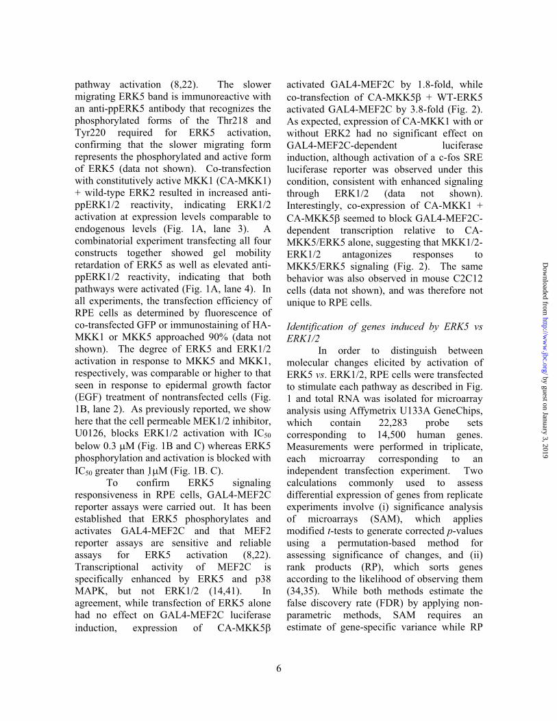
One mechanism by which cellsrespond transcriptionally to hypoxia involves posttranslational modifications that elevateHIF-1 protein by blocking its proteolytic turnover. In order to determine the effect of ERK5 and ERK1/2 signaling on HIF-1expression, Western blots were carried out on nuclear extracts prepared from serum-starvedRPE cells transfected with CA-MKK5 / +ERK5 or CA-MKK1+ERK2. Under non-hypoxic conditions, HIF-1 was almost undetectable in cells transfected with control vector (pCDNA3) (Fig. 5, lane 1), and induced above background in cells co-transfected with CA-MKK5 + ERK5 or CA-MKK5 + ERK5 (Fig. 5, lanes 2 and 3). HIF-1 protein levels were also induced in the presence of CA-MKK1 + ERK2 (Fig. 5, lane 4), as well as the combination of CA-MKK5, CA-MKK1, ERK2 and ERK5 (Fig. 5, lane 5). As previously reported (61), the hypoxia mimetic, CoCl2 induced HIF-1 to very high levels (Fig. 5, lane 6), and was inhibited by pretreatment of cells with the cell permeablecompound, U0126 (Fig. 5, lane 6), which inhibits both MKK1/2 and MKK5 at higher concentrations (61). Regulation of protein
8
by guest on January 3, 2019http://w
ww
.jbc.org/D
ownloaded from

expression appeared to be controlled by post-transcriptional mechanisms, because HIF-1mRNA levels were unaffected by activation of either ERK5 or ERK1/2 in the microarraydatasets (data not shown).
Regulation of ERK5 by Hypoxia To determine the significance of ERK5
signaling under hypoxic conditions, RPE cells were treated with or without hypoxia (3% O2),monitoring the phosphorylation of ERK5 and ERK1/2 by Western blotting. Figure 6 shows that while ERK5 and ERK1/2 were activatedby EGF in a U0126-dependent manner (lanes 1-3, upper panels); ERK5 and ERK1/2 were not significantly activated by hypoxia treatment for 1, 5 or 24 hours (lanes 4-9 vs. 10-15, upper panels). As a positive control to ensure hypoxic conditions were achieved, HIF-1 protein levels were monitored by Western blotting, and was found to be induced at 5 and 24 h after incubation at 3% O2 (Fig. 6, lanes 4-9 vs. 10-15). Thus, the regulation of hypoxia-responsive genes by ERK5 appears to be important under normoxic rather than hypoxic conditions in RPE cells. Likewise,the induction of HIF-1 is not affected byU0126 (Fig. 6, lanes 12-15), consistent with our findings that transcription from pGalHIF and EPO-HRE reporters was unaffected by U0126 following hypoxia (data not shown). Taken together, these data indicate that hypoxia induced transcription does not require ERK5 and ERK1/2 signaling in RPE cells.
We conclude that MKK5-ERK5 signaling induces HIF-1 protein expression and HRE transactivation to a similar extent asthat induced by the MKK1/2-ERK1/2 pathway. On the other hand, MKK5-ERK5, but not MKK1/2-ERK1/2, enhancestranscription from a GAL4-HIF-1 reporter system. Taken together, these studies indicate that the control of HRE gene expression via induction of HIF-1 represents a mechanismthat appears to be shared with ERK1/2, whereas alternative mechanisms involving
regulation of HIF-1 are controlled by ERK5 in a manner distinct from ERK1/2.
DISCUSSIONIn this study we used global gene
expression measurements to profile responses to activation of separate ERK pathways.Selective stimulation of ERK5 and ERK1/2respectively increased expression of 36 and 39 genes, respectively. None of the ERK5 responses were previously identified as regulated by this pathway, in contrast toERK1/2 responses, where 11 genes were previously identified as downstream targets. In general, the magnitude of changes responsive to ERK5 were lower compared to ERK1/2, and interestingly, no gene whose message was decreased in response to ERK5 or ERK1/2 passed our filters.
The profiling results revealed new insight into the specificity of signalingthrough each pathway. Of the responses observed, 47% of ERK5 targets and 51% of ERK1/2 targets were uniquely regulated by each pathway. Significantly, of the 17 genes controlled by ERK5 alone, 14 were found by literature searching to be regulated in response to hypoxia (Table 1). Most of these were previously validated by experimentsdemonstrating a link to transcription by HIF-1, a basic helix-loop-helix factor that promotestranscription under conditions of low oxygen tension (55,62-65). For example, of the set of 14 ERK5-responsive genes, nine were induced or blocked upon WT HIF-1 or dominant-negative HIF-1 overexpression, respectively, in cell lines (bHLHB3, PFKFB3, PFKFB4, ADM, CA9, PPP1R3C, SLC2A3, HIG2, andCCNG2, Table 1), and four were downregulated upon homologous recombination to delete expression of HIF-1 or (CCNG2, ADM, DDIT4, SLC2A3,Table 1). In addition, six genes had promotersequences which were verified as hypoxia- or HIF-inducible elements or which bound to HIF-1 (bHLHB3, ADM, PFKFB3, PFKFB4,
9
by guest on January 3, 2019http://w
ww
.jbc.org/D
ownloaded from

CA9, and DDIT4, Table 1), and of the remaining, one showed conserved E box elements containing potential HREs with HIF-1 consensus sequences (5’-RCGTG-3’;CALR).
The preferential representation of hypoxia-responsive genes in the set of ERK5 targets was striking, and strongly suggested a positive role for the ERK5 pathway in thecontrol of HIF-1-dependent HREtranscription. This was confirmedexperimentally by demonstrating elevated transcription from a promoter containing three repeats of the EPO-HRE, in response to ERK5 pathway activation. This finding provides evidence that ERK5 is able to promotetransactivation from an HRE-dependent promoter, and that this regulation is relevant under normoxic conditions.
Mechanisms for regulation were thentested by examining the potential control ofHIF-1 expression by this pathway. The role of the transcription factor HIF-1 in adaptive hypoxia-mediated signaling is well established (66,67), and known to be controlled by hypoxia at the level of protein stability. Undernormoxic conditions, human HIF-1 is hydroxylated at Pro402 and Pro564 which promotes ubiquitination by the E3-ligase, von-Hippel-Lindau (VHL), and proteasomemediated degradation (68-70). HIF-1 is alsohydroxylated at Asn803 within its C-terminaltransactivation domain, which prevents binding of the co-activator p300/CBP (71,72). Under conditions of low oxygen tension hydroxylation is repressed, HIF-1 is stabilized, and gene expression is activated by formation of dimeric HIF-1 / as well asderepression of p300/CBP binding (68-70).
We found that ERK5 signaling resulted in a significant increase of HIF-1expression compared to controls, concomitantwith the upregulation of HRE-dependent transcription. In addition, ERK5 activation led to stimulation of GAL4 transcriptionalactivity in the presence of a GAL4-HIF-1
fusion protein. Previous studies have shown levels of this fusion protein are stable and remain unchanged even under conditions of hypoxia (31). This was confirmed in our system by controls showing no effect of ERK5 on GAL4-HIF-1 protein (data not shown). The ability of ERK5 to activate GAL4-luciferase transcription indicates a mechanismindependent of expression, conceivably through GAL4-HIF-1 post-translational events although at this point we find no evidence for regulated phosphorylation. Taken together, our results indicate that ERK5 may control hypoxia responsive genes by at least two mechanisms, one involving elevated protein stability or synthesis, and another that is independent of protein expression control.
In contrast to ERK5, many of the 20 genes found to be selectively regulated by ERK1/2 had been previously characterized astargets for this pathway, including genes known to control cellular events related to proliferation and tissue remodeling. Of these, nine were previously reported to be associatedwith hypoxia signaling (Table 2). However, few of these genes were well characterized as hypoxia targets, the majority being identified in a single microarray study which profiled gene expression changes at low oxygen (55), and none have been explicitly linked to HIF-1regulation or shown to depend on HRE-controlled transcription. Thus, global responses to ERK1/2 differed substantively from ERK5 responses, in that a mechanistic link between ERK1/2 and HIF-1/HRE could not be established on the basis of target gene regulation in our system.
This result implies that ERK5 andERK1/2 might differ in their respectivemechanisms of gene regulation. ERK1/2 is known to promote expression of HIF-1 andhas also been shown to increase its transactivation either by direct phosphorylation within its C-terminaltransactivation domain (73-75), or by indirect phosphorylation of p300/CBP (76). However,
10
by guest on January 3, 2019http://w
ww
.jbc.org/D
ownloaded from

the fact that both ERK5 and ERK1/2 elevate HIF-1 protein levels to the same degree suggests that induced expression is irrelevant to the mechanism by which well characterizedhypoxia genes are selectively upregulated by ERK5. Conceivably, the relevant mechanismreflects the events involved in preferentialactivation of GAL4-HIF-1 by ERK5, and does not involve control of HIF-1 protein. Wemay expect these two MAPK pathways to have very different mechanisms for activation of hypoxia-responsive genes in cells. Wefurther note that many previous studies implicating ERK1/2 in the regulation of HIF-1 are based on repression of transcription using cell permeable inhibitors of MKK1/2, such as compounds U0126 and PD98059, which have been used at concentrationsranging from 10-100 M (61,75,77). As demonstrated by Cohen’s laboratory in HeLa cells (78) as well as our study (Fig. 1 and data not shown), ERK5/MEK5 signaling is sensitive to inhibition by U0126 at concentrations above 10 M. Thus, manyprevious studies cannot preclude the involvement of ERK5 rather than ERK1/2 in the regulation of HIF-1 , highlighting the importance of our studies supporting a role for ERK5-MKK5 in the regulation of HIF-1transactivation.
Significant overlap was seen in the regulation of gene expression by these pathways, observed in approximately half of the genes responsive to ERK5 and ERK1/2, respectively, and representing a wide range of biological function. This indicates that the ERK5 and ERK1/2 pathways converge on more targets than had been previously recognized (12,14,21). This was intriguing, given the different signaling mechanisms used by ERK5 vs ERK1/2, which often differ even in cases where the two pathways share a common target. For example, both ERK5 and ERK1/2 phosphorylate the transcription factor, CREB, but the localization of ERK5 activation specifies its preferential
involvement in CREB phosphorylation and neuronal survival (12). Additionally, in regulating CYP24 gene expression, ERK5 directly phosphorylates and activates the nuclear factor, Ets-1, whereas ERK1/2 phosphorylates and activates the retinoid Xreceptor to control CYP24 promotertransactivation (13). On the other hand, ERK5 and ERK1/2 sometimes target the samemolecule to elicit specific responses. For example, ERK5 and ERK1/2 cooperate to transform NIH3T3 cells, possibly through synergy between p90RSK and NF B (21). Interestingly 14 of the 19 genes upregulated by both ERK5 and ERK1/2 were previously reported to be responsive to hypoxia (Table 3). Literature searching showed two that wereregulated by HIF-1 Thus, like ERK1/2, hypoxia responsive genes controlled by both ERK5 and ERK1/2 appear to be in a separate class than those regulated by ERK5 alone, suggesting control by independentmechanisms. Six genes in the former class are known to be transcribed in response to hypoxia via NF B (Table 3), implicating thistranscription factor in the regulation ofERK1/2 responsive genes.
Finally, we speculate on the importance of ERK5 signaling in mediatingresponses to hypoxia. Two previous studies have shown that ERK5 is phosphorylated in response to low oxygen tension and also functions to promote angiogenesis (79,80). Interestingly, both reports showed that ERK5 activation inhibited hypoxia-induced VEGF expression, most likely by suppression of HIF-1 (79,80). These studies, which were carried out in bovine lung microvascular endothelial cells (BLMECs) and mouse embryonicfibroblast cells, showed behavior opposite to that of the RPE system used in our study, where ERK5 instead promotes HIF-1/HRE signaling. Such differences are most likely explained by cell function. Epithelial cells are more vulnerable to ischemia, responding by increasing cellular permeability and
11
by guest on January 3, 2019http://w
ww
.jbc.org/D
ownloaded from

breakdown of barrier function (81). RPE cells maintain the blood-retinal barrier by facilitating macromolecular transport betweenthe choroids and the outer neural retina, and abnormal behavior of RPE cells has been shown to contribute to age-related maculardegeneration and ischemic retinopathies (82,83). A reasonable hypothesis is that regulation of genes by ERK5 may be relevant to the adaptive response to hypoxia, which is instantaneous and involves Hif-1 . In
contrast, convergent regulation of genes by ERK1/2 with or without ERK5 maypreferentially regulate targets relevant to an inflammatory hypoxia response in epithelialcells which is known to involve NF B,CREB, and AP1 (84). Thus, the participation of different ERK pathways may provide a mechanism to respond selectively to hypoxia in RPE cells.
FIGURE LEGENDS
Figure 1. Expression of CA-MKK5 and CA-MKK1 and activation of ERK5 and ERK1/2.(A) RPE cells (2 x 10⁵) were transfected with the indicated combinations of rat CA-MKK5 ,CA-MKK1, ERK5, or ERK2 and harvested 24 h after transfection. Expression of CA-MKK1 and CA-MKK5 was monitored by Western blotting with antibodies against HA (1:2000).Activation of ERK5 and ERK1/2 was monitored by probing Western blotting with antibodies against ERK5 (1:1000) and ppERK1/2 (1:2000), respectively. (B) RPE cells (2 x 10⁵) were pretreated with the indicated amounts of U0126 for 1 h prior to stimulation with 20 ng/ml EGF for 10 min. Cells were harvested and ERK5 and ERK1/2 activity was monitored as in 1A except that the gel running time was increased to maximize the ppERK5 phosphorylation shift. Similarresults were seen in two additional experiments. (C) ERK5 and ERK1/2 inhibition by U0126 (Fig. 1B) was quantitfied from Western blots using Adobe Photoshop. ERK5 phosphorylation was quantified as the ratio of gel mobility retarded species to the faster mobility species. ERK5 and ERK1/2 stimulated by EGF in the absence of U0126 was set to 100% and the degree of basal ERK5 phosphorylation observed under control conditions (no stimulation, 5%) was subtractedfrom each measurement. Results presented are the mean +/- S.D. of two experiments.
Figure 2. MKK5 and MKK1 activation of MEF2C in RPE cells. RPE cells (2 x 10⁴) were transfected with the Gal4MEF2C-luciferase reporter along with the indicated effector or control plasmids, as in Fig. 1A, and pRLnull Renilla. Cells were harvested 24 h after transfection and firefly luciferase activity was normalized to Renilla. Results are the mean +/- standard deviation of 3 transfections done in duplicate.
Figure 3. MKK5 and MKK1 activation of the EPO HRE. RPE cells were transfected with the pGL(HRE)3-luciferse promoter, the indicated effector plasmids, along with pRLnull Renilla as in Fig. 2 except hMKK were used. Cells were harvested and assayed for luciferase and Renilla as in Fig. 2. Results are the mean +/- standard deviation of 3 transfections done in duplicate.
Figure 4. MKK5 and MKK1 activation of Hif-1 in RPE cells. RPE cells were transfectedwith the pGal4-Hif luciferase reporter along with the indicated effector or control plasmids, and pRLnull Renilla as in Fig. 3. Cells were harvested and assayed for luciferase and Renilla as in Fig. 2. Results are the mean +/- standard deviation of 3 transfections done in duplicate.
12
by guest on January 3, 2019http://w
ww
.jbc.org/D
ownloaded from

13
Figure 5. Western blot analysis of Hif-1 . RPE cells (3 x 10⁶) were transfected using FuGene with pCDNA3 (lane 1), human CA-MKK5 /ERK5 (lane 2), human CA-MKK5 /ERK5 (lane 3), CA-MKK1/ERK2 (lane 4), or CA-hMKK5 /CA-MKK1/ERK5/ERK2 (lane 5). At the time of transfection, cells were also pre-treated with 10 M U0126 (lane 6) or vehicle (lane 7) for 1 h prior to treatment with 100 M CoCl2. Cells were serum starved and harvested at 24 h for nuclear extract preparation, as described under “Experimental Procedures”. Expression and activation of Hif-1 , was monitored using antibodies recognizing Hif-1 (1:1000).
Figure 6. ERK5 is not activated under conditions of hypoxia. RPE cells (8 x 10⁵) were incubated with 10 M U0126 for 1 h prior to treatment with or without hypoxia (3% O2) for the indicated times. Expression and activation of ERK5, ERK1/2 and HIF-1 was monitored by Western blotting as described in Figs. 1 and 4. Where indicated, RPE cells were treated with 20 ng/ml EGF +/- 10 M U0126, as described in Fig. 1, as a positive control for ERK5 and ERK1/2 activation. Statistical analysis (Student’s t test) of Western blots quantified as in Fig. 1C showed that ERK5 phosphorylation did not significantly differ between normoxic vs. hypoxic conditions.
ACKNOWLEDGEMENTSWe thank the University of Colorado Health Sciences Gene Expression Core for assistance with processing the Affymetrix Genechips. We thank the Department of Cardiology at the University of Colorado Health Sciences Center for the use of their hypoxia chamber and Sandi Walchak for assistance with the chamber. We also thank Drs. Gretchen Argast and Elisabeth Roberts for critical reading of the manuscript. R.E.S. is a Howard Hughes Medical Institute Fellow of the Life Sciences Research Foundation. by guest on January 3, 2019
http://ww
w.jbc.org/
Dow
nloaded from

14
REFERENCES
1. Chen, Z., Gibson, T., Robinson, F., Silvestro, L., Pearson, G., Xu, B., Wright, A., Vanderbilt, C., and Cobb, M. (2001) Chem. Rev. 101, 2449-2476
2. Whitmarsh, A., and Davis, R. (1996) J. Mol. Med. 74, 589-607 3. Lee, J., Ulevitch, R., and Han, J. (1995) Biochem. Biophys. Res. Comm. 213, 715-724 4. Zhou, G., Bao, Z., and Dixon, J. (1995) J. Biol. Chem. 270, 12665-12669 5. Bogoyevitch, M., and Court, N. (2004) Cell Signal. 16, 1345-1354 6. Abe, J., Takahashi, M., Ishida, M., Lee, J., and Berk, B. (1997) J. Biol. Chem. 272,
20389-203947. Kato, Y., Tapping, R., Huang, C., Watson, M., Ulevitch, R., and Lee, J. (1998) Nature
395, 713-716 8. Sun, W., Wei, X., Kesavan, K., Garrington, T., Fan, R., Mei, J., Anderson, S., Gelfand,
E., and Johnson, G. (2003) Mol Cell Biol. 23, 2298-2308 9. Chao, T., Hayashi, M., Tapping, R., Kato, Y., and Lee, J.-D. (1999) J. Biol. Chem. 274,
36305-3630810. English, J., Pearson, G., Hockenberry, T., Shivakumar, L., White, M., and Cobb, M.
(1999) J. Biol. Chem. 274, 31588-31592 11. Kamakura, S., Moriguchi, T., and Nishida, E. (1999) J. Biol. Chem. 274, 26563-26571 12. Watson, F., Heerssen, H., Bhattacharyya, A., Klesse, L., Lin, M., and Segal, R. (2001)
Nat. Neurosci. 4, 981-988 13. Dwivedi, P., Hii, C., Ferrante, A., Tan, J., Der, C., Omdahl, J., Morris, H., and May, B.
(2002) J. Biol. Chem. 277, 29643-29653 14. Kato, Y., Kravchenko, V., Tapping, R., Han, J., Ulevitch, R., and Lee, J.-D. (1997)
EMBO J. 16, 7054-7066 15. Yang, C., Ornatsky, O., McDermott, J., Cruz, T., and Prody, C. (1998) Nucleic Acids Res.
26, 4771-4777 16. Pi, X., Yan, C., and Berk, B. (2004) Circ. Res. 94, 362-369 17. Dinev, D., Jordan, B., Neufeld, B., Lee, J., Lindemann, D., Rapp, U., and Ludwig, S.
(2001) EMBO Rep. 2, 829-834 18. Kasler, H., Victoria, J., Duramad, O., and Winoto, A. (2000) Mol Cell Biol. 20, 8382-
838919. Zheng, Q., Yin, G., Yan, C., Cavet, M., and Berk, B. (2004) J. Biol. Chem. 279, 8787-
879120. Hii, C., Anson, D., Costabile, M., Mukaro, V., Dunning, K., and Ferrante, A. (2004) J.
Biol. Chem. 279, 49825-49834 21. Pearson, G., English, J., White, M., and Cobb, M. (2001) J. Biol. Chem. 276, 7927-7931 22. Barros, J., and Marshall, C. (2005) J. Cell Sci. 118, 1663-1671 23. Pages, G., Guerin, S., Grall, D., Bonino, F., Smith, A., Anjuere, F., Auberger, P., and
Pouyssegur, J. (1999) Science 286, 1374-1377 24. Hatano, M., Mori, Y., Oh-hora, M., Kosugi, A., Fujikawa, T., Nakai, N., Niwa, H.,
Miyazaki, J., Hamaoka, T., and Ogata, M. (2003) Genes Cells 8, 847-856 25. Regan, C., Li, W., Boucher, D., Spatz, S., Su, M., and Kuida, K. (2002) Proc. Natl. Acad.
Sci. USA 99, 9248-9253 26. Yang, J., Boerm, M., McCarty, M., Bucana, C., Fidler, I., Zhuang, Y., and Su, B. (2000)
Nat. Genet. 24, 309-313
by guest on January 3, 2019http://w
ww
.jbc.org/D
ownloaded from

15
27. Bi, W., Drake, C., and Schwarz, J. (1999) Dev. Biol. 211, 255-267 28. Hayashi, M., Kim, S., Imanaka-Yoshida, K., Yoshida, T., Abel, E., Eliceiri, B., Yang, Y.,
Ulevitch, R., and Lee, J. (2004) J. Clin. Invest. 113, 1138-1148 29. Mansour, S., Candia, J., Gloor, K., and Ahn, N. (1996) Cell Growth & Diff. 7, 243-250 30. Mansour, S., Matten, W., Hermann, A., Candia, J., Rong, S., Fukasawa, K., Vande, G.,
Woude, V., and Ahn, N. (1994) Science 265, 966-970 31. Jiang, B., Zheng, J., Leung, S., Roe, R., and Semenza, G. (1997) J. Biol. Chem. 272,
19253-1926032. Wanner, R., Spielmann, P., Stroka, D., Camenisch, G., Camenisch, I., Scheid, A., Houck,
D., Bauer, C., Gassmann, M., and Wenger, R. (2000) Blood 96, 1558-1565 33. Bolstad, B., RA, I., Astrand, M., and Speed, T. (2003) Bioinformatics 19, 185-193 34. Tusher, V., Tibshirani, R., and Chu, G. (2001) Proc. Natl. Acad. Sci. USA 98, 5116-5121 35. Breitling, R., Armengaud, P., Amtmann, A., and Herzyk, P. (2004) FEBS Lett 573, 83-92 36. Hecquet, C., Lefevre, G., Valtink, M., Engelmann, K., and Mascarelli, F. (2002) Invest.
Opthalmol. Vis. Sci. 43, 3091-3098 37. Guillonneau, X., Bryckaert, M., Launay-Longo, C., Courtois, Y., and Mascarelli, F.
(1998) J. Biol. Chem. 273, 22367-22373 38. Hecquet, C., Lefevre, G., Valtink, M., Engelmann, K., and Mascarelli, F. (2002)
Oncogene 21, 6101-6112 39. Defoe, D., and Grindstaff, R. (2004) Exp. Eye Res. 79, 51-59 40. Weigel, A., Handa, J., and Hjelmeland, L. (2002) Free Radic Biol Med 33, 1419-1432 41. Zhao, M., New, L., Kravchenko, V., Kato, Y., Gram, H., diPadova, F., Olson, E.,
Ulevitch, R., and Han, J. (1999) Mol. Cell. Biol. 19, 21-30 42. Hipskind, R., Baccarini, M., and Nordheim, A. (1994) Mol Cell Biol. 14, 6219-6231 43. Su, Y., Denegre, J., Wigglesworth, K., Pendola, F., O'Brien, M., and Eppig, J. (2003)
Dev. Biol. 263, 126-138 44. Aklilu, F., Gladu, J., Goltzman, D., and Rabbani, S. (2000) Cancer Res. 6045. Nishihara, H., Hwang, M., Kizaka-Kondoh, S., Eckmann, L., and Insel, P. (2004) J. Biol.
Chem. 279, 26176-16183 46. Ni, C., Wang, D., Lien, S., Cheng, J., Chao, Y., and Hsieh, H. (2003) J. Cell. Physiol.
195, 428-434 47. Takaya, K., Koya, D., Isono, M., Sugimoto, T., Sugaya, T., Kashiwaga, A., and Haneda,
M. (2003) Am. J. Physiol. Renal Physiol. 284, F1037-1045 48. Kawaguchi, M., Kokubu, F., Matsukura, S., Ieki, K., Odaka, M., Watanabe, S., Suzuki,
S., Adachi, M., and Huang, S. (2003) J. Pharmacol. Exp. Ther. 307, 1213-1220 49. Zampetaki, A., Mitsialis, S., Pfeilschifter, J., and Kourembanas, S. (2004) FASEB J50. Luo, G., Hershko, D., Robb, B., Wray, C., and Hasselgren, P. (2003) Am. J. Physiol.
Regul. Integr. Comp. Physiol. 284, R1249-1254 51. Li, J., Kartha, S., Iasvovskaia, S., Tan, A., Bhat, R., Manaligod, J., Page, K., Brasier, A.,
and Hershenson, M. (2002) Am. J. Physiol. Lung Cell Mol. Physiol. 283, L690-699 52. Yan, Z., Stapleton, P., Freeman, T., Fuortes, M., and Daly, J. (2004) Cell. Immunol. 232,
116-12653. Wang, Z., Chen, X., Yang, G., and Zhou, L. (2004) Chin. Med. Sci. J. 19, 270-275 54. Muller, J., Kruass, B., Kaltschmidt, C., Baeuerle, P., and Rupec, R. (1997) J. Biol. Chem.
272, 23435-23439 55. Scheurer, S., Raybak, J., Rosli, C., Neri, D., and Elia, G. (2004) Proteomics 4, 1737-1760
by guest on January 3, 2019http://w
ww
.jbc.org/D
ownloaded from

16
56. Lal, A., Peters, H., St. Croix, B., Haroon, Z., Dewhirst, M., Strausberg, R., Kaanders, J., J. van der Kogel, A., and Riggins, G. (2001) J. Natl. Cancer Inst. 93, 1337-1343
57. Berra, E., Pages, G., and Pouyssegur, J. (2000) Cancer Metasis Rev. 19, 139-145 58. Chen, C., Pore, N., Behrooz, A., Ismail-Beigi, F., and Maity, A. (2001) J. Biol. Chem.
276, 9519-9525 59. Kopacek, J., Barathova, M., Dequiedt, F., Sepelakova, J., Kettmann, R., Pastorek, J., and
Pastorekova, S. (2005) Biochim Biophys Acta 1729, 41-49 60. Prusiner, S., Bolton, D., Groth, D., Bowman, K., Cochran, S., and McKinley, M. (1982)
Biochemistry 21, 6942-6950 61. Treins, C., Giorgeti-Peraldi, S., Murdaca, J., and Van Obberghen, E. (2001) J. Biol.
Chem. 276, 43836-43841 62. Wykoff, C., Beasley, N., Watson, P., Turner, K., Pastorek, J., Sibtain, A., Wilson, G.,
Turley, H., Talks, K., Maxwell, P., Pugh, C., Ratcliffe, P., and Harris, A. (2000) CancerRes 60, 7075-7083
63. Sonna, L., Cullivan, M., Sheldon, H., Pratt, R., and Lilly, C. (2003) Physiol. Genomics12, 195-207
64. Minchenko, O., Opentanova, I., Michenko, D., Ogur, T., and Esumi, H. (2004) FEBS Lett576, 14-20
65. Minchenko, A., Leshchinsky, I., Opentanova, I., Sang, N., Srinivas, V., Armstead, V., and Caro, J. (2002) J. Biol. Chem. 277, 6183-6187
66. Brahimi-Horn, C., Mazure, N., and Pouyssegur, J. (2005) Cell Signal. 17, 1-9 67. Semenza, G. (2004) Physiology 19, 176-182 68. Jaakkola, P., Mole, D., Tian, Y., Wilson, M., Gielbert, J., Gaskell, S., Kriegsheim, A.,
Hebestreit, H., Mukherji, M., Schofield, C., Maxwell, P., Pugh, C., and Ratcliffe, P. (2001) Science 292, 468-472
69. Ivan, M., Kondo, K., Yang, H., Kim, W., Valiando, J., Ohh, M., Salic, A., Asara, J., Lane, W., and Kaelin, W. J. (2001) Science 292, 464-468
70. Masson, N., Willam, C., Maxwell, P., Pugh, C., and Ratcliffe, P. (2001) EMBO J. 20,5197-5206
71. Lando, D., Peet, D., Whelan, D., Gorman, J., and Whitelaw, M. (2002) Science 295, 858-861
72. Mahon, P., Hirota, K., and Semenza, G. (2001) Genes Dev. 15, 2675-2686 73. Sodhi, A., Montaner, S., Miyazaki, H., and Gutkind, J. (2001) Biochem. Biophys. Res.
Comm. 287, 292-300 74. Richard, D., Berra, E., Gothie, E., Roux, D., and Pouyssegur, J. (1999) J. Biol. Chem.
274, 32631-32637 75. Lee, E., Yim, S., Lee, S.-K., and Park, H. (2002) Mol. Cells 14, 9-15 76. Sang, N., Stiehl, D., Bohensky, J., Leshchinsky, I., Srinivas, V., and Caro, J. (2003) J.
Biol. Chem. 278, 14013-14019 77. Hur, E., Chang, K., Lee, E., Lee, S., and Park, H. (2001) Mol. Pharmacol. 59, 1216-1224 78. Mody, N., Leitch, J., Armstrong, C., Dixon, J., and Cohen, P. (2001) FEBS Lett 502, 21-
2479. Sohn, S., Sarvis, B., Cado, D., and Winoto, A. (2002) J. Biol. Chem. 277, 43344-43351 80. Pi, X., Garin, G., Xie, L., Zheng, Q., Wei, H., Abe, J., Yan, C., and Berk, B. (2005) Circ.
Res. 96, 1145-1151
by guest on January 3, 2019http://w
ww
.jbc.org/D
ownloaded from

17
81. Karhausen, J., Furuta, G., Tomaszewski, J., Johnson, R., Colgan, S., and Haase, V. (2004) J. Clin. Invest. 114, 1098-1106
82. Cunha-Vaz, J. (2004) Exp. Eye Res. 78, 715-721 83. Wangsa-Wirawan, N., and Linsenmeier, R. (2003) Arch. Ophthalmol. 121, 547-557 84. Zampetaki, A., Mitsialis, S., Pfeilschifter, J., and Kourembanas, S. (2004) FASEB J 18,
1090-109285. Miyazaki, K., Kawamoto, T., Tanimoto, K., Nishiyama, M., Honda, H., and Kato, Y.
(2002) J. Biol. Chem. 277, 47014-47021 86. Wykoff, C., Pugh, C., Maxwell, P., Harris, A., and Ratcliffe, P. (2000) Oncogene 19,
6297-230587. Blais, J., Filipenko, V., Bi, M., Harding, H., Ron, D., Coumenis, C., Wouters, B., and
Bell, J. (2004) Mol Cell Biol. 24, 7469-7482 88. Wang, V., Davis, D., Haque, M., Huang, L., and Yarchoan, R. (2005) Cancer Res 65,
3299-330689. Obach, M., Navarro-Sabate, A., Caro, J., Kong, X., Duran, J., Gomez, M., Perales, J.,
Ventura, F., Rosa, J., and Bartrons, R. (2004) J. Biol. Chem. 279, 53562-53570 90. Leonard, M., Cottell, D., Godson, C., Brady, H., and Taylor, C. (2003) J. Biol. Chem.
278, 40296-40304 91. Lee, M., Bikram, M., Oh, S., Bull, D., and Kim, S. (2004) Pharm. Res. 21, 736-741 92. Maxwell, P., Dachs, G., Gleadle, J., Nicholls, L., Harris, A., Stratford, L., Hankinson, O.,
Pugh, C., and Ratcliffe, P. (1997) Proc. Natl. Acad. Sci. USA 94, 8104-8109 93. O'Rourke, J., Pugh, C., Bartlett, S., and Ratcliffe, P. (1996) Eur. J. Biochem. 241, 403-
41094. Bobrovnikova-Marjon, E., Marjon, P., Barbash, O., Vander Jagt, D., and Abcouwer, S.
(2004) Cancer Res 64, 4858-4869 95. Combe, C., Burton, C., Dufourco, P., Weston, S., Horsburgh, T., Walls, J., and Harris, K.
(1997) Kidney Int. 51, 1703-1709 96. Muraoka, K., Shimizu, K., Sun, X., Zhang, Y., Tani, T., Hashimoto, T., Yagi, M.,
Miyazaki, I., and Yamamoto, K. (1997) Transplantation 63, 466-470 97. Matsui, H., Ihara, Y., Fujio, Y., Kunisada, K., Akira, S., Kishimoto, T., and Yamauchi-
Takihara, K. (1999) Cardiovasc Res 42, 104-112 98. Yan, S., Zou, Y., Mendelsohn, M., Gao, Y., Naka, Y., Du Yan, S., Pinsky, D., and Stern,
D. (1997) J. Biol. Chem. 272, 4287-4294 99. Shi, Q., Le, X., Abbruzzese, J., Wang, B., Mujaida, N., Matsushima, K., Huang, S.,
Xiong, Q., and Xie, K. (1999) J. Interferon Cytokine Res 19, 1363-1371 100. Cangul, H. (2004) BMC Genet. 5, 27 101. Salnikow, K., Kluz, T., Costa, M., Piquemal, D., Demidenko, Z., Xie, K., and
Blagosklonny, M. (2002) Mol. Cell. Biol. 22, 1734-1741 102. Plummer, S., Holloway, K., Manson, M., Munks, R., Kaptein, A., Farrow, S., and
Howells, L. (1999) Oncogene 18, 6013-6020 103. Lukiw, W., Ottlecz, A., Lambrou, G., Grueninger, M., Finley, J., Thompson, H., and
Bazan, N. (2003) Invest. Opthalmol. Vis. Sci. 44, 4163-4170 104. Ji, Y., Xu, Q., and Schmedtje, J. J. (1998) Circ. Res. 83, 295-304
by guest on January 3, 2019http://w
ww
.jbc.org/D
ownloaded from

Table 1. Genes upregulated by CA-MKK5 but not CA-MKK1 Gene Title Gene Symbol Average Fold
ChangeHypoxia
TF involved Genes previously reported to be upregulated by hypoxia
MKK1MKK5Adrenomedullin ADM 2.3 HIF1-1 (55)1.2basic helix-loop-helix domain containing, class B, 3 (DEC2) BHLHB3 2 1.1 HIF1-1 (85)chromosome 10 open reading frame 10 (DEPP) C10orf10 2.2 1.2carbonic anhydrase IX CA9 2.2 1.1 HIF-1 (62)Calreticulin CALR 1.87.4Cyclin G2 CCNG2 2.1 1.2 HIF1- (86,87)hypothetical protein DKFZp434K1210 DKFZp434K1210 2.3 1.2Hypoxia-inducible protein 2 HIG2 5 1.1 HIF1- (88)MAX interacting protein 1 MXI1 2.7 16-phosphofructo-2-kinase/fructose-2,6-biphosphatase 3 PFKFB3 3.8 1.5 HIF-1 (89) 6-phosphofructo-2-kinase/fructose-2,6-biphosphatase 4 PFKFB4 2 1.3 HIF-1 (63,64)Protein phosphatase 1, regulatory (inhibitor) subunit 3C PPP1R3C 2.4 1DNA-damage inducible transcript 4/REDD/RTP801 DDIT4 5.2 1.9 HIF-1 (90), Sp1 (91) solute carrier family 2, member 3 (GLUT3) SLC2A3 2 1.3 HIF-1 (90,92,93)
Other genes upregulated by MKK5 but not linked to hypoxia
chemokine (C-X-C motif) ligand 6 CXCL6 2.1 1.5Insulin induced gene 2 INSIG2 2.1 1.1Tumor necrosis factor, alpha-induced protein 2 TNFAIP2 2.4 1.6
18
by guest on January 3, 2019 http://www.jbc.org/ Downloaded from

19
Table 2. Genes upregulated by CA-MKK1 but not CA-MKK5 Gene Title Gene Symbol Average Fold
ChangeTF
involvedMKK1 MKK5
Genes previously reported to be upregulated by hypoxiadual specificity phosphatase 6 DUSP6 3 0.8endothelial cell-specific molecule 1
ESM1 5.5 1.2v-fos FBJ murine osteosarcoma viral oncogene homolog FOS 2.7 1.1 SRF/Elk-1 (54)hyaluronan synthase 2 HAS2 2 0.8parathyroid hormone-like hormone PTHLH 5.1 1serine (or cysteine) proteinase inhibitor, clade B (ovalbumin), member 2
SERPINB2 14 1.2
stanniocalcin 1 STC1 2.2 1.3tissue factor pathway inhibitor 2 TFPI2 2.5 1.5Thrombomodulin THBD 2.6 1
Other genes significantly upregulated by MKK1
AFFX-r2-Hs18SrRNA-M_x_at --- 2 1.3amphiregulin (schwannoma-derived growth factor) AREG 4.2 0.92,3-bisphosphoglycerate mutase BPGM 1.6 1.1chromosome 20 open reading frame 42 C20orf42 2.3 1dual specificity phosphatase 5 DUSP5 2 0.9early growth response 1 EGR1 2.2 0.9early growth response 3
EGR3 2.2 1.1
interleukin 11 IL11 2.4 1.1matrix metalloproteinase 1 (interstitial collagenase) MMP1 16.3 1.2matrix metalloproteinase 10 (stromelysin 2) MMP10 4 1pleckstrin homology-like domain, family A, member 1 PHLDA1 2 1
by guest on January 3, 2019 http://www.jbc.org/ Downloaded from

20
Table 3. Genes commonly upregulated by CA-MKK5 and CA-MKK1 Gene Title Gene
SymbolFold Change TF involved
Genes previously reported to be regulated by hypoxia
MKK5 MKK1baculoviral IAP repeat-containing 3 BIRC3 3.3 3bone morphogenetic protein 2 BMP2 1.6 5.4,
3.6chemokine (C-C motif) ligand 2 CCL2 2.3 2colony stimulating factor 2 (granulocyte-macrophage) CSF2 4.3 3.8chemokine (C-X-C motif) ligand 1 CXCL1 21.6 18.9 NF B (94) chemokine (C-X-C motif) ligand 2 CXCL2 17.2 18.6 NF B (84) chemokine (C-X-C motif) ligand 3 CXCL3 8.9 9.2Ephrin-A1 EFNA1 5.7 2.8intercellular adhesion molecule 1 (CD54) ICAM1 3.3, 2.5 2.1 NF B (95) interleukin 6 (interferon, beta 2) IL6 3.7 4.1 NF B, NF-IL6 (96-98) interleukin 8 IL8 12.3,
31.211.4,22.1
AP1, NF B (94,99)
N-myc downstream regulated gene 1 NDRG1 4.4 1.6 HIF1 (100), AP1(101) nuclear factor of kappa light polypeptide gene enhancer in B-cells inhibitor, alpha
NFKBIA 2.3 2.1
prostaglandin-endoperoxide synthase 2 PTGS2 2.7 10.4 NF B (102), HIF1 (103), HMGI(Y) (104)
pentaxin-related gene, rapidly induced by IL-1 beta PTX3 2.2 2.7superoxide dismutase 2, mitochondrial SOD2 2.6 1.8
Other genes significantly upregulated
hypothetical protein FLJ23231 FLJ23231 2.7 2interleukin 1, beta IL1B 3.6 4.9tumor necrosis factor, alpha-induced protein 3 TNFAIP3 5.1 4.1
by guest on January 3, 2019 http://www.jbc.org/ Downloaded from

21
Table S1. PCR Primers used in this study Gene Symbol PCR Primers Product size
MMP1 5’-GGAGATCATCGGGACAACTC-3’5’-AGTTCATGAGCTGCAACACG-3’
164 bp
SERPINB2
5’-TCAAACCAAAGGCAAAATCC-3’5’-ACAGCATTCACCAGGACCAT-3’
81 bp
EFNA1 5’-ACATCTCCAAACCCATCCAC-3’5’-ATGTAGAACCCGCACCTCTG-3’
149 bp
ICAM 5’-CACCTATGGCAACGACTCCT-3’5’-CCGGAAAGCTGTAGATGGTC-3’
146 bp
DDIT4* 5’-GTGGAGGTGGTTTGTGTATC-3’5’-CACCCCTTGCTACTCTTAC-3’
150 bp
ACTB 5’-CACCCAGCACAATGAAGATC-3’5’-CCTGCTTGCTGATCCACATC-3’
120 bp
TBP 5’-TATAATCCCAAGCGGTTTGC-3’5’-CTCCTGTGCACACCATTTTC-3’
143 bp
*primers were designed against the Affymetrix probe sequence using GenAct primer design program (http://enhancer.colorado.edu:6400/~hudakg/home.html).
Table S2. Confirmation of changes induced by ERK5 and ERK1/2Gene Symbol Affymetrix Fold Change RTQ RT-PCR
MKK5 MKK1 MKK5 MKK1MMP11
1.2 16.3 2.3 195SERPINB22 1.2 14 3 24EFNA11 5.7 2.8 1.38 2.93ICAM1 3.3, 2.5 2.1 5.4 1.2DDIT4 5.2 1.9 1.62 2.16
1normalized to -actin (ACTB) 2normalized to TATA binding protein (TBP)
by guest on January 3, 2019 http://www.jbc.org/ Downloaded from

pCDNA3
CA-MKK5β
/ERK5
CA-MKK1/E
RK2
CA-MKK5/M
KK1/ERK2/5
Schweppe et al Fig. 1
1 2 43
CA-MKK1HACA-MKK5βHA
ppERK5ERK5
ppERK1/2
ERK2
A
B
ppERK5ERK5
ppERK1/2
Con
trol
0 0.3 1 3 10 μM U0126
EGF
1 2 543 6
C
0.3 3
%In
hibi
tion
0 1 100
20
40
60
80
100 ERK5ERK1/2
U0126 [μM]
by guest on January 3, 2019http://w
ww
.jbc.org/D
ownloaded from

Schweppe et al Fig. 2
ERK2
CA-MKK5β CA-MKK1 CA-MKK5β +CA-MKK1
+ + ++ + +
−− −
−− −
− −−
−−−
ERK5
Fold
Act
ivat
ion
0
1
2
3
4
5
6
by guest on January 3, 2019http://w
ww
.jbc.org/D
ownloaded from

ERK2ERK5
Fold
Act
ivat
ion
Schweppe et al Fig. 3
0
1
2
3
4
5
6
7
MKK5α MKK5β +MKK1
+ + +− −− − − −+ + +− −− − −−
MKK1
−+
MKK5β
−−
MKK5α +MKK1
−−
++
by guest on January 3, 2019http://w
ww
.jbc.org/D
ownloaded from

Schweppe et al Fig. 4
ERK2
MKK5α MKK5β +MKK1
+ + +− −− − − −+ + +− −− − −−ERK5
Fold
Act
ivat
ion
MKK1
−+
00.5
11.5
22.5
33.5
44.5
MKK5β
−−
MKK5α +MKK1
−−
++
by guest on January 3, 2019http://w
ww
.jbc.org/D
ownloaded from
Schweppe et al Fig. 5
CA
-MK
K5β
/ER
K5
CA
-MK
K1/
ER
K2
CA
-MK
K5β
/MK
K1/
ER
K2/
5
pCD
NA
3
CoC
l 2C
oCl 2
+U
0126
1 2 543 76
HIF-1α115 kDa
A
CA
-MK
K5α
/ER
K5
by guest on January 3, 2019http://w
ww
.jbc.org/D
ownloaded from

1 2 43
ppERK5ERK5
Con
trol
- + U0126EGF
- + - + - + - + - + - +
Normoxic Hypoxic
1 h 5 h 24 h 1 h 5 h 24 h
ppERK1/2
HIF-1α
ERK2
5 6 87 109 11 12 1413 15
Schweppe et al Fig. 6
by guest on January 3, 2019http://w
ww
.jbc.org/D
ownloaded from

Rebecca E. Schweppe, Tom Hiu Cheung and Natalie G. AhnHIF-1 in ERK5-meidated responses
Global gene expression analysis of ERK5 and ERK1/2 signaling reveals a role for
published online May 30, 2006J. Biol. Chem.
10.1074/jbc.M604208200Access the most updated version of this article at doi:
Alerts:
When a correction for this article is posted•
When this article is cited•
to choose from all of JBC's e-mail alertsClick here
Supplemental material:
http://www.jbc.org/content/suppl/2006/05/31/M604208200.DC1
by guest on January 3, 2019http://w
ww
.jbc.org/D
ownloaded from