protein kinase a regulates hyphal growth by relieving the
TRANSCRIPT

Purdue UniversityPurdue e-Pubs
Open Access Dissertations Theses and Dissertations
January 2016
Protein Kinase A Regulates Hyphal Growth byRelieving the Interaction of MoSfl1 with theCyc8-Tup1 Co-repressor in Magnaporthe oryzaeYang LiPurdue University
Follow this and additional works at: https://docs.lib.purdue.edu/open_access_dissertations
This document has been made available through Purdue e-Pubs, a service of the Purdue University Libraries. Please contact [email protected] foradditional information.
Recommended CitationLi, Yang, "Protein Kinase A Regulates Hyphal Growth by Relieving the Interaction of MoSfl1 with the Cyc8-Tup1 Co-repressor inMagnaporthe oryzae" (2016). Open Access Dissertations. 1256.https://docs.lib.purdue.edu/open_access_dissertations/1256

Graduate School Form 30 Updated 12/26/2015
PURDUE UNIVERSITY GRADUATE SCHOOL
Thesis/Dissertation Acceptance
This is to certify that the thesis/dissertation prepared
By Yang Li
Entitled Protein Kinase A Regulates Hyphal Growth by Relieving the Interaction of MoSfl1 with the Cyc8-Tup1 Co-repressor in Magnaporthe oryzae
For the degree of Doctor of Philosophy
Is approved by the final examining committee:
Dr. Jin-Rong Xu
Chair
Dr. Charles P. Woloshuk
Dr. Stephen B. Goodwin
Dr. Steven R. Scofield
To the best of my knowledge and as understood by the student in the Thesis/Dissertation Agreement, Publication Delay, and Certification Disclaimer (Graduate School Form 32), this thesis/dissertation adheres to the provisions of Purdue University’s “Policy of Integrity in Research” and the use of copyright material.
Approved by Major Professor(s): Dr. Jin-Rong Xu
Approved by:
Dr. Christopher J. Staiger 7/20/2016
Head of the Departmental Graduate Program Date

i
i
PROTEIN KINASE A REGULATES HYPHAL GROWTH BY RELIEVING THE
INTERACTION OF MOSFL1 WITH THE CYC8-TUP1 CO-REPRESSOR IN
MAGNAPORTHE ORYZAE
A Dissertation
Submitted to the Faculty
of
Purdue University
by
Yang Li
In Partial Fulfillment of the
Requirements for the Degree
of
Doctor of Philosophy
August 2016
Purdue University
West Lafayette, Indiana

ii
ii
ACKNOWLEDGEMENTS
I am grateful to my advisor Dr. Jin-Rong Xu, for his continuous support and
patience during my Ph.D. study. Jin-Rong was always there to give me great advice and
guidance directing my research. I would also like to thank members of my advisory
committee, Drs. Charles P. Woloshuk, Stephen B Goodwin, and Steven R. Scofield at
Purdue University for their constructive suggestions.
I also thank the past and present members of the Xu Lab, including Xue Zhang and
Drs. Xiaoying Zhou, Guotian Li, Guanghui Wang, Linlu Qi, and Cong Jiang, who are
always supportive and willing to help.
Last, but not least, I thank my husband Xiaofeng Zhou and my parents for their
generous love and support.

iii
TABLE OF CONTENTS
Page
LIST OF TABLES ............................................................................................................. vi
LIST OF FIGURES .......................................................................................................... vii
ABSTRACT ....................................................................................................................... ix
CHAPTER 1. INTRODUCTION .................................................................................... 1
1.1 The Rice Blast Fungus Magnaporthe oryzae ............................................................. 1
1.1.1 Rice blast disease ............................................................................................. 1
1.1.2 Biology of Magnaporthe oryzae ...................................................................... 2
1.1.3 Disease cycle of the rice blast fungus .............................................................. 2
1.2 The cAMP-PKA Pathway .......................................................................................... 4
1.2.1 Protein kinase A ............................................................................................... 4
1.2.2 Upstream components of the cAMP-PKA signaling in M. oryzae .................. 5
1.2.3 Downstream targets of PKA in M. oryzae ....................................................... 7
1.3 The Cyc8-Tup1 Co-repressor .................................................................................... 8
1.3.1 Genes repressed by the Cyc8-Tup1 complex in yeast ..................................... 8
1.3.2 Structures of Tup1 and Cyc8 proteins ........................................................... 10
1.3.3 Mechanisms of the Cyc8-Tup1 co-repressor ................................................. 11
1.3.4 Study of the Cyc8-Tup1 co-repressor in filamentous fungi .......................... 12
1.4 Research Justification and Objectives ..................................................................... 13
1.5 List of References .................................................................................................... 14
CHAPTER 2. CAMP-DEPENDENT PROTEIN KINASE A REGULATES GROWTH
AND PATHOGENIC DIFFERENTIATION IN THE RICE BLAST FUNGUS
MAGNAPORTHE ORYZAE .............................................................................................. 20
2.1 Abstract .................................................................................................................... 20

iv
Page 2.2 Introduction .............................................................................................................. 21
2.3 Materials and Methods ............................................................................................. 24
2.3.1 Stains and growth media ................................................................................ 24
2.3.2 Generation of the cpk2 mutant and the cpkA cpk2 double mutant ................ 25
2.3.3 Appressorium formation, penetration, and plant infection assays ................. 25
2.3.4 qRT-PCR analyses ......................................................................................... 26
2.3.5 Generation of S or 3×FLAG fusion constructs .............................................. 26
2.3.6 Protein isolation and western blot analysis .................................................... 27
2.3.7 Co-immunoprecipitation and affinity purification ......................................... 27
2.4 Results ...................................................................................................................... 29
2.4.1 CPKA and CPK2 have functional redundancy in morphogenesis and
pathogenicity .............................................................................................................. 29
2.4.2 The cpkA cpk2 double mutant has severe growth defects and it is unstable . 34
2.4.3 Conidiation is significantly reduced in the cpkA cpk2 double mutant ........... 36
2.4.4 PKA catalytic subunits are essential for appressoria formation .................... 36
2.4.5 The cpkA cpk2 double mutant failed to infect intact leaves or through
wounding sites ........................................................................................................... 39
2.4.6 Both CpkA and Cpk2 interact with the Sum1 PKA regulatory subunit ........ 41
2.4.7 The intracellular cAMP level is significantly increased in the cpkA cpk2
mutant. ....................................................................................................................... 43
2.4.8 Deletion of PKA catalytic subunits interferes with multiple MAPK signaling
pathways .................................................................................................................... 45
2.5 Discussion ................................................................................................................ 47
2.6 List of References .................................................................................................... 51
CHAPTER 3. PHOSPHORYLATION OF MOSFL1 BY PKA RELIEVES ITS
INTERACTION WITH THE CYC8-TUP1 CO-REPRESSOR ....................................... 55
3.1 Abstract .................................................................................................................... 55
3.2 Introduction .............................................................................................................. 56
3.3 Materials and Methods ............................................................................................. 58

v
v
Page 3.3.1 Strains and culture conditions ........................................................................ 58
3.3.2 Spontaneous suppressors of the cpkA cpk2 mutant ....................................... 60
3.3.3 Appressorium formation and plant infection assays ...................................... 60
3.3.4 Generation of the gene-replacement mutants ................................................ 60
3.3.5 Affinity purification and mass spectrometry analysis ................................... 61
3.3.6 Co-immunoprecipitation (Co-IP) assay ......................................................... 62
3.3.7 Construction of the MoSFL1S211D, MoSFL1T441D, MoSFL1S554D allele
vectors… .................................................................................................................... 62
3.4 Results ...................................................................................................................... 65
3.4.1 Defects of the cpkA cpk2 mutant are partially recovered in the spontaneous
suppressors ................................................................................................................. 65
3.4.2 Identification of suppressor mutations in MoSFL1 ....................................... 69
3.4.3 Either deletion of MoSFL1 or its C-terminal region rescues the cpkA cpk2
mutant. ....................................................................................................................... 71
3.4.4 Identification of MoSfl1- and MoSfl1ΔCT-interacting proteins by affinity
purification and mass spectrometry analysis ............................................................. 74
3.4.5 The C-terminal region of MoSfl1 is important for its interaction with the
Cyc8-Tup1 co-repressor ............................................................................................ 78
3.4.6 The interaction of MoSfl1 with the Cyc8-Tup1 co-repressor is negatively
regulated by PKA ...................................................................................................... 80
3.4.7 Phosphorylation of S211 in MoSfl1 rescues the growth rate of the cpkA cpk2
mutant. ....................................................................................................................... 82
3.5 Discussion ................................................................................................................ 84
3.6 List of References .................................................................................................... 89
VITA ................................................................................................................................. 93
PUBLICATIONS .............................................................................................................. 94

vi
vi
LIST OF TABLES
Table .............................................................................................................................. Page
Table 2.1 Wild-type and mutant strains of Magnaporthe oryzae used in this study ........ 24
Table 2.2 PCR primers used in this study ......................................................................... 28
Table 2.3 Growth rate and conidiation ............................................................................. 32
Table 3.1 Wild-type and mutant strains of Magnaporthe oryzae used in this study ........ 59
Table 3.2 PCR primers used in this study ......................................................................... 63
Table 3.3 Genes selected for sequencing analysis in suppressor strains .......................... 70
Table 3.4 Suppressor mutations identified in MoSFL1 .................................................... 70
Table 3.5 Growth rate and conidiation of the cpkA cpk2 Mosfl1CT and cpkA cpk2 Mosfl1
mutants .............................................................................................................................. 72
Table 3.6 Putative MoSFL1- and MoSFL1ΔCT-interacting genes identified by affinity
purification ........................................................................................................................ 76
vii
LIST OF FIGURES
Figure ............................................................................................................................. Page
Figure 1.1 The DNA-binding proteins interacting with Cyc8-Tup1 repress genes that
function in a variety of cellular pathways ........................................................................... 9
Figure 1.2 Domain organization of Tup1 co-repressor family proteins. .......................... 10
Figure 2.1 Gene replacement of CPKA and CPK2.. ......................................................... 31
Figure 2.2 Domain structures of CPKA and CPK2 and qRT-PCR assays for their
expression levels ............................................................................................................... 33
Figure 2.3 Growth rate and colony morphology of the single and double mutant of CPKA
and CPK2 .......................................................................................................................... 35
Figure 2.4 Appressorium formation assays ...................................................................... 38
Figure 2.5 Rice infection assays.. ..................................................................................... 40
Figure 2.6 The physical interactions of Sum1-CpkA and Sum1-Cpk2 were confirmed by
co-IP. ................................................................................................................................. 42
Figure 2.7 The intracellular cAMP level in the wild type and mutant strains. ................. 44
Figure 2.8 Western blot analysis for assaying the activation of Pmk1, Mps1 and Osm1. 46
Figure 3.1 Colony morphology of the 20 spontaneous suppressors of the cpkA cpk2
mutant. .............................................................................................................................. 66
Figure 3.2 Growth rate and conidiation of the spontaneous suppressors... ...................... 67

viii
Figure ............................................................................................................................. Page
Figure 3.3 Phenotypes of the suppressor strain CCS1 ...................................................... 68
Figure 3.4 Deletion of MoSFL1 and MoSFLCT in the cpkA cpk2 mutant.. ....................... 73
Figure 3.5 Co-IP assays for the MGG_06958-MoSfl1 and MGG_13806-MoSfl1
interactions. ....................................................................................................................... 77
Figure 3.6 Co-IP assays for interactions of MoSfl1 and MoSfl1ΔCT with the Cyc8-Tup1
co-repressor.. ..................................................................................................................... 79
Figure 3.7 Co-IP assays for interactions between MoSfl1 and Cyc8-Tup1 co-repressor
with cAMP and/or PKI treatment. .................................................................................... 81
Figure 3.8 Site-directed mutagenesis of the putative protein kinase A (PKA)
phosphorylation sites in MoSfl1. ...................................................................................... 83
Figure 3.9 A model of suppressor mutations in MoSfl1 bypassing the requirement for
phosphorylation by PKA................................................................................................... 88

ix
ix
ABSTRACT
Li, Yang. Ph.D., Purdue University, August 2016. Protein Kinase A Regulates Hyphal Growth by Relieving the Interaction of MoSfl1 with the Cyc8-Tup1 co-repressor in Magnaporthe oryzae. Major Professor: Jin-Rong Xu.
The cAMP-dependent protein kinase A (PKA) signal transduction pathway plays an
important role in morphogenesis and virulence in plant pathogenic fungi. In the rice blast
fungus Magnaporthe oryzae, it regulates surface recognition, appressorium turgor
generation, and invasive growth. Two genes in M. oryzae named CPKA and CPK2
encode the catalytic subunits of cAMP-dependent protein kinase A. Previous studies
have shown that deletion of CPKA failed to block response to exogenous cAMP,
suggesting the involvement of CPK2 in cAMP signaling. To further characterize the
function of the catalytic subunits of PKA in infection-related development in M. oryzae,
we generated the cpkA cpk2 double mutant. The double mutant had severe growth and
conidiation defects. It was non-pathogenic though the intracellular cAMP level and
activation of the Pmk1 MAP kinase were increased. Interestingly, the double mutant
spontaneously produced fast-growing suppressors after cultivation on oatmeal agar plates
over ten days. Twenty fast-growing suppressors were isolated and characterized.
Sequencing analysis showed that loss-of-function mutations in MoSFL1 were responsible
for the rescue of growth defects of the cpkA cpk2 mutant. MoSfl1 acts as a transcription
x
x
repressor by interacting with the Cyc8-Tup1 co-repressor. The interaction between
MoSfl1 and Cyc8-Tup1 is relieved by phosphorylation of MoSfl1 by PKA, which is
important for normal hyphal growth. In the suppressor strains, loss-of-function mutations
in MoSfl1 bypassed the requirement of PKA phosphorylation to release its inhibitory
binding with the Cyc8-Tup1 co-repressor complex. In this study, we provide new
insights into the role of the catalytic subunits of PKA in growth and development and
implicate that its negative effect on the transcription repressor MoSfl1 is required for
hyphal growth in M. oryzae.

1
1
CHAPTER 1. INTRODUCTION
1.1 The Rice Blast Fungus Magnaporthe oryzae
Magnaporthe oryzae is the causal agent of rice blast disease. The rice blast fungus
has been developed as a model for studying fungal-plant interactions because of its
economic importance and experimental tractability (Wilson and Talbot 2009). It contains
seven chromosomes and an estimated genome size of 38.8 Mb encoding 11,109 protein-
coding genes (Dean, Talbot et al. 2005). To date, over 100 genes have been shown to be
important for its pathogenicity in M. oryzae.
1.1.1 Rice blast disease
Rice is the stable food for 50% of the world’s population (Ashkani, Rafii et al.
2015). Rice blast caused by Magnaporthe oryzae is a devastating disease on rice and
renders 30-50% yield loss in major rice-producing areas (Skamnioti and Gurr 2009). The
rice blast fungus is able to infect all parts of rice plants, including leaves, collars, necks,
panicles, seeds, and roots. On infected leaves, M. oryzae produces diamond-shaped blast
lesions that are gray, light tan, or dark brown depending on the age of rice plants.
Dieback of infected leaves can result in dramatic decrease of energy production and
causes seedling blight. However, neck blast is often the major cause of significant yield
losses because of its impact on rice seed development and filling.

2
2
1.1.2 Biology of Magnaporthe oryzae
Magnaporthe oryzae (formerly Magnaporthe grisea) is an ascomycete that produces
sexual spores (ascospores) in unitunicate asci developed inside fruiting bodies called
perithecia. It has been classified in the newly erected family Magnaporthaceae.
Vegetative hyphae of M. oryzae have regular septation and contain a single haploid
nucleus in each hyphal compartment. Although the teleomorph stage has not been found
in nature, sexual reproduction can be observed in the laboratory when strains of opposite
mating genotypes are paired. Besides genes at the mating type locus, additional genes are
found to be required for regulating sexual reproduction in M. oryzae (Yan, Li et al. 2011,
Saleh, Xu et al. 2012, Jeon, Choi et al. 2015). M. oryzae is also able to produce two types
of asexual spores: microconidia and macroconidia (Zhang, Wu et al. 2014). Both
microconidia and macroconidia can be germinated on artificial and plant surfaces, and
are infectious although the germination rate of microconidia is less than 5%. Three-
celled pyriform macroconidia are produced on conidiophores. Unicellular, crescent-
shaped microconidia are produced on phialides. Phylogenetic analyses suggest that
isolates from rice and other grasses including Eragrostris curvula, Eleusine coracana,
Lolium perenne, and Setaria species are M. oryzae, isolates from Digitaria sanguinalis
(crabgrass) is M. grisea (Choi, Park et al. 2013).
1.1.3 Disease cycle of the rice blast fungus
The disease cycle of the rice blast fungus begins with the landing of a three-celled
conidium on the leaf surface (Wilson and Talbot 2009). After attaching to the
hydrophobic cuticle, a conidium starts germination and forming a narrow germ tube (Xu

3
3
and Hamer 1996). The tip of the germination tube is able to flatten and develop a knob-
like structure, then forms a single-celled appressorium. Later, the breakdown of the
three-celled conidium occurs in a process of programmed cell death regulated by
autophagy (Kershaw and Talbot 2009). Maturation of appressoria requires melanization
on the inner side of the cell wall and production of glycerol and other polyols to develop
appressorium turgor (Ryder and Talbot 2015). Mature appressoria then form a
penetration peg that provides enough physical force to puncture the cuticle of rice leaves
and allows the emergence of invasive hyphae (Wilson and Talbot 2009). An F-actin
toroidal scaffolding consisting of four septin guanosine triphosphatases is essential for
growth of the penetration peg from the appressorium pore (Dagdas, Yoshino et al. 2012).
Successful infection is achieved through invading epidermal cells by bulbous, invasive
hyphae and an invaginating plasma membrane of rice mesophyll cells (Zhang and Xu
2014). To successfully invade living cells and manipulate plant immunity, M. oryzae
deploys distinct secretion systems to deliver various effectors into plant cells (Giraldo,
Dagdas et al. 2013). The biotrophic interfacial complex (BIC), a novel plant membrane-
rich structure associated with invasive hyphae, is involved in the translocation of
cytoplasmic effectors, which are delivered into plant cells by exocyst complex or t-
SNAREs. The Golgi-dependent secretory system is responsible for the delivery of
apoplastic effectors, which is independent of the BIC. Cell-to-cell movement occurs via
plasmodesmata. Typical lesions are developed between 72 and 96 hours after infection.
Humid conditions induce sporulation, therefore facilitate disease spreading through
newly-generated aerial conidiophores carried by dewdrop splash landing on new host
surfaces (Wilson and Talbot 2009).

4
4
1.2 The cAMP-PKA Pathway
In M. oryzae, several signaling pathways have been identified that regulate surface
recognition, appressorium formation, and invasive growth (Xu and Hamer 1996, Xu,
Urban et al. 1997, Xu, Staiger et al. 1998, Dixon, Xu et al. 1999). The conserved cyclic
AMP-dependent protein kinase A (cAMP-PKA) signaling pathway is involved in surface
recognition and appressorium turgor generation (Zhao, Kim et al. 2005).
1.2.1 Protein kinase A
The cAMP-dependent protein kinase A (PKA) is the main cAMP effector for signal
transduction in response to various hormones and physical stimuli (Tamaki 2007, Stefan,
Malleshaiah et al. 2011). It is evolutionarily conserved from yeast to humans. cAMP is a
secondary messenger synthesized by adenylate cyclase. In the absence of cAMP, the
PKA holoenzyme acts as an inactive hetero-tetramer consisting of two regulatory (R)
subunits and two catalytic (C) subunits. In the presence of cAMP, each of the regulatory
subunits binds with one cAMP molecule and releases two monomeric catalytic subunits,
which can phosphorylate downstream target proteins and regulate gene expression (Yan,
Li et al. 2011). In Saccharomyces cerevisiae, there are three genes, TPK1, TPK2, and
TPK3, coding the catalytic subunits of PKA. The triple mutants lacking the TPK1, TPK2,
and TPK3 catalytic subunits are lethal (Pan and Heitman 1999). In general, filamentous
fungi have two genes encoding the catalytic subunits, one of which has a major function
for PKA activity (Lee, D'Souza et al. 2003, Banno, Ochiai et al. 2005, Ni, Rierson et al.
2005, Schumacher, Kokkelink et al. 2008, Hu, Zhou et al. 2014). In phytopathogenic
fungi, the major PKA catalytic subunit gene may vary in its function during vegetative

5
5
growth or asexual reproduction but has a conserved role in plant infection as a critical
virulence factor (Shimizu and Keller 2001, Choi and Xu 2010, Kim, Park et al. 2011).
For examples, deletion of FgCPK1 in F. graminearum, FoCPKA in F. oxysporum, or
CPK1 in F. verticillioides results in significant reductions in growth rate, conidiation, as
well as virulence (Shimizu and Keller 2001, Choi and Xu 2010, Kim, Park et al. 2011,
Hu, Zhou et al. 2014). In Verticillium dahliae, the growth rate of the Vdpkac1 mutant is
similar to wild-type strains, but conidiation and virulence are significantly reduced
(Tzima, Paplomatas et al. 2010). Deletion of CPKA in M. oryzae has no obvious effects
on growth rate and conidiation but it causes defects in appressorium formation and plant
infection (Mitchell and Dean 1995, Choi and Dean 1997, Adachi and Hamer 1998). In
Botrytis cinerea, deletion of BcPKA1 causes defects in growth and lesion development
but has no effect on conidiation (Shimizu and Keller 2001).
1.2.2 Upstream components of the cAMP-PKA signaling in M. oryzae
During the last two decades, several components of the cAMP-PKA signaling
pathway have been identified and functionally characterized in M. oryzae. cAMP
signaling is stimulated by G protein-coupled receptors (GPCRs), which are anchored in
membranes, sensing a variety of extracellular stimuli. In M. oryzae, PTH11 encodes a
putative GPCR that responds to surface cues, including soluble plant cutin monomers and
surface hydrophobicity (DeZwaan, Carroll et al. 1999). Deletion of PTH11 results in
defects of appressorium differentiation and pathogenicity (DeZwaan, Carroll et al. 1999).
But the defects can be suppressed by adding exogenous cAMP (DeZwaan, Carroll et al.
1999). When extracellular signals activate a GPCR, a conformational change is induced

6
6
in the GPCR. This change affects its interaction with Gα subunits of the heterotrimeric
G-proteins. Several components of heterotrimeric G-proteins have been characterized in
M. oryzae, including three Gα proteins MagA, MagB and MagC, one Gβ protein Mgb1,
and one Gγ protein Mgg1 (Li, Zhou et al. 2012). Among the three Gα proteins, MagB
may be responsible for stimulating cAMP synthesis (Fang and Dean 2000). MGB1 and
MGG1 may be also involved in cAMP signaling because they are required for
appressorium formation and plant infection (Nishimura, Park et al. 2003, Li, Que et al.
2015).
M. oryzae has eight RGS (Regulators of G-protein signaling) proteins. RGS1 is a
negative regulator of all Gα subunits and important for asexual and pathogenic
development (Liu, Suresh et al. 2007). RGS3, RGS4, and RGS7 also are required for full
virulence (Zhang, Tang et al. 2011). Adenylyl cyclase is a target of activated G proteins,
which is activated by Gα proteins to synthesize cAMP. Deletion of M. oryzae adenylate
cyclase gene MAC1 results in mutants that failed to form appressoria (Choi and Dean
1997). Besides Gα proteins, a small GTP-binding protein Ras2 also promotes the
activation of adenylate cyclase (Zhou, Zhao et al. 2014). RAS2 is an essential gene in M.
oryzae, and expression of the dominant active MoRAS2G18V allele results in over-
activation of the cAMP pathway, which leads to the formation of abnormal appressoria
without surface recognition (Zhou, Zhao et al. 2014). The cyclase-associated protein
Cap1 facilitates RAS2 activation of adenylate cyclase (Zhou, Zhang et al. 2012). Deletion
of CAP1 causes a reduced intracellular cAMP level and pathogenicity, and suppresses the
over-activation effect of MoRas2G18V (Zhou, Zhang et al. 2012). Two cAMP
phosphodiesterase genes, PDEL and PDEH, function to balance intracellular cAMP
7
7
levels. In M. oryzae, PDEH, a high-affinity cAMP phosphodiesterase, is crucial for
asexual and pathogenic differentiation (Ramanujam and Naqvi 2010).
1.2.3 Downstream targets of PKA in M. oryzae
To date, several putative downstream targets of PKA have been identified in M.
oryzae. The transcription factors SOM1 and CDTF1 are characterized to be downstream
targets of PKA because their expression levels are regulated by the cAMP-PKA signaling
pathway (Yan, Li et al. 2011). Furthermore, a weak interaction between Som1 and CpkA
was detected by yeast two-hybrid assays. Both SOM1 and CDTF1 are important for
sporulation, appressoria formation, and pathogenicity (Yan, Li et al. 2011). MSTU1 is an
APSES transcription factor. Its ortholog in the budding yeast, SOK2, is involved in
cAMP-mediated signaling (Ward, Gimeno et al. 1995). MSTU1 is required for
appressorium-mediated infection (Nishimura, Fukada et al. 2009). Similar to MSTU1,
the ortholog of the yeast transcription factor MSN2 is also identified as a substrate of
PKA. In the budding yeast, PKA phosphorylation of Msn2 inhibits its nuclear
localization (Gorner, Durchschlag et al. 1998). In M. oryzae, MSN2 plays an important
role in cell-wall biosynthesis and resistance to osmotic stress (Zhang, Zhao et al. 2014).
A serine-threonine protein kinase Yak1 is negatively regulated by the PKA pathway in
the presence of glucose in S. cerevisiae (Lee, Paik et al. 2011). Its ortholog in M. oryzae
is also related to cAMP signaling because the defect in appressorium development caused
by deletion of MoYAK1 was restored by exogenous cAMP (Han, Lee et al. 2015). Last
but not the least, the Pmk1-interacting transcription factor MoSFL1 is important for plant
infection although its yeast ortholog is negatively regulated by PKA (Li, Zhou et al.

8
8
2011), suggesting that MoSFL1 is a common downstream target of the Pmk1 and cAMP-
PKA pathways.
1.3 The Cyc8-Tup1 Co-repressor
The Cyc8-Tup1 (also known as Ssn6-Tup1) complex from budding yeast is one of
the best-studied co-repressors. The complex is composed of four Tup1 molecules and
one Cyc8 molecule (Varanasi, Klis et al. 1996). As a co-repressor, the Cyc8-Tup1
complex lacks intrinsic DNA-binding activity and is recruited to the target genes by
interaction with sequence-specific DNA-binding transcriptional repressors to the
regulatory region of target genes (Smith and Johnson 2000). In many cases, Tup1 is the
protein responsible for the repression, and Cyc8 functions as an adaptor protein between
the Tup1 and particular DNA-binding transcription factors (Tzamarias and Struhl 1994).
The Cyc8-Tup1 complex represses target genes by different molecular mechanisms,
including modulation of chromatin structures by interacting with histone deacetylases and
interference directly with the RNA polymerase II mediators (Edmondson, Smith et al.
1996, Lee, Chatterjee et al. 2000, Watson, Edmondson et al. 2000, Wu, Suka et al. 2001).
In yeast, both Cyc8 and Tup1 are nonessential genes, but deletion of either or both genes
results in abnormal growth and development.
1.3.1 Genes repressed by the Cyc8-Tup1 complex in yeast
In S. cerevisiae, the Cyc8-Tup1 complex interacts with many different DNA-binding
transcription factors to regulate as many as 3% of the yeast genes involved in a wide
variety of physiological processes (Smith and Johnson 2000, Courey and Jia 2001). One

9
9
of these transcription factors is MATα2, which acts with MCM1 to repress a-specific
genes in haploid cells or along with MATA1 to repress haploid-specific genes in diploid
cells (Komachi and Johnson 1997). SFL1 is another one of them that is involved in the
repression of flocculation-related genes (Conlan and Tzamarias 2001). Other
transcription factors regulated by the Cyc8-Tup1 complex include MIG1 that regulates
glucose-repressible genes that encode enzymes involved in the utilization of maltose,
sucrose, galactose, or other sugars (Treitel and Carlson 1995, Carlson 1999); CRT1 that
represses DNA-damage-regulated genes (Huang, Zhou et al. 1998); ROX1 that regulates
hypoxia-induced genes (Mennella, Klinkenberg et al. 2003); NRG1 that is a negative
regulator of glucose-repressed genes (Zhou and Winston 2001); and SKO1 that is
involved in stress responses (Proft, Pascual-Ahuir et al. 2001) (Figure 1.1).
DNA-binding protein Gene sets repressed by Cyc8-Tup1 α2/Mcm1 a-specific genes α2/a1 Haploid-specific genes Sfl1 Flocculation-related genes Mig1 Glucose-repressible genes Crt1 DNA-damage-regulated genes Rox1 Hypoxia induced genes Nrg1 Starch-degrading enzymes Sko1 Osmotic-stress inducibed genes
Figure 1.1 The DNA-binding proteins interacting with Cyc8-Tup1 represses genes that function in a variety of cellular pathways (Smith and Johnson 2000).

10
10
1.3.2 Structures of Tup1 and Cyc8 proteins
The Tup1 protein contains a helical structure at the N-terminal region that is
important for interaction with Cyc8 (as called Ssn6). The C-terminal region of Tup1
contains seven repeats of a WD40 motif that fold into a β-transducin-like propeller
structure required for its tetramerization and interaction with other proteins (Figure 1.2)
(Sprague, Redd et al. 2000, Green and Johnson 2005). The Tup1 co-repressor family is
evolutionarily conserved in fungi, plants and animals, including the Drosophila Groucho
(Gro) and its mammalian homolog Transducin-like enhancer of split (TLE) proteins, as
well as Arabidopsis LUG and TPL-TPR-WSIP, all of which are characterized by
conserved C-terminal WD-repeats (Figure 1.2) (Liu and Karmarkar 2008). Cyc8
comprises 10 tandem copies of a tetratricopeptide (TPR) motif, which is required for
interaction with Tup1 (Jabet, Sprague et al. 2000). Distinct TPR motifs of Cyc8 are
involved in its interaction with different DNA-binding proteins for repression of specific
genes (Tzamarias and Struhl 1995).
Figure 1.2 Domain organization of Tup1 co-repressor family proteins (Liu and Karmarkar 2008).

11
11
1.3.3 Mechanisms of the Cyc8-Tup1 co-repressor
Several models that are not mutually exclusive have been proposed to explain how
the Cyc8-Tup1 complex represses transcription once it is recruited to the promoter of its
target genes in yeast.
(1) Recruitment of histone deacetylases (HDACs) to alter the local chromatin
structure. Both Cyc8 and Tup1 have been shown to interact with histone deacetylase
(HDACs), including Rpd3, Hos1, and Hos2, which in turn deacetylates histones at target
promoters (Davie, Edmondson et al. 2003). In general, decreased acetylation of histone
proteins diminishes the level of transcription.
(2) Nucleosome positioning at the promoter. This model is proposed based on the
finding that positioned nucleosomes in the promoter region of Tup1-regulated genes
occlude the transcription initiation site and TATA box to limit the accessibility of trans-
acting factors (Shimizu, Roth et al. 1991, Patterton and Simpson 1994). Nucleosome
positioning is observed at RNR2, RNR3, FLO11, ANB1 and some a-specific genes when
they are repressed by the Cyc8-Tup1 complex (Malave and Dent 2006).
(3) Interaction with components of the RNA Pol II mediator complex. Mediator is a
multiprotein complex that regulates gene transcription by interacting with transcription
regulatory factors and assembling the RNA polymerase II (Pol II) subunits (Allen and
Taatjes 2015). Several observations have suggested that genetic association between the
Cyc8-Tup1 co-repressor and mediator components Srb8, Srb9, Srb10, Srb11, Sin4, Rgr1,
Rox3, and Hrs1 play roles in transcriptional repression (Lee, Chatterjee et al. 2000). In
addition, it is found that Tup1 directly interacts with the Srb10 (Cdk8) subunit to recruit a
mediator containing the Srb8-Srb11 module (Zaman, Ansari et al. 2001).

12
12
(4) Masking the activation domain of DNA-bound proteins. The study by Wong and
Struhl (2011) suggests that the Cyc8-Tup1 complex represses transcription by masking
and inhibiting the activation domain of the DNA-bound transcription factors to prevent
recruitment of co-activators such as the mediator complex (Wong and Struhl 2011).
1.3.4 Study of the Cyc8-Tup1 co-repressor in filamentous fungi
In Neurospora crassa, the RCM-1 and RCO-1 genes, which are orthologous to yeast
CYC8 and TUP1, are required for hyphal fusion, normal hyphal morphology, and sexual
development (Aldabbous, Roca et al. 2010). In Aspergillus nidulans, deletion of rcoA,
the ortholog of TUP1, resulted in gross defects in vegetative growth, asexual spore
production and sterigmatocystin (ST) biosynthesis (Hicks, Lockington et al. 2001).
However, unlike yeast, rcoA is not involved in carbon catabolite repression (Garcia,
Mathieu et al. 2008) and deletion of the putative SSN6 ortholog, SsnF, is lethal in A.
nidulans (Garcia, Mathieu et al. 2008). In Ustilago maydis, Tup1 plays a key role in
morphology switch of the yeast to hypha transition. Deletion of TUP1 significantly
impairs the mating and filamentation capacity of U. maydis, which in turn leads to
reduced virulence (Elias-Villalobos, Fernandez-Alvarez et al. 2011). In V. dahliae, CYC8
is required for normal growth, sporulation, microsclerotium formation, and full virulence
(Li, Liu et al. 2015).

13
13
1.4 Research Justification and Objectives
In many plant pathogenic fungi, cAMP signaling plays an important role in various
differentiation and infection processes. M. oryzae has two PKA catalytic subunits, CPKA
and CPK2, which are thought to have overlapping functions. Although CPKA regulates
surface recognition and turgor generation in appressoria, it is dispensable for growth,
conidiation, and infection through wounding (Xu and Hamer 1996). Deletion of both
PKA catalytic subunit genes, CPKA and CPK2, is assumed to be lethal in M. oryzae and
other filamentous ascomycetes. It is not clear what is the impact without PKA activity
and how PKA regulates growth-related genes in M. oryzae. This study aims to further
characterize the function of PKA by deletion of both PKA catalytic subunit genes and
identification of the downstream targets by characterization of suppressor mutations that
can rescue the growth defects of the cpkA cpk2 mutant. The following three objectives
will be addressed in this dissertation:
Objective 1: Investigate the phenotype of the cpkA cpk2 mutant. This objective is
covered in Chapter 2. The hypothesis in Objective 1 is that PKA plays important a role
in infection-related morphogenesis.
Objective 2: Identify and analyze genetic mutations occurred in the fast-growing
suppressors. This objective is covered in Chapter 3. The hypothesis in Objective 2 is
that the genetic mutations in MoSfl1 are responsible for the suppressors of the cpkA cpk2
double mutant.
Objective 3: Determine the function and activation of MoSfl1 by the cAMP-PKA
pathway. This objective is covered in Chapter 3. The hypothesis in Objective 3 is that
PKA negatively regulates the interaction of MoSfl1 with the Cyc8-Tup1 co-repressor.
14
14
1.5 List of References
Adachi, K. and J. E. Hamer (1998). "Divergent cAMP signaling pathways regulate growth and pathogenesis in the rice blast fungus Magnaporthe grisea." Plant Cell 10(8): 1361-1374.
Aldabbous, M. S., M. G. Roca, A. Stout, I. C. Huang, N. D. Read and S. J. Free (2010). "The ham-5, rcm-1 and rco-1 genes regulate hyphal fusion in Neurospora crassa." Microbiology 156(Pt 9): 2621-2629.
Allen, B. L. and D. J. Taatjes (2015). "The Mediator complex: a central integrator of transcription." Nat Rev Mol Cell Biol 16(3): 155-166.
Ashkani, S., M. Y. Rafii, M. Shabanimofrad, G. Miah, M. Sahebi, P. Azizi, F. A. Tanweer, M. S. Akhtar and A. Nasehi (2015). "Molecular Breeding Strategy and Challenges Towards Improvement of Blast Disease Resistance in Rice Crop." Front Plant Sci 6: 886.
Banno, S., N. Ochiai, R. Noguchi, M. Kimura, I. Yamaguchi, S. Kanzaki, T. Murayama and M. Fujimura (2005). "A catalytic subunit of cyclic AMP-dependent protein kinase, PKAC-1, regulates asexual differentiation in Neurospora crassa." Genes Genet Syst 80(1): 25-34.
Carlson, M. (1999). "Glucose repression in yeast." Curr Opin Microbiol 2(2): 202-207. Choi, J., S. Y. Park, B. R. Kim, J. H. Roh, I. S. Oh, S. S. Han and Y. H. Lee (2013).
"Comparative analysis of pathogenicity and phylogenetic relationship in Magnaporthe grisea species complex." PLoS One 8(2): e57196.
Choi, W. and R. A. Dean (1997). "The adenylate cyclase gene MAC1 of Magnaporthe grisea controls appressorium formation and other aspects of growth and development." Plant Cell 9(11): 1973-1983.
Choi, Y. E. and J. R. Xu (2010). "The cAMP signaling pathway in Fusarium verticillioides is important for conidiation, plant infection, and stress responses but not fumonisin production." Mol Plant Microbe Interact 23(4): 522-533.
Conlan, R. S. and D. Tzamarias (2001). "Sfl1 functions via the co-repressor Ssn6-Tup1 and the cAMP-dependent protein kinase Tpk2." J Mol Biol 309(5): 1007-1015.
Courey, A. J. and S. Jia (2001). "Transcriptional repression: the long and the short of it." Genes Dev 15(21): 2786-2796.
Dagdas, Y. F., K. Yoshino, G. Dagdas, L. S. Ryder, E. Bielska, G. Steinberg and N. J. Talbot (2012). "Septin-mediated plant cell invasion by the rice blast fungus, Magnaporthe oryzae." Science 336(6088): 1590-1595.
Davie, J. K., D. G. Edmondson, C. B. Coco and S. Y. Dent (2003). "Tup1-Ssn6 interacts with multiple class I histone deacetylases in vivo." J Biol Chem 278(50): 50158-50162.
Dean, R. A., N. J. Talbot, D. J. Ebbole, M. L. Farman, T. K. Mitchell, M. J. Orbach, M. Thon, R. Kulkarni, J. R. Xu, H. Pan, N. D. Read, Y. H. Lee, I. Carbone, D. Brown, Y. Y. Oh, N. Donofrio, J. S. Jeong, D. M. Soanes, S. Djonovic, E. Kolomiets, C. Rehmeyer, W. Li, M. Harding, S. Kim, M. H. Lebrun, H. Bohnert, S. Coughlan, J. Butler, S. Calvo, L. J. Ma, R. Nicol, S. Purcell, C. Nusbaum, J. E. Galagan and B. W. Birren (2005). "The genome sequence of the rice blast fungus Magnaporthe grisea." Nature 434(7036): 980-986.
15
15
DeZwaan, T. M., A. M. Carroll, B. Valent and J. A. Sweigard (1999). "Magnaporthe grisea pth11p is a novel plasma membrane protein that mediates appressorium differentiation in response to inductive substrate cues." Plant Cell 11(10): 2013-2030.
Dixon, K. P., J. R. Xu, N. Smirnoff and N. J. Talbot (1999). "Independent signaling pathways regulate cellular turgor during hyperosmotic stress and appressorium-mediated plant infection by Magnaporthe grisea." Plant Cell 11(10): 2045-2058.
Edmondson, D. G., M. M. Smith and S. Y. Roth (1996). "Repression domain of the yeast global repressor Tup1 interacts directly with histones H3 and H4." Genes Dev 10(10): 1247-1259.
Elias-Villalobos, A., A. Fernandez-Alvarez and J. I. Ibeas (2011). "The general transcriptional repressor Tup1 is required for dimorphism and virulence in a fungal plant pathogen." PLoS Pathog 7(9): e1002235.
Fang, E. G. and R. A. Dean (2000). "Site-directed mutagenesis of the magB gene affects growth and development in Magnaporthe grisea." Mol Plant Microbe Interact 13(11): 1214-1227.
Garcia, I., M. Mathieu, I. Nikolaev, B. Felenbok and C. Scazzocchio (2008). "Roles of the Aspergillus nidulans homologues of Tup1 and Ssn6 in chromatin structure and cell viability." FEMS Microbiol Lett 289(2): 146-154.
Giraldo, M. C., Y. F. Dagdas, Y. K. Gupta, T. A. Mentlak, M. Yi, A. L. Martinez-Rocha, H. Saitoh, R. Terauchi, N. J. Talbot and B. Valent (2013). "Two distinct secretion systems facilitate tissue invasion by the rice blast fungus Magnaporthe oryzae." Nat Commun 4: 1996.
Gorner, W., E. Durchschlag, M. T. Martinez-Pastor, F. Estruch, G. Ammerer, B. Hamilton, H. Ruis and C. Schuller (1998). "Nuclear localization of the C2H2 zinc finger protein Msn2p is regulated by stress and protein kinase A activity." Genes Dev 12(4): 586-597.
Green, S. R. and A. D. Johnson (2005). "Genome-wide analysis of the functions of a conserved surface on the corepressor Tup1." Mol Biol Cell 16(6): 2605-2613.
Han, J. H., H. M. Lee, J. H. Shin, Y. H. Lee and K. S. Kim (2015). "Role of the MoYAK1 protein kinase gene in Magnaporthe oryzae development and pathogenicity." Environ Microbiol 17(11): 4672-4689.
Hicks, J., R. A. Lockington, J. Strauss, D. Dieringer, C. P. Kubicek, J. Kelly and N. Keller (2001). "RcoA has pleiotropic effects on Aspergillus nidulans cellular development." Mol Microbiol 39(6): 1482-1493.
Hu, S., X. Y. Zhou, X. Y. Gu, S. L. Cao, C. F. Wang and J. R. Xu (2014). "The cAMP-PKA Pathway Regulates Growth, Sexual and Asexual Differentiation, and Pathogenesis in Fusarium graminearum." Molecular Plant-Microbe Interactions 27(6): 557-566.
Huang, M., Z. Zhou and S. J. Elledge (1998). "The DNA replication and damage checkpoint pathways induce transcription by inhibition of the Crt1 repressor." Cell 94(5): 595-605.

16
16
Jabet, C., E. R. Sprague, A. P. VanDemark and C. Wolberger (2000). "Characterization of the N-terminal domain of the yeast transcriptional repressor Tup1. Proposal for an association model of the repressor complex Tup1 x Ssn6." J Biol Chem 275(12): 9011-9018.
Jeon, J., J. Choi, G. W. Lee, S. Y. Park, A. Huh, R. A. Dean and Y. H. Lee (2015). "Genome-wide profiling of DNA methylation provides insights into epigenetic regulation of fungal development in a plant pathogenic fungus, Magnaporthe oryzae." Sci Rep 5: 8567.
Kershaw, M. J. and N. J. Talbot (2009). "Genome-wide functional analysis reveals that infection-associated fungal autophagy is necessary for rice blast disease." Proc Natl Acad Sci U S A 106(37): 15967-15972.
Kim, H. S., S. Y. Park, S. Lee, E. L. Adams, K. Czymmek and S. Kang (2011). "Loss of cAMP-dependent protein kinase A affects multiple traits important for root pathogenesis by Fusarium oxysporum." Mol Plant Microbe Interact 24(6): 719-732.
Komachi, K. and A. D. Johnson (1997). "Residues in the WD repeats of Tup1 required for interaction with alpha2." Mol Cell Biol 17(10): 6023-6028.
Lee, M., S. Chatterjee and K. Struhl (2000). "Genetic analysis of the role of Pol II holoenzyme components in repression by the Cyc8-Tup1 corepressor in yeast." Genetics 155(4): 1535-1542.
Lee, N., C. A. D'Souza and J. W. Kronstad (2003). "Of smuts, blasts, mildews, and blights: cAMP signaling in phytopathogenic fungi." Annu Rev Phytopathol 41: 399-427.
Lee, P., S. M. Paik, C. S. Shin, W. K. Huh and J. S. Hahn (2011). "Regulation of yeast Yak1 kinase by PKA and autophosphorylation-dependent 14-3-3 binding." Mol Microbiol 79(3): 633-646.
Li, G., X. Zhou, L. Kong, Y. Wang, H. Zhang, H. Zhu, T. K. Mitchell, R. A. Dean and J. R. Xu (2011). "MoSfl1 is important for virulence and heat tolerance in Magnaporthe oryzae." PLoS One 6(5): e19951.
Li, G., X. Zhou and J. R. Xu (2012). "Genetic control of infection-related development in Magnaporthe oryzae." Curr Opin Microbiol 15(6): 678-684.
Li, Y., Y. Que, Y. Liu, X. Yue, X. Meng, Z. Zhang and Z. Wang (2015). "The putative Ggamma subunit gene MGG1 is required for conidiation, appressorium formation, mating and pathogenicity in Magnaporthe oryzae." Curr Genet 61(4): 641-651.
Li, Z. F., Y. J. Liu, Z. L. Feng, H. J. Feng, S. J. Klosterman, F. F. Zhou, L. H. Zhao, Y. Q. Shi and H. Q. Zhu (2015). "VdCYC8, Encoding CYC8 Glucose Repression Mediator Protein, Is Required for Microsclerotia Formation and Full Virulence in Verticillium dahliae." PLoS One 10(12): e0144020.
Liu, H., A. Suresh, F. S. Willard, D. P. Siderovski, S. Lu and N. I. Naqvi (2007). "Rgs1 regulates multiple Galpha subunits in Magnaporthe pathogenesis, asexual growth and thigmotropism." EMBO J 26(3): 690-700.
Liu, Z. and V. Karmarkar (2008). "Groucho/Tup1 family co-repressors in plant development." Trends Plant Sci 13(3): 137-144.

17
17
Malave, T. M. and S. Y. Dent (2006). "Transcriptional repression by Tup1-Ssn6." Biochem Cell Biol 84(4): 437-443.
Mennella, T. A., L. G. Klinkenberg and R. S. Zitomer (2003). "Recruitment of Tup1-Ssn6 by yeast hypoxic genes and chromatin-independent exclusion of TATA binding protein." Eukaryot Cell 2(6): 1288-1303.
Mitchell, T. K. and R. A. Dean (1995). "The cAMP-dependent protein kinase catalytic subunit is required for appressorium formation and pathogenesis by the rice blast pathogen Magnaporthe grisea." Plant Cell 7(11): 1869-1878.
Ni, M., S. Rierson, J. A. Seo and J. H. Yu (2005). "The pkaB gene encoding the secondary protein kinase A catalytic subunit has a synthetic lethal interaction with pkaA and plays overlapping and opposite roles in Aspergillus nidulans." Eukaryot Cell 4(8): 1465-1476.
Nishimura, M., J. Fukada, A. Moriwaki, T. Fujikawa, M. Ohashi, T. Hibi and N. Hayashi (2009). "Mstu1, an APSES transcription factor, is required for appressorium-mediated infection in Magnaporthe grisea." Biosci Biotechnol Biochem 73(8): 1779-1786.
Nishimura, M., G. Park and J. R. Xu (2003). "The G-beta subunit MGB1 is involved in regulating multiple steps of infection-related morphogenesis in Magnaporthe grisea." Mol Microbiol 50(1): 231-243.
Pan, X. and J. Heitman (1999). "Cyclic AMP-dependent protein kinase regulates pseudohyphal differentiation in Saccharomyces cerevisiae." Molecular and Cellular Biology 19(7): 4874-4887.
Patterton, H. G. and R. T. Simpson (1994). "Nucleosomal location of the STE6 TATA box and Mat alpha 2p-mediated repression." Mol Cell Biol 14(6): 4002-4010.
Proft, M., A. Pascual-Ahuir, E. de Nadal, J. Arino, R. Serrano and F. Posas (2001). "Regulation of the Sko1 transcriptional repressor by the Hog1 MAP kinase in response to osmotic stress." EMBO J 20(5): 1123-1133.
Ramanujam, R. and N. Naqvi (2010). "PdeH, a high-affinity cAMP phosphodiesterase, is a key regulator of asexual and pathogenic differentiation in Magnaporthe oryzae." Plos Pathogens 6(5).
Ryder, L. S. and N. J. Talbot (2015). "Regulation of appressorium development in pathogenic fungi." Curr Opin Plant Biol 26: 8-13.
Saleh, D., P. Xu, Y. Shen, C. Li, H. Adreit, J. Milazzo, V. Ravigne, E. Bazin, J. L. Notteghem, E. Fournier and D. Tharreau (2012). "Sex at the origin: an Asian population of the rice blast fungus Magnaporthe oryzae reproduces sexually." Mol Ecol 21(6): 1330-1344.
Schumacher, J., L. Kokkelink, C. Huesmann, D. Jimenez-Teja, I. G. Collado, R. Barakat, P. Tudzynski and B. Tudzynski (2008). "The cAMP-dependent signaling pathway and its role in conidial germination, growth, and virulence of the gray mold Botrytis cinerea." Mol Plant Microbe Interact 21(11): 1443-1459.
Shimizu, K. and N. P. Keller (2001). "Genetic involvement of a cAMP-dependent protein kinase in a G protein signaling pathway regulating morphological and chemical transitions in Aspergillus nidulans." Genetics 157(2): 591-600.

18
18
Shimizu, M., S. Y. Roth, C. Szent-Gyorgyi and R. T. Simpson (1991). "Nucleosomes are positioned with base pair precision adjacent to the alpha 2 operator in Saccharomyces cerevisiae." EMBO J 10(10): 3033-3041.
Skamnioti, P. and S. J. Gurr (2009). "Against the grain: safeguarding rice from rice blast disease." Trends Biotechnol 27(3): 141-150.
Smith, R. L. and A. D. Johnson (2000). "Turning genes off by Ssn6-Tup1: a conserved system of transcriptional repression in eukaryotes." Trends Biochem Sci 25(7): 325-330.
Sprague, E. R., M. J. Redd, A. D. Johnson and C. Wolberger (2000). "Structure of the C-terminal domain of Tup1, a corepressor of transcription in yeast." EMBO J 19(12): 3016-3027.
Stefan, E., M. Malleshaiah, B. Breton, P. Ear, V. Bachmann, M. Beyermann, M. Bouvier and S. Michnick (2011). "PKA regulatory subunits mediate synergy among conserved G-protein-coupled receptor cascades." Nature Communications 2: 598.
Tamaki, H. (2007). "Glucose-stimulated cAMP-protein kinase A pathway in yeast Saccharomyces cerevisiae." Journal of Bioscience and Bioengineering 104(4): 245-250.
Treitel, M. A. and M. Carlson (1995). "Repression by SSN6-TUP1 is directed by MIG1, a repressor/activator protein." Proc Natl Acad Sci U S A 92(8): 3132-3136.
Tzamarias, D. and K. Struhl (1994). "Functional dissection of the yeast Cyc8-Tup1 transcriptional co-repressor complex." Nature 369(6483): 758-761.
Tzamarias, D. and K. Struhl (1995). "Distinct TPR motifs of Cyc8 are involved in recruiting the Cyc8-Tup1 corepressor complex to differentially regulated promoters." Genes Dev 9(7): 821-831.
Tzima, A., E. J. Paplomatas, P. Rauyaree and S. Kang (2010). "Roles of the catalytic subunit of cAMP-dependent protein kinase A in virulence and development of the soilborne plant pathogen Verticillium dahliae." Fungal Genetics and Biology 47(5): 406-415.
Varanasi, U. S., M. Klis, P. B. Mikesell and R. J. Trumbly (1996). "The Cyc8 (Ssn6)-Tup1 corepressor complex is composed of one Cyc8 and four Tup1 subunits." Mol Cell Biol 16(12): 6707-6714.
Ward, M. P., C. J. Gimeno, G. R. Fink and S. Garrett (1995). "SOK2 may regulate cyclic AMP-dependent protein kinase-stimulated growth and pseudohyphal development by repressing transcription." Mol Cell Biol 15(12): 6854-6863.
Watson, A. D., D. G. Edmondson, J. R. Bone, Y. Mukai, Y. Yu, W. Du, D. J. Stillman and S. Y. Roth (2000). "Ssn6-Tup1 interacts with class I histone deacetylases required for repression." Genes Dev 14(21): 2737-2744.
Wilson, R. A. and N. J. Talbot (2009). "Under pressure: investigating the biology of plant infection by Magnaporthe oryzae." Nat. Rev. Microbiol. 7(3): 185-195.
Wong, K. H. and K. Struhl (2011). "The Cyc8-Tup1 complex inhibits transcription primarily by masking the activation domain of the recruiting protein." Genes Dev 25(23): 2525-2539.

19
19
Wu, J., N. Suka, M. Carlson and M. Grunstein (2001). "TUP1 utilizes histone H3/H2B-specific HDA1 deacetylase to repress gene activity in yeast." Mol Cell 7(1): 117-126.
Xu, J. and J. Hamer (1996). "MAP kinase and cAMP signaling regulate infection structure formation and pathogenic growth in the rice blast fungus Magnaporthe grisea." Genes & Development 10(21): 2696-2706.
Xu, J.-R., M. Urban, J. A. Sweigard and J. E. Hamer (1997). "The CPKA gene of Magnaporthe grisea is essential for appressorial penetration." Molecular Plant-Microbe Interactions 10(2): 187-194.
Xu, J. R., C. J. Staiger and J. E. Hamer (1998). "Inactivation of the mitogen-activated protein kinase Mps1 from the rice blast fungus prevents penetration of host cells but allows activation of plant defense responses." Proc Natl Acad Sci U S A 95(21): 12713-12718.
Yan, X., Y. Li, X. Yue, C. Wang, Y. Que, D. Kong, Z. Ma, N. J. Talbot and Z. Wang (2011). "Two novel transcriptional regulators are essential for infection-related morphogenesis and pathogenicity of the rice blast fungus Magnaporthe oryzae." PLoS Pathog 7(12): e1002385.
Zaman, Z., A. Z. Ansari, S. S. Koh, R. Young and M. Ptashne (2001). "Interaction of a transcriptional repressor with the RNA polymerase II holoenzyme plays a crucial role in repression." Proc Natl Acad Sci U S A 98(5): 2550-2554.
Zhang, H., W. Tang, K. Liu, Q. Huang, X. Zhang, X. Yan, Y. Chen, J. Wang, Z. Qi, Z. Wang, X. Zheng, P. Wang and Z. Zhang (2011). "Eight RGS and RGS-like proteins orchestrate growth, differentiation, and pathogenicity of Magnaporthe oryzae." PLoS Pathog 7(12): e1002450.
Zhang, H., Z. Wu, C. Wang, Y. Li and J. R. Xu (2014). "Germination and infectivity of microconidia in the rice blast fungus Magnaporthe oryzae." Nat Commun 5: 4518.
Zhang, H., Q. Zhao, X. Guo, M. Guo, Z. Qi, W. Tang, Y. Dong, W. Ye, X. Zheng, P. Wang and Z. Zhang (2014). "Pleiotropic function of the putative zinc-finger protein MoMsn2 in Magnaporthe oryzae." Mol Plant Microbe Interact 27(5): 446-460.
Zhang, S. and J. R. Xu (2014). "Effectors and effector delivery in Magnaporthe oryzae." PLoS Pathog 10(1): e1003826.
Zhao, X., Y. Kim, G. Park and J.-R. Xu (2005). "A mitogen-activated protein kinase cascade regulating infection-related morphogenesis in Magnaporthe grisea." The Plant cell 17(4): 1317-1329.
Zhou, H. and F. Winston (2001). "NRG1 is required for glucose repression of the SUC2 and GAL genes of Saccharomyces cerevisiae." BMC Genet 2: 5.
Zhou, X., H. Zhang, G. Li, B. Shaw and J. R. Xu (2012). "The Cyclase-associated protein Cap1 is important for proper regulation of infection-related morphogenesis in Magnaporthe oryzae." PLoS Pathog 8(9): e1002911.
Zhou, X., X. Zhao, C. Xue, Y. Dai and J. R. Xu (2014). "Bypassing both surface attachment and surface recognition requirements for appressorium formation by overactive ras signaling in Magnaporthe oryzae." Mol Plant Microbe Interact 27(9): 996-1004
20
20
CHAPTER 2. CAMP-DEPENDENT PROTEIN KINASE A REGULATES GROWTH AND PATHOGENIC DIFFERENTIATION IN THE RICE BLAST FUNGUS
MAGNAPORTHE ORYZAE
2.1 Abstract
The conserved cyclic AMP-dependent protein kinase A (cAMP-PKA) signaling
pathway plays a critical role in the regulation of a variety of cellular processes in
eukaryotic cells responding to extracellular cues. In Magnaporthe oryzae, components of
the cAMP-PKA pathway have been shown to be required for surface recognition,
appressorium morphogenesis, and pathogenicity. However, the role of PKA is not well
characterized because of the functional redundancy of the two PKA catalytic subunits
CPKA and CPK2 in M. oryzae. To identify the critical roles of PKA in growth and
pathogenicity, we generated the cpkA cpk2 double mutant. Unlike the cpkA and cpk2
single mutants, the cpkA cpk2 double mutant had severe defects in growth and
conidiation. Although deletion of both CPKA and CPK2 results in an increased
intracellular cAMP level and overactivation of the Pmk1 MAP kinase, the double mutant
failed to form appressoria and was non-pathogenic. However, the rate of conidium
germination was reduced on hydrophilic surfaces compared with that on hydrophobic
surfaces in the double mutant, indicating that M. oryzae still can sense surface signals
without PKA activities. In this study, we obtain new insights into the role of PKA in

21
21
growth and development and reveal the interconnection between the cAMP-PKA and
MAPK pathways.
2.2 Introduction
Magnaporthe oryzae is the causal agent of rice blast, which is one of the most
important diseases on rice worldwide (Dean, Talbot et al. 2005). In addition, a wheat-
adapted population of M. oryzae recently has appeared in South America to cause wheat
blast, which now poses a threat to global wheat production (Christian, William et al.
2012). It has been considered as a major threat to global food security (Fisher, Henk et al.
2012). M. oryzae has been developed as a model organism to study fungal-plant
interactions because of its economic importance and the experimental tractability (Dean,
Talbot et al. 2005, Wilson and Talbot 2009). For plant infection, the fungus forms a
highly specialized infection cell called an appressorium to penetrate the plant cuticle and
cell wall (Dagdas, Yoshino et al. 2012). After penetration, the narrow penetration peg
differentiates into bulbous invasive hyphae (Kankanala, Czymmek et al. 2007).
Formation of appressoria requires a hard, hydrophobic surface and can be induced by
cAMP, cutin monomers, or primary alcohols (Wilson and Talbot 2009, Liu, Zhou et al.
2011).
During the past two decades, several signal transduction pathways have been
identified that regulate surface recognition, appressorium formation, and invasive growth
in M. oryzae (Xu and Hamer 1996, Xu, Urban et al. 1997, Xu, Staiger et al. 1998, Dixon,
Xu et al. 1999). The cAMP signaling pathway is involved in surface recognition and
appressorium turgor generation (Zhao, Kim et al. 2005). Deletion of the MAC1 gene,

22
22
which encodes adenylate cyclase, results in mutants that cannot form appressoria (Choi
and Dean 1997). The CAP1 gene was recently reported to be involved in Mac1
activation (Zhou, Zhang et al. 2012). Several components of heterotrimeric G-proteins,
and their negative regulator Rgs1, have been identified as upstream components of the
cAMP-PKA pathway (Wilson and Talbot 2009, Zhang, Tang et al. 2011). PdeH, as a
high-affinity cAMP phosphodiesterase to dynamically regulate cAMP signaling during M.
oryzae development, is crucial for successful establishment and spread of the blast
disease (Ramanujam and Naqvi 2010). Two of the downstream transcription factors of
the cAMP-PKA pathway in M. oryzae are SOM1 and CDTF1 that are important for
sporulation and appressorium formation (Yan, Li et al. 2011).
In M. oryzae, the Pmk1 MAP kinase, a homolog of yeast MAPK FUS3/KSS1, is
essential for appressorium formation and infectious hyphal growth (Xu and Hamer 1996).
Msb2 and Sho1 have been reported to function as upstream sensors of the Pmk1 pathway
for recognizing surface chemical signals (Liu, Zhou et al. 2011). Ras2 is an essential
gene in M. oryzae and it may function upstream from both the Pmk1 and cAMP signaling
pathways (Zhou, Zhang et al. 2012). Both the MEK Mst7 and MEK kinase Mst11
interact with the adaptor protein Mt50 (Park, Xue et al. 2006, Zhao and Xu 2007) to
activate the Pmk1 MAP kinase. Several downstream targets of the Pmk1 pathway have
been identified, including Mst12, Mcm1, Sfl1, MoHox7 and Pth12 (Park, Xue et al. 2002,
Kim, Park et al. 2009, Li, Zhou et al. 2011, Zhou, Liu et al. 2011).
The cAMP-dependent protein kinase A (PKA) is the main and evolutionarily
conserved cAMP effector of signal transduction in response to an assortment of chemical
and physical stimuli (Tamaki 2007, Stefan, Malleshaiah et al. 2011). The PKA

23
23
holoenzyme consists of two regulatory subunits and two catalytic subunits. cAMP
interacts with the PKA regulatory subunit, resulting in the detachment and activation of
the catalytic subunits (Kim, Xuong et al. 2005). In M. oryzae, CPKA has been identified
to encode the PKA catalytic subunit (Xu and Hamer 1996). However, deletion of CPKA
does not show obvious effects on growth rate and conidiation in M. oryzae. In addition,
the cpkA mutant still forms appressoria as efficiently as the wild-type strains after
incubation on hydrophobic surfaces for 24 h and responds to exogenous cAMP for
appressorium formation on hydrophilic surfaces (Xu, Urban et al. 1997). Thus, it has
been proposed that a second gene encoding the PKA catalytic subunit must exist in M.
oryzae and it likely plays a role in surface recognition and infection-related
morphogenesis.
The budding yeast Saccharomyces cerevisiae has three genes encoding the PKA
catalytic subunits, TPK1, TPK2, and TPK3. The triple mutants lacking the TPK1, TPK2,
and TPK3 catalytic subunit genes are inviable (Pan and Heitman 1999). The fission yeast
Schizosaccharomyces pombe has only one gene, PKA1, encoding the catalytic subunit.
Deletion of PKA1 results in slow cell growth but is not lethal (Maeda, Watanabe et al.
1994). Aspergillus fumigatus contains two PKA catalytic genes in its genome, PKAC1
and PKAC2. The pkaC1 pkaC2 double deletion mutant is delayed in conidium
germination in response to environmental nutrients and is significantly reduced in
virulence (Fuller, Richie et al. 2011). In Fusarium graminearum, deletion of both PKA
catalytic subunit genes CPK1 and CPK2 caused severe defects in growth and conidiation.
The cpk1 cpk2 double mutant was sterile in sexual reproduction and nonpathogenic (Hu,
Zhou et al. 2014).

24
24
To test our hypothesis that PKA in M. oryzae has important function in infection-
related development, we generated the cpk2 mutant and cpkA cpk2 double mutant in this
study. Our results demonstrate that CPKA and CPK2 have functional redundancy and
PKA is essential for growth and infection-related differentiation. In addition, the cAMP-
PKA signaling pathway may cross-talk with the MAP kinase pathways.
2.3 Materials and Methods
2.3.1 Stains and growth media
Wild-type strains Guy11 (MAT1-2), and all related mutants (Table 2.1) were
cultured on oatmeal agar (OTA) plates at 25°C under fluorescent light for conidiation and
stored on desiccated Whatman #1 filter paper at -20°C. Transformants were selected on
medium with 250 µg/ml of hygromycin B (CalBiochem) or geneticin G418 (Sigma) as
described (Park, Xue et al. 2006). Monoconidial culture isolation, measurement of
growth rate and conidiation were performed as previously described (Li, Xue et al. 2004,
Park, Xue et al. 2006).
Table 2.1 Wild-type and mutant strains of Magnaporthe oryzae used in this study
Strain Genotype description Reference Guy11 Wild type (MAT1-2) (Leung, Borromeo et al. 1988) nn78 pmk1 mutant (Xu and Hamer, 1996) M3H51 mps1 mutant (Xu et al., 1998) I-27 cpkA deletion mutant (Xu, Urban et al. 1997) YP18 cpk2 deletion mutant This study CAC2 cpkA cpk2 deletion mutant This study KAS5 SUM1-S and CPKA-3×FLAG transformant of
Guy11 This study
KZS2 SUM1-S and CPK2-3×FLAG transformant of Guy11 This study
25
25
2.3.2 Generation of the cpk2 mutant and the cpkA cpk2 double mutant
The double-joint PCR method (Yu, Hamari et al. 2004) was used to generate the
CPK2 gene-replacement vectors. The 1.2-kb upstream and downstream flanking
sequences of CPK2 were amplified with primer pairs 1F/ 2R and 3F/4R, respectively
(Figure 2.1A). The hph cassette was amplified with primers Hyg/F and Hyg/R from
pCX63 (Zhao, Kim et al. 2005). The resulting products of double-joint PCR were
transformed into protoplasts of the wild-type strain Guy11. Putative cpk2 mutants were
screened by PCR with primers 5F and 6R and further confirmed by Southern blot
analyses with the CPK2 gene and its upstream flanking sequence as the probes. Genomic
DNA was extracted from mycelia by the CTAB protocol (Xu and Hamer 1996). All the
primers used in this study were listed in Table 2.2.
The same strategy was used to generate the CPKA gene-replacement vector. The
1.2-kb upstream and downstream flanking sequences of CPKA were amplified with
primer pairs A1F/A2R and A3F/A4R, respectively (Figure 2.1A). The G418 cassette was
amplified with primers G418/F and G418/R from pFl7. The products of double-joint
PCR were transformed into protoplasts of the cpk2 mutant. Putative CPKA deletion
mutants were screened by PCR and confirmed by Southern blot analysis.
2.3.3 Appressorium formation, penetration, and plant infection assays
Conidia were harvested from 10-day-old OTA cultures and resuspended to 5×10⁴
conidia/ml in sterile distilled water. For appressoria formation assays, 50 µl droplets of
conidial suspensions were placed on glass cover slips (Fisher Scientific) or GelBond
membranes (Cambrex) and incubated at 25oC for 24h as described (Zhou, Zhang et al.

26
26
2012). To assay its stimulatory effect on appressoria formation, IBMX, cAMP, and cutin
monomer (1,16-hexadecanediol) were added to the final concentrations of 2.5 mM, 10
mM, and 10 µM, respectively, to conidium suspensions (Kong, Li et al. 2013). For
infection assays, conidia were resuspended to 5×10⁴ conidia/ml in 0.25% gelatin. Two-
week-old seedlings of CO-39 were used for spray or injection infection assays as
described (Li, Xue et al. 2004, Zhou, Liu et al. 2011). Lesion formation was examined 7
days post inoculation (dpi).
2.3.4 qRT-PCR analyses
RNA was isolated from vegetative hyphae harvested from 2-day-old liquid CM
cultures (Zhou, Liu et al. 2011). The resulting RNA samples were used to synthesize
first-strand cDNA with the AccuScript first-strand cDNA synthesis kit (Stratagene). RT-
PCR was performed with the Stratagene Gene MX 3000 PM using the RT2 Real-
TimeTM SYBR Green/ROX PCR master mixture (SABiosciences). Primers used for
qRT-PCR assays for CPKA and CPK2 were listed in Table 2.2. The relative expression
level of each gene was calculated by the 2-ΔΔCT method (Livak and Schmittgen 2001)
with the β-tubulin gene MGG_00604.6 as the internal control. Mean and standard
deviation were determined with data from three biological replicates.
2.3.5 Generation of S or 3×FLAG fusion constructs
The entire SUM1, CPKA and CPK2 genes were amplified and cloned into the
pXY203 or pFl7 (Zhou, Li et al. 2011, Zhou, Liu et al. 2011) vectors by the yeast gap
repair approach (Bruno, Tenjo et al. 2004). All of the resulting SUM1-Stag, CPKA-

27
27
3×Flag, CPK2-3×Flag fusion constructs were confirmed by sequencing analysis and
transformed into Guy11.
2.3.6 Protein isolation and western blot analysis
Vegetative hyphae were harvested from 2-day-old CM cultures and used for protein
extraction as described (Bruno, Tenjo et al. 2004, Ding, Liu et al. 2010). Total proteins
(approximately 20 mg) were separated on a 12.5% SDS-PAGE gel and transferred to
nitrocellulose membranes for western blot analysis (Liu, Zhou et al. 2011). TEY and
TGY specific phosphorylations of MAP kinases were detected with the PhophoPlus
p44/42 and p38 MAP kinase antibody kits (Cell Signaling Technology) following the
manufacturer’s instructions.
2.3.7 Co-immunoprecipitation and affinity purification
To confirm the interaction between Sum1 and CpkA, and Sum1 and Cpk2 in vivo,
the CPKA-3×FLAG and CPK2-3×FLAG were co-transformed with SUM1-S into
protoplasts of the wild-type strain Guy11. The resulting transformant KAS5 were
detected to express Sum1-S and CpkA-3×FLAG by Western blot, and the transformant
KZS2 was detected to express Sum1-S and Cpk2-3×FLAG by Western blot. Total
proteins were isolated from KAS5 and KZS2, and incubated with anti-S-Tag Antibody
Agarose (Bethyl Laboratories). Proteins bound to anti-S-Tag agarose were eluted after a
series of washing steps as described (Ding, Liu et al. 2010, Zhou, Zhang et al. 2012).
Western blots of the total and immunoprecipitated proteins were detected with anti-

28
28
FLAG (Sigma-Aldrich) and anti-S (Abcam) antibodies using the ECL Super-signal
system (Pierce).
Table 2.2 PCR primers used in this study
Name Sequence (5’-3’) Applications A1F GTGACACCAACAAGCATCCAACTT CPKA knockout A2R CAGATACGGCAGAGAAATCGCAACCTCCGAGGCGACA
ATGGGGATTCCTGC CPKA knockout
A3F GTTTAGATTCCAAGTGTCTACTGCTGGCGAGGCTATGATTTGTATTCCACCGG CPKA knockout
A4R TCGTGCGAAAAAATCCCTCCCCTG CPKA knockout A5F ACTATTGATCCGCAACAAAGCCTG CPKA knockout A6R AACTTACCCCCGATTTTCTCAGTA CPKA knockout G418/F TCATTGTCAGATACGGCAGAGAAA CPKA knockout G418/R CTCTTAATACATCAGACAGTACATGC CPKA knockout 1F GAGTCGGGATATGCACCAAGTT CPK2 knockout
2R TTGACCTCCACTAGCTCCAGCCAAGCCGGAAGGAATCATCCCCGACGAA CPK2 knockout
3F GAATAGAGTAGATGCCGACCGCGGGTTGTAAAAGCTCAAGCGGAAATCC CPK2 knockout
4R CTTCTTTTTCCTTGCGGTGGTT CPK2 knockout 5F GAGATTCATCTGCACAAAGCTCAAAT CPK2 knockout 6R TTTTGTTAAACTGCTTTGCCCATTGT CPK2 knockout HYG/F GGCTTGGCTGGAGCTAGTGGAGGTCAA CPK2 knockout HYG/R AACCCGCGGTCGGCATCTACTCTATTC CPK2 knockout
Sum1-S/F ACTCACTATAGGGCGAATTGGGTACTCAAATTGGTTGCCCGTGCTGGGGTTTCGGAGC SUM1-S
Sum1-S/R TTCGAATTTAGCAGCAGCGGTTTCTTTGGCAGCCTGTAGTGGATCCATT SUM1-S
CpkA/FL7F CGACTCACTATAGGGCGAATTGGGTACTCAAATTGGAGCCCCTCGTTGTCGACTGCTTGT CPKA-3×FLAG
CpkA/FL7F CTTTATAATCACCGTCATGGTCTTTGTAGTCGAATCCAGGGAACAAATTCCCGTA CPKA-3×FLAG
Cpk2/FL7F CGACTCACTATAGGGCGAATTGGGTACTCAAATTGGGGCCATCTCTCACACCGATTCTAG CPK2-3×FLAG
Cpk2/FL7R CTTTATAATCACCGTCATGGTCTTTGTAGTCAAAGTCCTGAAAGTAGTGGTCGTACT CPK2-3×FLAG

29
29
2.4 Results
2.4.1 CPKA and CPK2 have functional redundancy in morphogenesis and
pathogenicity
In the budding yeast, the catalytic subunits of PKA are encoded by the TPK1, TPK2,
and TPK3 genes that have redundant functions for viability (Toda, Cameron et al. 1987).
In M. oryzae, the second PKA catalytic subunit CPK2 (MGG_02832) was identified by
analysis with the Basic Local Alignment Search Tool Blastp Program. CpkA and Cpk2
share 48% amino acid identity. They are heterogeneous in length but highly conserved in
the C-terminal region. Both of them contain a protein kinase domain and an AGC-kinase
C-terminal domain (Figure 2.2A). To determine its function, we generated the cpk2
deletion mutant in M. oryzae. The CPK2 gene replacement vector (Figure 2.1A) was
constructed by replacing the CPK2 open reading frame (ORF) with the hph gene and
transformed it into the wild-type strain Guy11. One cpk2 gene replacement mutant was
identified by PCR and confirmed by Southern blot analysis (Figure 2.1B). The growth
rate of the cpkA (strain I-27, Table 2.1) and cpk2 mutants was measured on complete
medium (CM) plates and under different nutritional conditions, including on minimal
medium (MM) plates and complete medium plates without nitrogen (CM-N) or carbon
(CM-C). The cpkA mutant had similar growth rate with the wild-type strain Guy11 on
CM, MM, and CM-N plates. But it had a reduced growth rate on CM-C plates in
comparison with Guy11. The growth rate of the cpk2 mutant was slightly reduced in
comparison with that of the wild-type strain Guy11 on all the media tested (Table 2.3).
The growth rate of the cpk2 mutant was more significantly reduced than that of the wild
type or cpkA mutants when cultured on the nutritional starvation condition, especially on

30
30
CM-C plates. However, deletion of CPK2 had no effects on conidiation, appressorium
formation, or pathogenicity (Table 2.3, Figure 2.5).
To further characterize the function of PKA, we also generated the cpkA cpk2
double mutants in M. oryzae. The CPKA (MGG_06368) gene replacement construct
(Figure 2.1A) was generated with the split-marker approach and transformed into the
cpk2 mutant. The cpkA cpk2 double mutant was obtained and confirmed by Southern
blot analysis (Figure 2.1B). Unlike in yeast, deletion of both PKA catalytic subunit genes
CPKA and CPK2 was not lethal in M. oryzae but resulted in more defects in growth and
infection, suggesting that CPKA and CPK2 have partial functional redundancy.
To determine whether deletion of one catalytic subunit gene affects the expression
of the other one, quantitative RT-PCR (qRT-PCR) was performed to measure the
expression levels of CPKA and CPK2 in the corresponding mutants. In RNA samples
isolated from 48h CM cultures, the expression levels of CPKA and CPK2 were similar
between the wild type and the cpk2 or cpkA single mutants (Figure 2.2B), indicating that
the expression of CPKA was not affected by deletion of CPK2 or vice versa.

31
31
Figure 2.1 Gene replacement of CPKA and CPK2. (A) The CPKA gene-replacement construct was constructed by amplifying the upstream and downstream flanking sequences with primers A1F/A2R and A3F/A4R and ligated into the G418 cassette. H, HindIII; K, KpnI. The CPK2 gene-replacement construct was constructed by amplifying the upstream and downstream flanking sequences with primers 1F/2R and 3F/4R and ligated into the hph cassette. H, HindIII; N, NcoI. Probe 1 and probe 2 are PCR fragments amplified with primers A5F/A6R and 5F/6R, respectively. (B) DNA gel blot analyses of the wild-type strain (Guy11) and the cpkA cpk2 double mutant (CAC2). For the blot at left, DNA samples were digested with HindIII and KpnI and hybridized with probe 1. For the blot at right, DNA samples were digested with HindIII and NcoI and hybridized with probe 2. The cpkA cpk2 double mutant had a 4.0-kb instead of the 3.0-kb band that hybridized with probe 1 and a 2.6-kb instead of the 3.3-kb band hybridized with probe 2.
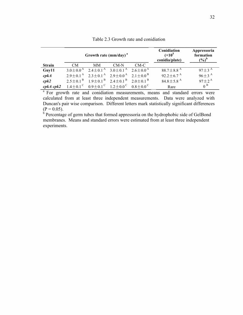
32
32
Table 2.3 Growth rate and conidiation
Growth rate (mm/day) a
Conidiation (×105
conidia/plate)
Appressoria formation
(%)b Strain CM MM CM-N CM-C Guy11 3.0±0.0 A 2.4±0.1 A 3.0±0.1 A 2.6±0.0 A 88.7±8.8 A 97±3 A
cpkA 2.9±0.1 A 2.3±0.1 A 2.9±0.0 A 2.1±0.0 B 92.2±6.7 A 96±3 A
cpk2 2.5±0.1 B 1.9±0.1 B 2.4±0.1 B 2.0±0.1 B 84.8±5.8 A 97±2 A
cpkA cpk2 1.4±0.1 C 0.9±0.1 C 1.2±0.0 C 0.8±0.0 C Rare 0 B
a For growth rate and conidiation measurements, means and standard errors were calculated from at least three independent measurements. Data were analyzed with Duncan's pair wise comparison. Different letters mark statistically significant differences (P = 0.05). b Percentage of germ tubes that formed appressoria on the hydrophobic side of GelBond membranes. Means and standard errors were estimated from at least three independent experiments.

33
33
Figure 2.2 Domain structures of CPKA and CPK2 and qRT-PCR assays for their expression levels. (A) Domain structures of CpkA and Cpk2. The hatched and block closed boxes in the protein kinase domain represent Protein kinases ATP-binding region and Serine/Threonine protein kinase active-sites, respectively. (B) Expression of the CPKA and CPK2 genes in the cpkA and cpk2 mutants measured by qRT-PCR. The relative expression in Guy11 was arbitrarily set to 1. Error bars represent the standard deviation.

34
34
2.4.2 The cpkA cpk2 double mutant has severe growth defects and it is unstable
Compared to the wild-type strain Guy11 and the single mutants, the growth rate of
the cpkA cpk2 double mutant was significantly reduced (Table 2.3, Figure 2.3A). Similar
to the cpk2 mutant, the cpkA cpk2 double mutant was more sensitive to nutritional
starvation, especially for carbon starvation (Table 2.3, Figure 2.3A). These results
suggest that PKA catalytic subunits are involved in regulating metabolism processes and
responses to nutritional stresses in M. oryzae.
Interestingly, the cpkA cpk2 mutants were unstable when cultured on the oatmeal
agar plates, and the fast-growing spontaneous suppressors became visible in some cpkA
cpk2 colonies older than 10 days (Figure 2.3B). Under the culture conditions, the cpkA
and cpk2 mutants were stable and never produced sectors. Interestingly, the cpkA cpk2
mutants appeared to be stable when cultured on CM plates and fast-growing spontaneous
suppressors were not or rarely observed.

35
35
Figure 2.3 Growth rate and colony morphology of the single and double mutant of CPKA and CPK2. (A) Colony morphology of Guy11, I-27 (the cpkA mutant), YP18 (the cpk2 mutant), and CAC2 (the cpkA cpk2 mutant) when cultured on the CM, MM, CM-N and CM-C plates at room temperature for 7 days. (B) Spontaneous suppressors appeared on oatmeal agar plates of the cpkA cpk2 mutant.

36
36
2.4.3 Conidiation is significantly reduced in the cpkA cpk2 double mutant
Because the cpkA cpk2 double mutant is unstable on OTA plates, we measured
conidiation with CM cultures or OTA cultures before spontaneous suppressors occurred.
Unlike the cpkA and cpk2 single mutants, the cpkA cpk2 double mutant rarely produced
conidia. After inducing for conidiation for two days, only a few conidia were observed in
each culture plate. Under the same conditions, over 1×10⁷ conidia/plate were produced
by the wild type.
2.4.4 PKA catalytic subunits are essential for appressoria formation
Previous studies have showed that cAMP signaling regulates surface recognition in
M. oryzae. To determine the impact of PKA on surface recognition, we examined
conidium germination and appressorium formation of the double mutant on the
hydrophilic and hydrophobic surfaces. On artificial hydrophobic surfaces, almost all the
conidia of the cpkA cpk2 double mutant germinated but failed to form appressoria at 24
hpi. Under the same experimental conditions, over 97% of wild-type germ tubes formed
appressoria (Figure 2.4A). Therefore, deletion of two PKA catalytic subunits completely
blocked appressoria formation. Even after prolonged incubation (over 72hpi), no
appressorium formation was observed in the double mutant. Interestingly, we observed
that over 90% of the cpkA cpk2 germ tubes were curved and were wider than the germ
tubes produced by Guy11. The majority (over 83%) of these curved germ tubes curled
clockwise (viewed from the germination site on conidium) (Figure 2.4A).
On the hydrophilic surface of Gelbond membranes, only 23% of conidia germinated
in the cpkA cpk2 mutant at 24 h. After 72 h, the germination rate of the cpkA cpk2

37
37
mutant is the same. Unlike on the hydrophobic surface, the cpkA cpk2 mutant, similar to
the wild-type strain, formed straight germ tubes, but these germ tubes were much shorter
than those of Guy11.
Because exogenous cAMP, IBMX, and cutin monomers are known to trigger
appressorium formation in M. oryzae, we assayed their effects on the cpkA cpk2 mutant.
In the presence of 10 mM cAMP, over 95% of the wild-type germ tubes formed
appressoria on hydrophilic surfaces. However, exogenous cAMP had no effects on either
conidium germination or appressorium formation in the cpkA cpk2 mutant (Figure 2.4B).
The same results were obtained when the double mutant was treated with IBMX or cutin
monomers, indicating that exogenous cAMP, IBMX, and cutin monomers that target the
upstream components of the cAMP-PKA pathway failed to rescue the appressorium
formation defect in the cpkA cpk2 mutant.

38
38
Figure 2.4 Appressorium formation assays. (A) Conidia from the wild-type strain Guy11 and the cpkA cpk2 double mutant were incubated on the hydrophobic and hydrophilic surfaces of GelBond membranes for 24 h. Bar = 10 µm. (B) Appressorium formation assays with Guy11 and the cpkA cpk2 mutant in the presence of 10 mM cAMP on the hydrophilic surface of GelBond membranes. Bar = 10 µm.

39
39
2.4.5 The cpkA cpk2 double mutant failed to infect intact leaves or through wounding
sites
To determine the role of PKA catalytic subunits in plant infection, we conducted
spray infection assays with Guy11, the cpkA and cpk2 single mutants, and the cpkA cpk2
double mutant with two-week-old rice seedlings of cultivar CO-39. Whereas numerous
blast lesions were observed on leaves sprayed with Guy11 or the cpk2 mutant, no typical
blast lesions were caused by the cpkA and cpkA cpk2 mutants (Figure 2.5). Because the
cpkA cpk2 mutant failed to form appressoria to penetrate the plant surface, we further
examined its virulence by injecting spore suspensions with needles into an area a few
centimeters below the rice leaf whorl. Consistent with the previous study, the cpkA
mutant can infect through wounding sites, whereas the cpkA cpk2 mutant failed to cause
lesions at the wounding sites (Figure 2.5). These results indicate that PKA plays a
critical role in invasive growth.

40
40
Figure 2.5 Rice infection assays. Leaves of two-week-old rice seedlings were sprayed or injected with conidial suspensions of Guy11, I-27 (the cpkA mutant), YP18 (the cpk2 mutant) and CAC2 (the cpkA cpk2 mutant). Inoculation with 0.25% gelatin was used as the negative control. Typical leaves were photographed 7 dpi.

41
41
2.4.6 Both CpkA and Cpk2 interact with the Sum1 PKA regulatory subunit
In M. oryzae, the PKA regulatory subunit is encoded by SUM1. To confirm the
interaction between the PKA catalytic subunits and regulatory subunit, co-
immunoprecipitation (co-IP) assays were performed. The SUM1-S, CPKA-3×FLAG and
CPK2-3×FLAG constructs were generated and transformed into the wild-type strain
Guy11 in pairs. The resulting transformants KAS5 (SUM1-S/CPKA-3×FLAG) and
KZS2 (SUM1-S/CPK2-3×FLAG) (Table 2.1) were confirmed by PCR and western blot
analyses to express the transforming fusion constructs. For co-IP assays, total proteins
were extracted from fresh hyphae and incubated with anti-S-Tag antibody agarose beads
(Bethyl Laboratories). The immunoprecipitated proteins were eluted from anti-S-Tag
agarose beads and analyzed by western blot detection. In the transformant KAS5, CpkA-
3Flag was detected with an anti-FLAG antibody in both the total and immunoprecipitated
proteins (Figure 2.6A). In the transformant KZS2, the Cpk2-3Flag band was also
detected with an anti-FLAG antibody in both the total proteins and proteins precipitated
with anti-S-Tag agarose beads (Figure 2.6B). These results indicate that both CpkA and
Cpk2 interact with the regulatory subunit Sum1.

42
42
Figure 2.6 The physical interactions of Sum1-CpkA and Sum1-Cpk2 were confirmed by co-IP. Western blots of total proteins (Input) and the proteins immunoprecipitated (IP) by anti-S-Tag antibody agarose from transformants (A) KAS5 (Sum1-S and CpkA-3×FLAG) and (B) KZS2 (Sum1-S and Cpk2-3×FLAG) were detected with anti-S, anti-FLAG, or anti-actin antibodies. Total proteins isolated from the wild-type strain Guy11 were included as the control.

43
43
2.4.7 The intracellular cAMP level is significantly increased in the cpkA cpk2 mutant
To determine whether deletion of CPKA and CPK2 has a feedback effect on the
upstream components of the cAMP-PKA pathway, we measured the intracellular cAMP
level of the single and double mutants in vegetative hyphae cultured in liquid CM. In the
cpk2 mutant, the intracellular cAMP level was similar to that of the wild-type strain.
However, the cpkA mutant and cpkA cpk2 double mutant were increased approximately
1.5-fold and 3-fold in the intracellular level in comparison with the wild type,
respectively (Figure 2.7).

44
44
Figure 2.7 The intracellular cAMP level in the wild type and mutant strains. The intracellular cAMP levels were assayed with vegetative hyphae of the wild-type strain Guy11 and the cpkA (I-27), cpk2 (YP18), and cpkA cpk2 (CAC2) mutants. Mean and standard deviation were calculated with results from three independent biological replicates.

45
45
2.4.8 Deletion of PKA catalytic subunits interferes with multiple MAPK signaling
pathways
In M. oryzae, both the PMK1 MAP kinase and cAMP-PKA signaling pathways
regulate appressorium formation and plant infection. PMK1 may function downstream
from cAMP signaling because the pmk1 mutant still responded to exogenous cAMP to
initiate germ tube tip deformation but failed to complete the formation of mature
appressoria. To determine the activation level of Pmk1, we assayed the phosphorylation
level of Pmk1 with an anti-TpEY specific antibody. Surprisingly, although Pmk1
expression was not affected, the activation of Pmk1 was increased in the cpk2 and cpkA
cpk2 mutants compared to the wild-type strain (Figure 2.8A). The phosphorylation level
of Pmk1 was higher in the cpkA cpk2 double mutant than in the cpkA mutant. Under the
same experimental conditions, the phosphorylation level of Mps1 was decreased in the
cpk2 and cpkA cpk2 mutants (Figure 2.8A). Osmoregulation is mediated by the Osm1
MAP kinase, the third MAPK in M. oryzae (Dixon 1999). When detected with an anti-
TpGY antibody, the cpkA and cpkA cpk2 mutants had a reduced level of Osm1
phosphorylation (Figure 2.8B).

46
46
Figure 2.8 Western blot analysis for assaying the activation of Pmk1, Mps1 and Osm1. (A) Total proteins were isolated from mycelia of Guy11, and the pmk1, mps1, cpkA, cpk2, and cpkA cpk2 mutant cultured for 2 days in liquid CM. The bottom panel was detected with an anti-actin antibody to show similar amount of total proteins was loaded in each lane. The band detected with an anti-Pmk1 antibody (middle panel) showed the Pmk1 expression level in all the strains, with the pmk1 deletion mutant as the negative control. The band detected with an anti-TpEY (top panel) antibody showed the phosphorylation level of Pmk1 (42-kD) or Mps1 (46-kD). (B) The 41-kD Osm1 band was detected with the anti-P38 antibody and anti-TpGY phosphorylation-specific antibody.

47
47
2.5 Discussion
The cAMP-PKA signal transduction pathway is known to be involved in infection-
related morphogenesis and plant infection in the rice blast fungus M. oryzae. Protein
kinase A, which functions as core a component in the cAMP signaling pathway, contains
two catalytic subunits CpkA and Cpk2. The function of CPKA has been characterized in
previous studies (Xu and Hamer 1996). However, CPKA is dispensable in growth and
conidiation. The deletion of CPKA only results in delayed formation of appressoria (Xu
and Hamer 1996). This phenotype cannot explain the important role of PKA in the
cAMP signaling pathway. In this study, we identified the second PKA catalytic subunit
CPK2 and confirmed that both CpkA and Cpk2 interact with the regulatory subunit Sum1
by co-IP assays.
Compared with the CPK single mutant, the double mutant showed significant
deficiency in growth and conidiation, indicating that the existence of one PKA catalytic
subunit can complement the deficiency of growth and conidiation caused by absence of
another one. And this complementation is not due to transcriptional regulation of each
other because our qRT-PCR results show that the deletion of one CPK gene does not
cause overexpression of the other CPK gene. Even though the two PKA catalytic
subunits have overlapping functions, they also have distinguishing features. For example,
the cpk2 mutant had a slightly reduced growth rate, whereas the cpkA mutant does not
show obvious defects in growth under rich medium conditions but it is sensitive to carbon
resource starvation. The cpkA mutant is defective in appressoria formation and
pathogenicity, but deletion of CPK2 has no effect on the pathogenicity-related process.
This functional specialization of PKA catalytic subunits is also found in yeast and other

48
48
organisms, which may be attributable to subtle differences in the conserved catalytic
regions and to differences in their substrates.
Lacking both CPKA and CPK2 absolutely blocked appressorium formation on a
hydrophobic surface. Our observation also showed that spore germination was reduced
on the hydrophilic surface compare with that on the hydrophobic surface. The reason for
the reduced germination is unclear, but it suggests that the hydrophobic signal plays an
important role in the spore germination process. Without PKA activity, the spores
showed low efficiency in surface attachment and germ tube emergence on the hydrophilic
surface. The cAMP-PKA pathway is known to function in surface recognition. And
adding exogenous cAMP is able to bypass the hydrophobic signals in the wild-type strain
and partially restore the deficiency in appressoria formation caused by deletion of some
MAPK components. In our study, the appressoria formation assay showed the cpkA cpk2
mutant is still able to sense a hydrophobic surface, suggesting that the hydrophobic
signals initiating surface recognition may be involved in multiple signal transduction
pathways besides the cAMP-PKA signaling. Neither PMK1 nor PKA can simply block
the surface recognition. It is probably because of two reasons at least. First, they have
overlapping downstream substrates. In M. oryzae, the transcription factor MoSFL1 is
phosphorylated by both PMK1 and PKA (Li, Zhou et al. 2011). Second, their
downstream substrates may share redundant functions. So far, we lack evidence to prove
this point in M. oryzae. But in S. cerevisiae, STE12 and FLO8 are the downstream
targets of Kss1 (orthologous to Pmk1 in M. oryzae) in the MAPK pathway and cAMP
pathway, respectively. Overexpression of one of them can suppress the loss of the other
(Rupp, Summers et al. 1999).

49
49
We measured the cAMP level in the CPK single mutants and double mutant.
Interestingly, the cpkA single mutant had an elevated cAMP level, and the elevated
cAMP level was enhanced in the double mutant. This result further confirms that the
strains lacking PKA activity are unable to respond to cAMP to form appressoria. In
Aspergillus nidulans, it is reported that cAMP levels are elevated by about 250-fold in a
strain that lacks PkaA (Fillinger, Chaveroche et al. 2002). The elevation of cAMP level
may be due to the disruption of PKA-independent feedback regulation. In
Saccharomyces cerevisiae, PKA exerts feedback systematically in several ways to
regulate itself activity. The negative feedback mechanisms include activating Pde1p and
deactivating Ras2 via phosphorylation of Cdc25 (Williamson, Schwartz et al. 2009). The
low affinity cAMP phosphodiesterase Pde1p, which is orthologous to PdeL in M. oryzae,
is the target of PKA for feedback inhibition of cAMP accumulation by hydrolysis of
cAMP. Pde1p can be phosphorylated by bovine PKA and leads to increased
phosphodiesterase activity (Ma, Wera et al. 1999). Without PKA activity, Pde1p may
lose the function to decrease the cAMP level. In addition, PKA phosphorylates Cdc25p
to dissociate it from Ras2p, which leads to inactivation of adenylate cyclases (Gross,
Goldberg et al. 1992). Therefore, the absence of PKA activity failed to balance cAMP
accumulation. And our result suggests that CPKA plays primary role in the negative
feedback regulation in M. oryzae.
In the MAPK/PMK1 pathway, deletion of MAP kinase kinase kinase (MEKK)
MST11 and MAP kinase kinase (MEK) MST7 caused much less conidia production and
defects in appressorium formation. However, their defects were able to be restored by
either stimulating with exogenous cAMP or expressing the MST7S212D T216E dominant

50
50
allele (Zhao, Kim et al. 2005). However, in our study, expressing the MST7S212D T216E
dominant allele failed to rescue any deficiency in the cpkA cpk2 double mutant. And in
fact, our TEY assay showed that Pmk1 was hyper-activated in the cpkA cpk2 mutant.
Therefore, we conclude that activating the cAMP-PKA pathway can partially recover the
function of MAPK/PMK1 pathway, but activation of the MAPK/PMK1 pathway is
unable to rescue the defect caused by absence of PKA. It suggests that the PKA
signaling pathway operates in parallel with the PMK1 pathway to regulate appressorium
formation independently but it is more essential for growth and infection-related
differentiation.
In this study, the crosstalks between the cAMP and MAPK signaling pathways were
the first time detected in M. oryzae. In mammalian cells, ERK can be activated or
inhibited by cAMP in a cell-specific manner (Stork and Schmitt 2002). In M. oryzae,
PMK1 is closely related to ERK-type kinase (Wilson and Talbot 2009). Deletion of
CPK2 or both CPKA and CPK2, but not CPKA alone, results in increased activation of
Pmk1. In contrast, cAMP-PKA seems to have opposite effects on another MAP kinase,
Osm1, in M. oryzae. However, we need more clues to reveal the mechanism of cross-talk
between the cAMP-PKA and MAPK pathways.

51
51
2.6 List of References
Bruno, K., F. Tenjo, L. Li, J. Hamer and J.-R. Xu (2004). "Cellular localization and role of kinase activity of PMK1 in Magnaporthe grisea." Eukaryotic Cell 3(6): 1525-1532.
Choi, W. and R. A. Dean (1997). "The adenylate cyclase gene MAC1 of Magnaporthe grisea controls appressorium formation and other aspects of growth and development." Plant Cell 9(11): 1973-1983.
Christian, D. C., W. B. William, P. S. James, T. Xiaoyan, V. Barbara, F. P. Kerry and L. P. Gary (2012). "Preliminary Assessment of Resistance Among U.S. Wheat Cultivars to the Triticum Pathotype of Magnaporthe oryzae." Plant Disease 96.
Dagdas, Y. F., K. Yoshino, G. Dagdas, L. S. Ryder, E. Bielska, G. Steinberg and N. J. Talbot (2012). "Septin-mediated plant cell invasion by the rice blast fungus, Magnaporthe oryzae." Science 336(6088): 1590-1595.
Dean, R. A., N. J. Talbot, D. J. Ebbole, M. L. Farman, T. K. Mitchell, M. J. Orbach, M. Thon, R. Kulkarni, J. R. Xu, H. Pan, N. D. Read, Y. H. Lee, I. Carbone, D. Brown, Y. Y. Oh, N. Donofrio, J. S. Jeong, D. M. Soanes, S. Djonovic, E. Kolomiets, C. Rehmeyer, W. Li, M. Harding, S. Kim, M. H. Lebrun, H. Bohnert, S. Coughlan, J. Butler, S. Calvo, L. J. Ma, R. Nicol, S. Purcell, C. Nusbaum, J. E. Galagan and B. W. Birren (2005). "The genome sequence of the rice blast fungus Magnaporthe grisea." Nature 434(7036): 980-986.
Ding, S. L., W. Liu, A. Iliuk, C. Ribot, J. Vallet, A. Tao, Y. Wang, M. H. Lebrun and J. R. Xu (2010). "The tig1 histone deacetylase complex regulates infectious growth in the rice blast fungus Magnaporthe oryzae." Plant Cell 22(7): 2495-2508.
Dixon, K. P., J. R. Xu, N. Smirnoff and N. J. Talbot (1999). "Independent signaling pathways regulate cellular turgor during hyperosmotic stress and appressorium-mediated plant infection by Magnaporthe grisea." Plant Cell 11(10): 2045-2058.
Fillinger, S., M.-K. Chaveroche, K. Shimizu, N. Keller and C. d'Enfert (2002). "cAMP and ras signalling independently control spore germination in the filamentous fungus Aspergillus nidulans." Molecular Microbiology 44(4): 1001-1016.
Fisher, M., D. Henk, C. Briggs, J. Brownstein, L. Madoff, S. McCraw and S. Gurr (2012). "Emerging fungal threats to animal, plant and ecosystem health." Nature 484(7393): 186-194.
Fuller, K. K., D. L. Richie, X. Feng, K. Krishnan, T. J. Stephens, K. A. Wikenheiser-Brokamp, D. S. Askew and J. C. Rhodes (2011). "Divergent Protein Kinase A isoforms co-ordinately regulate conidial germination, carbohydrate metabolism and virulence in Aspergillus fumigatus." Molecular Microbiology 79(4): 1045-1062.
Gross, E., D. Goldberg and A. Levitzki (1992). "Phosphorylation of the S. cerevisiae Cdc25 in response to glucose results in its dissociation from Ras." Nature 360(6406): 762-765.

52
52
Hu, S., X. Zhou, X. Gu, S. Cao, C. Wang and J. R. Xu (2014). "The cAMP-PKA pathway regulates growth, sexual and asexual differentiation, and pathogenesis in Fusarium graminearum." Mol Plant Microbe Interact 27(6): 557-566.
Kankanala, P., K. Czymmek and B. Valent (2007). "Roles for rice membrane dynamics and plasmodesmata during biotrophic invasion by the blast fungus." Plant Cell 19(2): 706-724.
Kim, C., N.-H. Xuong and S. Taylor (2005). "Crystal structure of a complex between the catalytic and regulatory (RIalpha) subunits of PKA." Science (New York, N.Y.) 307(5710): 690-696.
Kim, S., S. Y. Park, K. S. Kim, H. S. Rho, M. H. Chi, J. Choi, J. Park, S. Kong, J. Goh and Y. H. Lee (2009). "Homeobox transcription factors are required for conidiation and appressorium development in the rice blast fungus Magnaporthe oryzae." Plos Genetics 5(12): e1000757.
Kong, L.-A., G.-T. Li, Y. Liu, M.-G. Liu, S.-J. Zhang, J. Yang, X.-Y. Zhou, Y.-L. Peng and J.-R. Xu (2013). "Differences between appressoria formed by germ tubes and appressorium-like structures developed by hyphal tips in Magnaporthe oryzae." Fungal genetics and biology : FG & B 56: 33-41.
Leung, H., E. S. Borromeo, M. A. Bernardo and J. L. Notteghem (1988). "Genetic analysis of virulence in the rice blast fungus Magnaporthe grisea." Phytopathology 78(9): 1227-1233.
Li, G., X. Zhou, L. Kong, Y. Wang, H. Zhang, H. Zhu, T. K. Mitchell, R. A. Dean and J. R. Xu (2011). "MoSfl1 is important for virulence and heat tolerance in Magnaporthe oryzae." PLoS One 6(5): e19951.
Li, L., C. Xue, K. Bruno, M. Nishimura and J. R. Xu (2004). "Two PAK kinase genes, CHM1 and MST20, have distinct functions in Magnaporthe grisea." Mol Plant Microbe Interact 17(5): 547-556.
Liu, W., X. Zhou, G. Li, L. Li, L. Kong, C. Wang, H. Zhang and J.-R. Xu (2011). "Multiple plant surface signals are sensed by different mechanisms in the rice blast fungus for appressorium formation." Plos Pathogens 7(1).
Livak, K. and T. Schmittgen (2001). "Analysis of relative gene expression data using real-time quantitative PCR and the 2(-Delta Delta C(T)) Method." Methods (San Diego, Calif.) 25(4): 402-408.
Ma, P., S. Wera, P. Van Dijck and J. Thevelein (1999). "The PDE1-encoded low-affinity phosphodiesterase in the yeast Saccharomyces cerevisiae has a specific function in controlling agonist-induced cAMP signaling." Molecular Biology of the Cell 10(1): 91-104.
Maeda, T., Y. Watanabe, H. Kunitomo and M. Yamamoto (1994). "Cloning of the pka1 gene encoding the catalytic subunit of the cAMP-dependent protein kinase in Schizosaccharomyces pombe." Journal of Biological Chemistry 269(13): 9632-9637.
Pan, X. and J. Heitman (1999). "Cyclic AMP-dependent protein kinase regulates pseudohyphal differentiation in Saccharomyces cerevisiae." Molecular and Cellular Biology 19(7): 4874-4887.
53
53
Park, G., C. Xue, X. Zhao, Y. Kim, M. Orbach and J.-R. Xu (2006). "Multiple upstream signals converge on the adaptor protein Mst50 in Magnaporthe grisea." The Plant cell 18(10): 2822-2835.
Park, G., C. Xue, L. Zheng, S. Lam and J. R. Xu (2002). "MST12 regulates infectious growth but not appressorium formation in the rice blast fungus Magnaporthe grisea." Molecular plant-microbe interactions : MPMI 15(3): 183-192.
Ramanujam, R. and N. Naqvi (2010). "PdeH, a high-affinity cAMP phosphodiesterase, is a key regulator of asexual and pathogenic differentiation in Magnaporthe oryzae." Plos Pathogens 6(5).
Rupp, S., E. Summers, H. J. Lo, H. Madhani and G. Fink (1999). "MAP kinase and cAMP filamentation signaling pathways converge on the unusually large promoter of the yeast FLO11 gene." Embo Journal 18(5): 1257-1269.
Stefan, E., M. Malleshaiah, B. Breton, P. Ear, V. Bachmann, M. Beyermann, M. Bouvier and S. Michnick (2011). "PKA regulatory subunits mediate synergy among conserved G-protein-coupled receptor cascades." Nature Communications 2: 598.
Stork, P. and J. Schmitt (2002). "Crosstalk between cAMP and MAP kinase signaling in the regulation of cell proliferation." Trends in cell biology 12(6): 258-266.
Tamaki, H. (2007). "Glucose-stimulated cAMP-protein kinase A pathway in yeast Saccharomyces cerevisiae." Journal of Bioscience and Bioengineering 104(4): 245-250.
Toda, T., S. Cameron, P. Sass, M. Zoller and M. Wigler (1987). "Three different genes in S. cerevisiae encode the catalytic subunits of the cAMP-dependent protein kinase." Cell 50(2): 277-287.
Williamson, T., J.-M. Schwartz, D. Kell and L. Stateva (2009). "Deterministic mathematical models of the cAMP pathway in Saccharomyces cerevisiae." Bmc Systems Biology 3: 70.
Wilson, R. and N. Talbot (2009). "Under pressure: investigating the biology of plant infection by Magnaporthe oryzae." Nature reviews. Microbiology 7(3): 185-195.
Xu, J. and J. Hamer (1996). "MAP kinase and cAMP signaling regulate infection structure formation and pathogenic growth in the rice blast fungus Magnaporthe grisea." Genes & Development 10(21): 2696-2706.
Xu, J.-R., M. Urban, J. A. Sweigard and J. E. Hamer (1997). "The CPKA gene of Magnaporthe grisea is essential for appressorial penetration." Molecular Plant-Microbe Interactions 10(2): 187-194.
Xu, J. R., C. J. Staiger and J. E. Hamer (1998). "Inactivation of the mitogen-activated protein kinase Mps1 from the rice blast fungus prevents penetration of host cells but allows activation of plant defense responses." Proc Natl Acad Sci U S A 95(21): 12713-12718.
Yan, X., Y. Li, X. Yue, C. Wang, Y. Que, D. Kong, Z. Ma, N. Talbot and Z. Wang (2011). "Two novel transcriptional regulators are essential for infection-related morphogenesis and pathogenicity of the rice blast fungus Magnaporthe oryzae." Plos Pathogens 7(12).

54
54
Yu, J. H., Z. Hamari, K. H. Han, J. A. Seo, Y. Reyes-Dominguez and C. Scazzocchio (2004). "Double-joint PCR: a PCR-based molecular tool for gene manipulations in filamentous fungi." Fungal Genet Biol 41(11): 973-981.
Zhang, H., W. Tang, K. Liu, Q. Huang, X. Zhang, X. Yan, Y. Chen, J. Wang, Z. Qi, Z. Wang, X. Zheng, P. Wang and Z. Zhang (2011). "Eight RGS and RGS-like proteins orchestrate growth, differentiation, and pathogenicity of Magnaporthe oryzae." Plos Pathogens 7(12).
Zhao, X., Y. Kim, G. Park and J.-R. Xu (2005). "A mitogen-activated protein kinase cascade regulating infection-related morphogenesis in Magnaporthe grisea." The Plant cell 17(4): 1317-1329.
Zhao, X. and J. R. Xu (2007). "A highly conserved MAPK-docking site in Mst7 is essential for Pmk1 activation in Magnaporthe grisea." Molecular Microbiology 63(3): 881-894.
Zhou, X., G. Li and J. R. Xu (2011). "Efficient approaches for generating GFP fusion and epitope-tagging constructs in filamentous fungi." Computational Drug Discovery and Design 722: 199-212.
Zhou, X., W. Liu, C. Wang, Q. Xu, Y. Wang, S. Ding and J.-R. Xu (2011). "A MADS-box transcription factor MoMcm1 is required for male fertility, microconidium production and virulence in Magnaporthe oryzae." Molecular Microbiology 80(1): 33-53.
Zhou, X., H. Zhang, G. Li, B. Shaw and J.-R. Xu (2012). "The Cyclase-associated protein Cap1 is important for proper regulation of infection-related morphogenesis in Magnaporthe oryzae." Plos Pathogens 8(9).

55
55
CHAPTER 3. PHOSPHORYLATION OF MOSFL1 BY PKA RELIEVES ITS INTERACTION WITH THE CYC8-TUP1 CO-REPRESSOR
3.1 Abstract
Identification and characterization of suppressor mutations is a powerful genetic
method to identify genes that are functionally related to the gene of interest. In
Maganporthe oryzae, the cpkA cpk2 double mutant had severe growth defects and often
produced fast-growing suppressors. To further characterize the cAMP-PKA pathway, in
this study, a total of 20 fast-growing suppressors of the cpkA cpk2 mutant were isolated.
Four of them were selected to identify suppressor mutations by sequencing analysis. In
the suppressor strains CCS1 and CCS7, genetic mutations were identified in the
transcription factor gene that is orthologous to yeast SFL1, which is involved in the
suppression of flocculation. Both deletion of MoSFL1 and truncation of the C-terminal
region (CT, residues 496-588) suppressed the growth defect of the cpkA cpk2 mutant,
suggesting that the C-terminal region is important to its function. Affinity purification
and co-IP analyses showed that truncation of the C-terminal region released the
interaction of MoSfl1 with Cyc8 or Tup1. Site-directed mutagenesis showed that
phosphorylation of MoSfl1 at the S211 residue by PKA negatively regulates the
interaction between MoSfl1 and the Cyc8-Tup1 complex. Therefore, we conclude that
the loss-of-function mutation in MoSfl1 can bypass PKA activity to suppress the growth
defects of the cpkA cpk2 mutant.

56
56
3.2 Introduction
In Magnaporthe oryzae and many other phytopathogenic fungi, cAMP signaling
plays an important role in various differentiation and infection processes (Adachi and
Hamer 1998, Ni, Rierson et al. 2005, Schumacher, Kokkelink et al. 2008, Choi and Xu
2010, Tzima, Paplomatas et al. 2010, Kim, Park et al. 2011, Hu, Zhou et al. 2014). Two
genes, named CPKA and CPK2, encode the catalytic subunits of cAMP-dependent
protein kinase A (PKA) and have been characterized in M. oryzae. Deletion of both
CPKA and CPK2 results in significant deficiency in growth, conidiation, appressorium
formation and pathogenicity. Interestingly, the cpkA cpk2 double mutant is unstable and
produces fast-growing spontaneous suppressors after cultivation on oatmeal plates over
10 days. It is likely that suppressor mutations may occur at some of the conserved
downstream targets of the cAMP-PKA pathway in fungi.
In yeast, Sfl1is the transcription factor that directly targets PKA. SFL1 is a heat
shock factor-like transcription factor that can act as either a repressor or an activator
(Fujita, Kikuchi et al. 1989, Galeote, Alexandre et al. 2007). As a repressor, it is
involved in repression of flocculation-related genes, including FLO11 and SUC2 (Song
and Carlson 1998, Rupp, Summers et al. 1999). As an activator, SFL1 is involved in the
activation of stress-responsive genes such as HSP30 (Galeote, Alexandre et al. 2007). In
S. cerevisiae, Sfl1 forms homodimers to promote DNA binding (Pan and Heitman 2002).
The PKA catalytic subunit Tpk2 negatively regulates the Sfl1 repressor function by
prohibiting its dimerization and DNA binding (Pan and Heitman 2002). In M. oryzae, a
previous study has shown that MoSfl1 could also be phosphorylated by the PMK1
mitogen-activated protein (MAP) kinase (Li, Zhou et al. 2011). Deletion of MoSFL1

57
57
does not affect vegetative growth but results in reduced virulence and heat tolerance (Li,
Zhou et al. 2011).
Several Sfl1-interacting proteins have been identified in budding yeast, including the
Cyc8-Tup1 co-repressor and mediator proteins (Song and Carlson 1998, Conlan and
Tzamarias 2001). The Cyc8-Tup1 (also known as Ssn6-Tup1) complex is a well-studied
co-repressor regulating transcription of many genes in the budding yeast (Parnell and
Stillman 2011). The Tup1 co-repressor family is evolutionarily conserved in fungi,
plants, and animals, which share similarities not only in domain structures but also in
repression functions (Braun and Johnson 1997, Fisher and Caudy 1998, Liu and
Karmarkar 2008). As a co-repressor, the Cyc8-Tup1 complex lacks intrinsic DNA-
binding activities and is recruited by sequence-specific DNA-binding transcription
factors to the regulatory region of target genes (Smith and Johnson 2000). In yeast, Cyc8
(also called Ssn6) functions as an adaptor protein required for interaction between Tup1
tetramers and DNA-binding transcription factors (Tzamarias and Struhl 1994). To
determine the specificity of genes to be repressed, the Cyc8-Tup1 complex interacts with
different transcription factors with sequence-specific DNA binding, including Sfl1, Mig1,
Crt1 and α2 (Smith and Johnson 2000, Conlan and Tzamarias 2001).
Several models have been proposed to explain how the Cyc8-Tup1 complex may
repress transcription in yeast. First, Cyc8-Tup1 was found to interact with histone
deacetylases (HDACs), including Rpd3, Hos1 and Hos2, to modify chromatin structure at
target promoters (Davie, Edmondson et al. 2003). Second, the repression of the Cyc8-
Tup1 complex could be achieved by nucleosome repositioning, which limits the
accessibility of transcription activators to promoters (Malave and Dent 2006). Third,

58
58
Cyc8-Tup1 interacts with the mediator complex to prevent RNA polymerase II
machinery from initiating transcription (Smith and Johnson 2000). And fourth, Cyc8-
Tup1 masks and inhibits the activation domain of DNA-bound proteins to prevent the
recruitment of transcription co-activators (including mediator) (Wong and Struhl 2011).
In this study, two suppressor mutations in MoSFL1 were found to rescue the growth
defects of the cpkA cpk2 mutant in M. oryzae. We demonstrated that MoSfl1 interacts
with the Cyc8-Tup1 co-repressor to negatively regulate vegetative growth in M. oryzae.
This interaction can be relieved by PKA phosphorylation of MoSfl1. However, without
PKA activity in the cpkA cpk2 mutant, loss-of-function mutations in MoSfl1 also resulted
in its dissociation from Cyc8-Tup1. Furthermore, site-directed mutagenesis analysis
indicates that S211 is an important PKA phosphorylation site in MoSfl1.
3.3 Materials and Methods
3.3.1 Strains and culture conditions
The wild-type strain Guy11 (MAT1-2) and all the transformants generated in this
study (Table 3.1) were cultured on oatmeal agar (OTA) plates or complete medium (CM)
plates at 25°C and stored on desiccated Whatman #1 filter paper at -20°C (Park, Xue et al.
2002). Protoplast preparation and PEG-mediated transformation were performed as
described (Park, Xue et al. 2006). Transformants were selected on regeneration medium
with 250 µg/ml hygromycin B (CalBiochem), 250 µg/ml geneticin G418 (Sigma), or 200
µg/ml zeocin (Invitrogen). Growth rate and conidiation were assayed with OTA cultures
as described (Li, Xue et al. 2004, Park, Xue et al. 2006).

59
59
Table 3.1 Wild-type and mutant strains of Magnaporthe oryzae used in this study Strain Genotype description Reference Guy11 Wild type (MAT1-2) (Leung, Borromeo
et al. 1988) CAC2 cpkA cpk2 deletion mutant This study Suppressor strains CCS1-CCS20
Spontaneous suppressor mutants of cpkA cpk2 This study
CTD2 cpkA cpk2 Mosfl1CT deletion mutant This study TKO4 cpkA cpk2 Mosfl1 deletion mutant This study SFL2 3×FLAG-MoSFL1 transformant of the cpkA cpk2 mutant This study SCT7 3×FLAG-MoSFL1ΔCT transformant of the cpkA cpk2
mutant This study
HYS4 MGG_06958-S and 3×FLAG-MoSFL1 transformant of the cpkA cpk2 mutant
This study
MCS49 MGG_13806-S and 3×FLAG-MoSFL1 transformant of the cpkA cpk2 mutant
This study
CYS15 CYC8-S and 3×FLAG-MoSFL1 transformant of the cpkA cpk2 mutant
This study
CNC19 CYC8-S and 3×FLAG-MoSFL1ΔCT transformant of the cpkA cpk2 mutant
This study
TSL6 TUP1-S and 3×FLAG-MoSFL1 transformant of the cpkA cpk2 mutant
This study
TCS28 TUP1-S and 3×FLAG-MoSFL1ΔCT transformant of the cpkA cpk2 mutant
This study
GCS1 CYC8-S and 3×FLAG-MoSFL1 transformant of Guy11 This study GTS9 TUP1-S and 3×FLAG-MoSFL1 transformant of Guy11 This study ASD5 MoSFL1S211D transformant of the cpkA cpk2 mutant This study HTD17 MoSFL1T441D transformant of the cpkA cpk2 mutant This study GSD22 MoSFL1S554D transformant of the cpkA cpk2 mutant This study
60
60
3.3.2 Spontaneous suppressors of the cpkA cpk2 mutant
Fast-growing suppressors of the cpkA cpk2 mutant were transferred with sterile
toothpicks to fresh oatmeal agar plates. After single-spore isolation, each subculture of
spontaneous suppressors was assayed for defects in growth, conidiation, and plant
infection (Li, Wang et al. 2016). To identify suppressor mutations in the candidate genes,
PCR products amplified with primers listed in Table 3.2 were sequenced at Purdue
Genomics Core Facility. Mutation sites were identified by sequence alignment.
3.3.3 Appressorium formation and plant infection assays
Conidia were harvested from 10-day-old OTA cultures and resuspended to 5×10⁴
conidia/ml in sterile distilled water. For appressorium formation assays, droplets of 50 µl
conidial suspensions were placed on glass cover slips (Fisher Scientific) or GelBond
membranes (Cambrex) and incubated at 25oC for 24h as described (Zhou, Zhang et al.
2012). For infection assays, conidia were resuspended to 5×10⁴ conidia/ml in 0.25%
gelatin. Two-week-old seedlings of rice cultivar CO-39 were used for spray or injection
infection assays as described (Li, Xue et al. 2004, Zhou, Liu et al. 2011). Lesion
formation was examined 7 days post inoculation (dpi).
3.3.4 Generation of the gene-replacement mutants
The double-joint PCR method (Yu, Hamari et al. 2004) was used to generate the
MoSFL1 gene-replacement vectors. The 1.2-kb upstream and a 1.2-kb downstream
flanking sequences of MoSFL1 were amplified with primer pairs Sfl1ko1F/Sfl1ko2R
(Table 3.2) and Sfl1ko3F/ Sfl1ko4R (Table 3.2), respectively. The ble cassette was

61
61
amplified with primers Ble/F and Ble/R (Table 3.2) from pFL6. The resulting products
of double-joint PCR were transformed into protoplasts of the cpkA cpk2 mutant. Putative
cpkA cpk2 Mosfl1 triple deletion mutants were screened by PCR analysis to be deleted of
MoSFL1. Genomic DNA was extracted from mycelia by the CTAB protocol (Xu and
Hamer 1996).
The same strategy was used to generate the MoSFL1ΔCT construct and cpkA cpk2
Mosfl1ΔCT transformants. The upstream and downstream flanking sequences were
amplified with primer pairs CCSko1F/CCSko2R (Table 3.2) and Sfl1ko3F/ Sfl1ko4R
(Table 3.2), respectively. After ligation into the ble cassette, the resulting products of
double-joint PCR were transformed into protoplasts of the cpkA cpk2 mutant. Putative
cpkA cpk2 Mosfl1ΔCT triple-deletion mutants were screened by PCR analysis to be deleted
of the C-terminal domain (496-588 aa).
3.3.5 Affinity purification and mass spectrometry analysis
The MoSFL1 and MoSFL1ΔCT constructs were amplified and cloned into pFL6 by
the yeast gap repair approach (Zhou, Li et al. 2011). The resulting 3×FLAG-MoSFL1
and 3×FLAG-MoSFL1ΔCT fusion constructs were transformed into the cpkA cpk2 mutant.
All of the primers used in the constructions of these tagged proteins are listed in Table
3.2. Affinity purification and mass spectrometry analysis was performed as described
(Liu, Iliuk et al. 2011). The resulting MS data were used to search against the non-
redundant M. oryzae protein database at NCBI.

62
62
3.3.6 Co-immunoprecipitation (Co-IP) assay
The yeast gap repair was used to generate the MGG_06958-S, MGG_013806-S,
Cyc8-S and Tup1-S fusion constructs as described (Zhou, Li et al. 2011). Primers used to
amplify the MGG_06958, MGG_013806, CYC8 and TUP1 genes are listed in Table 3.2.
The resulting S-tagged fusion constructs were co-transformed with the Flag-tagged fusion
constructs into protoplasts of Guy11 or the cpkA cpk2 mutant in pairs. Transformants
containing the 3×FLAG-MoSFL1/MGG_06958-S, 3×FLAG-MoSFL1/MGG_13806-S,
3×FLAG-MoSFL1/CYC8-S, 3×FLAG-MoSFL1/TUP1-S, 3×FLAG-MoSFL1ΔCT/CYC8-S
and 3×FLAG-MoSFL1ΔCT/TUP1-S constructs were identified by PCR. The expression of
S and Flag fusion proteins was further confirmed by Western blot. For co-IP assays, total
proteins were isolated and incubated with the anti-S-Tag Antibody Agarose (Bethyl
Laboratories) as described (Liu, Iliuk et al. 2011). The immunoprecipitated (IP) proteins
were analyzed by Western blot with the anti-FLAG (Sigma-Aldrich) and anti-S (Abcam)
antibodies.
3.3.7 Construction of the MoSFL1S211D, MoSFL1T441D, MoSFL1S554D allele vectors
All MoSFL1 alleles were generated using overlapping PCR-mediated methods
(Heckman and Pease 2007) and cloned into pFL5 by the yeast gap repair approach (Zhou,
Li et al. 2011). All of the primers used in the construction of these mutation alleles are
listed in Table 3.2. All of the resulting constructs were confirmed by sequence analysis
and transformed into the cpkA cpk2 mutant.

63
63
Table 3.2 PCR primers used in this study Name Sequence (5’-3’) Applications Sfl1ko1F CAGCAGGCCAAGAATTGCAAGC MoSFL1 knockout
Sfl1ko2R GTGTTGACCTCCACTAGCTCCAGCCACTTGAGACGTACAACAAAGGTCCACG MoSFL1 knockout
Sfl1ko3F ACGAATTGCTTGCAGGCATCTCATACTGCAGTGACAAAAGAGCCAGAT MoSFL1 knockout
Sfl1ko4R AACAGCCGCCACAGCCGTAAA MoSFL1 knockout CCSko1F AAGCAGTACCCCAAAGGCCGA MoSFL1CT knockout
CCSko2R GTGTTGACCTCCACTAGCTCCAGCCACTTATCGAGTATGTGGAAAATGAGTCCCTG MoSFL1CT knockout
MoSfl1/FL6F TCGATTACAAGGATGACGATGACAAGGCTACCGCCATCCAAACAGC 3×FLAG-MoSFL1
MoSfl1/FL6R ACACTGTAACCCGCAACGAAGAACTCCTCCCCACTCCTTTGCCTGATCTGTAA 3×FLAG-MoSFL1
MoCCS/FL6R CACTGTAACCCGCAACGAAGAACTCCTCCCTATATCGAGTATGTGGAAAATGAGTCCCTG 3×FLAG-MoSFL1ΔCT
MoSfl1/FL5F CAGATCTTGGCTTTCGTAGGAACCCAATCTTCAGCTACCGCCATCCAAACAGC MoSFL1S211D
MoSfl1/FL5R CTTTATAATCACCGTCATGGTCTTTGTAGTCCTGCAGTCGCTTGCGCTTCCT MoSFL1S211D
MoSfl1/S211D1R TTGTGCACAAGGGCGTGGCGGTCGGCACGGCGCTTGATCTCTCGCAA MoSFL1S211D
MoSfl1/S211D2F GAGAGATCAAGCGCCGTGCCGACCGCCACGCCCTTGTGCACAA MoSFL1S211D
MoSfl1/T441D1R GTGACCGCGTATATCTGCCGAGTCGTGTCGTCGAGCTAAGCTACTTGG MoSFL1T441D
MoSfl1/T441D2F AAGTAGCTTAGCTCGACGACACGACTCGGCAGATATACGCGGTCAC MoSFL1T441D
MoSfl1/S554D1R TCGGGTTAAGTATGTGGGCCATGTCTCCCCTCCTCGTCGGTGGTGCT MoSFL1S554D
MoSfl1/S554D2F GCACCACCGACGAGGAGGGGAGACATGGCCCACATACTTAACCCGA MoSFL1S554D
Rp6958stag/F CAGATCTTGGCTTTCGTAGGAACCCAATCTTCAATGGCGCCCGCTGTTGGTAT MGG_06958-S
Rp6958stag/R TTCGAATTTAGCAGCAGCGGTTTCTTTGTCGACCTCCTCGACAGTGG MGG_06958-S
6958check/F AATGACCAGGGTAACCGAACAA MGG_06958-S 6958check/R GAGGAAAACCTACCTTCTTGTGC MGG_06958-S
Rp13806stag/F CAGATCTTGGCTTTCGTAGGAACCCAATCTTCAATGGCCACCGAGGTACGTCAAA MGG_13806-S
Rp13806stag/R TTCGAATTTAGCAGCAGCGGTTTCTTTCTCGTCAGCCTTGGCCTCAGTCTT MGG_13806-S
13806check/F CCTTCTCAGCGAGTGACCATTG MGG_13806-S 13806check/R TCCAACACGGCAGCTGAAGAAA MGG_13806-S
Cyc8stag/F ACTCACTATAGGGCGAATTGGGTACTCAAATTGGTTCTGCTAAGACCTGCACCGAACT CYC8-S
Cyc8stsg/R TTCGAATTTAGCAGCAGCGGTTTCTTTGGAGCTCTCAACCTTGGGACCCAT CYC8-S
Cyc8check/F AAATCCTTGGCAACCTCGAAGACG CYC8-S Cyc8check/R CAACAGGTTGAGACGGGCTTTGAT CYC8-S

64
64
Table 3.2 continued
Tup1stag/F ACTCACTATAGGGCGAATTGGGTACTCAAATTGGTTAATTCGTGTCAGTGCCAGCCATCA TUP1-S
Tup1stag/R TTCGAATTTAGCAGCAGCGGTTTCTTTCGCCATACGGTTGTCCATGC TUP1-S
Tup1check/F ATCAATGAGATGCACATGGTCCGA TUP1-S Tup1check/R GTTCTCAATATCCCAAAGCCTGAC TUP1-S RIM15seq1F GGCTCTGCTTCTACCAGACA RIM15 sequence RIM15seq2F GGAGAAAGCCATGTAAGTTG RIM15 sequence RIM15seq3F AAGCTCGAATTGTCGAACTA RIM15 sequence RIM15seq4F CTGAGTATCTCCGGTCACAC RIM15 sequence RIM15seq5R CTAGAGGAGGAGATTGCACA RIM15 sequence RIM15seq6F AAGGCCTGGCAAACATGCTC RIM15 sequence RIM15seq7F ATACGCTACTTCAAGATGAA RIM15 sequence RIM15seq8F TCTTCCAGCTTTTCACAGCC RIM15 sequence RIM15seq9F AATGGTGCCGATGTTGCCAG RIM15 sequence RIM15seq10R TTAGATAAAGTTCATGGGCT RIM15 sequence YAK1seq1 CTAAGGCGTTGCTGGTGCTG YAK1 sequence YAK1seq2R GGCAAAGATGCTGTCTATGAAT YAK1 sequence YAK1seq3 CTCGACAAGAACGACGATCA YAK1 sequence YAK1seq4R GACTCCATCGGCCGCCAATA YAK1 sequence MSN2seq1 GGGGATGCTAATACCTTTTCAG MSN2 sequence MSN2seq2 TGGTCTAGGTTGGCAGTAGAAC MSN2 sequence MSN2seq3 CCAAGATGGATTCTGCCACCAA MSN2 sequence MSN2seq3R CTCCCTTAGGTCCCGCAAACTCTG MSN2 sequence SOK1seq1F GCCGGTGAAGCTTTACAGTTCTCG SOK1 sequence SOK1seq2R TGCGGTCCTCGTCGAATACA SOK1 sequence SOK1seq3R GAAGGGAAGCCAGAGTGAAGGGAG SOK1 sequence Sch9seq1 AACATCCCGACAGCCCAAGAATAT SCH9 sequence Sch9seq2 CGAGCTGATATCAGGTGGCCCGAG SCH9 sequence Sch9seq3 GTTCGGACAGCAACGTCAAATTCG SCH9 sequence Sch9seq4 CGTGAAACGGACTAGAACAAAATCG SCH9 sequence MoSfl1seq1F AGAATTGCAAGCCCTGTCCG SFL1 sequence MoSfl1seq2R GTGTCCTGCGAGCTAAAGGT SFL1 sequence MoSfl1seq3F TCTGTTGCATGAGACTCGAC SFL1 sequence MoSfl1seq4R GCAAGCCAATGATGTCTCCC SFL1 sequence MoSfl1seq5F TCAGCGACGTCTTCCACAAC SFL1 sequence MoSfl1seq6R CAGTCGCTTGCGCTTCCTGT SFL1 sequence MoSfl1seq7R TCAGGTACTTTGGCATCTCC SFL1 sequence Som1seq1 CTTGCATCCAACCGAGAACACACA SOM1 sequence Som1seq2 GCATCGCATCGCTTCCGGAGTA SOM1 sequence Som1seq3 TTACAGGCTATCGCGAATGCATCC SOM1 sequence Som1seq4 CGCACCGCACTTCTTCAAAGGTA SOM1 sequence Som1seq5R GGGTATGATGAGATTTGTGATGAC SOM1 sequence Cdtf1seq1 TTCGACTAGGAACGAACGAATGAG CDTF1 sequence Cdtf1seq2 GTTCCAAAACCAGGCTATCAACCA CDTF1 sequence Cdtf1seq3 ACCCTCAGGTCATGTTGCTCCTTC CDTF1 sequence Cdtf1seq4 GAACTAGCTCGGAACTTGCCTCAG CDTF1 sequence Cdtf1seq5 ACGCAAGAACAGTTGTCCAAGCTC CDTF1 sequence Cdtf1seq6 CGGAAGAGAACTTTGAGGCAAACA CDTF1 sequence Cdtf1seq7R GATCAAAACATGCTGGTTGCCAAA CDTF1 sequence

65
65
3.4 Results
3.4.1 Defects of the cpkA cpk2 mutant are partially recovered in the spontaneous
suppressors
Unlike the cpkA mutant, the cpkA cpk2 double mutant was unstable. Fast-growing
spontaneous suppressors became visible in oatmeal cultures of the cpkA cpk2 mutant
older than 10 days. A total of 20 suppressor strains of the double mutant were isolated
and purified by single-spore isolation. The colony morphology of the suppressors
appeared darker and flat compared to that of the wild-type strain or cpkA cpk2 double
mutant (Figure 3.1 and 3.3A). The growth rate of suppressor strains recovered to over 83%
of the wild-type strain (Figure 3.2A). Conidiation also was partially rescued in
comparison with the double mutant but only recovered to approximately 3% of the wild-
type strain (Figure 3.2B).
In infection assays with two-week-old seedling of rice cultivar CO-39, none of the
suppressor strains was able to cause lesions in intact or wounded rice leaves (Figure
3.3C). These results indicate that suppressor mutations could suppress the growth defects
but not defects of the cpkA cpk2 mutant in plant infection. Close examination revealed
that suppressor strains had melanized hyphal tips (Figure 3.3B). After incubation for 24
h, conidia of the suppressor strains were able to germinate and form appressoria on both
the hydrophobic and hydrophilic surfaces of GelBond membranes (Figure 3.3D).
However, approximately 40% of appressoria formed by suppressor strains were abnormal
in morphology no matter on hydrophilic or hydrophobic surfaces (Figure 3.3E).

66
66
Figure 3.1 Colony morphology of the 20 spontaneous suppressors of the cpkA cpk2 mutant. Oatmeal agar plates were photographed after incubation at room temperature for 5 days.

67
67
Figure 3.2 Growth rate and conidiation of the spontaneous suppressors. (A) Growth rate and (B) conidiation of the wild-type strain Guy11, the cpkA cpk2 mutant, and 20 spontaneous suppressor strains (CCS1-CCS20) were measured with 7-day-old oatmeal agar cultures. Mean and standard errors were calculated from three independent measurements.

68
68
Figure 3.3 Phenotypes of the suppressor strain CCS1. (A) Seven-day-old OTA cultures of the wild-type strain Guy11, cpkA cpk2 mutant, and suppressor strain CCS1. (B) Hyphal tips of Guy11 and CCS1 from 7-day-old OTA cultures. Bar = 10 µm. (C) Rice leaves inoculated with conidia of Guy11 and CCS1 were examined 7 days post-inoculation. (D) Appressoria formed by Guy11 and CCS1 on hydrophobic and hydrophilic surfaces after incubation for 24 h. Bar = 10 µm. (E) Abnormal appressoria formed by CCS1 on hydrophobic surfaces after incubation for 24 h. Bar = 10 µm.

69
69
3.4.2 Identification of suppressor mutations in MoSFL1
To identify suppressor mutations, we selected eight genes (Table 3.3) that are
orthologous to the known downstream targets of PKA or genes that can suppress the
mutants lacking PKA activity in the budding yeast. These selected genes were amplified
from suppressor strains CCS1, CCS4, CCS7 and CCS14 and sequenced. Whereas
suppressor stains CCS4 and CCS14 had no mutations in these candidate genes, both
CCS1 and CCS7 had mutations in MoSFL1 (Table 3.4). In suppressor stain CCS1, 10
extra nucleotides CCCCCGCCGC were inserted at the position between 1556 and 1557
in the coding region of MoSFL1, resulting in a frameshift change at residue P414. In
suppressor strain CCS7, a 1241-bp deletion occurred in the coding region of MoSFL1
(405-1645), resulting in amino acid changes after residue M131.

70
70
Table 3.3 Genes selected for sequencing analysis in suppressor strains Gene Notes Reference RIM15 Deletion of RIM15 rescues spore viability in the tpk1 tpk2 tpk3
triple mutant (Reinders, Bürckert et al. 1998)
YAK1 Overexpression of YAK1 rescues growth defects of the tpk1 tpk2 tpk3 triple mutant
(Garrett and Broach 1989)
MSN2 Deletion of MSN2 and MSN4 increases growth in the tpk2 mutant (Smith, Ward et al. 1998)
SOK1 Overexpression of SOK1 suppresses the growth defect of mutants lacking PKA activity
(Ward and Garrett 1994)
SCH9 Overexpression SCH9 can functionally rescue a triple TPK deletion mutant
(Toda, Cameron et al. 1988)
SFL1 Downstream target of PKA (Conlan and Tzamarias 2001)
SOM1 Downstream target of PKA (Yan, Li et al. 2011) CDTF1 Downstream target of PKA (Yan, Li et al. 2011)
Table 3.4 Suppressor mutations identified in MoSFL1
Suppressor strain Mutations in MoSFL1 a Amino acid changes in MoSfl1 b
CCS1 1556_1557insCCCCCGCCGC P414fs CCS7 405_1645del M131fs
a ins, insertion; del, deletion. b fs, frameshift.

71
71
3.4.3 Either deletion of MoSFL1 or its C-terminal region rescues the cpkA cpk2 mutant
MoSFL1 is a transcription factor with a conserved HSF (heat shock factor) DNA-
binding domain in the N-terminal region (residues 124-225) (Li, Zhou et al. 2011). The
frame-shift mutation in suppressor strain CCS7 results in the truncation of 78% of amino
acids in MoSfl1, including parts of the HSF DNA-binding domain, which may
significantly affect function of the protein. To verify that loss-of-function mutations in
MoSFL1 can rescue the cpkA cpk2 mutant, the MoSFL1 gene-replacement construct
(Figure 3.4A) was generated and transformed into the cpkA cpk2 mutant. Bleomycin-
resistant transformants were screened by PCR for deletion of MoSFL1. Putative cpkA
cpk2 Mosfl1 mutants were confirmed by PCR analysis for the deletion of CPKA, CPK2,
and MoSFL1. The resulting cpkA cpk2 Mosfl1 triple mutant had similar phenotypes with
spontaneous suppressor strains isolated from the cpkA cpk2 mutant in M. oryzae (Figure
3.4C; Table 3.5).
In suppressor CCS1, the insertion mutation in MoSFL1 does not affect the HSF
DNA-binding domain but causes the truncation of its C-terminal region due to the
frameshift mutation. Sequence alignment showed that the C-terminal region of MoSfl1 is
conserved among its orthologs from other filamentous fungi, including Fusarium
graminearum, Neurospora crassa, Sclerotinia sclerotiorum, Botrytis cinerea, Aspergillus
nidulans, Microsporum canis, and Trichophyton rubrum (Figure 3.4B). To confirm that
truncation of the C-terminal region of MoSfl1 can also result in the phenotype recovery
of the cpkA cpk2 mutant, we generated a MoSFL1CT gene-replacement construct (Figure
3.4A) to delete the C-terminal region (CT, residues 496-588) of MoSfl1 in the cpkA cpk2
mutant. The resulting cpkA cpk2 Mosfl1CT triple mutants were confirmed by PCR

72
72
analyses. The phenotype of the cpkA cpk2 Mosfl1CT mutants was similar to that of the
cpkA cpk2 Mosfl1 mutants and spontaneous suppressors (Figure 3.4C; Table 3.5).
Table 3.5 Growth rate and conidiation of the cpkA cpk2 Mosfl1CT and cpkA cpk2 Mosfl1 mutants
Strain Growth rate (mm/day) * Conidiation (×105 conidia per plate) Guy11 (WT) 3.4±0.0 165.5±24.4 CAC2 (cpkA cpk2) 0.9±0.1 0.0±0.0 CT-5 (cpkA cpk2 Mosfl1CT) 3.1±0.1 1.5±0.3 SL-3 (cpkA cpk2 Mosfl1) 3.0±0.0 1.7±0.5 * Means and standard errors were calculated from at least three independent measurements.
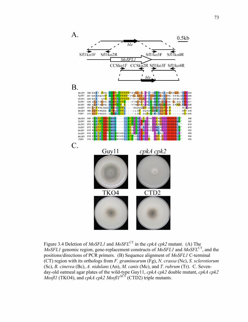
73
73
Figure 3.4 Deletion of MoSFL1 and MoSFLCT in the cpkA cpk2 mutant. (A) The MoSFL1 genomic region, gene-replacement constructs of MoSFL1 and MoSFLCT, and the positions/directions of PCR primers. (B) Sequence alignment of MoSFL1 C-terminal (CT) region with its orthologs from F. graminearum (Fg), N. crassa (Nc), S. sclerotiorum (Sc), B. cinerea (Bc), A. nidulans (An), M. canis (Mc), and T. rubrum (Tr). C. Seven-day-old oatmeal agar plates of the wild-type Guy11, cpkA cpk2 double mutant, cpkA cpk2 Mosfl1 (TKO4), and cpkA cpk2 Mosfl1ΔCT (CTD2) triple mutants.

74
74
3.4.4 Identification of MoSfl1- and MoSfl1ΔCT-interacting proteins by affinity purification
and mass spectrometry analysis
Several Sfl1-interacting proteins have been identified in budding yeast, including the
Cyc8-Tup1 co-repressor and mediator proteins (Song and Carlson 1998, Conlan and
Tzamarias 2001). To determine how mutations in MoSFL1 caused the suppressor effect,
affinity purification and mass spectrometry (MS) analysis were performed to identify
proteins interacting with MoSfl1 and MoSfl1ΔCT in the cpkA cpk2 mutant. The 3×FLAG-
MoSFL1 and 3×FLAG-MoSFL1ΔCT constructs were generated and transformed into the
cpkA cpk2 mutant. The resulting 3×FLAG-MoSFL1 transformants and 3×FLAG-
MoSFL1ΔCT transformants were confirmed by PCR and western blot analyses.
For affinity purification, total proteins were isolated from vegetative hyphae grown
in liquid CM medium and mixed with anti-FLAG M2 beads (Sigma-Aldrich). Proteins
bound to M2 beads were eluted and digested with trypsin. Proteins that co-purified with
3×FLAG-MoSfl1 and 3×FLAG-MoSfl1ΔCT were identified by MS analysis. Among the
MoSfl1-interacting proteins, MGG_06958, MGG_01588, MGG_08829 and MGG_03196
(Table 3.6) are orthologous to yeast SSA1, BMH1, TUP1, and CYC8, respectively, that
are known to interact with Sfl1 in S. cerevisiae. In comparison with Sfl1-interacting
proteins in yeast, the novel MoSfl1-interacting proteins include MGG_04191,
MGG_05193, MGG_02503, MGG_03286, MGG_13806, MGG_08142, MGG_01720,
MGG_08829, MGG_01268, MGG_09565, and MGG_03196 (Table 3.6). Similar to
MGG_06958 (SSA1), MGG_04191 and MGG_02503 are also Hsp70-like proteins that
are orthologs to yeast SSC1 and KAR2, respectively. The 14-3-3 family protein
MGG_13806 is orthologous to yeast BMH2, a paralog of BMH1. MGG_01842 and
75
75
MGG_01720 are orthologs of yeast THO1 and SWI3 that function as transcription
regulators. The putative pathway-specific nitrogen regulator MGG_03286 has no
homologous sequence in S. cerevisiae. The same set of proteins was identified as
MoSfl1ΔCT-interacting proteins except Cyc8 (Table 3.6).
The interaction between Pmk1 (MGG_09565) and MoSfl1 has been proved in the
previous study of our lab (Li, Zhou et al. 2011). To verify the pull-down data, we
selected two pairs of interactions, MGG_06958-MoSfl1 and MGG_13806-MoSfl1, for
co-immunoprecipitation (co-IP) assays. We generated MGG_06958-S and MGG_13806-
S constructs, and co-transformed them with 3×FLAG-MoSFL1 into the cpkA cpk2 mutant,
respectively. The resulting transformants HYS4 and MCS49 (Table 3.1) were confirmed
by PCR and further confirmed by western blot analysis with anti-FLAG and anti-S
antibodies. To confirm the interaction between MoSfl1 and MGG_06958, total proteins
were extracted from transformant HYS4 and incubated with anti-S-Tag Antibody
Agarose beads (Bethyl Laboratories). Proteins bound to the beads were then eluted and
used for western blot analysis. The 3×FLAG-MoSfl1 band was detected with an anti-
FLAG antibody in both total (In) and immunoprecipitated (IP) proteins (Figure 3.5A),
indicating that MoSfl1 interacts with MGG_06958.
Similarly, to confirm the interaction between MoSfl1 and MGG_13806, total
proteins were extracted from transformant MCS49 and incubated with anti-S-Tag
Antibody Agarose beads. The 3×FLAG-MoSfl1 band was detected in both total proteins
and proteins bound to anti-S-Tag Antibody Agarose beads (Figure 3.5B).
Unlike Cyc8 that interacts with MoSfl1 but not with MoSfl1ΔCT, we noticed that
although Tup1 were immunoprecipitated with both 3×FLAG-MoSfl1 and 3×FLAG-

76
76
MoSfl1ΔCT, the amount of Tup1 peptides detected by MS analysis was significantly
reduced in MoSfl1ΔCT samples compared to that in MoSfl1 samples (Table 3.6).
Table 3.6 Putative MoSFL1- and MoSFL1ΔCT-interacting genes identified by affinity purification
Genes Annotation % PSMs of *
MoSFL1 MoSFL1ΔCT MGG_06958 Hsp70-like protein 63 65 MGG_04191 Hsp70-like protein 36 38 MGG_02503 Glucose-regulated protein 17 16 MGG_03286 Pathway-specific nitrogen regulator 9 10 MGG_13806 14-3-3 family protein 8 12 MGG_01842 Uncharacterized protein 3 3 MGG_01720 Transcription regulatory protein SWI3 11 8 MGG_08829 Transcriptional repressor TUP1 26 4 MGG_01268 Nuclear localization sequence binding protein 3 4 MGG_01588 14-3-3 family protein 3 3 MGG_09565 Mitogen-activated protein kinase 2 3 MGG_03196 CYC8 10 0 * PSMs (peptide spectrum matches): The total number of identified peptide sequences for the protein.

77
77
Figure 3.5 Co-IP assays for the MGG_06958-MoSfl1 and MGG_13806-MoSfl1 interactions. Western blots of total proteins (Input) and proteins immunoprecipitated (IP) with anti-S-Tag antibody agarose beads from transformants (A) HYS4 (MGG_06958-S and 3×FLAG-MoSfl1) and (B) MCS49 (MGG_13806-S and 3×FLAG-MoSfl1) were detected with anti-S, anti-FLAG, or anti-actin antibodies. Total proteins isolated from the wild-type strain Guy11 were included as the control.

78
78
3.4.5 The C-terminal region of MoSfl1 is important for its interaction with the Cyc8-
Tup1 co-repressor
Our pull-down data suggest that MoSfl1 interacts with Cyc8 and Tup1, which is
consistent with studies in yeast (Conlan and Tzamarias 2001). In S. cerevisiae, Cyc8 and
Tup1 form a protein complex that functions as a transcription co-repressor (Smith and
Johnson 2000). To verify the interaction between MoSfl1 and Cyc8-Tup1 and test our
hypothesis that truncation of the C-terminal region of MoSfl1 affects its interactions with
the Cyc8-Tup1 complex, S-tagged CYC8 and TUP1 constructs were generated and co-
transformed with 3×FLAG-MoSFL1 and 3×FLAG-MoSFL1ΔCT into the cpkA cpk2 mutant.
The resulting transformants CYS15, CNC19 TSL6, and TCS28 (Table 3.1) were
confirmed by PCR and western blot analyses. To compare the interactions of MoSfl1
and MoSfl1ΔCT with Cyc8, the same amount of total proteins were extracted from
transformants CYS15 and CNC19 and incubated with anti-S-Tag antibody agarose for
co-IP assays. As expected, MoSfl1 was detected by an anti-FLAG antibody in both total
and immunoprecipitated proteins (Figure 3.6A). However, the 3×FLAG-MoSfl1ΔCT band
was not detected in proteins eluted from anti-S-Tag agarose beads in western blot
analysis (Figure 3.6A).
Similarly, to compare the interactions of MoSfl1 and MoSfl1ΔCT with Tup1, the
same amount of total proteins extracted from transformants TSL6 and TCS28 were used
for co-IP assays with anti-S-Tag antibody agarose beads. The amount of MoSfl1ΔCT
proteins co-precipitated with anti-S-Tag agarose was significantly reduced in comparison
with the amount that co-purified with MoSfl1 (Figure 2.6B).

79
79
Figure 3.6 Co-IP assays for interactions of MoSfl1 and MoSfl1ΔCT with the Cyc8-Tup1 co-repressor. Western blots of total proteins (Input) and proteins immunoprecipitated (IP) by anti-S-Tag antibody agarose beads from (A) CYS15 (Cyc8-S and 3×FLAG-MoSfl1) and CNC19 (Cyc8-S and 3×FLAG-MoSfl1ΔCT); B) TSL6 (Tup1-S and 3×FLAG-MoSfl1) and TCS28 (Tup1-S and 3×FLAG-MoSfl1ΔCT) were detected with anti-S, anti-FLAG, or anti-actin antibodies. Total proteins isolated from the wild-type strain Guy11 (WT) were included as the control.
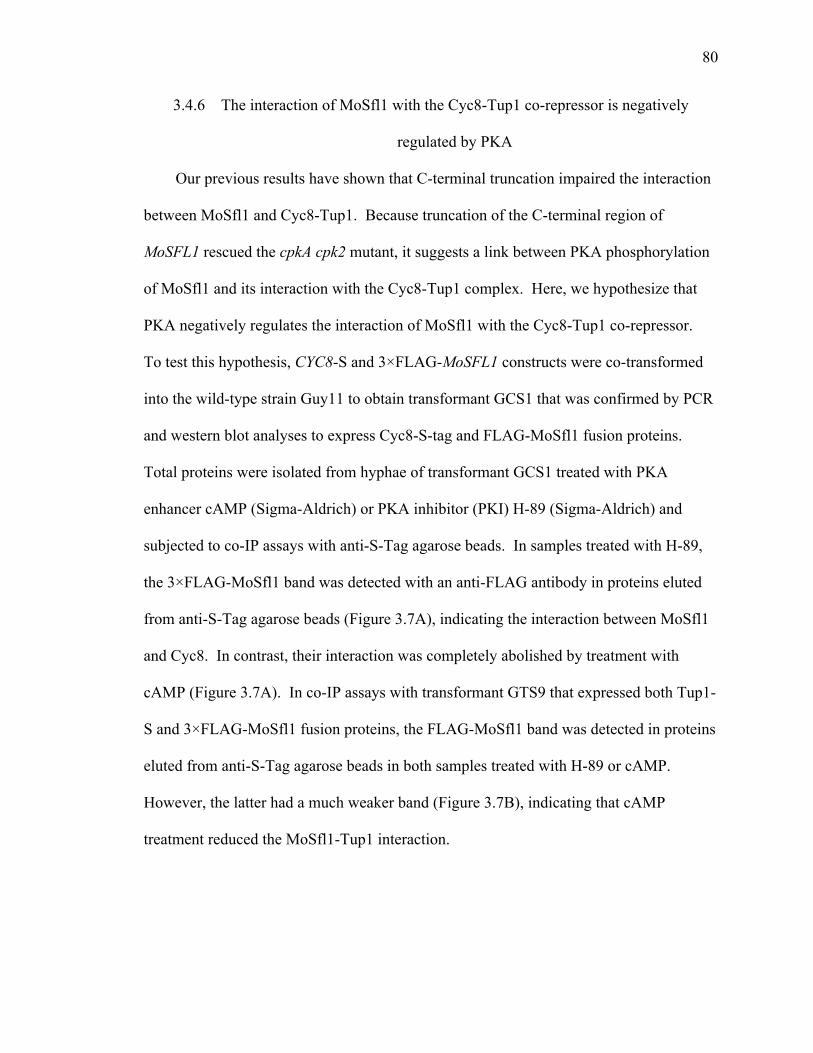
80
80
3.4.6 The interaction of MoSfl1 with the Cyc8-Tup1 co-repressor is negatively
regulated by PKA
Our previous results have shown that C-terminal truncation impaired the interaction
between MoSfl1 and Cyc8-Tup1. Because truncation of the C-terminal region of
MoSFL1 rescued the cpkA cpk2 mutant, it suggests a link between PKA phosphorylation
of MoSfl1 and its interaction with the Cyc8-Tup1 complex. Here, we hypothesize that
PKA negatively regulates the interaction of MoSfl1 with the Cyc8-Tup1 co-repressor.
To test this hypothesis, CYC8-S and 3×FLAG-MoSFL1 constructs were co-transformed
into the wild-type strain Guy11 to obtain transformant GCS1 that was confirmed by PCR
and western blot analyses to express Cyc8-S-tag and FLAG-MoSfl1 fusion proteins.
Total proteins were isolated from hyphae of transformant GCS1 treated with PKA
enhancer cAMP (Sigma-Aldrich) or PKA inhibitor (PKI) H-89 (Sigma-Aldrich) and
subjected to co-IP assays with anti-S-Tag agarose beads. In samples treated with H-89,
the 3×FLAG-MoSfl1 band was detected with an anti-FLAG antibody in proteins eluted
from anti-S-Tag agarose beads (Figure 3.7A), indicating the interaction between MoSfl1
and Cyc8. In contrast, their interaction was completely abolished by treatment with
cAMP (Figure 3.7A). In co-IP assays with transformant GTS9 that expressed both Tup1-
S and 3×FLAG-MoSfl1 fusion proteins, the FLAG-MoSfl1 band was detected in proteins
eluted from anti-S-Tag agarose beads in both samples treated with H-89 or cAMP.
However, the latter had a much weaker band (Figure 3.7B), indicating that cAMP
treatment reduced the MoSfl1-Tup1 interaction.
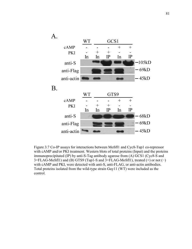
81
81
Figure 3.7 Co-IP assays for interactions between MoSfl1 and Cyc8-Tup1 co-repressor with cAMP and/or PKI treatment. Western blots of total proteins (Input) and the proteins immunoprecipitated (IP) by anti-S-Tag antibody agarose from (A) GCS1 (Cyc8-S and 3×FLAG-MoSfl1) and (B) GTS9 (Tup1-S and 3×FLAG-MoSfl1), treated (+) or not (−) with cAMP and PKI, were detected with anti-S, anti-FLAG, or anti-actin antibodies. Total proteins isolated from the wild-type strain Guy11 (WT) were included as the control.

82
82
3.4.7 Phosphorylation of S211 in MoSfl1 rescues the growth rate of the cpkA cpk2
mutant
In S. cerevisiae, Sfl1 has two putative PKA phosphorylation sites, S207 and S733
(Figure 3.8A). According to CLUSTALW2 alignment analysis, these two
phosphorylation sites are well conserved in MoSfl1 (S211 and S554) and FgSfl1 (S223
and S559) (Figure 3.8A). One additional putative PKA phosphorylation site was
identified in MoSfl1 (T441) and FgSfl1 (T452) (Figure 3.8A). However, the
corresponding site in yeast (RLYS, 568-571) (Figure 3.8A) does not fit with the
consensus PKA phosphorylation site motif [R/K][R/K]-X-[S/T], where X indicates any
amino acid (Zhou, Blechner et al. 1994).
To identify the PKA phosphorylation sites in MoSfl1 and further prove that it is
negatively regulated by PKA, the MoSFL1S211D, MoSFL1T441D and MoSFL1S554D alleles
were generated and transformed into the cpkA cpk2 double mutant to obtain
transformants ASD5, HTD17 and GSD22, respectively (Table 3.1). Changing from
serine or threonine (S/T) to aspartate (D) is assumed to mimic the phosphorylation status.
Of all the transformants, only ASD5 showed fast-growing phenotype compared to the
cpkA cpk2 mutant (Figure 3.8B).

83
83
Figure 3.8 Site-directed mutagenesis of the putative protein kinase A (PKA) phosphorylation sites in MoSfl1. (A) Schematic drawing of the MoSfl1 protein and alignment of the marked region with its orthologs from F. graminearum (Fg) and S. cerevisiae (Sc). The consensus PKA phosphorylation site motifs were boxed with red lines. The putative PKA phosphorylation residues were marked with stars. (B) Colony morphology of the wild-type strain Guy11, the cpkA cpk2 mutant, and the MoSFL1S211D (ASD5), MoSFL1T441D (HTD17) and MoSFL1S554D (GSD22) transformants. Oatmeal agar plates were photographed after incubating at room temperature for 7 days.

84
84
3.5 Discussion
In the fast-growing spontaneous suppressors CCS1 and CCS7, genetic mutations
were identified in MoSFL1, suggesting that these mutations are likely responsible for
suppressor effects. Although the growth rate of the cpkA cpk2 mutant was recovered, the
suppressor strains were still defective in plant infection, suggesting that suppressor
mutations in MoSFL1 could not bypass the PKA function in pathogenicity. In the other
two suppressor strains, CCS4 and CCS14, mutation sites were not identified in the genes
selected for sequencing analysis, indicating that suppressor mutations likely occurred in
other genes besides MoSFL1. It is possible that suppressor mutations occurred in genes
interacting with MoSfl1 or components of the MoSfl1 complex.
In yeast, SFL1 can function as both a transcriptional activator and repressor (Song
and Carlson 1998, Galeote, Alexandre et al. 2007). In M. oryzae, deletion of MoSFL1
did not affect vegetative growth but resulted in a significant reduction in pathogenicity
(Li, Zhou et al. 2011). In this study, we showed that loss-of-function mutations in
MoSFL1 recovered the growth rate but not pathogenicity in the cpkA cpk2 mutant. These
results suggest that MoSFL1 may play an activator role in regulating pathogenesis but
negatively regulate vegetative growth.
In MoSfl1, the only predicted domain is the putative DNA-binding domain of heat
shock transcription factor (HSF). Based on alignment analysis, the C-terminal regions of
Sfl1 orthologs are well conserved in filamentous fungi, suggesting that this region may
have special structures and functions in MoSfl1. Because the conserved C-terminal
region of MoSfl1 and its orthologs from other filamentous fungi is distinct from the yeast
Sfl1, its function may be evolutionarily different from that in yeast. In addition, because

85
85
the Mosfl1ΔCT mutant allele also suppressed the growth defects of the cpkA cpk2 double
mutant, we conclude that the C-terminal region of MoSFL1 is important for its repressor
function.
Previous studies in yeast have shown that Sfl1 inhibits gene transcription by
interacting with the Cyc8-Tup1 co-repressor (Conlan and Tzamarias 2001). In M. oryzae,
the interaction between MoSfl1 and Cyc8 or Tup1 was confirmed by co-IP assays.
Moreover, the conserved C-terminal region of MoSfl1 was shown to play a critical role in
its interaction with Cyc8 and Tup1, which may be directly related to the frameshift
mutation at residue P414, which resulted in a fast-growing suppressor of the cpkA cpk2
mutant.
Besides the Cyc1-Tup1 co-repressor, Sfl1 was also found to interact with
Srb/mediator proteins Ssn2, Ssn8, Sin4, and Rox3 in yeast, which have roles in
transcriptional repression (Song and Carlson 1998). The orthologs of yeast Ssn2, Ssn8,
Sin4, and Rox3 are MGG_06499, MGG_01071, MGG_04587, and MGG_07445 in M.
oryzae, respectively. However, none of them were identified in proteins co-
immunoprecipitated with MoSfl1 in affinity purification. Instead of direct interaction
with Sfl1, the mediator components may be recruited by Cyc8-Tup1 to serve as partners
of Sfl1 to fulfill repression functions. The Cyc8-Tup1-mediated association between Sfl1
and the mediator complex may be too dynamic to be detected in M. oryzae. To date, it is
unclear how Sin4 and Rox3 repress transcription but the role of Ssn2 (Srb9) and Ssn8
(Srb11) are well studied as components of the Srb8-Srb11 module. In fact, Tup1 is found
to directly interact with the Cdk8 (Srb10) subunit to recruit the mediator complex that
contains the Srb8-11 module (Smith and Johnson 2000, Zaman, Ansari et al. 2001).

86
86
Although it is not clear how MoSfl1 represses transcription by interacting with the Cyc8-
Tup1 complex in M. oryzae, one possible model is that MoSfl1 binds to specific
regulatory sequences in the promoter and then recruits the Cyc8-Tup1 co-repressor,
which in turn recruits the Srb8-Srb11 module (Figure 3.9B). In this model, the core
mediator containing the Srb8-Srb11 module represses transcription by preventing the
interactions of target promoters with RNA polymerase ΙΙ and the basal transcription
machinery (Figure 3.9B) (Borggrefe, Davis et al. 2002, Samuelsen, Baraznenok et al.
2003, Guglielmi, van Berkum et al. 2004).
In yeast, both genetic and biochemical evidences indicate that Sfl1 is the substrate of
PKA catalytic subunit Tpk2 (Robertson and Fink 1998, Pan and Heitman 2002) and it is
negatively regulated by PKA (Conlan and Tzamarias 2001). In M. oryzae, we showed
that the MoSfl1S211D phosphorylation mimic allele rescued the growth defect caused by
the deletion of PKA catalytic subunits CPKA and CPK2, indicating that MoSfl1 is the
downstream target of PKA. In addition, the interaction between MoSfl1 and the Cyc8-
Tup1 co-repressor was impaired by stimulating the cAMP-PKA signaling pathway,
suggesting that MoSfl is negatively regulated by PKA. In yeast, phosphorylation of Sfl1
by PKA prohibits its dimerization and reduces its DNA binding efficiency. In this study,
our results showed that PKA negatively regulates MoSfl1 and its interaction with the
Cyc8-Tup1 co-repressor.
How does the suppressor mutation in MoSfl1 bypass absence of PKA activity? In
the presence of PKA, MoSfl1 is negatively regulated to prohibit its interaction with the
Cyc8-Tup1 co-repressor. As a result, the repression function of MoSfl1 is inhibited by
PKA phosphorylation (Figure 3.9A). In suppressor strains CCS1 and CCS7, the same

87
87
effect is achieved by genetic mutation. Both P414fs and M131fs mutations result in the
disassociation of MoSfl1 from the Cyc8-Tup1 co-repressor without phosphorylation by
PKA (Figure 3.9C), which in turn leads to the release of the Srb8-Srb11 module.
Meanwhile, the core mediator complex lack of the Srb8-Srb11 module recruited by other
transcription activators, such as Flo8 (Kim, Lee et al. 2014), serves for the assembly of
the pol ΙΙ subunits to start gene transcription (Figure 3.9A and C) (Allen and Taatjes
2015).

88
88
Figure 3.9 A model of suppressor mutations in MoSfl1 bypassing the requirement for phosphorylation by PKA. (A) In the wild-type strain, phosphorylation of MoSfl1 by PKA prohibits its interaction with the Cyc8-Tup1 co-repressor. The core mediator complex (orange) combining with activator (ACT, red) recruits the RNA polymerase ΙΙ (pol ΙΙ) subunits (green) to initiate gene transcription (Allen and Taatjes 2015). (B) In the cpkA cpk2 mutant, un-phosphorylated MoSfl1 interacts with the Cyc8-Tup1 co-repressor, which in turn recruits the Srb8-Srb11 module (pink). The Srb8-Srb11-containing mediator represses transcription by preventing its interactions with pol ΙΙ (green) and the basal transcription machinery (II B, II D, II E, II F and II H, green) (Bjorklund and Gustafsson 2005). (C) MoSfl1 with suppressor mutations is dissociated from the Cyc8-Tup1 co-suppressor to bypass the absence of PKA activity, which recovers the transcription of genes repressed by MoSfl1.

89
89
3.6 List of References
Adachi, K. and J. E. Hamer (1998). "Divergent cAMP signaling pathways regulate growth and pathogenesis in the rice blast fungus Magnaporthe grisea." Plant Cell 10(8): 1361-1374.
Allen, B. L. and D. J. Taatjes (2015). "The Mediator complex: a central integrator of transcription." Nat Rev Mol Cell Biol 16(3): 155-166.
Bjorklund, S. and C. M. Gustafsson (2005). "The yeast Mediator complex and its regulation." Trends Biochem Sci 30(5): 240-244.
Borggrefe, T., R. Davis, H. Erdjument-Bromage, P. Tempst and R. D. Kornberg (2002). "A complex of the Srb8, -9, -10, and -11 transcriptional regulatory proteins from yeast." J Biol Chem 277(46): 44202-44207.
Braun, B. R. and A. D. Johnson (1997). "Control of filament formation in Candida albicans by the transcriptional repressor TUP1." Science 277(5322): 105-109.
Choi, Y. E. and J. R. Xu (2010). "The cAMP signaling pathway in Fusarium verticillioides is important for conidiation, plant infection, and stress responses but not fumonisin production." Mol Plant Microbe Interact 23(4): 522-533.
Conlan, R. S. and D. Tzamarias (2001). "Sfl1 functions via the co-repressor Ssn6-Tup1 and the cAMP-dependent protein kinase Tpk2." J Mol Biol 309(5): 1007-1015.
Davie, J. K., D. G. Edmondson, C. B. Coco and S. Y. Dent (2003). "Tup1-Ssn6 interacts with multiple class I histone deacetylases in vivo." J Biol Chem 278(50): 50158-50162.
Fisher, A. L. and M. Caudy (1998). "Groucho proteins: transcriptional corepressors for specific subsets of DNA-binding transcription factors in vertebrates and invertebrates." Genes Dev 12(13): 1931-1940.
Fujita, A., Y. Kikuchi, S. Kuhara, Y. Misumi, S. Matsumoto and H. Kobayashi (1989). "Domains of the SFL1 protein of yeasts are homologous to Myc oncoproteins or yeast heat-shock transcription factor." Gene 85(2): 321-328.
Galeote, V. A., H. Alexandre, B. Bach, P. Delobel, S. Dequin and B. Blondin (2007). "Sfl1p acts as an activator of the HSP30 gene in Saccharomyces cerevisiae." Curr Genet 52(2): 55-63.
Garrett, S. and J. Broach (1989). "Loss of Ras activity in Saccharomyces cerevisiae is suppressed by disruptions of a new kinase gene, YAKI, whose product may act downstream of the cAMP-dependent protein kinase." Genes & Development 3(9): 1336-1348.
Guglielmi, B., N. L. van Berkum, B. Klapholz, T. Bijma, M. Boube, C. Boschiero, H. M. Bourbon, F. C. Holstege and M. Werner (2004). "A high resolution protein interaction map of the yeast Mediator complex." Nucleic Acids Res 32(18): 5379-5391.
Heckman, K. L. and L. R. Pease (2007). "Gene splicing and mutagenesis by PCR-driven overlap extension." Nat. Protoc. 2(4): 924-932.
Hu, S., X. Zhou, X. Gu, S. Cao, C. Wang and J. R. Xu (2014). "The cAMP-PKA pathway regulates growth, sexual and asexual differentiation, and pathogenesis in Fusarium graminearum." Mol Plant Microbe Interact 27(6): 557-566.

90
90
Kim, H. S., S. Y. Park, S. Lee, E. L. Adams, K. Czymmek and S. Kang (2011). "Loss of cAMP-dependent protein kinase A affects multiple traits important for root pathogenesis by Fusarium oxysporum." Mol Plant Microbe Interact 24(6): 719-732.
Kim, H. Y., S. B. Lee, H. S. Kang, G. T. Oh and T. Kim (2014). "Two distinct domains of Flo8 activator mediates its role in transcriptional activation and the physical interaction with Mss11." Biochem Biophys Res Commun 449(2): 202-207.
Leung, H., E. S. Borromeo, M. A. Bernardo and J. L. Notteghem (1988). "Genetic analysis of virulence in the rice blast fungus Magnaporthe grisea." Phytopathology 78(9): 1227-1233.
Li, G., X. Zhou, L. Kong, Y. Wang, H. Zhang, H. Zhu, T. K. Mitchell, R. A. Dean and J. R. Xu (2011). "MoSfl1 is important for virulence and heat tolerance in Magnaporthe oryzae." PLoS One 6(5): e19951.
Li, L., C. Y. Xue, K. Bruno, M. Nishimura and J. R. Xu (2004). "Two PAK kinase genes, CHM1 and MST20, have distinct functions in Magnaporthe grisea." Mol. Plant Microbe. In. 17(5): 547-556.
Li, Y., G. Wang, J. R. Xu and C. Jiang (2016). "Penetration Peg Formation and Invasive Hyphae Development Require Stage-Specific Activation of MoGTI1 in Magnaporthe oryzae." Mol Plant Microbe Interact 29(1): 36-45.
Liu, W., A. Iliuk, A. Tao and S. Ding (2011). "Identifying protein complexes by affinity purification and mass spectrometry analysis in the rice blast fungus." Methods Mol Biol 722: 157-166.
Liu, Z. and V. Karmarkar (2008). "Groucho/Tup1 family co-repressors in plant development." Trends Plant Sci 13(3): 137-144.
Malave, T. M. and S. Y. Dent (2006). "Transcriptional repression by Tup1-Ssn6." Biochem Cell Biol 84(4): 437-443.
Ni, M., S. Rierson, J. A. Seo and J. H. Yu (2005). "The pkaB gene encoding the secondary protein kinase A catalytic subunit has a synthetic lethal interaction with pkaA and plays overlapping and opposite roles in Aspergillus nidulans." Eukaryot Cell 4(8): 1465-1476.
Pan, X. and J. Heitman (2002). "Protein kinase A operates a molecular switch that governs yeast pseudohyphal differentiation." Mol Cell Biol 22(12): 3981-3993.
Park, G., C. Xue, X. Zhao, Y. Kim, M. Orbach and J. R. Xu (2006). "Multiple upstream signals converge on the adaptor protein Mst50 in Magnaporthe grisea." Plant Cell 18(10): 2822-2835.
Park, G., G. Y. Xue, L. Zheng, S. Lam and J. R. Xu (2002). "MST12 regulates infectious growth but not appressorium formation in the rice blast fungus Magnaporthe grisea." Mol. Plant Microbe. In. 15(3): 183-192.
Parnell, E. J. and D. J. Stillman (2011). "Shields up: the Tup1-Cyc8 repressor complex blocks coactivator recruitment." Genes Dev 25(23): 2429-2435.
Reinders, A., N. Bürckert, T. Boller, A. Wiemken and C. De Virgilio (1998). "Saccharomyces cerevisiae cAMP-dependent protein kinase controls entry into stationary phase through the Rim15p protein kinase." Genes & Development 12(18): 2943-2955.

91
91
Robertson, L. S. and G. R. Fink (1998). "The three yeast A kinases have specific signaling functions in pseudohyphal growth." Proc Natl Acad Sci U S A 95(23): 13783-13787.
Rupp, S., E. Summers, H. J. Lo, H. Madhani and G. Fink (1999). "MAP kinase and cAMP filamentation signaling pathways converge on the unusually large promoter of the yeast FLO11 gene." EMBO J 18(5): 1257-1269.
Samuelsen, C. O., V. Baraznenok, O. Khorosjutina, H. Spahr, T. Kieselbach, S. Holmberg and C. M. Gustafsson (2003). "TRAP230/ARC240 and TRAP240/ARC250 Mediator subunits are functionally conserved through evolution." Proc Natl Acad Sci U S A 100(11): 6422-6427.
Schumacher, J., L. Kokkelink, C. Huesmann, D. Jimenez-Teja, I. G. Collado, R. Barakat, P. Tudzynski and B. Tudzynski (2008). "The cAMP-dependent signaling pathway and its role in conidial germination, growth, and virulence of the gray mold Botrytis cinerea." Mol Plant Microbe Interact 21(11): 1443-1459.
Smith, A., M. P. Ward and S. Garrett (1998). "Yeast PKA represses Msn2p/Msn4p-dependent gene expression to regulate growth, stress response and glycogen accumulation." Embo Journal 17(13): 3556-3564.
Smith, R. L. and A. D. Johnson (2000). "Turning genes off by Ssn6-Tup1: a conserved system of transcriptional repression in eukaryotes." Trends Biochem Sci 25(7): 325-330.
Song, W. and M. Carlson (1998). "Srb/mediator proteins interact functionally and physically with transcriptional repressor Sfl1." EMBO J 17(19): 5757-5765.
Toda, T., S. Cameron, P. Sass and M. Wigler (1988). "SCH9, a gene of Saccharomyces cerevisiae that encodes a protein distinct from, but functionally and structurally related to, cAMP-dependent protein kinase catalytic subunits." Genes Dev 2(5): 517-527.
Tzamarias, D. and K. Struhl (1994). "Functional dissection of the yeast Cyc8-Tup1 transcriptional co-repressor complex." Nature 369(6483): 758-761.
Tzima, A., E. J. Paplomatas, P. Rauyaree and S. Kang (2010). "Roles of the catalytic subunit of cAMP-dependent protein kinase A in virulence and development of the soilborne plant pathogen Verticillium dahliae." Fungal Genet Biol 47(5): 406-415.
Ward, M. P. and S. Garrett (1994). "Suppression of a yeast cyclic AMP-dependent protein kinase defect by overexpression of SOK1, a yeast gene exhibiting sequence similarity to a developmentally regulated mouse gene." Molecular and Cellular Biology 14(9): 5619-5627.
Wong, K. H. and K. Struhl (2011). "The Cyc8-Tup1 complex inhibits transcription primarily by masking the activation domain of the recruiting protein." Genes Dev 25(23): 2525-2539.
Xu, J. R. and J. E. Hamer (1996). "MAP kinase and cAMP signaling regulate infection structure formation and pathogenic growth in the rice blast fungus Magnaporthe grisea." Gene Dev. 10(21): 2696-2706.

92
92
Yan, X., Y. Li, X. Yue, C. Wang, Y. Que, D. Kong, Z. Ma, N. Talbot and Z. Wang (2011). "Two novel transcriptional regulators are essential for infection-related morphogenesis and pathogenicity of the rice blast fungus Magnaporthe oryzae." Plos Pathogens 7(12).
Yu, J. H., Z. Hamari, K. H. Han, J. A. Seo, Y. Reyes-Dominguez and C. Scazzocchio (2004). "Double-joint PCR: a PCR-based molecular tool for gene manipulations in filamentous fungi." Fungal Genet Biol 41(11): 973-981.
Zaman, Z., A. Z. Ansari, S. S. Koh, R. Young and M. Ptashne (2001). "Interaction of a transcriptional repressor with the RNA polymerase II holoenzyme plays a crucial role in repression." Proc Natl Acad Sci U S A 98(5): 2550-2554.
Zhou, S. Y., S. Blechner, N. Hoagland, M. F. Hoekstra, H. Piwnicaworms and L. C. Cantley (1994). "Use of an Oriented Peptide Library to Determine the Optimal Substrates of Protein-Kinases." Current Biology 4(11): 973-982.
Zhou, X., G. Li and J. R. Xu (2011). "Efficient approaches for generating GFP fusion and epitope-tagging constructs in filamentous fungi." Methods Mol Biol 722: 199-212.
Zhou, X. Y., W. D. Liu, C. F. Wang, Q. J. Xu, Y. Wang, S. L. Ding and J. R. Xu (2011). "A MADS-box transcription factor MoMcm1 is required for male fertility, microconidium production and virulence in Magnaporthe oryzae." Mol. Microbiol. 80(1): 33-53.
Zhou, X. Y., H. F. Zhang, G. T. Li, B. Shaw and J. R. Xu (2012). "The cyclase-associated protein Cap1 is important for proper regulation of infection-related morphogenesis in Magnaporthe oryzae." PLoS Pathog. 8(9).

93
93
VITA

93
93
VITA
Name: Yang Li
Data of Birth: 10/08/1985
Place of Birth: Hubei, China
EDUCATION
Present Ph.D. student in Department of Botany and Plant Pathology
Purdue University, West Lafayette, Indiana
2011 M.S. in Plant Pathology
China Agricultural University, Beijing, China
2008 Bachelor in Plant Protection
China Agricultural University, Beijing, China

PUBLICATIONS

94
94
PUBLICATIONS
1. Rikno Harmoko, Jae Yong Yoo, Ki Seong Ko, Nirmal Kumar Ramasamy, Bo Young
Hwang, Eun Ji Lee, Ho Soo Kim, Kyung Jin Lee, Doo-Byoung Oh, Dool-Yi Kim,
Sanghun Lee, Yang Li, Sang Yeol Lee, Kyun Oh Lee. 2016. N-glycan containing a
core alpha1,3-fucose residue is required for basipetal auxin transport and
gravitropic response in rice (Oryza sativa). The New phytologist
2. Xuli Gao, Qiaojun Jin, Cong Jiang, Yang Li, Chaohui Li, Huiquan Liu, Zhensheng
Kang, Jin-Rong Xu. 2016. FgPrp4 Kinase Is Important for Spliceosome B-Complex
Activation and Splicing Efficiency in Fusarium graminearum. PLoS genetics 12:
e1005973
3. Huiquan Liu, Qinhu Wang, Yi He, Lingfeng Chen, Chaofeng Hao, Cong Jiang, Yang
Li, Yafeng Dai, Zhensheng Kang, Jin-Rong Xu. 2016. Genome-wide A-to-I RNA
editing in fungi independent of ADAR enzymes. Genome research 26: 499-509
4. Yang Li, Guanghui Wang, Jin-Rong Xu, Cong Jiang. 2016. Penetration Peg
Formation and Invasive Hyphae Development Require Stage-Specific Activation of
MoGTI1 in Magnaporthe oryzae. Molecular plant-microbe interactions : MPMI 29:
36-45

95
95
5. Linlu Qi, Yangseon Kim, Cong Jiang, Yang Li, Youliang Peng, Jin-Rong Xu. 2015.
Activation of Mst11 and Feedback Inhibition of Germ Tube Growth in Magnaporthe
oryzae. Molecular plant-microbe interactions : MPMI 28: 881-91
6. Huili Zhang, Zhongshou Wu, Chenfang Wang, Yang Li, Jin-Rong Xu. 2014.
Germination and infectivity of microconidia in the rice blast fungus Magnaporthe
oryzae. Nat Commun 5: 4518