protein homeostasis in models of aging and age … · protein homeostasis in models of aging and...
TRANSCRIPT

Chapter 11
*Corresponding Author: Richard I. Morimoto—Department of Biochemistry, Molecular Biology and Cell Biology, Rice Institute for Biomedical Research, 2205 Tech Drive, Hogan 2-100,Northwestern University, Evanston, Illinois 60208. USA. Email: [email protected]
Protein Metabolism and Homeostasis in Aging, edited by Nektarios Tavernarakis. gg©2010 Landes Bioscience and Springer Science+Business Media.
Protein Homeostasis in Modelsof Aging and Age-Related Conformational DiseaseElise A. Kikis†, Tali Gidalevitz† and Richard I. Morimoto*
Abstract
The stability of the proteome is crucial to the health of the cell, and contributes significantly to the lifespan of the organism. Aging and many age-related diseases have in common the expression of misfolded and damaged proteins. The chronic expression of damaged
proteins during disease can have devastating consequences on protein homeostasis (proteosta-sis), resulting in disruption of numerous biological processes. This chapter discusses our currentunderstanding of the various contributors to protein misfolding, and the mechanisms by which misfolding, and accompanied aggregation/toxicity, is accelerated by stress and aging. Invertebrate models have been instrumental in studying the processes related to aggregation and toxicity of disease-associated proteins and how dysregulation of proteostasis leads to neurodegenerativediseases of aging.
Protein Folding Problem in AgingProteins are the predominant products of gene expression and contribute significantly to the
shape and functionality of the cell. The status of the expressed proteome has an important role in the health of individual cells and the lifespan of the organism. In addition to cell type- andtissue-specific regulation of protein expression, maintenance of the proteome depends on efficientprotein folding homeostasis, or proteostasis, that monitors and ensures folding, assembly and targeting of newly-synthesized proteins, repair of damaged proteins and clearance. Proteotoxic conditions arise by external stresses, or as a byproduct of normal cellular metabolic and signaling events dur-ing development and aging (Fig. 1). This is in addition to the intrinsic variation in the proteomedue to polymorphisms, which is further exacerbated by random errors that can occur at every step of protein biogenesis. Together, these factors contribute to a flux of metastable proteins that are at risk for misfolding and aggregation.1-4 In healthy young cells, these processes are balanced by the concerted action of molecular chaperones, detoxifying enzymes, degradation machinery andadaptive stress responses (Fig. 2).5-7 In aging and disease, damaged proteins accumulate, leading to both loss-of-function and gain-of-function toxicity as these homeostatic mechanisms fail andcontribute to pathology.
†These authors contributed equally

139Protein Homeostasis in Models of Aging and Age-Related Conformational Disease
Sources of Protein Damage—Oxidative ModificationsThe free radical theory‘ ’8 and the ‘oxidative stress theory’ 9,10 of aging postulate that aging and
many age-related diseases may be attributed to the generation of oxygen free radicals and reactive oxygen and nitrogen species (ROS and RNS), in excess of cellular antioxidants, resulting in oxida-tive damage to DNA, lipids and proteins. Early evidence connected many of the aging-associateddiseases, including atherosclerosis, arthritis, muscular dystrophy, cataracts, pulmonary dysfunction, neurological disorders and cancer with oxidative damage.11 Levels of oxidized proteins increaseexponentially in regions of aging brains, concomitant with a decrease in the activities of the enzymes glutamine synthetase and alcohol dehydrogenase, which are susceptible to oxidative damage.12 The decrease in glutamine synthetase activity was found to distinguish the brains of Alzheimer’s disease(AD) patients from age-matched individuals, leading to the suggestion that AD may represent a specific brain vulnerability to age-related oxidation. Furthermore, a functional connection betweenthe accumulation of oxidized proteins and the phenotypic manifestations of cellular disfunction in aging has been suggested.13
In addition to oxygen radicals, glucose, galactose, fructose and many glycolytic intermediatesparticipate in non-enzymatic protein glycosylation (glycation) and glycoxidation, contributing to age-related protein modifications. Some glycolytic intermediates can generate methylglyoxal (MG), which is a highly reactive glycating agent,14,15 leading to the formation of advanced glycation end products (AGEs), that have been implicated in age-related diseases, including Alzheimer’s disease and complications associated with diabetes.15-17 A decrease in circulating glucose and reduction of MG, resulting from dietary restriction or fasting, may explain some of the health-improving effects of these treatments.18 Recently, an inactivating mutation in a glycolytic enzyme triosephosphate isomerase (Tpi) was shown to cause progressive motor impairment, severely reduced lifespan and neurodegeneration in Drosophila, presumably by causing an accumulation of methylglyoxal and subsequent increase in generation of AGEs.19
Figure 1. Extrinsic and intrinsic factors detrimental to protein folding.

140 Protein Metabolism and Homeostasis in Aging
Loss of protein function due to oxidative modification can be due either to direct inhibition of activity of a protein (for example, covalent modification of a side chain in proximity of an active site), or to conformational changes or alteration of protein stability. Most modifications of the first type are irreversible, with the exception of cysteine and methionine modifications,which can be reversed by thiol transferases, thioredoxin, glutaredoxin and methionine sulfoxide reductases (MsrA and MsrB). The detoxification of methylglyoxal and glyoxal is achieved by the glutathione-dependent glyoxalase system. These and other enzymes are part of the cellular proteostasis machinery that is activated in response to various protein damaging conditions.For example, MrsA and B allow recovery of protein function in specific cases20,21 and theiroverexpression confers protection against oxidative damage in yeast, Drosophila and humanfibroblasts.21
In addition to functional inactivation of proteins, free radicals can have a local effect on charged side chains, or cause cleavage of the polypeptide backbone, leading to a spectrum of consequences, from complete unfolding, to local conformational changes,22-24 to targeting for disposal. If not degraded, modified proteins often become cross-linked, leading to the accumulation of nonspe-cific aggregated material, such as lipofuscin, ceroids and AGE-containing pigments. Nuclear and cytosolic oxidized proteins have been shown to be degraded by an ATP- and ubiquitin-independent 20S proteasome25,26 as well as by classical ubiquitin-26S proteasome system,27 whereas mito-chondrial oxidized proteins are degraded by the ATP-dependent Lon protease.28 Interestingly, the 20S proteasome appears to be resistant to oxidative damage, while 26S proteasome is not.29
Chaperone-mediated autophagy represents an alternative protein degradation pathway that isinduced under oxidative stress conditions.30 Under chronic oxidative stress conditions, there seems
Figure 2. Proteostasis pathways. Multiple interconnected pathways regulate expression of genes that contribute to maintenance of protein folding homeostasis during growth, development, and under various stress conditions. The complex genetic interactions among these pathways necessitate precise control over their activities. Many of these pathways also possess regula-tory feedback mechanisms, which are not well characterized/understood.
141Protein Homeostasis in Models of Aging and Age-Related Conformational Disease
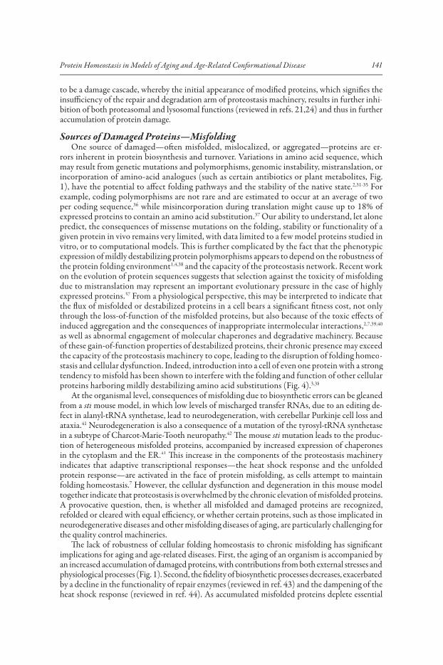
to be a damage cascade, whereby the initial appearance of modified proteins, which signifies theinsufficiency of the repair and degradation arm of proteostasis machinery, results in further inhi-bition of both proteasomal and lysosomal functions (reviewed in refs. 21,24) and thus in furtheraccumulation of protein damage.
Sources of Damaged Proteins—MisfoldingOne source of damaged—often misfolded, mislocalized, or aggregated—proteins are er-
rors inherent in protein biosynthesis and turnover. Variations in amino acid sequence, whichmay result from genetic mutations and polymorphisms, genomic instability, mistranslation, orincorporation of amino-acid analogues (such as certain antibiotics or plant metabolites, Fig.1), have the potential to affect folding pathways and the stability of the native state.2,31-35 For example, coding polymorphisms are not rare and are estimated to occur at an average of two per coding sequence,36 while misincorporation during translation might cause up to 18% of expressed proteins to contain an amino acid substitution.37 Our ability to understand, let alonepredict, the consequences of missense mutations on the folding, stability or functionality of a given protein in vivo remains very limited, with data limited to a few model proteins studied in vitro, or to computational models. This is further complicated by the fact that the phenotypicexpression of mildly destabilizing protein polymorphisms appears to depend on the robustness of the protein folding environment1,4,38 and the capacity of the proteostasis network. Recent work on the evolution of protein sequences suggests that selection against the toxicity of misfolding due to mistranslation may represent an important evolutionary pressure in the case of highly expressed proteins.37 From a physiological perspective, this may be interpreted to indicate that the flux of misfolded or destabilized proteins in a cell bears a significant fitness cost, not only through the loss-of-function of the misfolded proteins, but also because of the toxic effects of induced aggregation and the consequences of inappropriate intermolecular interactions,2,7,39,40
as well as abnormal engagement of molecular chaperones and degradative machinery. Because of these gain-of-function properties of destabilized proteins, their chronic presence may exceedthe capacity of the proteostasis machinery to cope, leading to the disruption of folding homeo-stasis and cellular dysfunction. Indeed, introduction into a cell of even one protein with a strong tendency to misfold has been shown to interfere with the folding and function of other cellularproteins harboring mildly destabilizing amino acid substitutions (Fig. 4).3,38
At the organismal level, consequences of misfolding due to biosynthetic errors can be gleanedfrom a sti mouse model, in which low levels of mischarged transfer RNAs, due to an editing de-fect in alanyl-tRNA synthetase, lead to neurodegeneration, with cerebellar Purkinje cell loss and ataxia.41 Neurodegeneration is also a consequence of a mutation of the tyrosyl-tRNA synthetasein a subtype of Charcot-Marie-Tooth neuropathy.42 The mouse sti mutation leads to the produc-tion of heterogeneous misfolded proteins, accompanied by increased expression of chaperonesin the cytoplasm and the ER.41 This increase in the components of the proteostasis machinery indicates that adaptive transcriptional responses—the heat shock response and the unfoldedprotein response—are activated in the face of protein misfolding, as cells attempt to maintainfolding homeostasis.7 However, the cellular dysfunction and degeneration in this mouse modeltogether indicate that proteostasis is overwhelmed by the chronic elevation of misfolded proteins. A provocative question, then, is whether all misfolded and damaged proteins are recognized,refolded or cleared with equal efficiency, or whether certain proteins, such as those implicated inneurodegenerative diseases and other misfolding diseases of aging, are particularly challenging forthe quality control machineries.
The lack of robustness of cellular folding homeostasis to chronic misfolding has significantimplications for aging and age-related diseases. First, the aging of an organism is accompanied by an increased accumulation of damaged proteins, with contributions from both external stresses and physiological processes (Fig. 1). Second, the fidelity of biosynthetic processes decreases, exacerbatedby a decline in the functionality of repair enzymes (reviewed in ref. 43) and the dampening of theheat shock response (reviewed in ref. 44). As accumulated misfolded proteins deplete essential

142 Protein Metabolism and Homeostasis in Aging
components of the proteostasis machinery45-47y and the quality control and repair mechanismsfail to respond and keep damaged proteins in check, the maintenance of the proteome becomes further compromised. With as much as 70% of rare missense alleles predicted to be mildly deleteri-ous in humans48 and approximately half of the genetic changes known to cause inherited disease (in OMIM and HGMD databases) being due to nonsynonymous changes,33 such compromiseshould manifest in a gradual increase in cellular dysfunction and onset of disease. In this scenario,the strength and composition of the proteostatic buffer, the specific complement of mutationsand polymorphisms in expressed genes and the accumulation of molecular damage, all serve toset a dynamic threshold for the onset of dysfunction, both in a cell-specific and an individual organism-specific manner.
Proteostasis Regulation in Aging and Late Onset DiseasesAdaptive transcriptional stress responses such as the heat shock response and the unfolded
protein response (UPR) serve to protect cells from extreme flux of misfolded and damaged proteins when cells or organism are exposed to an acute proteotoxic condition (reviewed in refs. 49,50). Parallel to the sharply increased transcription/translation of molecular chaperones andcomponents of degradative quality control machineries, mediated by the transcription factorHSF1, a generalized inhibition of protein synthesis allows the switch to refolding or degradation of existing proteins that become unstable during such proteotoxic shock.51 However, the factthat HSF1,52,53 the components of the UPR signaling,54-56 as well as some of the stress-induciblemolecular chaperones are essential for life or development suggests that these adaptive stressresponses are also necessary for the maintenance of proteostasis under normal, nonstress con-ditions. On the other hand, forced overexpression of molecular chaperones is deleterious to growth of normal cells,57,58 while the transformed phenotype of cancer cells depends on HSF1and on an increased expression of chaperones.59-61 It appears then that the precise regulation of proteostasis networks is essential for the health of the organism, while accumulating evidencepoints to its disregulation during aging as one of the main causes of cellular dysfunction and disease onset.
The association with aging is one of the most distinctive characteristics of protein confor-mation diseases. This connection is particularly striking in neurodegeneration where, for eachspecific disease, age is the strongest predictor of disease onset even for the familial variants. On the other hand, age appears to have a modifying, rather than causative, influence on disease onset, as each disease has its characteristic age of onset, with Alzheimer’s disease and Parkinson’s diseasebeing late onset, ALS occurring most frequently in early to mid-life and Huntington’s diseaseexhibiting a strong positive correlation between age of onset and polyQ length polymorphism.62
Aging has been shown to be a potent enhancer of aggregation/toxicity in C. elegans, in which length-dependent polyQ aggregation and toxicity phenotypes are enhanced during aging, but suppressed in animals rendered long-lived by mutations in the lifespan regulating insulin-likesignaling (ILS) pathway (Fig. 2).63
The modulatory effects of aging are most likely due to the functional and regulatory connectionsbetween aging and protein homeostasis. Activation of the kinase activity of IP3K (AGE-1), by downregulation of the insulin-like growth factor receptor DAF-2, in wild type C. elegans, initiates a signaling cascade that represses the fork head transcription factor DAF-16.64,65 De-repression of DAF-16 in age-1 or daf-2 mutant animals extends lifespan, while inactivating daf-16 mutations6suppress longevity. The daf-2/age-1 effects on polyQ aggregation toxicity also require DAF-16activation, revealing that the dual effects of daf-2/age-1 on longevity and polyglutamine toxicity share a common genetic pathway (ILS pathway, Fig. 2).66,67 The modulation of protein misfolding and aggregation by the ILS pathway appears to be a general feature and has also been observed in C. elegans expressing A� as a model for Alzheimer’s disease.68
How do genetic pathways that regulate lifespan suppress proteotoxicity? The molecular interactions between these pathways are mediated, in part, by factors that detect and respondto misfolded proteins—molecular chaperones, HSF1, DAF-16 and other transcription factors

143Protein Homeostasis in Models of Aging and Age-Related Conformational Disease
(Fig. 2), and molecular chaperones have been shown to accumulate in the long-lived mutantsof C. elegans. Downregulation of hsf-1 suppresses both the ILS-mediated lifespan extension and the protection against proteotoxicity. Moreover, hsf-1 downregulation leads to a decrease innormal lifespan and an accelerated aging phenotype in C. elegans, while overexpression of HSF1extends lifespan.66,67 The functional relationship between ILS and protein folding homeostasis can be demonstrated by the induction of both thermotolerance and life span extension not only by mutations in the ILS pathway, but also by a sublethal heat stress.69 Furthermore, cells from naturally long-lived70 or lifespan mutant71 rodents appear to be resistant to multiple proteotoxic stresses. The mechanistic relationship can be shown by the induction of stress resistance by ablation of cells making insulin-like ligands in wild type Drosophila,72 thus excluding indirect effects and adaptation to mutations. In C. elegans, inactivation of daf-16, 66 hsf-1, hsp-1 (the major cytosolic Hsp70 chaperone) and small heat shock proteins accelerates polyQ protein aggrega-tion (Fig. 3C), supporting the proposition that the ILS could coordinately influence aging and proteostasis through the action of DAF-16 and HSF1.66,67 The regulation of ILS pathway and of HSF1 and other transcription factors (such as SKN-1,73 the C. elegans Nrf2 homologue) in the context of adaptive stress responses must serve to integrate organismal growth and development with multiple networks that regulate different aspects of protein homeostasis.7,66-68 We are only now beginning to understand how such integration could be achieved, with recent evidenceshowing that the ability of individual cells in an organism to respond to proteotoxic conditionsis controlled by the activity of a subset of neurons.74
In addition to the control of the heat shock response by neuronal signaling, there have beennumerous observations in which the heat shock response is poorly or incompletely activated,including early in development.75 Of particular interest are studies of the deficiency of the heatshock response in the brain and during aging.76,77 Restricted expression of heat shock genes hasbeen observed in different regions of the brain, while neuronal cells in culture can exhibit selec-tive induction of chaperone genes. Human neuroblastoma Y79 cells, for example, respond to heat shock by induction of many chaperones including Hsp90, but not Hsp70, despite activation of HSF1.78 Intact primary hippocampal neurons from neonatal rat embryos express HSF2 but not HSF1 until later stages of development.79 Consequently, the heat shock response of primary hip-pocampal neurons is deficient, while astrocytes have a robust stress response. Similar observationshave been made in primary motor neurons that exhibit a deficient heat shock response thoughtto be due to a defect in activation of HSF1.80
The involvement of proteostasis networks in conformational diseases and aging is underscoredby a decrease in toxicity when individual molecular chaperones are overexpressed in various cell-based and animal models (reviewed in ref. 81). Overexpression of HSF1 and certain mo-lecular chaperones has been shown to extend lifespan.66,67,82,83 Furthermore, the ability to induce the heat shock response under conditions of environmental proteotoxic stress is predictive of the remaining poststress lifespan.84,85 Likewise, proteasomal adaptation (by modulation of substrateaccessibility to the proteasome core) to environmental stress in C. elegans ensures both resistanceto proteotoxic conditions and maintenance of lifespan under normal conditions, arguably through regulating the degradation of misfolded proteins.86 Moreover, because both HSF-1 and DAF-16 are regulated by the NAD-dependent sirtuin, SIRT1,87-89 in addition to the ILS-pathway, it isreasonable to suggest that regulation of proteostasis is intimately linked by multiple pathways tometabolic control and lifespan.
Late Onset Diseases are Mainly Protein Folding DiseasesCommon features of protein conformation diseases are the accumulation of protein deposits
(such as aggregates, inclusion bodies and plaques) and a presumably consequential “gain-of-func-tion” proteotoxicity.90,91 These features, which are characteristic of misfolded protein species, are present in neurodegenerative diseases of aging such as Parkinson’s disease, amyotrophic lateral sclerosis (ALS), prion disease, Alzheimer’s disease and the family of disorders generally referred toas the CAG-repeat/polyglutamine-expansion diseases (Huntington’s disease), Kennedy’s disease,

144 Protein Metabolism and Homeostasis in Aging
Figure 3. A) SOD1 mutant proteins aggregate in the body wall muscle cells of C. elegans.Fluorescent micrographs of SOD1transgenic embryos (i-iv) and adult animals (ix through xii)showing the distribution of the SOD-YFP fluorescence (green) and Rhodamine-phalloidinstained myofilaments (red). WT SOD1 protein exhibits diffuse, if patchy, fluorescence, while allmutant strains contain discrete fluorescent foci as well as some diffuse fluorescence. Panels vthrough viii show G85R animals in all larval stages (L1 through L4). Adapted from Gidalevitz et al, 2009 (ref. 38). B) Length-dependent aggregation of polyQ-YFP fusion proteins in C. el-egans. Epifluoresence micrographs of 3- to 4-day-old C. elegans expressing different lengths of polyQ-YFP (Q0, Q19, Q29, Q33, Q35, Q40, Q44, Q64, Q82). Adapted from Bieschke etal, 2006 (ref. 63). C) RNAi for hsp-1 and hsf-1 induces aggregate formation of polyglutamineYFP proteins in a Q stretch length-dependent manner. Fluorescence microscopy pictures of 4-day-old C. elegans adults expressing Q0-YFP or Q35-YFP. RNAi of either hsp-1 or hsf-1results in premature onset of aggregation in Q35 animals. Adapted from Nollen et al, 2004(ref. 100). B,C) Copyright National Academy of Sciences, USA. Used with permission. D)HSP70 overexpression suppresses polyglutamine-induced neurodegeneration in Drosophila.Eyes (i-iv) and retinal sections (v,vi) of flies expressing expanded polyglutamine protein and human HSPA1L are shown. i,v, Control fly expressing only the promoter transgene. ii,vi, Flies expressing HSPA1L, coding for Hsp70 protein. Eye structure appears grossly normal. Moredetailed analysis revealed abnormalities in nuclear position and photoreceptor rhabdomere morphology when HSPA1L is highly expressed. iii,vii, Flies expressing the expanded polyglu-tamine protein MJDtr-Q78. These flies have degenerate eyes that lack pigment and show severe loss of retinal structure. iv,viii, Flies expressing both MJDtr-Q78 and HSPA1L. Co-expressionof HSPA1L ameliorates the degenerative effects of MJDtr-Q78. The eye appears normalexternally. Internally, eye structure is largely restored, although photoreceptor rhabdomerespecializations are not made. Adapted from Warrick et al, 1999 (ref. 98).

145Protein Homeostasis in Models of Aging and Age-Related Conformational Disease
spinocerebellar ataxias). Each disease has the distinctive characteristic of age-dependent onset,a progressive, usually fatal, clinical course and selective neuronal vulnerability despite broaderexpression of the causative protein.
Despite the fact that the proteins that are now known to “cause” neurodegenerative diseaseslack similarities in primary sequence (other than the polyQ tract in CAG-repeat expansiondiseases), they all share the ability to form alternate unfolded or misfolded states that in turnaggregate and/or are toxic. Thus, misfolding and aggregation has been proposed to be the mo-lecular underpinning of disease. This is supported by evidence from transgenic model systemsincluding S. cerevisiae, C. elegans and D. melanogaster,92-97 in which it has been possible to reca-pitulate many molecular, cellular and behavioral phenotypes associated with neurodegenerative disease. The development of these nonmammalian models for protein conformational diseasehas been invaluable for the discovery of modifiers, pathways and underlying mechanisms of toxicity and for the testing of small molecules.63,98-101 Additionally, these models have solidified our understanding of the link between protein conformational disorders, molecular chaperonesand proteostasis regulators and aging.66,68
Invertebrate Models of Late Onset Conformational DiseasesThe link between protein misfolding and human neurodegenerative disease and the fact that
the machinery involved in proteostasis maintenance, is highly conserved amongst eukaryotes,has led to the widespread use of invertebrate models systems, such as C. elegans and Drosophila. Ultimately, modeling aspects of human neurodegenerative diseases in invertebrates allows for genetic manipulations, such as mutant screens/identification of modifiers and aging studies that would be prohibitively time consuming if carried out in vertebrate animals.
These studies in C. elegans and D. melanogaster benefit from the sequenced genomes, relatively rshort life cycles and abundant genetic tools of these well-characterized model organisms. C. elegansis particularly well suited for the study of neurodegenerative diseases of aging due to its a relatively simple (but still sufficiently complex) nervous system, its genetically defined aging pathway and the relative ease to perform live cell imaging of fluorescent proteins for studies of disease-causing protein aggregation dynamics. Likewise, in D. melanogaster the photoreceptor neurons have proven rto be highly amenable to these types of studies, as human proteins can be ectopically expressedand the readout for proteotoxicity is revealed by effects on eye morphology.
To date, a relatively large number of invertebrate models of human neurodegenerative diseaseshave been generated. Importantly, these models have solidified our understanding that protein misfolding underlies the mechanism(s) of disease action. Furthermore, the invertebrate models haveopened up new ways to study the molecular underpinnings of disease and to identify genes and genenetworks whose activities are involved, positively or negatively, in modulating aggregation/toxicity.We begin with an overview of the existing models and discuss what has been learned from themgenerally and in what ways they recapitulate key aspects of human disease.
PolyQHuntington’s disease (HD) and a number of related neurodegenerative diseases are caused
Q
genetically by expansions of polyQ-encoding CAG tracts in specific individual genes/proteins.Expressing these proteins, with wild-type or expanded polyQ tracts, in invertebrates has proven tobe an effective way to study the molecular underpinnings of protein misfolding/toxicity associated with polyQ disorders. Specifically, SCA3/MJD (Machado-Joseph disease) (Fig. 3D)92 and HD102
were modeled in Drosophila and shown to recapitulate many important aspects of disease including cell-type specific sensitivity to the expressed protein, nuclear inclusion formation and late-onset cellular degeneration. These findings were furthered by the expression of polyQ proteins in C. elegans (Fig. 3B,C). HD models have been generated via the expression, in neuronal subsets, of anN-terminal fragment of the protein Huntingtin (Htt) with the wild type or a long polyglutamine (polyQ) tract.93,97 Ultimately, it was shown that human Htt with an expanded polyglutamine tractled to aggregate formation in, and apoptotic cell death of, ASH sensory neurons, in a sensitized

146 Protein Metabolism and Homeostasis in Aging
genetic background.93 Additionally, Htt with 128 glutamines fused to YFP aggregated whenexpressed in PLM mechanosensory neurons and caused neuronal abnormalities but not death.97
Having such fluorescently tagged variants of disease-causing proteins in C. elegans has proven highly advantageous by allowing for the real-time monitoring of aggregation and using dynamic imaging techniques in this transparent organism.38,63,96,103
The overall similarities between various polyQ disorders suggested that the polyQ expansions are predominantly, if not solely, responsible for the observed disease pathologies. To address this, general polyQ models, in which only the polyQ tract, without any of its normal flanking protein sequences (but fused in some cases to YFP, or to another protein not usually containing a polyQ tract) were developed (Fig. 3B).63,96,104 The data from these models provided the first direct invivo evidence that polyQ tracts themselves, independently of their protein context, aggregate and are toxic.
The significance of recapitulating key aspects of disease by expressing a single human protein in an invertebrate should not be underestimated. It confirmed that the mutant proteins are generally toxic even across species and in different cell types. This, in turn, lead to the hypothesis that thehighly conserved cellular quality control system, involving protein folding/clearance, is inherently lacking the ability to prevent damage by these polyQ-containing disease-causing proteins.
A�Alzheimer’s disease (AD) is a relatively common neurodegenerative disorder affecting nearly
30% of all individuals older than 85 years of age. It is characterized by the accumulation of �-amyloid plaques and neurofibrillary tangles. The major proteinaceous components of �-amyloid plaquesin AD patients are A� peptides; specifically, A�1-40 and A�1-42, which are produced via proteolysis of APP, the amyloid precursor protein. To model A� aggregation/toxicity in an invertebratemodel system, A�1-42 was expressed in C. elegans body wall muscle cells and was shown to formamyloid plaques that have biochemical characteristics similar to those found in the brains of ADpatients.105 Additionally, A�1-42 and A�1-40 were expressed in Drosophila photoreceptor neurons.106
It was shown that A�1-42 is substantially more toxic than A�1-40,106 consistent with the idea that A�1-42 has a higher amyloidogenic propensity.
TauIn addition to amyloid plaques, AD and other neurodegenerative diseases are characterized
by the formation of neurofibrillary tangles comprised of the protein tau. Expression of mutant tau in specific neuronal subtypes or under pan-neuronal control in Drosophila resulted in neu-rodegeneration, but not neurofibrillary tangle/filamentous aggregate formation, as determined by electron microscopy, suggesting that the tangles themselves are not the source of mutant tautoxicity.107 Expression of tau in all C. elegans neurons resulted in motility defects indicative of tau proteotoxicity, providing further support to the idea that low levels of tau protein, not necessarily neurofibrillarly tau tangles or otherwise insoluble tau, are required for toxicity.108
Alpha-SynucleinFamilial forms of Parkinson’s disease (PD) are caused by mutations in the protein alpha-synu-
clein.109,110 Expression of alpha-synuclein in C. elegans neurons was shown to cause neurodegen-eration of neuronal subsets.111 Expression of alpha-synuclein in body wall muscle cells resulted inage-dependent aggregation, a phenotype that was used to perform a genetic screen for suppressorsof aggregation (see below).112
SOD1Amyotrophic Lateral Sclerosis (ALS) is a neurodegenerative disease that, in its inherited
form, is often associated with mutation of superoxide dismutase 1 (SOD1). A large number of mutations in SOD1 have been identified and shown to be linked to ALS.113 Evidence suggeststhat mutations in SOD1 cause disease not by loss of SOD1 function, but instead by gain of toxicfunction by the mutant protein. Therefore, to model SOD1 protein misfolding/toxicity, these

147Protein Homeostasis in Models of Aging and Age-Related Conformational Disease
mutations have been expressed in a number of genetic systems including mouse, Drosophila andC. elegans. To model SOD1 protein misfolding in Drosophila, wild type human SOD1 (hSOD1),two mutant forms of SOD1 (G85R and A4V) and Drosophila SOD1 (dSOD1) were overex-pressed in Drosophila motor neurons.114 It was shown that unlike dSOD1, wild type and mutanthSOD1 are toxic, but non-aggregating in Drosophila.114 This is in contrast to what was observed when hSOD1 was expressed in C. elegans body wall muscle cells,38 or neurons.115 Namely, expres-sion of YFP-tagged SOD1 (G85R) under the control of a pan-neuronal promoter resulted in motility defects and distinct neuronal subtype-type-specific aggregation phenotypes that wereconfirmed by FRAP analysis.115 Likewise, expression of a number of different disease-associated SOD1 mutants in C. elegans body wall muscle cells resulted in the formation of SOD1 protein aggregates (Fig. 3A).38 Interestingly, the toxic effects of mutant forms of SOD1 were subtlein body wall muscle cells, but enhanced in the background of metastable ts proteins.38 This isconsistent both with the finding that temperature sensitive proteins can exacerbate the toxiceffects of polyQ protein expression (Fig. 4)3 and with the idea that disease phenotypes and/or
Figure 4. Progressive disruption of cellular folding capacity by misfolded proteins. A,B) Confocal images showing cellular localization of ts mutant paramyosin in the absence (A) or presence(B) of Q40-YFP. Arrows, normal muscle sarcomeres, arrowheads, abnormal paramyosin(ts)assemblies in the presence of Q40, both under permissive conditions. C,D) Images of Q40-YFP fluorescence in L2 larval stage animals expressing wild type (C) or ts mutant paramyosin (D)under permissive conditions. Adapted from: Gidalevitz T et al. Science 2006; 311:1471-1474;3
©2006 with permission from AAAS.

148 Protein Metabolism and Homeostasis in Aging
proteotoxicity are highly sensitive to changes in the folding environment, presumably due to limited folding resources (see discussion below).
Why are these disease-causing proteins toxic to cells? In other words, why can’t cells, or more specifically, affected neurons, combat the toxic effect of their expression either by activating the machinery responsible for assisting in the folding of misfolded protein species or in clearing them via autophagy and/or proteasome-mediated degradation?
Failure of Homeostasis in Conformational Disease of AgingFailure of Adaptive Stress Responses
The cytoplasmic heat shock response and the ER and mitochondrial unfolded protein responses are the adaptive responses employed by cells to combat proteotoxic stress. The heat shock responseis a multistep process that results in the immediate induction of transcription and translation of genes encoding molecular chaperones, proteases and other proteostasis regulators and in a general-ized translational silencing. Although recent evidence indicates that additional signals may activate heat shock response,116-118 appearance of misfolded proteins is thought to serve as the predominant signal for its induction.119-123 Misfolded proteins are thought to titrate HSF1-associated chaperones,releasing the inhibition of HSF1and enabling it to trimerize and translocate into the nucleus. This results in the activation of Hsp gene transcription, rebalancing of proteostasis and, finally, the attenuation of the heat shock response (reviewed in ref. 124). The main function of heat shock proteins during stress is to prevent inappropriate protein interactions and aggregation, mainly through binding to the exposed hydrophobic areas on a variety of cellular proteins, followed by the facilitation of refolding, or targeting to degradation during the recovery phase. Many of these proteins also function as molecular chaperones in the absence of stress, by guiding conformationaltransitions during synthesis, folding, translocation, assembly and degradation of proteins.124-126 As chaperones affect other cellular functions, such as signaling,127 the proper regulation of chaperone expression is critical to the health of the cell.
The ability of cells to manage general/widespread protein misfolding during heat shock andother proteotoxic stress conditions suggests that the combination of the abundant expression of chaperones under basal conditions,50,128 together with the adaptive stress responses, provides suf-ffficient ‘folding capacity’ to buffer unexpected folding requirements. Thus, the accumulation of misfolded and aggregated proteins associated with aging-related conformational diseases, and the misfolding-associated toxicity, indicate a failure of folding homeostasis.
Understanding the molecular mechanisms underlying this dysfunction is essential to both defin-ing the mechanisms of toxicity and finding targets for corrective interventions in conformationaldisease and, perhaps, aging. Several possible, but not mutually exclusive, mechanisms are indicated by observations of dysregulation of proteostasis under these conditions. First, Hsp expression isoften not induced in symptomatic cells, despite the accumulation of misfolded and aggregated proteins; moreover, decrease in specific chaperone expression has been noted.129-134 In C. elegans,intracellular accumulation of misfolded and aggregated polyQ proteins only sporadically activatedHSP expression96 and in fact required the downregulation of ILS signaling for modulation of itstoxicity.66,68 This points to a potential override of the cellular stress response at the organismal level,74 which traditionally has been considered to be cell-autonomous. Alternatively, it is possible that the accumulation of misfolded proteins in models of conformational diseases is either toogradual, or does not reach the threshold for heat shock activation. Second, molecular chaperones, components of degradative machinery and other proteins are often found trapped in aggregates,potentially mimicking hypomorphic phenotypes.96,135,136 Third, there is evidence that the accumu-lation of misfolded proteins leads to the inactivation of components of proteostasis networks, inparticular the proteasome45,137,138 and of the cell’s ability to induce a heat shock response139 through the disease-associated misfolded proteins. The downregulation of specific chaperones and theinhibition of heat shock induction could potentially indicate that cells (or organisms) adapt tothe chronic expression of misfolded proteins by actively preventing stress induction.

149Protein Homeostasis in Models of Aging and Age-Related Conformational Disease
Whatever the specific mechanism of proteostasis dysfunction, it is clear that our ability to po-tentially adjust, if not correct, proteostatic networks to cope with the chronic protein misfolding may be essential to our ability to manage, or even prevent, conformational diseases of aging.7
Disruption of Proteostasis by Chronic MisfoldingOur knowledge of how protein misfolding and the disruption of proteostasis translate into
cell-specific and disease-specific toxicity is still quite limited. Evidence from C. elegans suggests that the toxicity associated with expression of expanded polyQ and mutant SOD1 proteins iscaused, in part, by the global disruption of the cellular protein folding homeostasis, resulting in a destabilization or misfolding of various metastable proteins.3,38 The potential sources of metastableproteins, as described above, range from expressed protein polymorphisms, to protein damage from environmental stresses, to what has been termed ‘activities of daily living’, for example cellsignaling, glycolysis, or respiration. When temperature-sensitive (ts) metastable proteins wereused to mimic naturally occurring mildly destabilizing polymorphisms, they acted both as sen-sors of cellular protein folding capacity and contributors to cellular dysfunction. Expression of polyQ or mutant SOD1 in muscle or neuronal cells of C. elegans leads to the exposure of thetemperature-sensitive phenotype at permissive conditions, mediated by misfolding and loss-of function of ts mutant protein present in the same cell (Fig. 4A).3,38 Furthermore, the misfolding of ts proteins not only directed the specific phenotypes, but further increased aggregation of thepolyQ proteins (Fig. 4B). The misfolding of ts proteins was most likely due to the depletion, by the polyQ or mutSOD1 proteins, of folding resources, necessary for maintaining these ts proteins in their folded and functional conformations.140,141 A recent finding that many of the modifiersof toxicity of polyQ-expanded ataxin-3 in Drosophila also rescue the generic toxicity of proteinmisfolding due to the reduced function of Hsp70142 strongly supports the disruption of proteostasisas a mechanism of toxicity. Furthermore, both the functionality of metastable proteins and the aggregation of polyglutamine expansions can be compromised by neuronally mediated overexcita-tion of the muscle cells in C. elegans.101
These findings parallel the evidence discussed above, that the selection against the toxicity of misfolding due to mistranslation exerts strong evolutionary pressure specifically on the highly expressed proteins.37 This indicates that the flux of destabilized proteins in a cell bears a significant fitness cost and that folding homeostasis is indeed not robust, at least when it comes to chronically misfolded proteins. Importantly for this proposition, the toxic interaction between the destabilizedprotein polymorphisms (coding for ts proteins) and the disease-related polyQ or mutSOD1 was reversed not only by overexpression of HSF1, but also by the aging regulator DAF-16.38
The causative connection between proteostasis, aging and cellular dysfunction was furtherillustrated in C. elegans by showing that, unlike in young animals, the ts proteins in older adults kept at the permissive conditions begin to gradually misfold and lose function, coincident with a reduced ability to activate the heat shock response and the unfolded protein response.143 Increasing the activity of either HSF1, or DAF-16, suppressed the misfolding of metastable proteins andrestored cellular proteostasis. Thus, the dysregulation of protein folding homeostasis may represent a set of early molecular events in aging, with an ability to amplify the protein damage cascade inage-related conformational diseases, while the complement of mutations and polymorphisms,together with the life history of an organism (environmental stress exposure, metabolic state, etc.), set the threshold for the onset of dysfunction and direct specific phenotypes.
Consistent with the proposal that a disruption in proteostasis is a key element of mechanismof toxicity in conformational diseases, genetic screens in invertebrate models described above have revealed proteostasis components as modifiers of aggregation/toxicity.
Modifiers of Conformational DiseaseGenetic Screens for Modifiers of Disease-Related Phenotypes
The versatility of C. elegans as a model system to study molecular processes involved in human disease has been demonstrated via the implementation of genome-wide RNA interference (RNAi)
150 Protein Metabolism and Homeostasis in Aging
screens to identify genetic modifiers of disease-related phenotypes. Such screens have been facili-tated by the availability of RNAi libraries, consisting of E. coli clones containing IPTG-inducible double stranded RNAs for the majority of C. elegans genes.144 Feeding of dsRNA-producing E. coli to C. elegans has proven to be a highly efficient method for targeted gene silencing, making high-throughput RNAi screens relatively straightforward.145
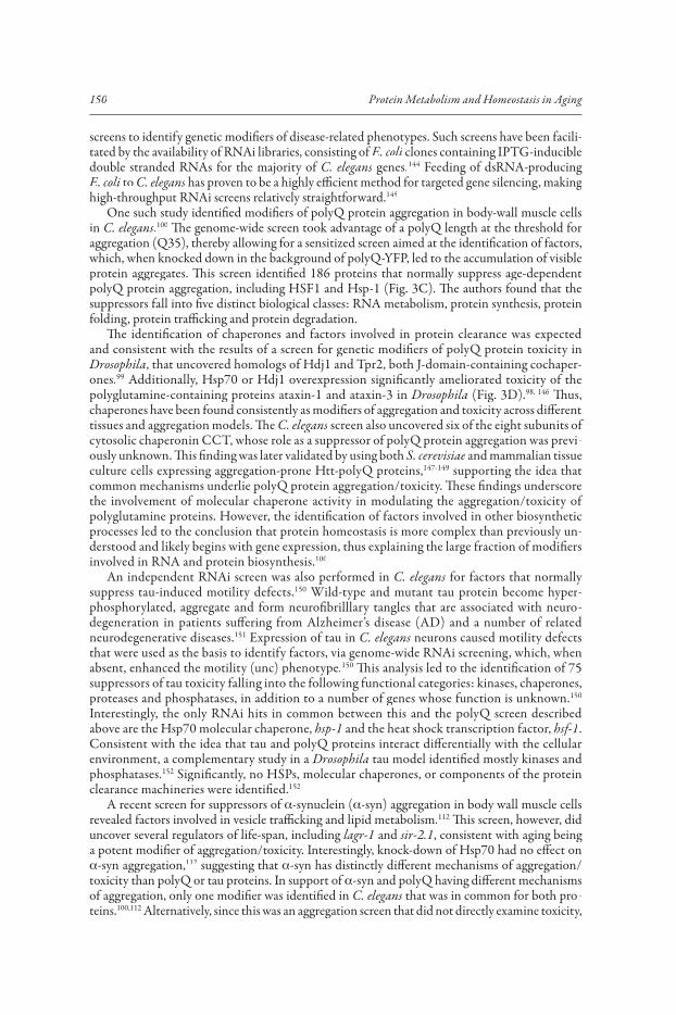
One such study identified modifiers of polyQ protein aggregation in body-wall muscle cells in C. elegans.100 The genome-wide screen took advantage of a polyQ length at the threshold foraggregation (Q35), thereby allowing for a sensitized screen aimed at the identification of factors,which, when knocked down in the background of polyQ-YFP, led to the accumulation of visibleprotein aggregates. This screen identified 186 proteins that normally suppress age-dependent polyQ protein aggregation, including HSF1 and Hsp-1 (Fig. 3C). The authors found that the suppressors fall into five distinct biological classes: RNA metabolism, protein synthesis, proteinfolding, protein trafficking and protein degradation.
The identification of chaperones and factors involved in protein clearance was expected and consistent with the results of a screen for genetic modifiers of polyQ protein toxicity inDrosophila, that uncovered homologs of Hdj1 and Tpr2, both J-domain-containing cochaper-ones.99 Additionally, Hsp70 or Hdj1 overexpression significantly ameliorated toxicity of the polyglutamine-containing proteins ataxin-1 and ataxin-3 in Drosophila (Fig. 3D).98, 146 Thus, chaperones have been found consistently as modifiers of aggregation and toxicity across differenttissues and aggregation models. The C. elegans screen also uncovered six of the eight subunits of cytosolic chaperonin CCT, whose role as a suppressor of polyQ protein aggregation was previ-ously unknown. This finding was later validated by using both S. cerevisiae and mammalian tissue culture cells expressing aggregation-prone Htt-polyQ proteins,147-149 supporting the idea that common mechanisms underlie polyQ protein aggregation/toxicity. These findings underscorethe involvement of molecular chaperone activity in modulating the aggregation/toxicity of polyglutamine proteins. However, the identification of factors involved in other biosyntheticprocesses led to the conclusion that protein homeostasis is more complex than previously un-derstood and likely begins with gene expression, thus explaining the large fraction of modifiers involved in RNA and protein biosynthesis.100
An independent RNAi screen was also performed in C. elegans for factors that normally suppress tau-induced motility defects.150 Wild-type and mutant tau protein become hyper-phosphorylated, aggregate and form neurofibrilllary tangles that are associated with neuro-degeneration in patients suffering from Alzheimer’s disease (AD) and a number of related neurodegenerative diseases.151 Expression of tau in C. elegans neurons caused motility defectsthat were used as the basis to identify factors, via genome-wide RNAi screening, which, when absent, enhanced the motility (unc) phenotype.150 This analysis led to the identification of 75suppressors of tau toxicity falling into the following functional categories: kinases, chaperones,proteases and phosphatases, in addition to a number of genes whose function is unknown.150
Interestingly, the only RNAi hits in common between this and the polyQ screen describedabove are the Hsp70 molecular chaperone, hsp-1 and the heat shock transcription factor, hsf-1. Consistent with the idea that tau and polyQ proteins interact differentially with the cellular environment, a complementary study in a Drosophila tau model identified mostly kinases andphosphatases.152 Significantly, no HSPs, molecular chaperones, or components of the proteinclearance machineries were identified.152
A recent screen for suppressors of �-synuclein (�-syn) aggregation in body wall muscle cells revealed factors involved in vesicle trafficking and lipid metabolism.112 This screen, however, did uncover several regulators of life-span, including lagr-1 and sir-2.1, consistent with aging being a potent modifier of aggregation/toxicity. Interestingly, knock-down of Hsp70 had no effect on �-syn aggregation,112 suggesting that �-syn has distinctly different mechanisms of aggregation/toxicity than polyQ or tau proteins. In support of �-syn and polyQ having different mechanismsof aggregation, only one modifier was identified in C. elegans that was in common for both pro-teins.100,112 Alternatively, since this was an aggregation screen that did not directly examine toxicity,

151Protein Homeostasis in Models of Aging and Age-Related Conformational Disease
it is possible that aggregation and toxicity of �-syn are uncoupled in the presence of Hsp70 RNAi, a finding that would be consistent with one from Drosophila.153
The striking lack of overlap between modifiers of aggregation/toxicity for uniqueaggregation-prone proteins could presumably be due to different factors acting on different mis-folded species, in addition to different factors acting in different tissue types, for example, body wall muscle cells as compared to neurons. Certainly, the identification of kinases and phosphatases in the screen for suppressors of tau toxicity provides support for the hypothesis that tau hyper-phosphorylation is a prerequisite for disease. Consequently, these data also provide evidence that C. elegans and D. melanogaster are valid genetic models for the identification of factors which arercapable of acting on particular human disease-causing proteins to suppress aggregation/toxicity.
In contrast to �-syn, a screen for modifiers of SOD1 aggregation and toxicity in C. elegansneurons yielded mostly molecular chaperones and other factors generally involved in protein quality control,115 more consistent with what was seen previously with respect to modifiers of polyQ protein aggregation/toxicity. Furthermore, the hits obtained by an RNAi screen aimed atthe identification of suppressors of osmotic stress-induced gene expression overlapped to a greatextent with those identified as suppressors of polyQ protein aggregation.154 In that study, the au-thors identified genes, including gpdh-1, that are upregulated in response to osmotic stress, whichis known to cause generalized, nonspecific protein misfolding. Although gpdh-1 expression does not respond to stresses other than osmotic stress,154 almost 30% of all the genes identified in this study overlap with those identified in the screen for suppressors of polyQ protein aggregation.Furthermore, 73% of the overlapping genes are predicted to fall into biological classes usually associated with the regulation of proteostasis, including RNA processing, protein synthesis, pro-tein folding and degradation.154 Finally, many of the modifiers of ataxin-3 toxicity in Drosophilawere also able to rescue the toxic phenotypes due to the reduced function of HSP70, which also causes generalized protein misfolding.142 Ultimately, these data suggest that a core set of factorsfunction generally in response to stress-induced protein damage and others respond specifically to a particular stress, for example the stress of a misfolded, disease-causing protein, to tailor theresponse to the situation at hand.
However, the extent to which the molecular mechanisms of disease are conserved betweenaggregation-prone proteins is unknown. To address this, it will be necessary to express unrelatedaggregation-prone proteins, such as A�, SOD1, tau, �-syn and a polyQ-containing proteins, inthe same cell type in a given model organism, for a direct comparison of modifiers of aggregation/toxicity. The expectation is that common modifiers would be those whose molecular function isin the general folding/clearance of misfolded proteins. On the other hand, modifiers that act onone or the other aggregation-prone protein would most likely be more closely associated with thespecific function, or mode of disease progression, of a particular protein. For example, a screenperformed in yeast expressing mutant Htt or �-synuclein, revealed almost entirely non-overlapping sets of genes, many with human homologs, acting as modifiers either of mutant Htt or �-synuclein toxicity.155 The authors speculate that their modifiers likely define mechanisms or pathways thatare specific for particular disease-causing proteins, such as vesicle transport playing a role in�-synuclein toxicity.155
In addition to RNAi and overexpression screens, forward genetic screens have also beenperformed to identify modulators of polyQ protein aggregation/toxicity in C. elegans. One suchscreen revealed a novel gene, pqe-1, which normally functions to suppress the proteotoxicity of anHtt exon 1 fragment with an expanded polyQ tract.156 Another forward genetic screen was aimedat the identification of genes that normally function to suppress the aggregation of polyQ-YFP in body wall muscle cells.101 This screen uncovered mutations in unc-30, which encodes thetranscription factor that regulates the synthesis of the neurotransmitter GABA.101 The findings described in101 are of particular interest, because they demonstrate that the ability of an organismto manage proteotoxic stress is not a cell autonomous process as previously thought, but may beaffected by cell-cell communication, for example via neuronal cholinergic signaling. Consistent with this, treating Q35-expressing C. elegans with small molecules, acting positively or negatively

152 Protein Metabolism and Homeostasis in Aging
on neuronal signaling, suppressed or enhanced, respectively, polyQ protein aggregation101 in postsynaptic cells. The identification, via chemical genetic screens, of additional small molecules that alleviate disease-causing protein aggregation/toxicity will be an important step in the devel-opment of pharmaceuticals.
Small Molecule Drug ScreensThe search for therapeutics to treat neurodegeneration is turning to the identification of small
molecule proteostasis regulators. Such small molecules could function by enhancing the expressionor activities of molecular chaperones, thereby effectively increasing the rates of folding of clientproteins, or, alternatively, they could act by enhancing degradation/clearance157 or by modulating protein translation rates.
A number of small molecule regulators of the heat shock response have been identified. These compounds include proteasome inhibitors, serine protease inhibitors, Hsp90 inhibitors, non-steroidal anti-inflammatory drugs (NSAIDS), triterpenoids such as celastrol and inhibitors of HSF1, including triptolide.49,158 Protease inhibitors (DCIC, TPCK and TLCK) and proteasome inhibitors (MG132, lactacystin) induce the heat-shock response by elevating the effective concen-trations of misfolfed, damaged, or otherwise no longer needed, proteins that are normally targeted for degradation.159,160 In contrast, other inducers of the heat shock response act as inhibitors of Hsp90. These include the fungal antibiotic radicicol, the benzoquinone ansamycins geldanamycin and 17AAG. They activate HSF1, in part because Hsp90 is a negative regulator of HSF1.61,161,162
NSAIDS, including sodium salicylate, have multiple properties. At higher concentrations they partially activate HSF1, while at lower concentrations they synergize with other stress conditions to induce the heat-shock response.163 Specifically, exposure of human tissue culture cells to sodium salicylate results in activation of HSF1 with respect to in vivo binding to the Hsp70 gene; however,transcriptional induction fails to occur. Salicylate-treated cells, however, are sensitized to stress and readily activate HS genes upon exposure to other mild stress conditions, often not sufficient themselves to activate the heat shock response. In a similar manner, indomethacin induces HSF1 DNA binding with full Hsp70 transcription upon exposure to a secondary stress.164 Of the inflam-matory modulators, arachidonic acid and the cyclopentenone prostaglandins, including PGA1,PGA2 and PGJ2, all induce the full complement of HSF1 activities.165,166 The triterpenoid celas-trol isolated from the Chinese plant Triptergium wilfordii represents an herbal medicine class of bioactive molecules that induces two protective stress responses, the heat shock response and the anti-oxidant response.167,168 The effects of celastrol are rapid like heat shock; however, unlike theheat-shock response that self-attenuates, the celastrol induction of HS genes persists for an extended period.167 Consistent with small molecule inducers of HSF1 acting to alleviate the toxic effects of misfolded disease-causing proteins, geranylgeranylacetone (GGA)169 and celastrol170 treatment of polyQ protein-expressing cells inhibited polyQ-associated cell death in tissue culture and mousemodels of polyQ protein misfolding.
The modulation of protein translation rates also plays an important role in inducible stress responses. A screen for small molecule inhibitors of ER stress-induced apoptosis in PC-12 cellsyielded salubrinol, a selective inhibitor of eIF2� dephosphorylation.171 eIF2� is a translationinitiation factor that, when in its phosphorylated state, either under conditions of heat stress or salubrinol treatment, is responsible for mediating a general decrease, but a selective increase in the translation of chaperones and other stress-associated proteins. Like small molecule inducers of HSF1, salubrinol, or other translational regulators, might be expected to modulate the toxic effects of misfolded disease-associated proteins.
Most of the chemical genetic analyses that have yielded the small molecules described above have been performed in tissue culture cells. A limited number of studies aimed at the identification of small molecules that act in invertebrate models to alleviate the toxicity of misfolding-prone disease-associated proteins have been performed. More commonly, small molecules that were identified in tissue culture systems as having the properties of inducing the HSR have been tested in invertebrates. For example, it was shown that the treatment of
153Protein Homeostasis in Models of Aging and Age-Related Conformational Disease
Htt-expressing C. elegans with the antioxidant resveratrol reduced Htt toxicity in a mannerdependent on the sirtuin, sir2.1.172
A small-scale chemical genetics analysis was performed in a C. elegans Htt-polyQ model, us-ing a screening approach which circumvented the inherent problems associated with screening based on motility defects. The authors examined treated worms for lack of neuronal cell death by visualizing the loss, or lack thereof, of GFP fluorescence in ASH neurons of a sensitized line thatrapidly undergoes polyQ-dependent neurodegeneration.173 These conditions were used to validatecandidate compounds identified previously in a large-scale, tissue culture-based, screen174,175 andrevealed two compounds, lithium chloride and mithramycin, which suppressed HD neurotoxicity in the C. elegans Htt-polyQ model.173 The use of this and similar assays, should make it possibleto rapidly screen large chemical libraries for their effect on toxicity in C. elegans models of neu-rodegenerative diseases.
Because C. elegans is a multicellular organism, we would expect that the successful implementa-tion of large-scale chemical genetics screens will be highly effective in identifying novel therapeutic compounds, not previously identified in cell culture models, that act either cell autonomously or cell non-autonomously. Finally, fluorescent labeling of candidate molecules will be instrumental in elucidating the mode of drug action and to determine whether the drug is acting directly orindirectly on the disease-causing proteins. This will be relatively straightforward in C. elegans due the easy visualization of fluorescent markers.
ConclusionThe work discussed here highlights the utility of invertebrate models in the study of neuro-
degenerative diseases of aging. Recent findings from these model systems have strengthened ourunderstanding of protein conformational diseases. Specifically, they have led to the proposal thatprotein folding homeostasis, while sufficiently robust to manage protein damage/misfolding caused by acute environmental stress such as heat shock, is apparently ineffective when faced with thechronic expression of an aggregation-prone disease-causing protein. This limited capacity of the cellto manage chronic misfolding is especially pronounced under conditions of additional stress on the proteome caused by the expression of metastable proteins, partially compromised in their folding and providing a relatively high demand for folding resources. To combat this inherent limitationfor the purpose of treating patients, small molecule drugs are being sought that will enhance the ability of a cell, or organism, to deal with the expression of chronically misfolded proteins.
AcknowledgementsE.A.K was supported by an individual postdoctoral fellowship from the National Institutes
of Health (NINDS); research in the laboratory of R.I.M. was supported by grants from the National Institutes of Health (NIGMS and NIA), the HDSA Coalition for the Cure and the ALS Association.
References1. Rutherford SL, Lindquist S. Hsp90 as a capacitor for morphological evolution. Nature 1998;
396:336-342.2. Stevens FJ, Argon Y. Pathogenic light chains and the B-cell repertoire. Immunol Today 1999;
20:451-457.3. Gidalevitz T, Ben-Zvi A, Ho KH et al. Progressive disruption of cellular protein folding in models of
polyglutamine diseases. Science 2006; 311:1471-1474.4. Yeyati PL, Bancewicz RM, Maule J et al. Hsp90 selectively modulates phenotype in vertebrate develop-
ment. PLoS Genet 2007; 3:e43.5. Michels AA, Kanon B, Konings AW et al. Hsp70 and Hsp40 chaperone activities in the cytoplasm and
the nucleus of mammalian cells. J Biol Chem 1997; 272:33283-33289.6. Parsell DA, Lindquist S. The function of heat-shock proteins in stress tolerance: degradation and reac-
tivation of damaged proteins. Annu Rev Genet 1993; 27:437-496.7. Balch WE, Morimoto RI, Dillin A et al. Adapting proteostasis for disease intervention. Science 2008;
319:916-919.

154 Protein Metabolism and Homeostasis in Aging
8. Harman D. Aging : a theory based on free radical and radiation chemistry. J Gerontol 1956; 11:298-300.
9. Sohal RS, Weindruch R. Oxidative stress, caloric restriction and aging. Science 1996; 273:59-63.10. Berlett BS, Stadtman ER. Protein oxidation in aging, disease and oxidative stress. J Biol Chem 1997;
272:20313-20316.11. Stadtman ER, Oliver CN. Metal-catalyzed oxidation of proteins. Physiological consequences. J Biol
Chem 1991; 266:2005-2008.12. Smith CD, Carney JM, Starke-Reed PE et al. Excess brain protein oxidation and enzyme dysfunction
in normal aging and in Alzheimer disease. Proc Natl Acad Sci USA 1991; 88:10540-10543.13. Stadtman ER. Protein oxidation and aging. Science 1992; 257:1220-1224.14. Ahmed N, Dobler D, Dean M et al. Peptide mapping identifies hotspot site of modification in human
serum albumin by methylglyoxal involved in ligand binding and esterase activity. J Biol Chem 2005;280:5724-5732.
15. Lo TW, Westwood ME, McLellan AC et al. Binding and modification of proteins by methylglyoxal under physiological conditions. A kinetic and mechanistic study with N alpha-acetylarginine,N alpha-acetylcysteine and N alpha-acetyllysine and bovine serum albumin. J Biol Chem 1994;269:32299-32305.
16. Kalapos MP. Methylglyoxal in living organisms: chemistry, biochemistry, toxicology and biologicalimplications. Toxicol Lett 1999; 110:145-175.
17. Kuhla B, Boeck K, Schmidt A et al. Age- and stage-dependent glyoxalase I expression and its activity in normal and Alzheimer’s disease brains. Neurobiol Aging 2007; 28:29-41.
18. Hipkiss AR. On the mechanisms of ageing suppression by dietary restriction-is persistent glycolysis theproblem? Mech Ageing Dev 2006; 127:8-15.
19. Gnerer JP, Kreber RA, Ganetzky B. Wasted away, a Drosophila mutation in triosephosphate isomerase, causes paralysis, neurodegeneration and early death. Proc Natl Acad Sci USA 2006; 103:14987-14993.
20. Moskovitz J. Roles of methionine suldfoxide reductases in antioxidant defense, protein regulation andsurvival. Curr Pharm Des 2005; 11:1451-1457.
21. Friguet B. Oxidized protein degradation and repair in ageing and oxidative stress. FEBS Lett 2006;580:2910-2916.
22. Stadtman ER. Oxidation of free amino acids and amino acid residues in proteins by radiolysis and by metal-catalyzed reactions. Annu Rev Biochem 1993; 62:797-821.
23. Hanson SR, Hasan A, Smith DL et al. The major in vivo modifications of the human water-insoluble lens crystallins are disulfide bonds, deamidation, methionine oxidation and backbone cleavage. Exp EyeRes 2000; 71:195-207.
24. Breusing N, Grune T. Regulation of proteasome-mediated protein degradation during oxidative stressand aging. Biol Chem 2008; 389:203-209.
25. Davies KJ. Degradation of oxidized proteins by the 20S proteasome. Biochimie 2001; 83:301-310.26. Shringarpure R, Grune T, Mehlhase J et al. Ubiquitin conjugation is not required for the degradation
of oxidized proteins by proteasome. J Biol Chem 2003; 278:311-318.27. Shang F, Nowell TR Jr, Taylor A. Removal of oxidatively damaged proteins from lens cells by the
ubiquitin-proteasome pathway. Exp Eye Res 2001; 73:229-238.28. Bota DA, Davies KJ. Lon protease preferentially degrades oxidized mitochondrial aconitase by an
ATP-stimulated mechanism. Nat Cell Biol 2002; 4:674-680.29. Reinheckel T, Grune T, Davies KJ. The measurement of protein degradation in response to oxidative
stress. Methods Mol Biol 2000; 99:49-60.30. Kiffin R, Christian C, Knecht E et al. Activation of chaperone-mediated autophagy during oxidative
stress. Mol Biol Cell 2004; 15:4829-4840.31. Schultz SC, Richards JH. Site-saturation studies of beta-lactamase: production and characterization
of mutant beta-lactamases with all possible amino acid substitutions at residue 71. Proc Natl Acad SciUSA 1986; 83:1588-1592.
32. Pakula AA, Sauer RT. Genetic analysis of protein stability and function. Annu Rev Genet 1989;23:289-310.
33. Ng PC, Henikoff S. Predicting the effects of amino acid substitutions on protein function. Annu Rev Genomics Hum Genet 2006; 7:61-80.
34. DePristo MA, Weinreich DM, Hartl DL. Missense meanderings in sequence space: a biophysical view of protein evolution. Nat Rev Genet 2005; 6:678-687.
35. Suckow J, Markiewicz P, Kleina LG et al. Genetic studies of the Lac repressor. XV: 4000 single aminoacid substitutions and analysis of the resulting phenotypes on the basis of the protein structure. J Mol Biol 1996; 261:509-523.

155Protein Homeostasis in Models of Aging and Age-Related Conformational Disease
36. Sachidanandam R, Weissman D, Schmidt SC et al. A map of human genome sequence variation con-taining 1.42 million single nucleotide polymorphisms. Nature 2001; 409:928-933.
37. Drummond DA, Wilke CO. Mistranslation-induced protein misfolding as a dominant constraint on coding-sequence evolution. Cell 2008; 134:341-352.
38. Gidalevitz T, Krupinski T, Garcia S et al. Destabilizing protein polymorphisms in the genetic backgrounddirect phenotypic expression of mutant SOD1 toxicity. PLoS Genet 2009; 5:e1000399.
39. Stefani M, Dobson CM. Protein aggregation and aggregate toxicity: new insights into protein folding, misfolding diseases and biological evolution. J Mol Med 2003; 81:678-699.
40. Kopito RR, Ron D. Conformational disease. Nat Cell Biol 2000; 2:E207-209.41. Lee JW, Beebe K, Nangle LA et al. Editing-defective tRNA synthetase causes protein misfolding and
neurodegeneration. Nature 2006; 443:50-55.42. Jordanova A, Irobi J, Thomas FP et al. Disrupted function and axonal distribution of mutant ty-
rosyl-tRNA synthetase in dominant intermediate Charcot-Marie-Tooth neuropathy. Nat Genet 2006;38:197-202.
43. Martin I, Grotewiel MS. Oxidative damage and age-related functional declines. Mech Ageing Dev 2006;127:411-423.
44. Calderwood SK, Murshid A, Prince T. The Shock of Aging: Molecular Chaperones and the Heat Shock Response in Longevity and Aging—A Mini-Review. Gerontology 2009.
45. Holmberg CI, Staniszewski KE, Mensah KN et al. Inefficient degradation of truncated polyglutamineproteins by the proteasome. EMBO J 2004; 23:4307-4318.
46. Kim S, Nollen EA, Kitagawa K et al. Polyglutamine protein aggregates are dynamic. Nat Cell Biol 2002; 4:826-831.
47. Suhr ST, Senut MC, Whitelegge JP et al. Identities of sequestered proteins in aggregates from cells with induced polyglutamine expression. J Cell Biol 2001; 153:283-294.
48. Kryukov GV, Pennacchio LA, Sunyaev SR. Most rare missense alleles are deleterious in humans: implica-tions for complex disease and association studies. Am J Hum Genet 2007; 80:727-739.
49. Westerheide SD, Morimoto RI. Heat shock response modulators as therapeutic tools for diseases of protein conformation. J Biol Chem 2005; 280:33097-33100.
50. Ron D, Walter P. Signal integration in the endoplasmic reticulum unfolded protein response. Nat Rev Mol Cell Biol 2007; 8:519-529.
51. Banerji SS, Theodorakis NG, Morimoto RI. Heat shock-induced translational control of HSP70 andglobin synthesis in chicken reticulocytes. Mol Cell Biol 1984; 4:2437-2448.
52. Xiao X, Zuo X, Davis AA et al. HSF1 is required for extra-embryonic development, postnatal growth and protection during inflammatory responses in mice. EMBO J 1999; 18:5943-5952.
53. Santos SD, Saraiva MJ. Enlarged ventricles, astrogliosis and neurodegeneration in heat shock factor 1 null mouse brain. Neuroscience 2004; 126:657-663.
54. Zhang P, McGrath B, Li S et al. The PERK eukaryotic initiation factor 2 alpha kinase is required for the development of the skeletal system, postnatal growth and the function and viability of the pancreas. Mol Cell Biol 2002; 22:3864-3874.
55. Reimold AM, Iwakoshi NN, Manis J et al. Plasma cell differentiation requires the transcription factor XBP-1. Nature 2001; 412:300-307.
56. Zhang K, Wong HN, Song B et al. The unfolded protein response sensor IRE1alpha is required at 2 distinct steps in B-cell lymphopoiesis. J Clin Invest 2005; 115:268-281.
57. Elefant F, Palter KB. Tissue-specific expression of dominant negative mutant Drosophila HSC70 causes developmental defects and lethality. Mol Biol Cell 1999; 10:2101-2117.
58. Feder JH, Rossi JM, Solomon J et al. The consequences of expressing hsp70 in Drosophila cells at normal temperatures. Genes Dev 1992; 6:1402-1413.
59. Nylandsted J, Rohde M, Brand K et al. Selective depletion of heat shock protein 70 (Hsp70) activatesa tumor-specific death program that is independent of caspases and bypasses Bcl-2. Proc Natl Acad SciUSA 2000; 97:7871-7876.
60. Whitesell L, Mimnaugh EG, De Costa B et al. Inhibition of heat shock protein HSP90-pp60v-srcheteroprotein complex formation by benzoquinone ansamycins: essential role for stress proteins in oncogenic transformation. Proc Natl Acad Sci USA 1994; 91:8324-8328.
61. Whitesell L, Lindquist SL. HSP90 and the chaperoning of cancer. Nat Rev Cancer 2005; 5:761-772.62. Duyao M, Ambrose C, Myers R et al. Trinucleotide repeat length instability and age of onset in Hun-
tington’s disease. Nat Genet 1993; 4:387-392.63. Morley JF, Brignull HR, Weyers JJ et al. The threshold for polyglutamine-expansion protein aggregation
and cellular toxicity is dynamic and influenced by aging in Caenorhabditis elegans. Proc Natl Acad SciUSA 2002; 99:10417-10422.
64. Morris JZ, Tissenbaum HA, Ruvkun G. A phosphatidylinositol-3-OH kinase family member regulating longevity and diapause in Caenorhabditis elegans. Nature 1996; 382:536-539.

156 Protein Metabolism and Homeostasis in Aging
65. Lin K, Dorman JB, Rodan A et al. daf-16: An HNF-3/forkhead family member that can function to double the life-span of Caenorhabditis elegans. Science 1997; 278:1319-1322.
66. Morley JF, Morimoto RI. Regulation of longevity in Caenorhabditis elegans by heat shock factor and molecular chaperones. Mol Biol Cell 2004; 15:657-664.
67. Hsu AL, Murphy CT, Kenyon C. Regulation of aging and age-related disease by DAF-16 and heat-shock factor. Science 2003; 300:1142-1145.
68. Cohen E, Bieschke J, Perciavalle RM et al. Opposing activities protect against age-onset proteotoxicity.Science 2006; 313:1604-1610.
69. Lithgow GJ, White TM, Melov S et al. Thermotolerance and extended life-span conferred by single-gene mutations and induced by thermal stress. Proc Natl Acad Sci USA 1995; 92:7540-7544.
70. Salmon AB, Sadighi Akha AA, Buffenstein R et al. Fibroblasts from naked mole-rats are resistant tomultiple forms of cell injury, but sensitive to peroxide, ultraviolet light and endoplasmic reticulum stress.J Gerontol A Biol Sci Med Sci 2008; 63:232-241.
71. Salmon AB, Murakami S, Bartke A et al. Fibroblast cell lines from young adult mice of long-lived mutantstrains are resistant to multiple forms of stress. Am J Physiol Endocrinol Metab 2005; 289:E23-29.
72. Broughton SJ, Piper MD, Ikeya T et al. Longer lifespan, altered metabolism and stress resistancein Drosophila from ablation of cells making insulin-like ligands. Proc Natl Acad Sci USA 2005; 102:3105-3110.
73. Tullet JM, Hertweck M, An JH et al. Direct inhibition of the longevity-promoting factor SKN-1 by insulin-like signaling in C. elegans. Cell 2008; 132:1025-1038.
74. Prahlad V, Cornelius T, Morimoto RI. Regulation of the cellular heat shock response in Caenorhabditis elegans by thermosensory neurons. Science 2008; 320:811-814.
75. Bienz M. Developmental control of the heat shock response in Xenopus. Proc Natl Acad Sci USA 1984;81:3138-3142.
76. Sprang GK, Brown IR. Selective induction of a heat shock gene in fibre tracts and cerebellar neurons of the rabbit brain detected by in situ hybridization. Brain Res 1987; 427:89-93.
77. Shamovsky I, Gershon D. Novel regulatory factors of HSF-1 activation: facts and perspectives regarding their involvement in the age-associated attenuation of the heat shock response. Mech Ageing Dev 2004;125:767-775.
78. Mathur SK, Sistonen L, Brown IR et al. Deficient induction of human hsp70 heat shock gene transcrip-tion in Y79 retinoblastoma cells despite activation of heat shock factor 1. Proc Natl Acad Sci USA 1994; 91:8695-8699.
79. Marcuccilli CJ, Mathur SK, Morimoto RI et al. Regulatory differences in the stress response of hip-pocampal neurons and glial cells after heat shock. J Neurosci 1996; 16:478-485.
80. Batulan Z, Shinder GA, Minotti S et al. High threshold for induction of the stress response in motor neurons is associated with failure to activate HSF1. J Neurosci 2003; 23:5789-5798.
81. Muchowski PJ, Wacker JL. Modulation of neurodegeneration by molecular chaperones. Nat Rev Neurosci2005; 6:11-22.
82. Walker GA, Lithgow GJ. Lifespan extension in C. elegans by a molecular chaperone dependent uponinsulin-like signals. Aging Cell 2003; 2:131-139.
83. YYokoyama K, Fukumoto K, Murakami T et al. Extended longevity of Caenorhabditis elegans by knocking in extra copies of hsp70F, a homolog of mot-2 (mortalin)/mthsp70/Grp75. FEBS Lett 2002;516:53-57.
84. Herndon LA, Schmeissner PJ, Dudaronek JM et al. Stochastic and genetic factors influence tissue-specificdecline in ageing C. elegans. Nature 2002; 419:808-814.
85. Yang J, Tower J. Expression of hsp22 and hsp70 transgenes is partially predictive of drosophila survivalunder normal and stress conditions. J Gerontol A Biol Sci Med Sci 2009; 64:828-838.
86. Yun C, Stanhill A, Yang Y et al. Proteasomal adaptation to environmental stress links resistance to pro-teotoxicity with longevity in Caenorhabditis elegans. Proc Natl Acad Sci USA 2008; 105:7094-7099.
87. Brunet A, Sweeney LB, Sturgill JF et al. Stress-dependent regulation of FOXO transcription factors by the SIRT1 deacetylase. Science 2004; 303:2011-2015.
88. Westerheide SD, Anckar J, Stevens SM et al. Stress-inducible regulation of heat shock factor 1 by thedeacetylase SIRT1. Science 2009; 323:1063-1066.
89. Lee SS, Kennedy S, Tolonen AC et al. DAF-16 target genes that control C. elegans life-span andmetabolism. Science 2003; 300:644-647.
90. Kakizuka A. Protein precipitation: a common etiology in neurodegenerative disorders? Trends Genet 1998; 14:396-402.
91. Chiti F, Dobson CM. Protein misfolding, functional amyloid and human disease. Annu Rev Biochem2006; 75:333-366.
92. Warrick JM, Paulson HL, Gray-Board GL et al. Expanded polyglutamine protein forms nuclear inclu-sions and causes neural degeneration in Drosophila. Cell 1998; 93:939-949.

157Protein Homeostasis in Models of Aging and Age-Related Conformational Disease
93. Faber PW, Alter JR, MacDonald ME et al. Polyglutamine-mediated dysfunction and apoptotic death of a Caenorhabditis elegans sensory neuron. Proc Natl Acad Sci USA 1999; 96:179-184.
94. Feany MB, Bender WW. A Drosophila model of Parkinson’s disease. Nature 2000; 404:394-398.95. Krobitsch S, Lindquist S. Aggregation of huntingtin in yeast varies with the length of the polyglutamine
expansion and the expression of chaperone proteins. Proc Natl Acad Sci USA 2000; 97:1589-1594.96. Satyal SH, Schmidt E, Kitagawa K et al. Polyglutamine aggregates alter protein folding homeostasis in
Caenorhabditis elegans. Proc Natl Acad Sci USA 2000; 97:5750-5755.97. Parker JA, Connolly JB, Wellington C et al. Expanded polyglutamines in Caenorhabditis elegans cause
axonal abnormalities and severe dysfunction of PLM mechanosensory neurons without cell death. Proc Natl Acad Sci USA 2001; 98:13318-13323.
98. Warrick JM, Chan HY, Gray-Board GL et al. Suppression of polyglutamine-mediated neurodegenerationin Drosophila by the molecular chaperone HSP70. Nat Genet 1999; 23:425-428.
99. Kazemi-Esfarjani P, Benzer S. Genetic suppression of polyglutamine toxicity in Drosophila. Science2000; 287:1837-1840.
100. Nollen EA, Garcia SM, van Haaften G et al. Genome-wide RNA interference screen identifies previously undescribed regulators of polyglutamine aggregation. Proc Natl Acad Sci USA 2004; 101:6403-6408.
101. Garcia SM, Casanueva MO, Silva MC et al. Neuronal signaling modulates protein homeostasis inCaenorhabditis elegans postsynaptic muscle cells. Genes Dev 2007; 21:3006-3016.
102. Jackson GR, Salecker I, Dong X et al. Polyglutamine-expanded human huntingtin transgenes inducedegeneration of Drosophila photoreceptor neurons. Neuron 1998; 21:633-642.
103. Brignull HR, Moore FE, Tang SJ et al. Polyglutamine proteins at the pathogenic threshold display neuron-specific aggregation in a pan-neuronal Caenorhabditis elegans model. J Neurosci 2006; 26:7597-7606.
104. Marsh JL, Walker H, Theisen H et al. Expanded polyglutamine peptides alone are intrinsically cytotoxic and cause neurodegeneration in Drosophila. Hum Mol Genet 2000; 9:13-25.
105. Link CD. Expression of human beta-amyloid peptide in transgenic Caenorhabditis elegans. Proc NatlAcad Sci USA 1995; 92:9368-9372.
106. Finelli A, Kelkar A, Song HJ et al. A model for studying Alzheimer’s Abeta42-induced toxicity inDrosophila melanogaster. Mol Cell Neurosci 2004; 26:365-375.
107. Wittmann CW, Wszolek MF, Shulman JM et al. Tauopathy in Drosophila: neurodegeneration withoutneurofibrillary tangles. Science 2001; 293:711-714.
108. Kraemer BC, Zhang B, Leverenz JB et al. Neurodegeneration and defective neurotransmission in a Caenorhabditis elegans model of tauopathy. Proc Natl Acad Sci USA 2003; 100:9980-9985.
109. Nussbaum RL, Polymeropoulos MH. Genetics of Parkinson’s disease. Hum Mol Genet 1997;6:1687-1691.
110. Chase TN. A gene for Parkinson disease. Arch Neurol 1997; 54:1156-1157.111. Lakso M, Vartiainen S, Moilanen AM et al. Dopaminergic neuronal loss and motor deficits in Caenorhab-
ditis elegans overexpressing human alpha-synuclein. J Neurochem 2003; 86:165-172.112. van Ham TJ, Thijssen KL, Breitling R et al. C. elegans model identifies genetic modifiers of alpha-sy-
nuclein inclusion formation during aging. PLoS Genet 2008; 4:e1000027.113. Rosen DR. Mutations in Cu/Zn superoxide dismutase gene are associated with familial amyotrophic
lateral sclerosis. Nature 1993; 364:362.114. Watson MR, Lagow RD, Xu K et al. A drosophila model for amyotrophic lateral sclerosis reveals motor
neuron damage by human SOD1. J Biol Chem 2008; 283:24972-24981.115. Wang J, Farr GW, Hall DH et al. An ALS-linked mutant SOD1 produces a locomotor defect associated
with aggregation and synaptic dysfunction when expressed in neurons of Caenorhabditis elegans. PLoS Genet 2009; 5:e1000350.
116. Ahn SG, Thiele DJ. Redox regulation of mammalian heat shock factor 1 is essential for Hsp gene activa-tion and protection from stress. Genes Dev 2003; 17:516-528.
117. Hahn JS, Thiele DJ. Activation of the Saccharomyces cerevisiae heat shock transcription factor under glucose starvation conditions by Snf1 protein kinase. J Biol Chem 2004; 279:5169-5176.
118. Thomson S, Hollis A, Hazzalin CA et al. Distinct stimulus-specific histone modifications at hsp70chromatin targeted by the transcription factor heat shock factor-1. Mol Cell 2004; 15:585-594.
119. Ananthan J, Goldberg AL, Voellmy R. Abnormal proteins serve as eukaryotic stress signals and triggerthe activation of heat shock genes. Science 1986; 232:522-524.
120. Hiromi Y, Okamoto H, Gehring WJ et al. Germline transformation with Drosophila mutant actin genes induces constitutive expression of heat shock genes. Cell 1986; 44:293-301.
121. Parsell DA, Sauer RT. Induction of a heat shock-like response by unfolded protein in Escherichia coli: dependence on protein level not protein degradation. Genes Dev 1989; 3:1226-1232.
122. Lindquist S, Craig EA. The heat-shock proteins. Annu Rev Genet 1988; 22:631-677.

158 Protein Metabolism and Homeostasis in Aging
123. Morimoto RI. Regulation of the heat shock transcriptional response: cross talk between a family of heat shock factors, molecular chaperones and negative regulators. Genes Dev 1998; 12:3788-3796.
124. Morimoto RI, Kline MP, Bimston DN et al. The heat-shock response: regulation and function of heat-shock proteins and molecular chaperones. Essays Biochem 1997; 32:17-29.
125. Hartl FU. Molecular chaperones in cellular protein folding. Nature 1996; 381:571-579.126. Bukau B, Horwich AL. The Hsp70 and Hsp60 chaperone machines. Cell 1998; 92:351-366.127. Pratt WB, Toft DO. Regulation of signaling protein function and trafficking by the hsp90/hsp70-based
chaperone machinery. Exp Biol Med (Maywood) 2003; 228:111-133.128. Bukau B, Weissman J, Horwich A. Molecular chaperones and protein quality control. Cell 2006;
125:443-451.129. Kieran D, Kalmar B, Dick JR et al. Treatment with arimoclomol, a coinducer of heat shock proteins,
delays disease progression in ALS mice. Nat Med 2004; 10:402-405.130. Katayama T, Imaizumi K, Honda A et al. Disturbed activation of endoplasmic reticulum stress transducers
by familial Alzheimer’s disease-linked presenilin-1 mutations. J Biol Chem 2001; 276:43446-43454.131. Cowan KJ, Diamond MI, Welch WJ. Polyglutamine protein aggregation and toxicity are linked to the
cellular stress response. Hum Mol Genet 2003; 12:1377-1391.132. Hay DG, Sathasivam K, Tobaben S et al. Progressive decrease in chaperone protein levels in a mouse
model of Huntington’s disease and induction of stress proteins as a therapeutic approach. Hum MolGenet 2004; 13:1389-1405.
133. Zourlidou A, Gidalevitz T, Kristiansen M et al. Hsp27 overexpression in the R6/2 mouse model of Huntington’s disease: chronic neurodegeneration does not induce Hsp27 activation. Hum Mol Genet 2007; 16:1078-1090.
134. Wen FC, Li YH, Tsai HF et al. Down-regulation of heat shock protein 27 in neuronal cells and non-neuronal cells expressing mutant ataxin-3. FEBS Lett 2003; 546:307-314.
135. Chai Y, Koppenhafer SL, Bonini NM et al. Analysis of the role of heat shock protein (Hsp) molecularchaperones in polyglutamine disease. J Neurosci 1999; 19:10338-10347.
136. Stenoien DL, Cummings CJ, Adams HP et al. Polyglutamine-expanded androgen receptors form ag-gregates that sequester heat shock proteins, proteasome components and SRC-1 and are suppressed by the HDJ-2 chaperone. Hum Mol Genet 1999; 8:731-741.
137. Bence NF, Sampat RM, Kopito RR. Impairment of the ubiquitin-proteasome system by protein ag-gregation. Science 2001; 292:1552-1555.
138. Nishitoh H, Matsuzawa A, Tobiume K et al. ASK1 is essential for endoplasmic reticulum stress-induced neuronal cell death triggered by expanded polyglutamine repeats. Genes Dev 2002; 16:1345-1355.
139. Tatzelt J, Zuo J, Voellmy R et al. Scrapie prions selectively modify the stress response in neuroblastoma cells. Proc Natl Acad Sci USA 1995; 92:2944-2948.
140. Van Dyk TK, Gatenby AA, LaRossa RA. Demonstration by genetic suppression of interaction of GroE products with many proteins. Nature 1989; 342:451-453.
141. Brown CR, Hong-Brown LQ, Welch WJ. Correcting temperature-sensitive protein folding defects. JClin Invest 1997; 99:1432-1444.
142. Bilen J, Bonini NM. Genome-wide screen for modifiers of ataxin-3 neurodegeneration in Drosophila.PLoS Genet 2007; 3:1950-1964.
143. Ben-Zvi A, Miller EA, Morimoto RI. Collapse of proteostasis represents an early molecular event in Caenorhabditis elegans aging. Proc Natl Acad Sci USA 2009.
144. Fraser AG, Kamath RS, Zipperlen P et al. Functional genomic analysis of C. elegans chromosome I by systematic RNA interference. Nature 2000; 408:325-330.
145. Kamath RS, Martinez-Campos M, Zipperlen P et al. Effectiveness of specific RNA-mediated in-terference through ingested double-stranded RNA in Caenorhabditis elegans. Genome Biol 2001;2:RESEARCH0002.
146. Ghosh S, Feany MB. Comparison of pathways controlling toxicity in the eye and brain in Drosophila models of human neurodegenerative diseases. Hum Mol Genet 2004; 13:2011-2018.
147. Kitamura A, Kubota H, Pack CG et al. Cytosolic chaperonin prevents polyglutamine toxicity with altering the aggregation state. Nat Cell Biol 2006; 8:1163-1170.
148. Behrends C, Langer CA, Boteva R et al. Chaperonin TRiC promotes the assembly of polyQ expansion proteins into nontoxic oligomers. Mol Cell 2006; 23:887-897.
149. Tam S, Geller R, Spiess C et al. The chaperonin TRiC controls polyglutamine aggregation and toxicity through subunit-specific interactions. Nat Cell Biol 2006; 8:1155-1162.
150. Kraemer BC, Burgess JK, Chen JH et al. Molecular pathways that influence human tau-induced pathol-ogy in Caenorhabditis elegans. Hum Mol Genet 2006; 15:1483-1496.
151. Lee VM, Goedert M, Trojanowski JQ. Neurodegenerative tauopathies. Annu Rev Neurosci 2001;24:1121-1159.

159Protein Homeostasis in Models of Aging and Age-Related Conformational Disease
152. Shulman JM, Feany MB. Genetic modifiers of tauopathy in Drosophila. Genetics 2003; 165:1233-1242.153. Auluck PK, Chan HY, Trojanowski JQ et al. Chaperone suppression of alpha-synuclein toxicity in a
Drosophila model for Parkinson’s disease. Science 2002; 295:865-868.154. Lamitina T, Huang CG, Strange K. Genome-wide RNAi screening identifies protein damage as a regula-
tor of osmoprotective gene expression. Proc Natl Acad Sci USA 2006; 103:12173-12178.155. Willingham S, Outeiro TF, DeVit MJ et al. Yeast genes that enhance the toxicity of a mutant huntingtin
fragment or alpha-synuclein. Science 2003; 302:1769-1772.156. Faber PW, Voisine C, King DC et al. Glutamine/proline-rich PQE-1 proteins protect Caenorhab-
ditis elegans neurons from huntingtin polyglutamine neurotoxicity. Proc Natl Acad Sci USA 2002;99:17131-17136.
157. Powers ET, Morimoto RI, Dillin A et al. Biological and chemical approaches to diseases of proteostasisdeficiency. Annu Rev Biochem 2009; 78:959-991.
158. Westerheide SD, Kawahara TL, Orton K et al. Triptolide, an inhibitor of the human heat shock response that enhances stress-induced cell death. J Biol Chem 2006; 281:9616-9622.
159. Mathew A, Mathur SK, Morimoto RI. Heat shock response and protein degradation: regulation of HSF2 by the ubiquitin-proteasome pathway. Mol Cell Biol 1998; 18:5091-5098.
160. Rossi A, Elia G, Santoro MG. Activation of the heat shock factor 1 by serine protease inhibitors. Aneffect associated with nuclear factor-kappaB inhibition. J Biol Chem 1998; 273:16446-16452.
161. Bagatell R, Paine-Murrieta GD, Taylor CW et al. Induction of a heat shock factor 1-dependent stressresponse alters the cytotoxic activity of hsp90-binding agents. Clin Cancer Res 2000; 6:3312-3318.
162. Bagatell R, Whitesell L. Altered Hsp90 function in cancer: a unique therapeutic opportunity. MolCancer Ther 2004; 3:1021-1030.
163. Jurivich DA, Sistonen L, Kroes RA et al. Effect of sodium salicylate on the human heat shock response.Science 1992; 255:1243-1245.
164. Lee BS, Chen J, Angelidis C et al. Pharmacological modulation of heat shock factor 1 by antiinflamma-tory drugs results in protection against stress-induced cellular damage. Proc Natl Acad Sci USA 1995;92:7207-7211.
165. Amici C, Sistonen L, Santoro MG et al. Antiproliferative prostaglandins activate heat shock transcrip-tion factor. Proc Natl Acad Sci USA 1992; 89:6227-6231.
166. Jurivich DA, Sistonen L, Sarge KD et al. Arachidonate is a potent modulator of human heat shock gene transcription. Proc Natl Acad Sci USA 1994; 91:2280-2284.
167. Westerheide SD, Bosman JD, Mbadugha BN et al. Celastrols as inducers of the heat shock responseand cytoprotection. J Biol Chem 2004; 279:56053-56060.
168. Trott A, West JD, Klaić L et al. Activation of heat shock and antioxidant responses by the natural prod-uct celastrol: transcriptional signatures of a thiol-targeted molecule. Mol Biol Cell. 2008;19(3):1104-12.Epub 2008 Jan 16.
169. Katsuno M, Sang C, Adachi H et al. Pharmacological induction of heat-shock proteins alleviates polyglutamine-mediated motor neuron disease. Proc Natl Acad Sci USA 2005; 102:16801-16806.
170. Zhang YQ, Sarge KD. Celastrol inhibits polyglutamine aggregation and toxicity though induction of the heat shock response. J Mol Med 2007; 85:1421-1428.
171. Boyce M, Bryant KF, Jousse C et al. A selective inhibitor of eIF2alpha dephosphorylation protects cells from ER stress. Science 2005; 307:935-939.
172. Parker JA, Arango M, Abderrahmane S et al. Resveratrol rescues mutant polyglutamine cytotoxicity innematode and mammalian neurons. Nat Genet 2005; 37:349-350.
173. Voisine C, Varma H, Walker N et al. Identification of potential therapeutic drugs for huntington’s disease using Caenorhabditis elegans. PLoS ONE 2007; 2:e504.
174. Varma H, Cheng R, Voisine C et al. Inhibitors of metabolism rescue cell death in Huntington’s diseasemodels. Proc Natl Acad Sci USA 2007; 104:14525-14530.
175. Varma H, Voisine C, DeMarco CT et al. Selective inhibitors of death in mutant huntingtin cells. NatChem Biol 2007; 3:99-100.