project: ghana emergency medicine collaborative document title: skin emergencies author(s): john w....
TRANSCRIPT

Project: Ghana Emergency Medicine Collaborative
Document Title: Skin Emergencies
Author(s): John W. Martel (University of Michigan), MD PhD 2012
License: Unless otherwise noted, this material is made available under the terms of the Creative Commons Attribution Share Alike-3.0 License: http://creativecommons.org/licenses/by-sa/3.0/
We have reviewed this material in accordance with U.S. Copyright Law and have tried to maximize your ability to use, share, and adapt it. These lectures have been modified in the process of making a publicly shareable version. The citation key on the following slide provides information about how you may share and adapt this material.
Copyright holders of content included in this material should contact [email protected] with any questions, corrections, or clarification regarding the use of content.
For more information about how to cite these materials visit http://open.umich.edu/privacy-and-terms-use.
Any medical information in this material is intended to inform and educate and is not a tool for self-diagnosis or a replacement for medical evaluation, advice, diagnosis or treatment by a healthcare professional. Please speak to your physician if you have questions about your medical condition.
Viewer discretion is advised: Some medical content is graphic and may not be suitable for all viewers.
1

Attribution Key
for more information see: http://open.umich.edu/wiki/AttributionPolicy
Use + Share + Adapt
Make Your Own Assessment
Creative Commons – Attribution License
Creative Commons – Attribution Share Alike License
Creative Commons – Attribution Noncommercial License
Creative Commons – Attribution Noncommercial Share Alike License
GNU – Free Documentation License
Creative Commons – Zero Waiver
Public Domain – Ineligible: Works that are ineligible for copyright protection in the U.S. (17 USC § 102(b)) *laws in your jurisdiction may differ
Public Domain – Expired: Works that are no longer protected due to an expired copyright term.
Public Domain – Government: Works that are produced by the U.S. Government. (17 USC § 105)
Public Domain – Self Dedicated: Works that a copyright holder has dedicated to the public domain.
Fair Use: Use of works that is determined to be Fair consistent with the U.S. Copyright Act. (17 USC § 107) *laws in your jurisdiction may differ
Our determination DOES NOT mean that all uses of this 3rd-party content are Fair Uses and we DO NOT guarantee that your use of the content is Fair.
To use this content you should do your own independent analysis to determine whether or not your use will be Fair.
{ Content the copyright holder, author, or law permits you to use, share and adapt. }
{ Content Open.Michigan believes can be used, shared, and adapted because it is ineligible for copyright. }
{ Content Open.Michigan has used under a Fair Use determination. }
2

Lesion Definition
3
Perspective
• Skin conditions and related complaints ≈ 4-12% of all US emergency department visits.
• Three important factors: – onset and evolution of the skin problem – associated symptoms– prior treatment
• Cutaneous eruptions can be manifestations of primary dermatologic disease or can signal underlying systemic illness.
4

Growths vs. Rashes
• Growths – Epidermal, pigmented, and dermal or subcutaneous
proliferative processes.
• Rashes – Two groups depending on whether epidermis is involved.
(1) Lesions and rashes with epidermal involvement include eczematous rashes, scaling as well as vesicular, papular, pustular, and hypopigmented rashes.
(2) Rashes without epidermal involvement include erythema, purpura, and induration.
5

Diagnosis
• Occasionally, configuration disease-specific– Morphology of the primary lesion is
usually given more diagnostic weight
• Many skin diseases have preferential areas of involvement– Location may aid in diagnosis
• Nikolsky’s Sign– When lateral pressure on un-blistered
skin causes the epidermis to slide off, with separation of large sheets of epidermis from underlying dermis
6
Source Undetermined
Source Undetermined

Case #1• 72 year old male presents with
a one day history of lip swelling. He denies breathlessness and otherwise has no complaints. He also denies any change in medications, consumption of new foods or environmental exposures. – He has hypertension controlled
by a medication whose name he cannot recall that was initiated 8 years ago.
– Diagnosis/Disposition?
7
Source Undetermined
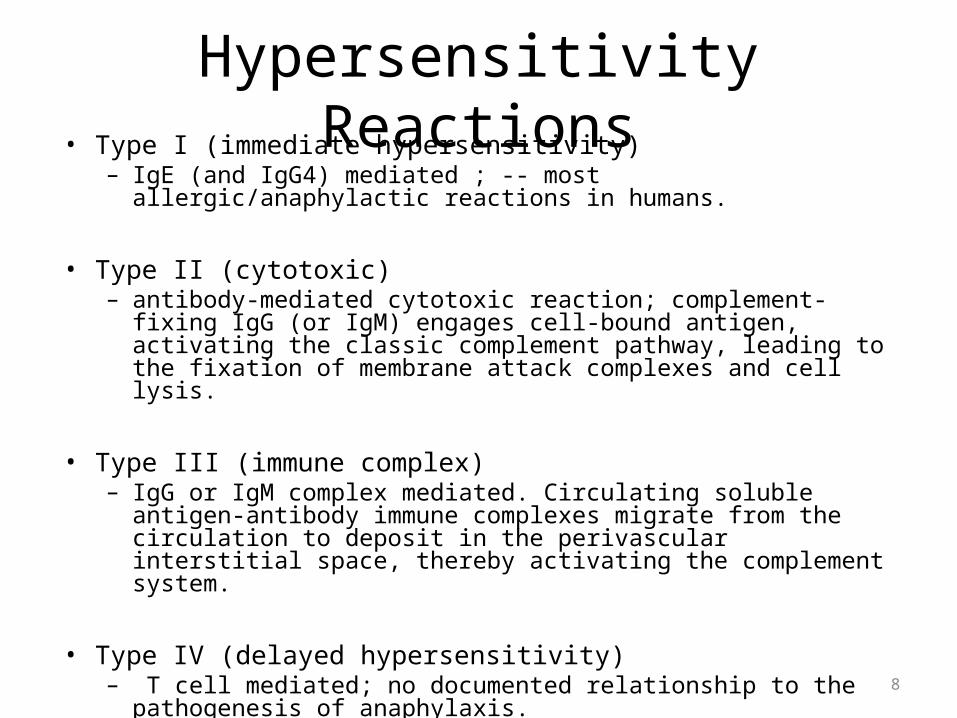
Hypersensitivity Reactions• Type I (immediate hypersensitivity)
– IgE (and IgG4) mediated ; -- most allergic/anaphylactic reactions in humans.
• Type II (cytotoxic) – antibody-mediated cytotoxic reaction; complement-fixing IgG (or
IgM) engages cell-bound antigen, activating the classic complement pathway, leading to the fixation of membrane attack complexes and cell lysis.
• Type III (immune complex) – IgG or IgM complex mediated. Circulating soluble antigen-antibody
immune complexes migrate from the circulation to deposit in the perivascular interstitial space, thereby activating the complement system.
• Type IV (delayed hypersensitivity)– T cell mediated; no documented relationship to the pathogenesis of
anaphylaxis.8

Hypersensitivity Reactions
9Source Undetermined

Urticaria & Angioedema
• Urticaria (hives)– reaction that consists of papules or wheals that are non-pitting, edematous,
pruritic, slightly erythematous, raised circular or annular, and range in size from millimeters to several centimeters.
– Centers are usually clear , borders can be serpiginous.
– Erythema due to dilation of blood vessels in the dermal layer of the skin and the edematous wheals are due to transudation from these blood vessels.
– Urticaria favors the extremities and trunk and is usually transient, with crops of hives appearing/resolving spontaneously in a matter of hours.
Heckat, Wikimedia Commons
10

Angioedema• Similar to urticaria
– But involves the deeper dermal and subcutaneous tissue.– (+) Urticaria = indicates a mast cell component to the reaction. – (-) Urticaria = kinin-related.
• Commonly involves the face, mouth, lips, tongue, extremities, and genitalia
• Acute – Episodes that last < 6 weeks (90%)
• Chronic– persist longer than 6 weeks (10%).
11Source Undetermined

Angioedema• (+) Urticaria (mast cell degranulation)
– IgE mediated, antihistamines (both H1 and H2) 1st Line
• Adrenaline can be considered for moderate to severe cases caution in patients older than 35 years to risk of precipitating an acute coronary syndrome.
• Steroids may help in preventing rebound Sx
• (-) Urticaria (10%)– usually kinin-related and causes include hereditary
angioedema (HAE), acquired C1 inhibitor deficiency (ACID), and ACE inhibitors. bradykinin levels.
– Treatment is mostly supportive, fewer Rx options• Active airway management is the mainstay of
treatment • ?Racemic Adrenaline (Epinephrine) aerosol for airway
edema• ?FFP (contains C1 Inhibitor)
– inhibition of ACE, one of the main inactivators of bradykinin, results in increased bradykinin levels. Rare reports of inciting worsened Sx
12
Source Undetermined

ACE Inhibitor-Induced• Incidence of 0.1-0.7% and has a predilection for the tongue,
lips, and laryngeal soft tissue.
• The highest incidence occurs in the first month of therapy, – But can occur as many as 10 years after therapy is started.
• Specific risk factors include African ancestry, smoking, older age, and female gender – some risk associated with DM.
• Pathophysiology– Prevention of bradykinin and substance P metabolism, both of
which are potent mediators of tissue inflammation. – Most patients who develop angioedema on ACE inhibitors
should be able to tolerate angiotensin receptor blocker drugs (ARBs).
13
Source Undetermined

Case #2
• 10 year old male with a history of recent TMP/SMX Rx for a peri-rectal abscess (resolved) presents with complaint of rash located primarily on the anterior thoraco-abdominal surface and palms. – Diagnosis/Disposition?
14
Source Undetermined

Erythema Multiforme
• Acute, usually self-limited disease precipitated by a variety of factors. – Sudden appearance of skin lesions that are
erythematous or violaceous macules, papules, vesicles, or bullae.
– Distribution often symmetrical, most commonly involving the soles and palms, the backs of the hands or feet, and the extensor surfaces of the extremities.
• Stevens-Johnson Syndrome (a severe form) is occasionally fatal.
– characterized by bullae, mucous membrane lesions, and multisystem involvement
15
Source Undetermined

• Initial Lesion Target Lesion – Three zones of color is the hallmark
• Central dark papule or vesicle that is surrounded by a pale zone and halo of erythema
• Commonly found on the hands or wrist.
16
Source Undetermined

Etiologies
• Most common precipitating factors – Exposure to medications (≤10%)
• NSAIDs, sulfonamides, multiple anti-epileptics• Antibiotics are responsible for the majority of cases
– HSV Infection• lesions appear approximately 10 days after an outbreak
– Other • Hepatitis and influenza A. • Less common causes
– fungal diseases, such as dermatophytosis, histoplasmosis, and coccidioidomycosis, and bacterial infections, especially streptococcal infections and tuberculosis.
– Various collagen vascular disorders have been known to precipitate erythema multiforme, particularly rheumatoid arthritis, systemic lupus erythematosus, dermato-myositis, and periarteritis nodosa.
– Pregnancy and various malignancies have also been associated with erythema multiforme. 17

Management
• Address underlying cause
• Mild forms (Discharge)– Resolve spontaneously in 2-3 weeks– Oral antivirals (acyclovir, valacyclovir, or famciclovir) may be used to
prevent both HSV (oral and genital) and EM– With the distinctive clinical findings and no systemic symptoms, patients
can be discharged home.
• Severe cases (Admit)– May last up to 6 weeks – IV hydration, local skin care, systemic analgesia, and systemic
corticosteroid therapy (80-120 mg of prednisone daily in divided doses)– Bullous Lesions
• treated with the application of wet compresses soaked in a 1 : 16,000 solution of potassium permanganate or a 0.05% silver nitrate solution several times a day.
18
Complications
• Stevens-Johnson Syndrome– Infection and fluid loss. – Renal involvement and pneumonia are rare. – Severe conjunctivitis
• may result in corneal scarring and blindness.
19

Case #3
• 44 year old male reports worsening rash over the last 2 days with a 5 day history of sore throat, fever, myalgia.– Has not taken antibiotics
but has been using ibuprofen.
– Diagnosis/Disposition?20
Source Undetermined

Stevens-Johnson Syndrome/Toxic Epidermal Necrolysis
• Life-threatening, reactive diseases that represent two ends of a continuum– SJS
• <10% TBSA epidermal detachment and – TEN
• >30% TBSA epidermal detachment. • Overlap occurs in 10% to 30% TBSA.
• Two or more mucosal sites are usually affected.– The overall mortality of TEN approaches 30%.
21

SJS/TEN
• Prodromal Symptoms– Malaise, rhinitis, sore throat, body aches, and fever.– Followed by the abrupt development of a macular rash
that may or may not appear as target lesions.– Mucous membrane involvement may precedes rash in TEN– Macular exanthem usually starts centrally and then
spreads to the extremities
• Bullae form within the rash and large sheets of epidermis separate from the dermis. – Involved skin is exquisitely tender to palpation.– Nikolsky Sign (+)
22

Progression
• Mucous membrane involvement develops
• Conjunctival and corneal involvement may lead to permanent scarring and blindness. – Full thickness of epidermis involved
• Mortality rate 15-20%– as high as 50% in elderly patients
• Life-threatening metabolic derangements, sepsis, respiratory failure, and gastrointestinal hemorrhage may occur and are compounded by underlying comorbidities.
23

Causes
• Non-staph TEN usually assoc with drug exposure
• The Main Culprits– Over 200 medications
• Long-acting sulfa drugs, penicillin, quinolones, tetracycline, aspirin, barbiturates, phenytoin, phenobarbital, carbamazepine, allopurinol, and non-steroidal anti-inflammatory drugs (NSAIDs)
• TEN has occurred after vaccination and immunization against poliomyelitis, measles, smallpox, diphtheria, and tetanus
24

SJS
25
Source Undetermined

TEN
26
Source Undetermined
Source Undetermined

Bullous SJS/TEN
27
Source Undetermined

Treatment• Supportive Care
– Discontinue offending agent, fluid replacement, electrolyte balance, ophthalmologic assessment and aggressive infection control
– Systemic corticosteroids remain controversial• no benefit and associated with increased mortality secondary to infection
• Admission to a Burn ICU– Fluid replacement
• Per Parkland Formula with Ringer’s Lactate • Goal UOP 0.5cc/kg/hr
– Wound Care (no Silvadine = Sulfa)– Infection control – IVIG per local protocol
• Fas ligand (FasL) expression hypothesized as the “death ligand” induced for abnormal apoptosis of epidermal cells
– in SJS/TEN patients• Pooled human immunoglobulin contain anti-Fas antibodies that have been
shown in vitro to impede apoptosis when pre-incubated with keratinocytes
28

Pearls
1. The hair-bearing scalp is spared even in severe disease.
2. Neutropenia is associated with a poor prognosis.
3. Cross reactions within anticonvulsants are common. The first 8 weeks of treatment have the highest risk of TEN and previous anticonvulsant therapy portends a tenfold increased risk.
4. Patients with previous SJS/TEN to one medication class are higher risk for developing SJS/TEN to other medication classes. 29

Case #4
• 32 year old female with no past history presents with 5 days of fever, rash, vomiting and new altered mental status/profound hypotension. – Last menstrual period was
7 days ago– Intubated at outside
hospital and transferred with possible sepsis...
– Diagnosis/Disposition?30
Source Undetermined
Source Undetermined

Toxic Shock Syndrome
• Acute febrile illness characterized by a diffuse desquamating erythroderma.
• Classically composed of high fever, hypotension, constitutional symptoms, multi-organ involvement and rash
31
32Source Undetermined

Causes• Notoriety in the early 1980s because of
association with tampon – Overall incidence of toxic shock syndrome has since
decreased– Has been seen in both men and children as well as
after nasal packing placement for epistaxis • Linked to exotoxin-producing S. aureus.
• Most cases of non-menstrual TSS occur in the post-operative setting.
33

• In menstruating women with toxic shock syndrome– S. aureus is isolated more than 90% of the time.
• Clinical manifestations– Due to the exfoliative exotoxin produced by S. aureus.– Is the same exotoxin produced in bullous impetigo
• TSS from systemic circulating exotoxin• Bullous impetigo direct S. aureus inoculum.
– The exotoxin in both cases has exquisite specificity in causing loss of desmosome-mediated cell adhesion within the superficial epidermis only.
34

TSS Diagnosis
• Requires the presence of – (1) fever of at least 38.9°C– (2) hypotension, SBP <90 mmHg– (3) skin rash
• Diffuse, blanching, non-pruritic macular erythroderma rash• Desquamination 1-2 weeks after onset of illness
– esp. Palms/Soles
– (4) involvement of ≥ 3 organ systems.
– Systemic involvement may include the gastrointestinal (GI) tract, muscular system, or central nervous system (CNS) and laboratory evidence of renal, hepatic, or hematologic dysfunction
35

36Source Undetermined

Multi-System Issues• ≥ 3 three of the following
– GI• Profuse diarrhea/vomiting at illness onset
– MSK• Sever myalgia or > two-fold increase in CPK
– Mucosal Inflammation• Conjunctival, vaginal, pharyngeal hyperemia
– Renal• BUN or creatinine > two times upper limit
– Hepatic• TBili or LFTs > two times the upper limit
– Hematologic• Thrombocytopenia (PLT <100,000/mm3)
– CNS• Disorientation, confusion, hallucination
37

TSS Physical Findings• Subjective
– Headache, myalgias, arthralgia, alteration of consciousness, nausea, vomiting, and diarrhea
• Objective– Rash
• Diffuse, blanching, macular erythroderma desquamation• Accompanying nonexudative mucous membrane inflammation is
common.
– Pharyngitis• Sometimes accompanied by a “strawberry tongue”
– Other• Conjunctivitis, or vaginitis may be seen. • As a rule, the rash fades within 3 days of its appearance. • This is followed by a full-thickness desquamation, most commonly
involving the hands and feet.38

Investigations• Basic Labs
– FBC, PT/PTT, LFTs, Renal Panel, Blood Cultures– ABG– Urinalysis– Creatinine Phosphokinase– Consider LP
• Imaging– CXR
• Pelvic Exam – TSST-1 exotoxin assay– Commonly negative
39

Management• Hypotension
– 4-20L IVF • What is the appropriate fluid choice?• What may be required if the patient receives 20L of RL?
– Vaso-active medications prn
• Infection Management– Remove tampon (or nasal/wound packing)– Clindamycin:
• potent suppressor of bacterial toxin synthesis
– Other Antibiotic Therapy not clearly indicated unless source is clear• If needed then lactamase resistant penicillin (nafcillin or oxacillin)
– OBGYN-guided drainage if abscess source found
40
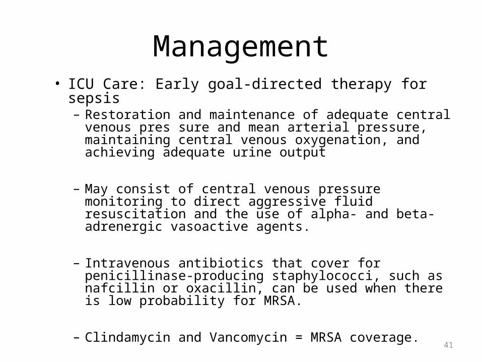
Management• ICU Care: Early goal-directed therapy for sepsis
– Restoration and maintenance of adequate central venous pres sure and mean arterial pressure, maintaining central venous oxygenation, and achieving adequate urine output
– May consist of central venous pressure monitoring to direct aggressive fluid resuscitation and the use of alpha- and beta- adrenergic vasoactive agents.
– Intravenous antibiotics that cover for penicillinase-producing staphylococci, such as nafcillin or oxacillin, can be used when there is low probability for MRSA.
– Clindamycin and Vancomycin = MRSA coverage. 41

What about Strep?
• Mid-1980s– Streptococcal toxic shock–like syndrome described
worldwide in younger otherwise healthy patients with serious soft tissue infections.
• 1987: two cases of shock due to isolated S. pyogenes soft tissue infections reported and postulated a toxin responsible (Cone)
• 1989: further characterized syndrome as involving otherwise healthy young patients who presented in shock or progressed to a shock state within 4 hours of admission (Stevens )
– Although these cases are predominantly caused by Lancefield group A strains, other groups have also been shown to cause streptococcal toxic shock syndrome.
42

Virulence
• Attributed to – Surface proteins, toxin production, host factors. – Strains that produce M protein associated with
overwhelming infection. • M protein binds to complement control factors and other
host proteins to prevent activation of the alternate complement pathway and thus evade phagocytosis and killing by polymorphonuclear neutrophil leukocytes.
– Extracellular toxins, including super antigenic streptococcal pyrogenic exotoxins, contribute to tissue invasion and initiate the cytokine storm believed to be responsible for illnesses such as necrotizing fasciitis and the highly lethal streptococcal toxic shock syndrome
43

Mechanism
• Toxins act as Super-Antigens– Activate a large population of T cells, bypassing the
antigen presentation phase and liberating massive amounts of cytokines, tumor necrosis factor alpha, interleukin-1, and interleukin-6.
– Cytokines induce the clinical signs of fever, hypotension, shock, rash, and ultimately multi-organ failure.
– Lack of host antibodies to the surface proteins and toxins predispose to infection and increased virulence.
44

Management
• Early goal-directed therapy for sepsis as described above for staphylococcal toxic shock syndrome is a mainstay of treatment.
• Even with appropriate antibiotic therapy and intensive supportive care, the mortality rate for this disease is 30-70%.
45

Antibiotics
• Choices– Clindamycin, erythromycin, or ceftriaxone + clindamycin. – Penicillin is only moderately effective against the large inoculum
of slow-growing Streptococcus seen in necrotizing fasciitis or myositis.
• Clindamycin – Thought to work by two mechanisms:
(1) effective killing of S. pyogenes organisms (2) decreasing production of extracellular products that play a role in the pathogenesis of systemic toxicity and/or tissue destruction.
• Resistance?Some clindamycin-resistant strains of S. pyogenes, so presumptive selection clindamycin and a β-lactam antibiotic would be prudent in the initial treatment 46

Case #5
• 4 month old male is brought to A&E with report of several days of fever, cough, congestion and new rash
– Disposition/Diagnosis?47
Source Undetermined

Staph Scalded Skin Syndrome • Generally occurs in children ≤ 6 years old• Caused by phage group 2 exotoxin-producing staphylococci
(Staphylococcus aureus).
• Prodrome– fever, malaise, and irritability following an upper respiratory
infection with pharyngitis or conjunctivitis.
• Rash– Begins with erythema and crusting around the mouth, then faint,
diffuse erythematous macular rash spreads peripherally, followed by flaccid bulla formation and desquamation in 2-3 days.
• exfoliation over large surface area fluid and electrolyte losses. – Mucous membranes are usually not involved.– After desquamation, lesions dry up with clinical resolution in 3-7
days.48

Management• Technically, most patients will recover without antibiotics
• Most group 2 toxin-producing organisms are penicillin resistant.
• Current Recommendations– IV therapy with 50-100 mg/kg of nafcillin daily or oral cloxacillin
50mg/kg/day or dicloxacillin (vs clindamycin)
• Admission usually necessary, especially infants.– Requires careful attention to fluid and electrolyte losses and the
prevention of secondary infection of the denuded skin. – Requires effective management of pain
49

Pearls
• The fluid in the bullae of staphylococcal scalded skin syndrome is sterile.– The toxin is produced at a remote site and delivered
to the skin via the bloodstream.
• Infants with involvement of large areas of the body surface are at increased risk for hypothermia.
• Corticosteroids are contraindicated in the treatment of staphylococcal scalded skin syndrome.
50

Case #6
• 35 year old female presents three days after having undergone I&D of right axillary abscess. Her wound was packed and she was discharged without antibiotics. She returns today for a wound check and dressing change. She reports increased pain at the site and is febrile in triage.
– Diagnosis/Disposition?51
Source Undetermined

Hidradenitis Suppurativa
• Disease of Apocrine Sweat Glands– Recurrent abscess formation in the axillae and
groin resembles localized furunculosis. – Tends to be recurrent and may be extremely
resistant to therapy• Treatment
– Incision & Drainage/Packing– Anti-staphylococcal antibiotics– Theatre/OR-drainage if refractory to A&E treatment
52

Case #7
• 21 year old female presents with several days of worsening external vaginal pain, swelling and fever without discharge.
– Diagnosis/Disposition?
53
Source Undetermined

Bartholin’s Cyst Abscess
• Obstructed Bartholin duct– Flora usually mixture of aerobic and anaerobic
flora from the vagina, with Chlamydia trachomatis and N. gonorrheae cultured ≈10% of the time.
– Patient usually has a painful cystic mass on the inferior lateral margin of the vaginal introitus.
– Signs and symptoms are usually localized, but septic shock can occur in rare cases.
54

Management• Incision & Drainage
– Drained from mucosal rather than cutaneous surface.
– Simple incision and drainage carries a high risk of recurrence, so the cavity should be packed open.
• Word Catheter– 10-Fr balloon catheter (specifically designed)– After incision and drainage, catheter is inserted and inflated
with 2-5 mL of water or saline – Remains for 4-6 weeks so that a sinus will have time to form.
• Sitz Baths– Warm H2O “hip bath” helps keep area clean and draining.
• Marsupialization– More complex treatment option, involving incision and
drainage of the cavity followed by suturing of the walls of the cyst to the skin 55

56Image: Micko, CNIS WikiCustom License: The authors and copyright holders grant to all users a free, irrevocable, worldwide, perpetual right of access to, and a license to copy, use, print, and distribute the work in any digital medium for any responsible purpose, subject to proper attribution of authorship, as long as all this is done for non-commercial reasons.

Case #8
• 5 year old boy with eczema complains of gluteal muscle pain after falling on a fence >48 hours ago but has no wounds or deficits and is otherwise well per his parents. – On exam you notice...
– Diagnosis/Disposition?
57
Source Undetermined
Source Undetermined
Source Undetermined

Bullous Impetigo• Focal bacterial infection of the superficial skin
– Streptococcus pyogenes (group A-hemolytic Streptococcus) and Staphylococcus aureus.
• Most commonly affects children 2-5 years of age and usually involves the face and extremities.
• Lesions– thin-walled, 1-2-cm bullae. When these rupture, they leave a thin
serous crust and remnant of the blister roof at the rim of the crust.
– The face, neck, and extremities are most often affected.– Gram’s stain of the fluid from a bulla reveals gram-positive cocci.
Cultures are positive in 95% of cases.
• The lesions may spread rapidly by autoinoculation secondary to scratching and coalesce to form larger areas of infection.– e.g., Super-infection of eczema sites
58

Management
• Good hand washing and personal hygiene– Emphasized to the patient and family– Highly contagious and spread by direct contact
• Treatment based on extent of lesions– Oral penicillinase-resistant semi-synthetic penicillin such as
dicloxacillin, 250 mg four times a day for 5 to 7 days for adults, or erythromycin, 250 mg four times a day in adults or 30 to 50 mg/kg/day in children.
– If the infection is limited to a small area, mupirocin 2% ointment three times a day may be applied.
– Without treatment, impetigo heals within 3 to 6 weeks
59

Case #9
• 60 year old Greek male with Rheumatoid Arthritis and Hypertension presents with mouth blisters for several months. – Blisters have now extended to
his neck/face over the last 4 days. Some of them have sheared with minimal pressure
– Diagnosis/Disposition?60
Source Undetermined

Pemphigus Vulgaris• Bullous disease, affecting both sexes equally.
• Most common in patients 40 to 60 years old.
• Mostly prevalent in people of Jewish, Mediterranean, or South Asian descent.
• Mortality rate before the use of steroids was approximately 95%.
• Cause largely unknown– Some studies suggest autoimmune mechanism. – Associated in a few instances with Rx use
• penicillamine , captopril
61

Pemphigus Vulgaris
• Mucous membrane lesions occur before skin lesions– 50-60% of patients have oral lesions that typically antedate the
cutaneous lesions by several months.– Most common site is in the mouth, especially the gums and
vermilion borders of the lips. – Oral lesions are bullous but commonly break, leaving painful,
denuded areas of superficial ulceration
• Initially small, flaccid bullae that break easily, forming superficial erosions and crusted ulcerations.
• Any area of the body may be involved.
• (+)Nikolsky’s Sign– Blisters may be extended or new bullae may be formed by firm
tangential pressure of a finger on the intact epidermis 62

Management
• Pain control and local wound care are essential components of therapy.
• Once the diagnosis is made, treatment with oral glucocorticoids– initial doses: Prednisone (or equivalent) 100-300 mg
qD• Despite localization to the skin and mucous
membranes, death the rule before steroid use– Related to uncontrolled spread of the disease,
secondary infection, dehydration and thromboembolism.
63

Case #10
• A 6 year boy and his 15 year old Rugby-playing brother presents with several days of a painful raised rash. Their mother is concerned that they have a bacterial infection.– Diagnosis/Disposition?
64
Source Undetermined
Source Undetermined

Herpes Simplex Virus
• Usually localized in a non-dermatomal distribution.
• HSV-1 (≈“above the waist”)– Primarily affects non-genital sites– Mouth is the most common site of HSV-1 infections. – Children are affected more commonly than adults.– Small clusters of vesicles appear but are soon broken,
leaving irregularly shaped, crusted erosions. • severity of gingivostomatitis varies from the presence of small
ulcers to extensive ulceration of the mouth, tongue, and gums accompanied by fever and cervical lymphadenopathy. The infection may be so severe that oral fluid intake is difficult, and dehydration may result.
• Healing typically occurs in 7 to 14 days, unless a secondary infection with streptococci or staphylococci occurs.
65

Non-Genital Herpes Simplex
• Herpetic whitlow– Painful HSV infection of the distal finger characterized by
edema, erythema, vesicles, pustules on erythematous base. – Fever, lymphangitis, and regional adenopathy
• Herpes gladiatorum– Spreads via direct skin-to-skin contact in sports such as
rugby and wrestling. – May become latent, and tends to recur at the site of the
primary lesion. – Cutaneous lesions of the primary infection may occur alone
or with fever, lymphadenopathy, myalgia, pharyngitis, malaise, blepharitis, or keratoconjunctivitis.
66

Progression• Incubation may take 2-14 days.• Symptoms
– Stinging, burning, and pruritus at the inoculation site typically precede the eruptions.
• Diagnosis– Made on identification of the characteristic lesions most
easily during the vesicular phase. – Later, crusted and dried vesicular fluid may be confused
with impetigo. – Viral cultures taken from scraping the base of an unroofed
vesicle may confirm the diagnosis. – Tzanck smear may provide more immediate confirmation.
67

Management
• Oral antivirals (Acyclovir, Famcyclovir) in addition to analgesics/antipyretics– To be most effective, antivirals must be started
within 72 hours of the eruption and should be initiated if prodromal symptoms (burning, stinging) occur.
– Topical antibiotic ointments help prevent secondary infection and speed healing.
– Do not I&D digital Whitlow Lesions
68

Case #11
• 42 year old female with a known (+)retroviral screen and anti-retroviral Rx non-compliance, complains of several days of a burning over her left forehead. As her vitals are normal and there is no evidence of rash she is discharged home with clinic follow-up.
69

• One week later she returns and now has an painful facial rash.
– Diagnosis/Disposition?
70
Source Undetermined

Herpes Zoster
• Varicella-Zoster Virus– Occurs exclusively in individuals who
have previously had chickenpox. – Before the rash appears, the patient
typically develops pain in a dermatomal distribution.
• Pain precedes eruption by 1-10 days• Rash consists of grouped vesicles on an
erythematous base involving one or more dermatomes.
– Thorax = most cases– Trigeminal distribution next most
71
Lipothymia, Wikimedia Commons
Mikael Häggström, Wikimedia Commons
Source Undetermined

Progression
• Caused by a reactivation of latent varicella-zoster virus present since the initial infection with chickenpox. – During the latent period between the two illnesses, the virus is
thought to reside in dorsal root ganglion cells.
• Very low mortality rate and rarely life threatening, even when dissemination to the visceral organs occurs. – Early systemic corticosteroid therapy may shorten the duration of
post-herpetic neuralgia but does not lessen the severity of pain.– Antiviral Tx (acyclovir, famciclovir, vidarabine, foscarnet, valacyclovir,
and interferon-α), has been shown to be effective for immunocompromised patients.
• Complications– CNS involvement, ocular infection, and neuralgia as well as rare
meningoencephalitis, myelitis, and peripheral neuropathy72

Herpes Zoster Ophthalmicus
• Ocular complications– Occur in 20-70% of cases involving the ophthalmic division
of the trigeminal nerve. – Severity varies from mild conjunctivitis to panophthalmitis– Eye involvement produces anterior uveitis, secondary
glaucoma, and corneal scarring.
• Hutchinson’s Sign – Correlation between eye involvement and vesicles located
at the tip/side of nose – Patch innervated by the infratrochlear and external nasal
branches of the nasociliary nerve (also innervates cornea)– The prognostic value of the Hutchinson sign has been
validated in one study.73

Immunocompromise
• More severe in immunosuppressed patients, especially those with AIDS, Hodgkin’s disease, or other lymphomas
• Cutaneous dissemination occurs more commonly in these patients than in the general population.
• Visceral and CNS dissemination is also more likely to occur in these patient.
74

Case #12• 65 year old man with poorly-
controlled DM and multiple prior pilonidal abscesses presents with 3 days of scrotal pain & swelling that started after he noticed a small, painful “spider bite” in the right gluteal fold.
• Transferred with Foley catheter from an outside hospital due to perineal involvement
– Diagnosis/Disposition?75
Source Undetermined

Necrotizing Fasciitis• Usually occurs in men in the lower extremities and may
evolve after only minimal local trauma.
• Type I– Polymicrobial and involves non- group A streptococci plus
anaerobes. – Abdomen and perineum
• Type II– Group A beta-hemolytic streptococci – Extremities
• Streptococci endotoxin separation of the dermal connective tissue, resulting in continued inflammation and necrosis.
• Streptococcal necrotizing fasciitis is frequently associated with streptococcal toxic shock syndrome. 76

Progression– Early clinical findings similar most infected wounds
• Involved site quickly becomes erythematous, tender, and edematous with formation of necrotic patches/bullae (days 2-4) productive of sero-sanguinous fluid
• Fever usually present• Deep structures and muscles are not involved. • Hypotension, tachycardia, leukocytosis, and
hypocalcemia ensue with systemic toxicity out of proportion to the clinical findings
– X-ray reveal gas in the tissues, but tissue gas is not a universal finding
• MRI better, but due to rapidly progressive and frequently fatal outcome of this condition, delaying treatment to wait for imaging is not justified even when available immediately
77

kidney, ureter and bladder showed mottled air density over the pelvic region and perirectal area.
Chen C , Su Y Emerg Med J 2011;28:174-174 78

Management
• Surgical debridement along fascial planes
• Fluid and critical care resuscitation
• Parenteral antibiotics against S. aureus, Streptococcus, gram-negative organisms, and anaerobes – Eventually directed by Gram stain/culture
79

Fournier’s Gangrene• Insidious necrotizing subcutaneous infection of the perineum that
occurs primarily in men usually 20-50 years of age– Usually involves the penis or scrotum.
• Pain or pruritus in the genitalia is followed by fever, chills, and impressive perineal swelling
• Inflammation may involve the entire abdomen, back, and thighs. – Frequently crepitus on palpation, indicating subcutaneous gas.
• Systemic Symptoms – Nausea and vomiting, changes in sensorium, and lethargy. – Eventually, gangrenous areas demarcate and become less painful as
destruction and sloughing of sensory nerves occur. The tissue breaks open, sloughs, and gives off an overwhelming feculent odor.
80

• Most common causal factors– Infection or trauma to the perianal area
• including anal intercourse, abscess/scratches, chemical or thermal injury, and diabetes.
– Local trauma and perianal disease precede approximately one third of all cases.
– Cultures demonstrate bacteria of the distal colon, with a complex picture of aerobic and anaerobic bacteria.
• B. fragilis the predominant anaerobe• E. coli the predominant aerobe.
81
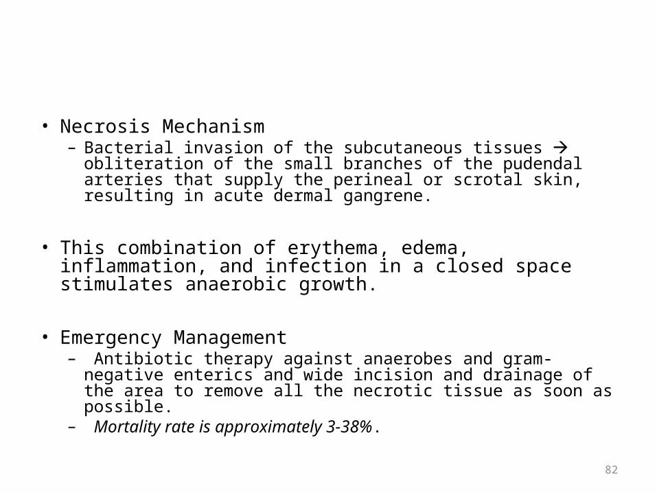
• Necrosis Mechanism– Bacterial invasion of the subcutaneous tissues obliteration of
the small branches of the pudendal arteries that supply the perineal or scrotal skin, resulting in acute dermal gangrene.
• This combination of erythema, edema, inflammation, and infection in a closed space stimulates anaerobic growth.
• Emergency Management– Antibiotic therapy against anaerobes and gram-negative
enterics and wide incision and drainage of the area to remove all the necrotic tissue as soon as possible.
– Mortality rate is approximately 3-38%.
82
Literature Cited• Chen et al. 2011. X-ray diagnosis of fatal Fournier's gangrene caused by
Klebsiella pneumoniae. Emerg Med J 2011;28:174
• Cone LA, Woodard DR, Schlievert PM. Clinical and bacteriologic observations of a toxic shock-like syndrome due to Streptococcus pyogenes. N Engl J Med. Jul 16 1987;317(3):146-9
• Cydulka RK and B Garber. Dermatology. In: Rosen’s Emergency Medicine, Concepts and Clinical Practice. 7th ed.
• Meislin HW and JA Guisto. Soft Tissue Infections. In: Rosen’s Emergency Medicine, Concepts and Clinical Practice. 7th ed.
• Stevens DL, Tanner MH, Winship J. Severe group A streptococcal infections associated with a toxic shock- like syndrome and scarlet fever toxin A. N Engl J Med. Jul 6 1989;321(1):1-7
83