project: ghana emergency medicine collaborative document title: musculoskeletal emergencies and the...
TRANSCRIPT

Project: Ghana Emergency Medicine Collaborative
Document Title: Musculoskeletal Emergencies and the Nursing Process
Author(s): Barbara Demman (University of Michigan), RN 2012
License: Unless otherwise noted, this material is made available under the terms of the Creative Commons Attribution Share Alike-3.0 License: http://creativecommons.org/licenses/by-sa/3.0/
We have reviewed this material in accordance with U.S. Copyright Law and have tried to maximize your ability to use, share, and adapt it. These lectures have been modified in the process of making a publicly shareable version. The citation key on the following slide provides information about how you may share and adapt this material.
Copyright holders of content included in this material should contact [email protected] with any questions, corrections, or clarification regarding the use of content.
For more information about how to cite these materials visit http://open.umich.edu/privacy-and-terms-use.
Any medical information in this material is intended to inform and educate and is not a tool for self-diagnosis or a replacement for medical evaluation, advice, diagnosis or treatment by a healthcare professional. Please speak to your physician if you have questions about your medical condition.
Viewer discretion is advised: Some medical content is graphic and may not be suitable for all viewers.
1

Attribution Key
for more information see: http://open.umich.edu/wiki/AttributionPolicy
Use + Share + Adapt
Make Your Own Assessment
Creative Commons – Attribution License
Creative Commons – Attribution Share Alike License
Creative Commons – Attribution Noncommercial License
Creative Commons – Attribution Noncommercial Share Alike License
GNU – Free Documentation License
Creative Commons – Zero Waiver
Public Domain – Ineligible: Works that are ineligible for copyright protection in the U.S. (17 USC § 102(b)) *laws in your jurisdiction may differ
Public Domain – Expired: Works that are no longer protected due to an expired copyright term.
Public Domain – Government: Works that are produced by the U.S. Government. (17 USC § 105)
Public Domain – Self Dedicated: Works that a copyright holder has dedicated to the public domain.
Fair Use: Use of works that is determined to be Fair consistent with the U.S. Copyright Act. (17 USC § 107) *laws in your jurisdiction may differ
Our determination DOES NOT mean that all uses of this 3rd-party content are Fair Uses and we DO NOT guarantee that your use of the content is Fair.
To use this content you should do your own independent analysis to determine whether or not your use will be Fair.
{ Content the copyright holder, author, or law permits you to use, share and adapt. }
{ Content Open.Michigan believes can be used, shared, and adapted because it is ineligible for copyright. }
{ Content Open.Michigan has used under a Fair Use determination. }
2

Function of Musculoskeletal System
• Support• Protection• Movement and leverage• Storage of mineral salts and fats• Red blood cell production
3

U.S. National Cancer Institute SEER Program, Wikimedia Commons 4

Components of Musculoskeletal system
• Bones• Nerves• Vessels- arteries, veins• Muscles• Tendons- attach muscle to bone• Ligaments-attach bone to bone• Joints
5

Brian0918, Wikimedia Commons 6

Assessment of Musculoskeletal System
• Primary– Form a general impression, “ABC”– AIRWAY: patency– BREATHING: adequate respirations– CIRCULATION: control bleeding, watch for shock
• Secondary– vital signs, physical exam, more detailed exam
7

Assessment of Musculoskeletal System
• Subjective Data: – what the patients says– Patient dialogue– History/information gathering
• Objective Data: – scientific data gathered from physical exam and
diagnostic exams
8

Assessment of MS System- Subjective
• Obtain history of injurymechanism of injury (twist, crush, stretch)Circumstances r/t injuryOnset- acute vs. chronicPrevious diagnostic examsMethods/duration of treatmentRelieving/aggravating factorsNeed for assistive devicesInterference with Activities of Daily Living (ADL’s)
9

Mechanism of Injury
• Significant force is generally required to cause fractures and dislocations.– Direct/Indirect/Twisting/High-energy forces
– Assists with anticipation of injuries
– A low-speed car accident is much less likely to cause a life-threatening injury than a rollover accident. A gunshot wound has more potential for serious injury than a fistfight.
10

Mechanism of Injury
-Was the arm or hand outstretched? • FOOSH- often distal radial fx• “Fall On Outstretched Hand”
–At what angle to the body was the arm, shoulder or hand on impact?–Did hyperflexion or hyperextension occur?–Fracture or dislocation of the area before?–Involved in rigorous athletic training (overuse injury)?
11

Assessment of MS System- Subjective
• SUBJECTIVE– Pain– Weakness– Deformity– Limitation of movement– Stiffness– Joint crepitation
• Medications– What medications
directly influence integrity of the MS system?
• Antiepileptics• Corticosteroids• chemotherapy
12
Assessment of MS System- Subjective
• Past Health Hx– What disease processes affect the MS system?– TB, poliomyelitis, infections-osteomyelitis– Diabetes mellitus, – Rickets– Rheumatoid arthritis, lupus– Gout, osteoarthritis– Autoimmune disease- steroid use
13

Assessment of MS System- Objective
• INSPECT- swelling/deformity/shortening of limb/bleeding
• PALPATE/FEEL for tenderness and crepitus with movement and temperature.
• Range of Motion- Passive and Active• Muscle-Strength Testing, 0-5 scale• Neurovascular exam*
– Color, temp, CRT, pulses, edema, sensation, motor function, nerve involvement
14

Muscle Strength Testing Scale
Source: www.pacificu.edu/optometry/ce/courses/15840/neuroexampg3.cfm
15

Assessment of Neurovascular StatusEnsures integrity of injured area
• Parasthesia• Pain• Pressure• Pallor- capillary refill time < 3 seconds• Paralysis• Pulses- distal to injured extremity
16

17Source Undetermined

Nursing Care Interventions for Musculoskeletal Emergencies
•Frequently assess, document, and report the six Ps (pain, pallor, pulses, pressure, paresthesia, and paralysis).
•REPORT STATUS CHANGES
18

Differential diagnosis
• the determination of which of two or more diseases with similar symptoms is the one from which the patient is suffering, by a systematic comparison and contrasting of the clinical findings.
• Gather all data and create differential diagnosis list
• Determines plan of care
19

Musculoskeletal Diagnostic Studies/Procedures to Aid in Diagnosis
• Radiology Studies– Standard X-Ray– Compute Tomography
(CT)• Endoscopy
-Arthroscopy• Arteriograms
• Complete blood count with differential (CBC)
• Electrolytes• Type and Cross Match• Urinalysis• Wound cultures• Mineral Metabolisms
– Calcium, Phosphate• Serological Studies
– ESR, RF, ANA
20

Skeletal X-RAY Studies
• Standard Anterior/Posterior and lateral radiographs, to include the joint above and below the fracture level, are the minimal requirement for most fractures.
21

Nursing Responsibilities for Diagnostic Exams
• Remove radiopaque objects• Pain management• Determine pregnancy status• Allergies to Contrast Medium/Iodine/Shell
Fish• Administer antianxiety if indicated
22
General Nursing Interventions/Responsibilities for MS
Emergencies• Assessment- report status changes• Physician (MD) orders• Pain Management• Preparation for diagnostic exams• Preparation for surgical intervention• Documentation• Patient Education
23

Nursing Care Interventions for Emergent Musculoskeletal Findings
BLEEDING
•Control bleeding by applying pressure with a sterile dressing. •Avoid hypovolemic shock by administering intravenous fluids and oxygen.
24

Nursing Care Interventions for Emergent Musculoskeletal Findings
DEFORMITY
•Immobilize above and below the injury site in the most comfortable position with a splint.•Do not attempt to straighten the limb or manipulate protruding bone.
25

Nursing Care Interventions for Emergent Musculoskeletal Findings
SWELLING
•Apply and intermittently reapply cool packs to the injured area for up to 48 hours if needed.•Elevate the extremity above the level of the heart.
26
Nursing Care Interventions for Emergent Musculoskeletal Findings
PAIN/ANXIETY
•Initiate oral or intravenous analgesia as soon as possible.•Communicate with patient plan of care and findings
27

Common Drugs used in Musculoskeletal Emergencies
• Pain Control: NSAIDS, Opiates• Infection Control:
Prophylaxis/Cephalosporin• Procedural Sedation: Fentanyl, Versed• Reversals: Naloxone, Flumazenil
28

Non-Steroidal Anti-inflammatory Drugs
• Aspirin, Ibuprofen, Naproxen, Indomethacin, ketorolac, diclofenac
• Use for mild to moderate pain, fever• Decrease pain, decrease inflammation• Non-opioid• Adverse reactions- GI effects, renal effects
29

OPIATES
• Morphine, Codeine, Hydrocodone, Fentanyl• Analgesic, IV, by mouth (PO), transdermal• decreased perception of pain, decreased
reaction, increased pain tolerance. • side effects: sedation, respiratory depression,
constipation, and a strong sense of euphoria• Opiate withdrawal symptoms with chronic
long term use
30

Procedural Sedation
• Procedural sedation refers to a technique of administering sedatives or dissociative agents, with or without analgesics, to induce a state that allows patients to tolerate unpleasant procedures while maintaining cardiorespiratory function and retaining the ability to respond purposefully to verbal commands and/or tactile stimulation.
• Appropriate for adult and pediatric patients.• Often used to sedate patients while reducing
fractures or dislocationshttp://emssa.org.za/documents/em013.pdf
31

SATS Policy on Procedural Sedation
• http://emssa.org.za/practice-guidelines/
32

Pre-Procedure Preparation and Equipment
• The following equipment should be present (refer to EMSSA Practice Guideline EM006):
• Oxygen and delivery devices (nasal, cannula and face mask)• Suction and suction catheters• Resuscitation trolley and defibrillator and intubation
equipment• Vital signs monitor (including BP, cardiac monitor and
saturation)• Positive pressure breathing device• Appropriate size oral airway• Reversal agents
33

MONITORING AND DOCUMENTATION for procedural sedation
• Assessment of the patient should be done at baseline and every five minutes once the first analgesia/sedation dose has been administered. The following should be documented:
• Vital signs (BP, HR, RR)• ECG rhythm• Oxygen saturation• Airway patency• Use of supplemental oxygen or not• Level of consciousness• Pain• Medications given including route, dose and person
administering.
34

Post-procedure Discharge Criteria• Vital signs, level of consciousness, cardiovascular and respiratory status have• returned to pre sedation levels.‐• A responsible, designated adult is able to accompany patient • The patient/caregiver has received appropriate verbal and written discharge• instructions.• Discharge forms are completed and discharge medication has been dispensed.• Pain is adequately controlled.• Nausea/vomiting is controlled.• Oxygen saturation is at pre intervention status.‐• No signs or symptoms that may jeopardize the safety of recovery (i.e. Bleeding,• swelling, extreme pain, dizziness etc.)• Follow up for extended care has been provided.‐• For children: age appropriate responses are present.
35

General MS Documentation• Documentation Neurovascular assessment and documentation shall
include: • a. date and time of assessment • b. extremity • c. sensation (sensory) • d. temperature (distal to pressure point) • e. movement (motor) • f. capillary refill (blanches) • g. pulses • h. color • i. any other pertinent observations (i.e. swelling)
• REPORT ANY PERTINENT CHANGES
36

Patient Education
• Strength exercises• Prevention• Pain management• Assist devices: cane, crutches, walker• Expected outcomes• When to return for emergencies
37

Review General Nursing Interventions/Responsibilities for MS
Emergencies• Assessment- report status changes• Physician (MD) orders• Pain Management• Education• Preparation for diagnostic exams• Preparation for surgical intervention• Documentation• Patient Education
38

Musculoskeletal Disease Topics
• Soft Tissue Injuries• Contusion/Hematoma• Overuse Injuries• Low back pain• Dislocations
• Fractures• Amputations• Joint Injuries• Arthritis
39

Soft Tissue Injuries• Contusion/Hematoma
• Dislocation- Displacement or separation of joint articular surfaces with severe ligamentous structure injury
• Subluxation- partial dislocation
• Shoulders, elbows, patella, hips, fingers • Forceful high impacts/transmissions
40

Contusion/Hematoma
• Contusion/Bruise: capillaries and veins are damaged by trauma, allowing blood to seep into the surrounding interstitial tissues. skin, subcutaneous tissue, muscle, or bone.– cerebral, myocardial, pulmonary
• Hematoma: localized collection or pocket of blood
41

Bruise versus Contusion
Ksuel, Wikimedia Commons Petr K, Wikimedia Commons 42

Treat a closed soft-tissue injury by applying the mnemonic RICE:
REST: keep patient quiet and comfortable ICE: constrict blood vessels and reduce pain COMPRESS: slow bleeding ELEVATE: raise injured part above level of the heart to decrease swelling
* Swelling hurts and delays healing time
43
Overuse Injuries
• subtle, occurs over time• Cumulative trauma• tissue damage, micro tears that results from
repetitive demand over the course of time.
• Stress fracture (runners)• Carpal Tunnel Syndrome• Manual labor occupations• Athletics
44

Sprains and Strains• Sprain- ligament injury
– Twisting– Ligament: bone to bone
• Strain- Muscle or Tendon injury– Overuse injury– Tendon: muscle to bone
• 1st, 2nd , 3rd degree – Fibrous and muscle tears, swelling, edema, pain, decrease in
function• Sprains/Strains not usually visible on x-ray but can be as
painful as and have similar healing time as a fracture.
45

Nursing Responsibilities for sprains and strains
• Assess neurovascular status*• RICE• Apply cold, ice pack acute phase/after 48
hours heat and cold• Immobilization• Anticipate x-rays• Analgesia as necessary• Education: strength exercises, prevention• Assist devices: crutches
46

Low Back Pain
Glitzy_queen00, Wikimedia Commons 47

Low Back Pain- Differential Diagnosis
• Trauma• Lifting something too heavy/overstretching• Nerve/Muscle Irritation• Arthritis, Osteoporosis, Bone disease/lesion• Infections- TB, pyelonephritis• Pelvic Fracture• Intravenous (IV) drug history use
48

• Pain accompanied by fever or loss of bowel or bladder control, pain when coughing, and progressive weakness in the legs may indicate a pinched nerve or other serious condition.
49

Low Back Pain ER Nursing Management
• Etiology of back pain/medical history• Physical exam• Circulation, movement, and sensation of all extremities • Pain scale assessment and reassessment within 2
hours after medication or other pain interventions.• Vital signs on admission. • Medicate for pain per MD order • Urine dipstick per MD order- + blood or +pregnancy
+White blood cells (WBC)• Diagnostic imaging per MD order
50
Low Back Pain Goals
• Accurate diagnosis• Relieve pain• Increase mobility
51

Dislocations
• Bones in a JOINT, become displaced or misaligned
• Obvious deformity- compare to other extremity• Loss of normal joint motion• Localized pain, swelling, tenderness
• Sometimes a dislocated joint will spontaneously reduce before your assessment.– Confirm the dislocation by taking a patient history.
52

Dislocation
– Ligaments usually damaged
– A dislocation that does not reduce is a serious problem.
– Numbness or impaired circulation to the limb or digit
• *Risk avascular Necrosis!• Orthopedic Emergency
53

Dislocation
• Assess pulses and cap refill distal to injury• Range of Motion• Obtain x-ray to verify dislocation and assess
for fractures related to dislocation• Pain management.
54

• The humeral head most commonly dislocates anteriorly.
• Shoulder dislocations are very painful.– Stabilization is difficult because any attempt to
bring the arm in toward the chest wall produces pain.
– Splint the joint in whatever position is more comfortable for the patient.
55

Anterior Shoulder Dislocation
Source undetermined, Pediatric Orthopedics Injuries

Dislocation: Nursing Management
• Goal: Realign dislocated joint-REDUCTION• Expose dislocated joint area• Start Intravenous Access (IV) and IV Fluids• Assist with reduction-CALM PATIENT!• Possible procedural sedation• Immobilization- sling, splint• Movement after reduction can lead to further
dislocation• Patient Education
57

Fractures
• Disruption or break in the continuity of bone structure
• Open fracture: skin integrity impaired/ bone exposed
• Closed fracture: skin integrity intact• Non-displaced: bone still in alignment• Displaced: bone not in alignment
58

Types of Fractures
Source undetermined, SayPeople.com 59

Suspect a Fracture If…
– Deformity– Tenderness– Pain– Guarding– Swelling– Bruising– Crepitus– Exposed fragments– Verified by X-ray
60

Fracture Goal and Care
Reduction and ImmobilizationImmobilize: to retain reduction or anatomical alignmentReduction: medical procedure to restore a fracture or dislocation to normal alignment. Needed for displaced fractures.
•Reduction:– Closed Reduction– Open Reduction
61

Anatomical Alignment of Fractures
• Closed Reduction– Nonsurgical– Traction/counter
traction– Under local anesthesia
(joint block) or conscious sedations
Bobjgalindo, Wikimedia Commons 62

Anatomical Alignment of Fractures
• Open Reduction– Surgical – Wires, pins, screws– Internal fixation– External fixation
– *ORIF: Open reduction internal fixation
Source undetermined, E-Radiography 63

External Fixation
• Pin care• Infection risk• Pain control
Source undetermined, Journal of Bone and Joint Surgery 64

Nursing Responsibilities: Fractures
• Neurovascular Assessment!• Compartment
Syndrome***• Pain Management• Immobilization
– Cast set up– Traction
• Pre-operative duties
• Open wound care• Tetanus Vaccination
status• IV antibiotics• Cover wound with
sterile dressing• If impaled object, do
not remove but stabilize
65

Anticipate Estimated Blood Loss with Fractures (EBL)
• Femur fx- 1.5 L• Pelvis fx- 2 L• Thorax- 2L• Humerus- 0.5 L
Hugh Dudley, Primary Surgery Textbook 66

Fractures of Pelvis• Often results from direct compression in the form of a
heavy blow– Can be caused by direct or indirect forces
• May be accompanied by life-threatening loss of blood• Open fractures are quite uncommon.• Suspect a fracture of the pelvis in any patient who has
sustained a high-velocity injury and complains of discomfort in the lower back or abdomen.
• If Pelvis is unstable or “open book” fracture apply pelvic binder.
67

Pelvic Binder
Source undetermined, Trauma.org68

Cast Set Up and Nursing Responsibilities
• Fiberglass• Plaster• Water bucket• Scissors• Towel• Stable patient positioning
69

70Mass Communication Specialist 3rd Class Eduardo Zaragoza, U.S. Navy, Wikimedia Commons

Patient Education on Cast Care
• Unusual odor beneath the cast• Tingling, burning, numbness of toes• Drainage through cast• Swelling or inability to move toes• Toes that are cold, blue or white• Sudden unexplained fever• Pain that is not relieved by comfort
measures• No smoking
71

Splinting
• Process of immobilizing and stabilizing painful, swollen, deformed extremities
• SOFT or RIGID• Soft: Pillows, Blankets, Dressings, slings• Hard: (very little flexibility)• – Plastic – Wood – Compressed cardboard
72

Why Do We Splint?
• Can reduce pain• Prevent further injury• Limit the damage to soft tissues• Limit internal & external bleeding• Help relieve pressure against blood vessels• closed fractures from becoming open
fractures• Easier for transport, transfer
73
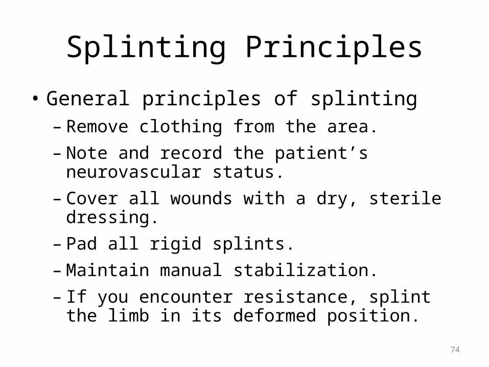
Splinting Principles
• General principles of splinting– Remove clothing from the area.– Note and record the patient’s neurovascular
status.– Cover all wounds with a dry, sterile dressing.– Pad all rigid splints.– Maintain manual stabilization.– If you encounter resistance, splint the limb in its
deformed position.
74

Assistive Walking Devices
• Crutches– Crutch Training to prevent further injury– Weight bearing versus non weight bearing
• Cane• Walker
75
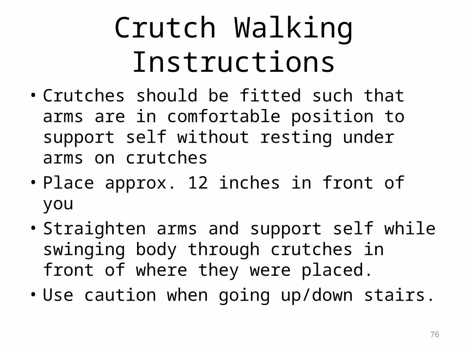
Crutch Walking Instructions
• Crutches should be fitted such that arms are in comfortable position to support self without resting under arms on crutches
• Place approx. 12 inches in front of you• Straighten arms and support self while
swinging body through crutches in front of where they were placed.
• Use caution when going up/down stairs.
76

Traumatic Amputations
• Complete- total severing of limb or appendage from rest of body
• Partial- some soft tissue remains attached• arms, ears, feet, fingers, hands, legs, and nose
77

Traumatic Amputation of Hand
Gabriel Mejia, trauma.org 78

• Surgeons can occasionally reattach amputated parts.
• Make sure to immobilize the part with bulky compression dressings.– Do not sever any partial amputations.
– Control any bleeding to the stump.
– If bleeding cannot be controlled, apply a tourniquet.
79

• With a complete amputation, wrap the severed part in a sterile dressing and place it in a plastic bag.– Put the bag in a cool container filled with ice.
– The goal is to keep the part cool without allowing it to freeze or develop frostbite.
80

Joint Injuries
• Joint Effusions• Costochondritis• Osteoarthritis• Septic Arthritis• Rheumatoid Arthritis
81

Joint Effusion
• Swollen joints happen when there's an increase of fluid in the tissues that surround the joints. Joint swelling is common with different types of arthritis, infections, and injuries.
82

Knee Effusion
James Heilman, Wikipedia 83

Costochondritis• Inflammation of junction where the ribs join the sternum• Localized chest pain/reproduce by pushing on cartilage in
front of ribcage. • Often has no definite cause/resolves without treatment• May be r/t chest infections or minor trauma to chest wall• Often in younger population, 12 -20 years of age• Diagnosis can be reached after excluding more serious
causes of chest pain- Differential diagnosis for chest pain/angina
• Supportive treatment- anti-inflammatory medications, heat application.
84

Costochondritis
Mikael Haggstrom, Wikimedia Commons85

Osteoarthritis (OA)
• Degenerative joint disease• Breakdown/erosion of cartilage in joints-
decreases shock absorbing • Often related to obesity, injury, overuse
syndromes, or hereditary factors• Most commonly in weight bearing joints
– Hips, knees, spine
86

OA Symptoms
• Pain in affected joint after repetitive use• Joint pain worse later in the day• Pain and stiffness after long periods of
inactivity (sitting)• Swelling, warmth, crepitus to affected joint• May walk with a limp• Bony enlargement of the joints from spur
formations is characteristic of osteoarthritis.
87

Heberden's & Bouchard's Nodes
Drahreg01, Wikimedia Commons88

Bunions
• Enlargement and repositioning of joints at the ball of the foot.
• Mostly women• Treatment of bunions
includes rest, alteration of footwear, foot supports, medications, and/or surgery.
Dr. Henri Lelièvre, Wikimedia Commons 89

OA Treatment
• Rehabilitative and supportive measures• Adjunctive drug therapy• Weight loss, low impact exercise, healthy diet,
assistive devices• Non-steroidal anti-inflammatory medications
90

Septic Arthritis
• inflammation of one or more joints as a result of infection by bacteria/viruses or less frequently, fungi or parasites.
• Most often, the infection begins at some other location in the body and travels via the bloodstream to the joint.
• Symptoms often include fever, chills, general weakness, and headaches, followed by inflammation and painful swelling of one or more joints of the body.
91

Septic Arthritis
• Anticipate x-ray of affected joint• Blood cultures• Aspiration of joint fluid for laboratory studies• Anticipate antibiotics, rest, and pain
management
92

Septic Arthritis of Right Index Finger
Chris Craig, Wikimedia Commons 93

Rheumatoid Arthritis (RA)• CHRONIC SYSTEMIC AUTOIMMUNE DISEASE• The inflammation in the joints causes pain,
stiffness, swelling, and loss of function. • The inflammation often affects other organs and
systems of the body, including the lungs, heart, and kidneys.
• Women more often than men• Usual onset 35-50 years of age, but it Can occur in children, teenagers, and elderly people
(juvenile rheumatoid arthritis)
94

Rheumatoid Arthritis (RA)
• Symmetrical polyarthritis• RA almost always affects the joints of the
hands (such as the knuckle joints), wrists, elbows, knees, ankles, and/or feet.
• Usually at least two or three different joints are involved on both sides of the body, often in a symmetrical (mirror image) pattern.
• Stiffness is most noticeable in the morning and improves later in the day.
95

Rheumatoid Arthritis (RA)
James Heilman, Wikimedia Commons 96

RA Treatment•Supportive measures:
– nutrition, rest, physical measures, analgesics
•NSAIDs, Steroids to decrease inflammation•Disease-Modifying drugs (slow progression of RA)
– e.g. Methotrexate, hydroxychloroquine, leflunomide, sulfasalazine, minocycline
•Immunosuppressants– Azathioprine, cyclosporine
•TNF-alpha inhibitors (tumor necrosis factor-alpha) reduces pain, stiffness, swelling in joints
– Etanercept, infliximab, adalimumab,
http://www.mayoclinic.com/health/rheumatoid-arthritis/ds00020/dsection=treatments-and-drugs97

RA Emergencies
-Atlanto-axial dislocation- pain in neck/neuro change-Scleromalacia perforans: thinning of sclera in eye, loss of vision- Vasculitis: obstruction of small blood vessels-Acute exacerbation of RA (synovitis)- Infections
98

Life Threatening Complications Associated with Orthopedic Injuries
• Hypovolemic Shock• Compartment syndrome• Venous Thromboembolism/Fat Embolism• Infection- Osteomyelitis• Acute Tubular Necrosis/Acute Kidney Injury
99

Hemorrhage 2nd to Long Bone FX
maximize oxygen delivery - completed by ensuring adequacy of ventilation, increasing oxygen saturation of the blood•control blood loss •fluid resuscitation.- two large bore IV•lactated Ringer solution or normal saline. An initial bolus of 1-2 L is given in an adult (20 mL/kg in a pediatric patient) and reassess•Possible PRBC transfusion•Prep for surgical intervention
100

Fat Embolism Syndrome
• A form of ARDS that follows major long bone fractures: 0.5-2% patients with multiple fx.
• Symptom: hypoxemia, recent long bone/pelvic fracture, possible petechial rash, change in LOC
• embolic marrow fat macroglobules damage small vessel perfusion leading to endothelial damage in pulmonary capillary beds leading to respiratory failure
101

Fat Embolism Syndrome Nursing Management
• Serial ABG’s• CXR- lung infiltrates, may be normal early
stages• EKG• Primarily supportive treatment- oxygen
delivery, possible tracheal intubation• Want early fracture fixation to decrease
incidence
102
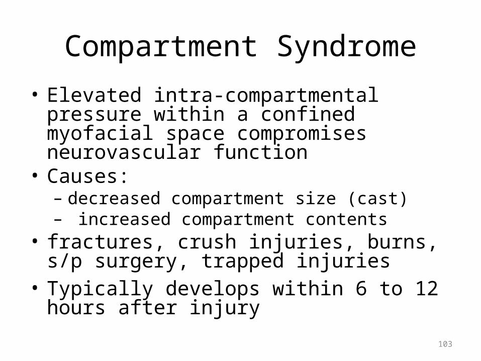
Compartment Syndrome• Elevated intra-compartmental pressure within a
confined myofacial space compromises neurovascular function
• Causes: – decreased compartment size (cast)– increased compartment contents
• fractures, crush injuries, burns, s/p surgery, trapped injuries
• Typically develops within 6 to 12 hours after injury
103

CarrieRocks, Wikimedia Commons 104

Compartment Syndrome signs and symptoms
• Pain- especially with passive flexion• This syndrome is characterized by:
– Pain that is out of proportion to the injury– Pain on passive stretching of muscles within the
compartment– Pallor– Decreased sensation– Decreased power
105

Nursing Management of Compartment Syndrome
• Remove restrictive dressing, cast, splint etc.• Prep for OR or Surgical decompression:
Fasciotomy• Elevate injured area• Notify MD STAT*
106

Compartment Pressure Measurement
Intermedichbo, Wikimedia Commons 107
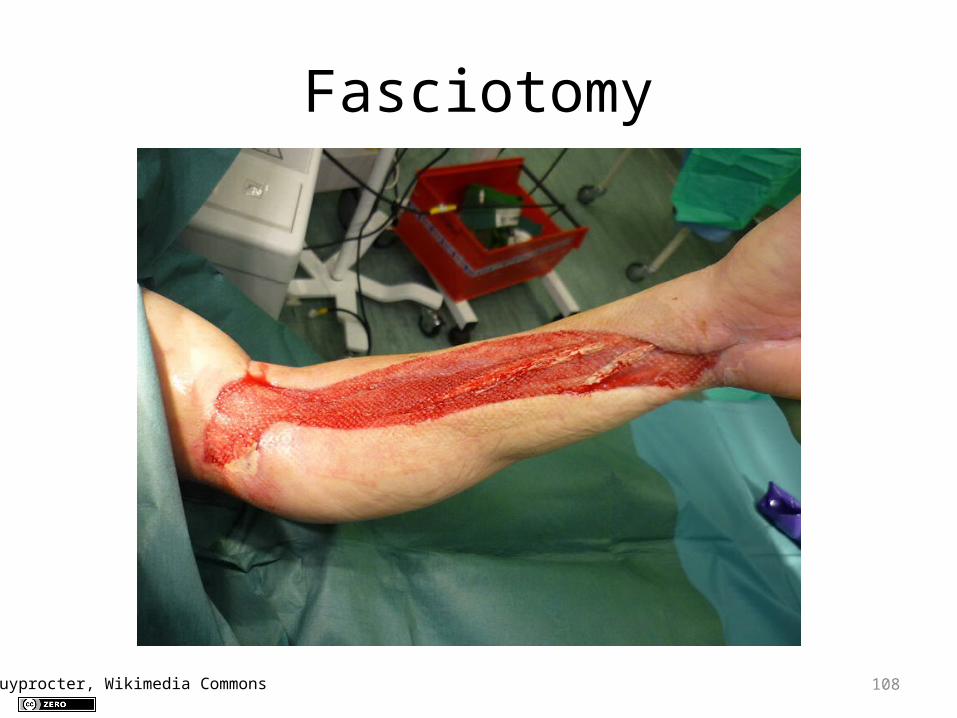
Fasciotomy
Guyprocter, Wikimedia Commons 108

Osteomyelitis• Acute or chronic bone infection
• Risk factors include:DiabetesHemodialysisInjected drug usePoor blood supplyRecent trauma
109

Osteomyelitis Symptoms
• Bone pain• Fever• General discomfort, ill-feeling (malaise)• Local swelling, redness, warmth• Chills• Excessive sweating• Low back pain
110

Osteomyelitis
FB Axelrod et al, Wikimedia CommonsSarindam7, Wikimedia Commons 111

Osteomyelitis Treatment
• IV access• Antibiotics IV• Possible surgical intervention• Debridement• Revascularization• Amputation
112

South African Triage Scale
VERY URGENT
•Threatened limb•Dislocation of larger joint•Fracture with break in skin (open)
URGENT
•Dislocation of finger or toe•fracture with no break in skin (closed)
113

Geriatric Considerations
• Bone fragility, frailness• Decrease in muscular strength and ROM• Chronic disease states-Osteoporosis• Loss of height with aging/kyphosis• Co-morbidities
114

Pediatric Considerations
• Infant bones are only 65% ossified• Long bones are porous and less dense and can
bend, buckle or break easily (Torus fx)• Presence of epiphyseal/growth plates, if these
are injured, can cause abnormal growth• Periosteum is thicker and more vascular, healing
occurs more quickly• Vigilance of abuse- injury inconsistent with
history
115

PARENT SUPPORT• Parents are trained and become active
participants in the physical therapy treatments and child’s stretching program
• Nurses need to help the parents understand the time commitment involved
• Assess the parents’ ability to monitor the child adequately for complications and confirm they understand the signs and symptoms of the
complications
116

Medical-Legal Aspects of Patient with Musculoskeletal Emergencies
• Abuse assessment-multiple stages of healing-injury inconsistent with history
• Informed consent pre surgical• Patient privacy• Occupational Safety• Health Risk Management Programs
117

1. A young male has a musculoskeletal injury and is unresponsive. You will NOT be able to assess:
A. Skin integrity.
B. distal pulses.
C. capillary refill.
D. sensory and motor functions.
118

2. The purpose of splinting a fracture is to:A. reduce the fracture if possible.
B. prevent motion of bony fragments.
C. reduce swelling in adjacent soft tissues.
D. force the bony fragments back into anatomic alignment.
119

3. Which of the following musculoskeletal injuries has the GREATEST risk for shock due to blood loss?
A. pelvic fracture
B. posterior hip dislocation
C. unilateral femur fracture
D. proximal humerus fracture
120

4. Tendons attach a. bone to bone
b. muscle to bonec. bone to adipose tissued. muscle to joints.
121

• 5. Which medication does not effect the integrity of the musculoskeletal system?
a. chemotherapyb. anti-epilepticsc. anti-emeticsd. corticosteroids
122

• 6. What are the 6 P’s of neurovascular assessment when evaluating a musculoskeletal injury?
123

• 7. What are the signs/symptoms of compartment syndrome? Select all that apply.a. Pallorb. Swellingc. Feverd. Pain out of proportion to injurye. Rednessf. Urinary retention
124

• 8. Which are diseases/conditions that may effect the musculoskeletal system?
a. Rheumatoid arthritis, Osteomyelitis, Goutb. Ricketts, Myocardial Infarction,
Pyelonephritisc. CVA, Lupus, GERDd. hypertension, high cholesterol, diabetes
125

9. What items do you want to have available and at bedside for a conscious sedation?
126

10. What does the acronym R.I.C.E. stand for and when would you use it?
127

Further Questions
128

Case Study
129