prognostic significance of tumor-associated macrophages in solid tumor a meta-analysis of the...
DESCRIPTION
Prognostic Significance of Tumor-Associated Macrophages in Solid Tumor a Meta-Analysis of the LiteratureTRANSCRIPT
Prognostic Significance of Tumor-AssociatedMacrophages in Solid Tumor: A Meta-Analysis of theLiteratureQiong-wen Zhang., Lei Liu*., Chang-yang Gong., Hua-shan Shi, Yun-hui Zeng, Xiao-ze Wang, Yu-
wei Zhao, Yu-quan Wei
Department of Medical Oncology, Cancer Center, State Key Laboratory of Biotherapy, West China Hospital, Sichuan University, Chengdu, P. R. China
Abstract
Purpose: Tumor associated macrophages (TAMs) are considered with the capacity to have both negative and positiveeffects on tumor growth. The prognostic value of TAM for survival in patients with solid tumor remains controversial.
Experimental Design: We conducted a meta-analysis of 55 studies (n = 8,692 patients) that evaluated the correlationbetween TAM (detected by immunohistochemistry) and clinical staging, overall survival (OS) and disease free survival (DFS).The impact of M1 and M2 type TAM (n = 5) on survival was also examined.
Results: High density of TAM was significantly associated with late clinical staging in patients with breast cancer [risk ratio(RR) = 1.20 (95% confidence interval (CI), 1.14–1.28)] and bladder cancer [RR = 3.30 (95%CI, 1.56–6.96)] and with early clinicalstaging in patients with ovarian cancer [RR = 0.52 (95%CI, 0.35–0.77)]. Negative effects of TAM on OS was shown in patientswith gastric cancer [RR = 1.64 (95%CI, 1.24–2.16)], breast cancer [RR = 8.62 (95%CI, 3.10–23.95)], bladder cancer [RR = 5.00(95%CI, 1.98–12.63)], ovarian cancer [RR = 2.55 (95%CI, 1.60–4.06)], oral cancer [RR = 2.03 (95%CI, 1.47–2.80)] and thyroidcancer [RR = 2.72 (95%CI, 1.26–5.86)],and positive effects was displayed in patients with colorectal cancer [RR = 0.64 (95%CI,0.43–0.96)]. No significant effect was showed between TAM and DFS. There was also no significant effect of two phenotypesof TAM on survival.
Conclusions: Although some modest bias cannot be excluded, high density of TAM seems to be associated with worse OSin patients with gastric cancer, urogenital cancer and head and neck cancer, with better OS in patients with colorectalcancer.
Citation: Zhang Q-w, Liu L, Gong C-y, Shi H-s, Zeng Y-h, et al. (2012) Prognostic Significance of Tumor-Associated Macrophages in Solid Tumor: A Meta-Analysisof the Literature. PLoS ONE 7(12): e50946. doi:10.1371/journal.pone.0050946
Editor: Mohammad O. Hoque, Johns Hopkins University, United States of America
Received June 19, 2012; Accepted October 29, 2012; Published December 28, 2012
Copyright: � 2012 Zhang et al. This is an open-access article distributed under the terms of the Creative Commons Attribution License, which permitsunrestricted use, distribution, and reproduction in any medium, provided the original author and source are credited.
Funding: This work was financially supported by National Natural Science Foundation of China (NSFC 81101729). The funders had no role in study design, datacollection and analysis, decision to publish, or preparation of the manuscript.
Competing Interests: The authors have declared that no competing interests exist.
* E-mail: [email protected]
. These authors contributed equally to this work.
Introduction
Macrophages are a population of innate myeloid cells that are
released from bone marrow as immature monocytic precursors
and, after circulating in the blood stream, migrate into different
tissues to undergo specific differentiation depending on local cues
in the tissue [1,2]. In response to different environment stimuli,
macrophages can appear a range of different phenotypes [3]. The
extremes of this range are recognized; the classically activated type
M1 phenotype and the alternative activated M2 phenotype. The
M1 macrophages are thought to be induced by interferon-c, with
or without lipopolysaccharide, tumor necrosis factor (TNF)-a, and
activate cells of the adaptive immune system [4]. Differentiation of
the M2 macrophages is induced by IL-4 or IL-13 and associated
with parasite clearance, wound healing and dampen immune
responses [5].
In 1863, it was fist found that a major leukocyte population was
present in tumor, the so-called tumor-associated macrophages
(TAM), which reflect the onset of cancer at site of previous chronic
inflammation [1,6]. These macrophages can induce neoplastic cell
(cytotoxicity, apoptosis) and/or elicit tumor destructive reactions
with the capacity to display both negative and positive effects on
tumor growth depending on environmental stimuli of the tumor
tissue [7,8].
For long a large number of studies have been focused on
identifying the prognostic value of TAM in solid tumors and most
studies suggest that TAM is beneficial for tumor growth and,
therefore, associated with poor prognosis [1]. However, there are
some exceptions with high density of macrophages correlating
with increased survival in different tumors [9–18] and even this
contradiction has come up in the one type of tumor [11,19]. This
meta-analysis focused on the identifying diverse roles and
PLOS ONE | www.plosone.org 1 December 2012 | Volume 7 | Issue 12 | e50946
functions of TAM and subpopulations of TAM for clinical
outcome in patients with solid tumors.
Materials and Methods
Identification and Eligibility of Relevant StudiesWe performed our meta-analysis according to a predetermined
written protocol. To be eligible for our meta-analysis, studies had
to deal with solid tumor at inclusion, to evaluate the correlation
between TAM and survival, and to be published in English or
Chinese languages. A computer-aided literature search of Pubmed
(MEDLINE) 1950-present and EMBASE was conduced by
combing search terms ‘‘cancer’’, ’’tumor’’, ‘‘neoplasm’’, ‘‘carcino-
ma’’, ‘‘tumor-associated macrophage’’, ‘‘tumor-infiltrating macro-
phage’’ and ‘‘intratumoral macrophage.’’ The deadline of the
included articles was April 20th, 2012. Reference list from primary
identified studies were also searched to prevent missing any studies
by the electronic search strategies.
Inclusion criteria for primary studies were as follows: (1) proven
diagnosis of solid tumor in humans, (2) using immunochemistry
method to evaluate TAM by anti-CD68, M1-type TAM by anti-
HLA-DR and M2-type TAM by anti-CD163, and (3) correlation
of TAM with TNM staging, OS or DFS. Two independent
reviewers processed primary assessment by identifying the
eligibility of abstracts from database. Full articles were retrieved
for further assessment if the eligibility was unclear from the
abstracts. Any disagreements were resolved by serious discussion.
We carefully examined the names of all authors and the medical
centers involved in each publication to avoid duplication data.
Whenever studies pertained to overlapping patients, we retained
the studies with highest number of patients.
Definitions and StandardizationsWe used preconcerted rules to standardize as much as possible
the definition of TAM positivity. As 20% was the used as a cutoff
value in majority of the included studies [18,20–26], we defined
TAM positivity as positive cell stain in at least 20% of tumor cells.
When different definitions were used, we contacted the primary
author of each articles to retrieve the cutoff value they used. When
cutoff value was not possible to retrieve, we accepted the cutoff was
closet to the 20% cutoff level. When cutoff value was closed to
20%, which ranged from 16.3% to 25%, we also accepted the
cutoff as 20%.
Data ExtractionData were carefully extracted from all of the included studies in
duplicate by two of us, using a standard information collection
form, with the following items, first author, year of publication,
study design, median follow-up time, country of origin, number of
patients involved, number of men included, mean or median age,
tumor location, histological type, tumor-node-metastasis (TNM)
staging, blinded reading, definition of TAM high, anti-cancer
treatment(s) during follow up, OS or DFS or both. The main
outcomes were tabulated in 262 tables showing the TNM staging
status, occurrence or not of death or disease during follow-up
according to TAM results.
Statistical AnalysesIncluded studies were divided into three groups for analysis:
those with data regarding TNM staging, those regarding OS and
those regarding DFS. A study was considered significant when the
P for comparing survival distribution between groups with high
and low TAM was inferior to 0.05. A study was termed ‘‘positive’’
when a high TAM predicted a late clinical staging or poorer
survival, ‘‘negative’’ when a high TAM predicted a early clinical
staging or better survival, ‘‘indeterminate’’ when no significant
relationship between TAM and clinical staging or survival was
found.
For the quantitative aggregation of survival result, impacts of
TAM on survival were reported for individual studies by
estimating RRs with 95% confidence interval values. We first
simply extracted RR and their 95%CI from the original article. If
not available, the published data including number of patients at
risk and total number of events in each groups from articles were
used to estimate RR according to the methods described by
Parmer et al [27]. When data were only available in the form of
figures, we read Kaplan-Meier curves by Engauge Digitizer
version 4.1 (free software down-loaded from http://sourceforge.
net) and extracted survival data to reconstruct RRs and its 95%CI.
An observed RR.1 indicated worse outcome for the TAM high
group relative to TAM low group and would be considered
statistically significant if the 95%CI did not overlap 1, with
p,0.05. Sensitivity analyses were performed to examine the TAM
effect of limiting the evaluation to studies using the 20% cutoff on
prognosis. The effect of publication bias on the outcomes was
assessed graphically using funnel plots, and funnel plot asymmetry
was assessed by Egger’s linear regression method. (p,0.05 was
considered statistically significant publication bias) [28]. Meta-
analyses were carried out by the Stata version 11.0 (Stata
Corporation, College Station, TX, USA).
Results
Studies Selection and CharacteristicsThe initial search algorithm retrieved a total of 3076 references
and we evaluated 144 candidate studies in full text. Upon further
review, 50 articles were eliminated due to inadequate data for
meta-analysis and another 29 articles were out of scope because of
evaluating other factors related to TAM (Figure 1). Overall, we
identified 55 articles (n = 8693) with TAM measurements in
patients with solid tumors.
The characteristics of included studies are listed in Table 1. The
median sample size for all studies was 158 patients (range = 24–
1902). The median sample size for staging was 189 patients, that
for OS was 164 patients and that for DFS was 202 patients. The
total proportion of male subjects was around 45% and that of
patients in grade I/II was 43%. All evaluated IHC staining in
formalin-fixed paraffin-embedded tissue blocks. The study design
was more often a retrospective (N = 25) than a prospective cohort
study (N = 5). Only 20% studies had performed blinded reading
during evaluating TAM. The median follow-up time for all
included studies ranged from 1.84 to 25 years. Of the 55 studies,
cut-off value for definition of TAM high only could be retrieved
from 32 original articles or by contacting authors.
Of the included studies, 19 studies focused on gastrointestinal
cancers, including colorectal cancer (N = 5), gastric cancer (N = 5),
liver cancer (N = 5), esophagus cancer (N = 2), pancreatic caner
(N = 1) and cholangiocarcinoma (N = 1). 20 studies analyzed the
impact of TAM on survival in patients with urogenital cancers,
including breast cancer (N = 5), endometrial cancer (N = 5),
prostate cancer (N = 3), cervical cancer (N = 2), bladder cancer
(N = 2), ovary cancer (N = 2) and urothelial cancer (N = 1). 5
studies mentioned the value of M2-type TAM on survival
[26,37,38,47,62].
Data Synthesis: Clinical StagingAs shown in Figure 2A, the combining data of TAM on clinical
staging showed a nonstatistically significant RR of 1.13 (95%CI
Prognostic Significance of TAM in Solid Tumor
PLOS ONE | www.plosone.org 2 December 2012 | Volume 7 | Issue 12 | e50946

0.97–1.31). For sub-group analysis, all of the studies were
dispatched into several classes according to the tumor location.
Our data showed that high density of TAM was significantly
higher in patients with advanced tumor stage (III+IV) than in the
patients with early stage (I+II), which occurred in breast cancer
[RR = 1.20 (95%CI, 1.14–1.28)], oral cancer [RR = 1.49 (95%CI,
1.17–1.89)] and bladder cancer [RR = 3.30 (95%CI, 1.78–
7.92)],whereas 1 article focused on ovary cancer found high
density of TAM was associated with early stage [RR = 0.52
(95%CI, 0.36–0.77)]. Other sub-group analysis found no relation
between TAM and clinical staging. In studies specific defining
TAM positivity as at least 20% positive staining cells in tumor
tissue, no significant relation was found between TAM and clinical
staging (RR = 1.23 (95%CI, 0.74–2.02). Analysis for M2-type
TAM on staging also showed no significant effect [RR = 1.43
(95%CI, 0.70–2.93)].
Data Synthesis: Overall SurvivalFor overall population, high density of TAM was associated
with a worse prognosis regarding overall survival (Figure 2B).
However, mortality was only 1.15-fold higher in high TAM
patients with solid tumor, which showed modest effect. In sub-
group analysis (Figure 3), high density of TAM was significantly
correlated with poor OS in patients with urogenital cancer
[RR = 1.95 (95%CI, 1.32–2.86)], including breast cancer
[RR = 8.62 (95%CI, 3.10–23.95)], endometrial cancer
[RR = 1.85 (95%CI, 0.10–34.63)], prostate cancer [RR = 1.16
(95%CI, 0.96–1.40)], bladder cancer [RR = 5.00 (95%CI, 1.98–
12.63)], ovary cancer [RR = 2.55 (95%CI, 1.60–4.06)] and
urothelial cancer [RR = 1.01 (95%CI, 1.32–2.86)]. In addition,
gastric cancer and oral cancer showed significant RR between
TAM and OS [RR = 1.54 (95%CI 1.24–2.16), 2.03 (95%CI 1.47–
2.80)]. However, analysis of studies on colorectal cancer showed
that there was a significant correlation between high density of
TAM and longer OS [RR = 0.64 (95%CI, 0.43–0.96)]. No
significant correlation between TAM and OS was found in other
sub-group analysis. Five studies reported that there was no
significant correlation between M2-type TAM and OS [RR = 0.98
(95%CI, 0.71–1.35)], one study reported that the M1-type TAM
density in the tumor islet is positively associated with extended
Figure 1. Flow chart of the literature search and selection of included studies.doi:10.1371/journal.pone.0050946.g001
Prognostic Significance of TAM in Solid Tumor
PLOS ONE | www.plosone.org 3 December 2012 | Volume 7 | Issue 12 | e50946

Ta
ble
1.
Ch
arac
teri
stic
so
fth
ee
ligib
lest
ud
ies.
Fir
sta
uth
or
(re
f)Y
ea
rS
tud
yd
esi
gn
n(M
/F)
Tu
mo
rlo
cati
on
Tu
mo
rst
ag
eI/
II(I
II/I
V)
Me
dia
nfo
llo
w-u
p(y
)B
lin
de
dre
ad
ing
Sta
inin
gfo
rT
AM
hig
hR
Re
stim
ati
on
An
aly
sis
Re
sult
Ga
stro
inte
stin
al
can
cer
(N=
19
)
Kh
ora
na
(29
)2
00
3P
rosp
.1
31
(69
/53
)1
31
Co
lon
11
(11
8)
5N
R.
2%
Dat
ae
xtra
po
late
d,
Surv
ival
curv
eSt
ag.,
OS
Ind
ete
rmin
ate
Tan
(12
)2
00
5R
etr
o.
60
(35
/25
)6
0C
olo
n2
6(3
4)
NR
NR
.2
5%
Dat
ae
xtra
po
late
d,
Surv
ival
curv
eSt
ag.,
OS
Ne
gat
ive
Bac
man
(30
)2
00
7N
R3
10
(18
9/1
21
)3
10
Co
lon
17
8(1
32
)7
.5N
RN
RD
ata
ext
rap
ola
ted
OS
Ind
ete
rmin
ate
Fors
sell
(15
)2
00
7R
etr
o.
48
8(2
71
/21
7)
48
8C
olo
n2
07
(25
4)
NR
NR
NR
Dat
ae
xtra
po
late
d,
Re
po
rte
din
text
Stag
.,O
SIn
de
term
inat
e,
ne
gat
ive
Zh
ou
(18
)2
01
0N
R1
60
(94
/66
)1
60
Co
lon
NR
5Y
es
20
%D
ata
ext
rap
ola
ted
,re
po
rte
din
text
Stag
.,O
SN
eg
ativ
e
Ish
igam
i(3
1)
20
03
Re
tro
.9
7(7
2/2
5)
97
Sto
m6
7(3
0)
NR
NR
.2
00
/H
PF
Dat
ae
xtra
po
late
d,
Surv
ival
curv
eSt
ag.,
OS
Ind
ete
rmin
ate
,p
osi
tive
Oh
no
(10
)2
00
5R
etr
o.
84
(57
/27
)8
4St
om
41
(43
)N
RY
es
.4
.7%
Surv
ival
curv
eD
FSN
eg
ativ
e
Has
s(3
2)
20
09
Pro
sp.
52
(40
/12
)5
0St
om
39
(13
)5
.9N
RN
RR
ep
ort
ed
inte
xtD
FSIn
de
term
inat
e
Kaw
ahar
a(3
3)
20
10
NR
11
1(7
7/3
4)
11
1St
om
36
(75
)N
RN
RN
RR
ep
ort
ed
inte
xtO
SP
osi
tive
Osi
nsk
y(3
4)
20
11
Pro
sp.
10
5(7
1/3
4)
10
5st
om
48
(57
)N
RN
R.
23
.0%
Re
po
rte
din
text
OS
Po
siti
ve
Zh
u(2
2)
20
08
Re
tro
.1
05
(96
/9)
10
5Li
ver
86
(19
)1
.84
NR
.2
0%
Dat
ae
xtra
po
late
d,
rep
ort
ed
inte
xtSt
ag.,
OS,
DFS
Ind
ete
rmin
ate
,p
osi
tive
Li(2
4)
20
09
Re
tro
.3
02
(26
0/4
2)
30
2Li
ver
23
7(6
5)
4.8
3N
R.
20
%D
ata
ext
rap
ola
ted
,re
po
rte
din
text
Stag
.,O
S,D
FSIn
de
term
inat
e,
ne
gat
ive
Din
g(3
5)
20
09
Re
tro
.1
07
(87
/20
)1
07
Live
r9
8(3
9)
2.5
MR
.2
3%
Re
po
rte
din
text
OS,
DFS
Po
siti
ve
Ku
ang
(36
)2
00
9R
etr
o.
26
2(N
R)
20
6Li
ver
24
9(1
3)
NR
NR
NR
Re
po
rte
din
text
OS,
DFS
Po
siti
ve
Ju(2
3)
20
09
Re
tro
.1
30
(11
2/1
8)
13
0Li
ver
66
(64
)2
.65
NR
.2
0%
Re
po
rte
din
text
Stag
.,O
S,D
FSP
osi
tive
Ko
ide
(9)
20
02
Re
tro
.5
6(4
2/1
4)
56
Eso
ph
agu
s3
0(2
6)
NR
NR
NR
Dat
ae
xtra
po
late
d,
surv
ival
cure
Stag
.,O
SIn
de
term
inat
e,
ne
gat
ive
Gu
o(1
9)
20
07
NR
13
7(1
03
/34
)1
37
Eso
ph
agu
s8
6(5
1)
NR
NR
.2
5%
Re
po
rte
din
text
Stag
.,O
SIn
de
term
inat
e,
po
siti
ve
Ku
rah
ara
(37
)2
01
2R
etr
o.
76
(52
/24
)5
2P
ancr
eas
29
(47
)N
RY
es
NR
Surv
ival
curv
eO
S,O
SbIn
de
term
inat
e
Has
ita
(38
)2
01
0N
R3
9(2
7/1
2)
55
Bile
du
ct2
9(1
0)
3.3
3Y
es
NR
Dat
ae
xtra
po
late
d,
surv
ival
curv
eSt
ag.,
OS,
DFS
,St
ag.b
,O
Sb,
DFS
bIn
de
term
inat
e
Uro
ge
nit
al
can
cer
(N=
20
)
Lee
k(3
9)
19
96
Re
tro
.1
01
(0/1
01
)5
Bre
ast
NR
NR
NR
MD
.=
12
Re
po
rte
din
text
OS,
DFS
Po
siti
ve
To
i(4
0)
19
99
NR
22
9(0
/22
9)
22
9B
reas
tN
R4
Ye
sN
RSu
rviv
alcu
rve
DFS
Ind
ete
rmin
ate
Val
kovi
c(4
1)
20
02
NR
97
(0/9
7)
97
Bre
ast
82
(15
)N
RN
RN
RD
ata
ext
rap
ola
ted
Stag
.In
de
term
inat
e
Bo
lat
(42
)2
00
6R
etr
o.
78
(0/7
8)
78
Bre
ast
38
(10
)N
RN
RN
RD
ata
ext
rap
ola
ted
Stag
.In
de
term
inat
e
Mah
mo
un
d(4
3)
20
12
Re
tro
.1
90
2(0
/19
02
)1
90
2B
reas
t2
20
(16
82
)N
RN
RN
RD
ata
ext
rap
ola
ted
,su
rviv
alcu
rve
Stag
.,D
FSP
osi
tive
,in
de
term
inat
e
Salv
ese
n(4
4)
19
99
Pro
sp.
60
(0/6
0)
60
End
om
etr
ium
50
(10
)1
1N
RN
RD
ata
ext
rap
ola
ted
,re
po
rte
din
text
Stag
.,O
SP
osi
tive
,in
de
term
inat
e
Prognostic Significance of TAM in Solid Tumor
PLOS ONE | www.plosone.org 4 December 2012 | Volume 7 | Issue 12 | e50946

Ta
ble
1.
Co
nt.
Fir
sta
uth
or
(re
f)Y
ea
rS
tud
yd
esi
gn
n(M
/F)
Tu
mo
rlo
cati
on
Tu
mo
rst
ag
eI/
II(I
II/I
V)
Me
dia
nfo
llo
w-u
p(y
)B
lin
de
dre
ad
ing
Sta
inin
gfo
rT
AM
hig
hR
Re
stim
ati
on
An
aly
sis
Re
sult
Has
him
oto
(45
)2
00
0N
R1
09
(0/1
09
)1
09
End
om
etr
ium
56
(53
)4
.58
NR
NR
Dat
ae
xtra
po
late
dSt
ag.,
DFS
Ind
ete
rmin
ate
Oh
no
(11
)2
00
4R
etr
o.
70
(0/7
0)
70
End
om
etr
ium
NR
NR
NR
MD
.1
0.7
Surv
ival
curv
eD
FSN
eg
ativ
e
Soe
da
(46
)2
00
8R
etr
o.
76
(0/7
6)
76
End
om
etr
ium
59
(17
)6
.83
NR
NR
Surv
ival
curv
eO
S,D
FSP
osi
tive
,in
de
term
inat
e
Esp
ino
sa(4
7)
20
10
NR
64
(0/6
4)
64
End
om
etr
ium
23
(26
)N
RN
RN
RD
ata
ext
rap
ola
ted
Stag
.bIn
de
term
inat
e
Liss
bra
nt
(48
)2
00
0R
etr
o.
85
(85
/0)
85
Pro
stat
e7
5(1
0)
NR
NR
.=
0.9
7%
Surv
ival
curv
eSt
ag.,
OS
Ind
ete
rmin
ate
Shim
ura
(49
)2
00
0N
R8
1(8
1/0
)8
1P
rost
ate
67
(13
)3
.9N
RN
RR
ep
ort
ed
inte
xtD
FSIn
de
term
inat
e
No
no
mu
ra(5
0)
20
11
NR
13
1(1
31
/0)
13
1P
rost
ate
30
(41
)2
.9N
RM
D.
=2
2Su
rviv
alcu
rve
DFS
Po
siti
ve
He
ller
(51
)2
00
2R
etr
o.
24
(0/2
4)
24
Ce
rvix
20
/4N
RN
RN
RD
ata
ext
rap
ola
ted
Stag
.In
de
term
inat
e
Kaw
anak
a(5
2)
20
08
Re
tro
73
(0/7
3)
73
Ce
rvix
22
/51
NR
Ye
sM
D.
=5
5Su
rviv
alcu
rve
DFS
Ind
ete
rmin
ate
Han
ada
(53
)2
00
0N
R6
3(5
1/1
2)
63
Bla
dd
er
42
/21
5.4
Ye
sM
D.
=6
7D
ata
ext
rap
ola
ted
,re
po
rte
din
text
Stag
.,O
SP
osi
tive
Tak
ayam
a(5
4)
20
09
NR
41
(38
/3)
41
Bla
dd
er
NR
25
NR
.=
4R
ep
ort
ed
inte
xtD
FSP
osi
tive
Tan
aka
(55
)2
00
4N
R8
9(0
/89
)8
9O
vary
22
/67
6.8
NR
.=
25
%D
ata
ext
rap
ola
ted
,re
po
rte
din
text
,su
rviv
alcu
rve
Stag
.,O
S,D
FSN
eg
ativ
e,
po
siti
ve,
ind
ete
rmin
ate
Wan
(25
)2
00
9N
R6
7(0
/67
)6
7O
vary
0/6
7N
RN
R.
20
%Su
rviv
alcu
rve
OS
Po
siti
ve
Ch
ai(2
0)
20
08
NR
99
(42
/57
)9
9U
roth
NR
NR
NR
.2
0%
Re
po
rte
din
text
OS,
DFS
Ind
ete
rmin
ate
,p
osi
tive
Lu
ng
can
cer
(N=
10
)
Tak
anam
i(5
6)
19
99
Re
tro
.1
13
(66
/47
)1
13
Lun
g6
1/5
2N
RN
RM
D.
32
Surv
ival
curv
eO
SP
osi
tive
Ch
en
(57
)2
00
5N
R4
1(2
7/1
4)
41
Lun
g2
3/1
8N
RN
RM
D.
=1
63
Surv
ival
curv
eD
FSIn
de
term
inat
e
We
lsh
(13
)2
00
5R
etr
o.
17
5(1
16
/59
)1
75
Lun
g1
23
/38
NR
Ye
sN
RD
ata
ext
rap
ola
ted
Stag
.,D
FSN
eg
ativ
e
Ze
ni
(58
)2
00
7N
R5
0(4
3/7
)4
3Lu
ng
NR
NR
NR
.1
6.3
%R
ep
ort
ed
inte
xt,
surv
ival
curv
eSt
ag.,
OS
Po
siti
ve
Kaw
ai(1
6)
20
08
NR
19
9(1
39
/60
)1
99
Lun
gN
RN
RN
RN
RR
ep
ort
ed
inte
xtO
SN
eg
ativ
e
Oh
ri(1
7)
20
09
Re
tro
.4
0(1
6/2
4)
40
Lun
g3
4/6
NR
NR
NR
Surv
ival
curv
eO
SN
eg
ativ
e
Al-
shib
li(5
9)
20
09
Re
tro
.3
71
(25
3/8
2)
37
1Lu
ng
30
3/3
28
NR
.=
25
%D
ata
ext
rap
ola
ted
,su
rviv
alcu
rve
OS,
DFS
Ind
ete
rmin
ate
Ma
(26
)2
01
0N
R5
0(4
0/1
0)
50
Lun
g3
3/1
7N
RN
R.
20
%R
ep
ort
ed
inte
xtO
Sa,
OSb
Ind
ete
rmin
ate
Oh
taki
(60
)2
01
0P
rosp
.1
70
(85
/85
)1
70
Lun
gN
R1
0.1
NR
NR
Dat
ae
xtra
po
late
dSt
ag.,
OS
Po
siti
ve
Zh
ang
(61
)2
01
1R
etr
o.
65
(38
/27
)6
5Lu
ng
38
/27
NR
Ye
sT
AM
cou
nts
.1
02
Surv
ival
curv
eO
SP
osi
tive
He
ad
an
dn
eck
can
cer
(N=
4)
Liu
(21
)2
00
8N
R1
12
(93
/19
)1
12
Ora
l5
0/6
2N
RN
R.
20
%D
ata
ext
rap
ola
ted
,su
rviv
alcu
rve
Stag
.,O
SP
osi
tive
Fujii
(62
)2
01
2N
R1
08
(31
/10
)1
08
Ora
l4
6/6
2N
RN
R.
=2
/HP
FD
ata
ext
rap
ola
ted
,re
po
rte
din
text
Stag
.,st
ag.b
,O
SbP
osi
tive
,in
de
term
inat
e,
po
siti
ve
Pe
ng
(14
)2
00
6N
R6
0(3
8/2
2)
60
Ora
l1
5/4
5N
RN
R.
=6
3.7
/HP
FD
ata
ext
rap
ola
ted
OS
Ne
gat
ive
Prognostic Significance of TAM in Solid Tumor
PLOS ONE | www.plosone.org 5 December 2012 | Volume 7 | Issue 12 | e50946
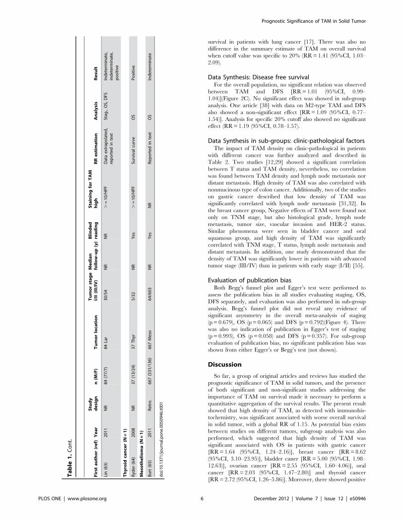
survival in patients with lung cancer [17]. There was also no
difference in the summary estimate of TAM on overall survival
when cutoff value was specific to 20% (RR = 1.41 (95%CI, 1.03–
2.09).
Data Synthesis: Disease free survivalFor the overall population, no significant relation was observed
between TAM and DFS [RR = 1.01 (95%CI, 0.99–
1.04)](Figure 2C). No significant effect was showed in sub-group
analysis. One article [38] with data on M2-type TAM and DFS
also showed a non-significant effect [RR = 1.09 (95%CI, 0.77–
1.54)]. Analysis for specific 20% cutoff also showed no significant
effect (RR = 1.19 (95%CI, 0.78–1.57).
Data Synthesis in sub-groups: clinic-pathological factorsThe impact of TAM density on clinic-pathological in patients
with different cancer was further analyzed and described in
Table 2. Two studies [12,29] showed a significant correlation
between T status and TAM density, nevertheless, no correlation
was found between TAM density and lymph node metastasis nor
distant metastasis. High density of TAM was also correlated with
nonmucinous type of colon cancer. Additionally, two of the studies
on gastric cancer described that low density of TAM was
significantly correlated with lymph node metastasis [31,32]. In
the breast cancer group, Negative effects of TAM were found not
only on TNM stage, but also histological grade, lymph node
metastasis, tumor size, vascular invasion and HER-2 status.
Similar phenomena were seen in bladder cancer and oral
squamous group, and high density of TAM was significantly
correlated with TNM stage, T status, lymph node metastasis and
distant metastasis. In addition, one study demonstrated that the
density of TAM was significantly lower in patients with advanced
tumor stage (III/IV) than in patients with early stage (I/II) [55].
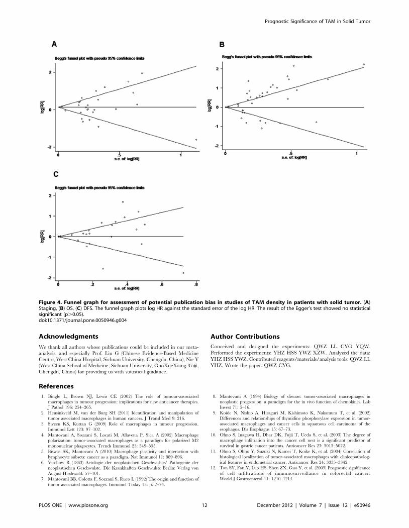
Evaluation of publication biasBoth Begg’s funnel plot and Egger’s test were performed to
assess the publication bias in all studies evaluating staging, OS,
DFS separately, and evaluation was also performed in sub-group
analysis. Begg’s funnel plot did not reveal any evidence of
significant asymmetry in the overall meta-analysis of staging
(p = 0.679), OS (p = 0.065) and DFS (p = 0.792)(Figure 4). There
was also no indication of publication in Egger’s test of staging
(p = 0.993), OS (p = 0.058) and DFS (p = 0.357). For sub-group
evaluation of publication bias, no significant publication bias was
shown from either Egger’s or Begg’s test (not shown).
Discussion
So far, a group of original articles and reviews has studied the
prognostic significance of TAM in solid tumors, and the presence
of both significant and non-significant studies addressing the
importance of TAM on survival made it necessary to perform a
quantitative aggregation of the survival results. The present result
showed that high density of TAM, as detected with immunohis-
tochemistry, was significant associated with worse overall survival
in solid tumor, with a global RR of 1.15. As potential bias exists
between studies on different tumors, subgroup analysis was also
performed, which suggested that high density of TAM was
significant associated with OS in patients with gastric cancer
[RR = 1.64 (95%CI, 1.24–2.16)], breast cancer [RR = 8.62
(95%CI, 3.10–23.95)], bladder caner [RR = 5.00 (95%CI, 1.98–
12.63)], ovarian cancer [RR = 2.55 (95%CI, 1.60–4.06)], oral
cancer [RR = 2.03 (95%CI, 1.47–2.80)] and thyroid cancer
[RR = 2.72 (95%CI, 1.26–5.86)]. Moreover, there showed positive
Ta
ble
1.
Co
nt.
Fir
sta
uth
or
(re
f)Y
ea
rS
tud
yd
esi
gn
n(M
/F)
Tu
mo
rlo
cati
on
Tu
mo
rst
ag
eI/
II(I
II/I
V)
Me
dia
nfo
llo
w-u
p(y
)B
lin
de
dre
ad
ing
Sta
inin
gfo
rT
AM
hig
hR
Re
stim
ati
on
An
aly
sis
Re
sult
Lin
(63
)2
01
1N
R8
4(7
7/7
)8
4La
r3
0/5
4N
RN
R.
=1
0/H
PF
Dat
ae
xtra
po
late
d,
rep
ort
ed
inte
xtSt
ag.,
OS,
DFS
Ind
ete
rmin
ate
,in
de
term
inat
e,
po
siti
ve
Th
yro
idca
nce
r(N
=1
)
Ryd
er
(64
)2
00
8N
R3
7(1
3/2
4)
37
Th
yr5
/32
NR
Ye
s.
=1
0/H
PF
Surv
ival
curv
eO
SP
osi
tive
Me
soth
eli
om
a(N
=1
)
Bu
rt(6
5)
20
11
Re
tro
.6
67
(53
1/1
36
)6
67
Me
so6
4/6
03
NR
Ye
sN
RR
ep
ort
ed
inte
xtO
SIn
de
term
inat
e
do
i:10
.13
71
/jo
urn
al.p
on
e.0
05
09
46
.t0
01
Prognostic Significance of TAM in Solid Tumor
PLOS ONE | www.plosone.org 6 December 2012 | Volume 7 | Issue 12 | e50946

effect in patients with colorectal cancer [RR = 0.64 (95%CI, 0.43–
0.96)]. However, no significant effect was seen between TAM and
DFS.
When comparing the results of different types of tumors, several
key differences were observed. As mentioned above, although
macrophages under certain conditions can kill tumor cells, they
can also play potential roles as tumor promoters to secrete a
variety of factors that directly stimulate tumor invasion and
metastasis. The combing effect of TAM on prognosis in patients
with different tumors depends on stimulating factors from two
opposite aspects in tumor environments. In this meta-analysis, we
reach a conclusion that high TAM infiltration is associated with
worsen prognosis in patients with urogenital cancer or gastric
cancer, not all cancer type. In other hand, TAM showed
antitumorigenic properties in combing 5 studies on colorectal
cancer, resulting in improved prognosis.
To further investigate the prognostic value of TAM in different
type of cancer, we analyzed the relation between the density of
TAM and clinic-pathological factors that was also associated with
outcome of cancer patients. As the density of TAM has a negative
effect on survival in patients with gastric cancer, breast cancer,
bladder cancer, ovary cancer, and oral squamous cell carcinoma,
negative effects are also seen in clinic-pathological factors such as
TNM staging (breast cancer, bladder cancer and oral squamous
cell carcinoma), T status (breast cancer and oral squamous cell
carcinoma), lymph node metastasis (breast cancer, bladder cancer
and oral squamous cell carcinoma), and distant metastasis (bladder
cancer), which contributed to tumor progression and patient
survival. Interestingly, there also demonstrated a positive effect of
TAM on lymph node metastasis in gastric and ovary cancer,
which indicated that high density of TAM was associated with less
probability of lymph node metastasis, however, significant negative
effect was shown on overall survival. Thus, more studies are
needed to clarify this ambivalent phenomenon. Contrary to
tumors we mentioned above, our data suggested that an
incremental increase in density of TAM improved overall survival
in patients with colon cancer, with a homodromous effect on T
status. There was also a trend towards lower rate of lymph node
metastasis and distant metastasis in TAM rich tumors. In addition,
a high density of TAM infiltration was found related to
nonmucious type of colon cancer.
The mechanisms behind the oncogenic and anti-tumorigenic
effects of TAMs have not been fully elucidated and a great number
of studies have focused on explaining these apparently contradic-
tory effects of TAM in different cancer outcome. The functions of
TAM in different type of tumors are concerned as the most
important determining factor to the prognosis, which are
profoundly affected by microenvironmental signals and can range
Figure 2. Forrest plots and meta-analysis of studies evaluating RR of high TAM counts as compared to low counts. Clinical staging andsurvival data are reported as (A) staging, (B) overall survival (OS) and (C) disease free survival (DFS).doi:10.1371/journal.pone.0050946.g002
Prognostic Significance of TAM in Solid Tumor
PLOS ONE | www.plosone.org 7 December 2012 | Volume 7 | Issue 12 | e50946

from powerful stimulation of inflammatory responses to induction
of immunosuppression [66]. Tumor necrosis factor (TNF)-a, nitric
oxide (NO), and monocyte chemoattractant protein (MCP)-1
released from TAM are major intermediate molecules for tumor
cell killing [67–69], and TAM associated vascular endothelial
growth factor (VEGF) and matrix metalloproteinases (MMPs) are
independent predictor of poor prognostic factor in cancer patients
[34]. In addition, a macrophage balance hypothesis between M1
and M2 type macrophages has been proposed and two different
macrophage populations range from polarized potent killer M1
cells to alternatively activated M2 macrophages with tumor-
promoting capability [70]. However, this study showed no
significant relation between the density of two phenotype of
TAM and survival of patients. Furthermore, histological classifi-
cation of the tumor should also be considered as a factor correlated
with the function of TAM. A previous study on colon cancer
demonstrated that a histologically more malignant phenotype was
associated with macrophage infiltration, disorganized matrix
deposition, and extensive stromal reaction [71]. In one included
study of this meta-analysis, the number of infiltrating TAM was
found to be significantly correlated with poor outcome in patients
with intestinal type of gastric cancer, but not in patients with
diffuse type, indicating that TAM could affect malignant
progression and prognosis on the basis of the histological type of
gastric cancer [33].
Although the results of meta-analysis are considered as gold
standards by authors worldwide [72], potential bias still exists
between studies and cannot be completely eliminated. Although
Begg’s funnel plot and Egger’s test were performed in this meta-
analysis and found no statistically significant publication bias,
results of this study should be interpreted very cautiously and
several aspects of importance in this field should be discussed. First
of all, we only included studies from which we could extract RR or
estimate RR, leading to data inaccessible for data aggregation
from studies, which only showed the conclusion on this topic
without data presented. Take one excluded study on evaluating
the prognostic value of TAM on oral cavity and oropharyngeal
squamous cell carcinoma for example, they found that macro-
phage content was an independent predictor of lymph node
metastasis, however, no data was accessible for meta-analysis from
this study [73]. Furthermore, considerable attention should be
paid to some cancers with few study included in this meta-analysis
Figure 3. Forrest plots and meta-analysis of studies evaluating RR of high TAM counts as compared to low counts in differentsubgroup of tumors. Clinical staging and survival data are reported as (A) gastrointestinal cancer, (B) urogenital cancer, (C) lung cancer and (D)head and neck cancer.doi:10.1371/journal.pone.0050946.g003
Prognostic Significance of TAM in Solid Tumor
PLOS ONE | www.plosone.org 8 December 2012 | Volume 7 | Issue 12 | e50946

Ta
ble
2.
Me
ta-a
nal
ysis
of
the
sub
gro
up
sb
ase
do
ncl
inic
-pat
ho
log
ical
fact
ors
rela
ted
toT
AM
de
nsi
tyfr
om
the
avai
lab
lep
ub
lish
ed
stu
die
s.
Ca
nce
rty
pe
Cli
nic
-pa
tho
log
ica
lfa
cto
rsN
um
be
ro
fst
ud
ies
Nu
mb
er
of
tota
lp
ati
en
tsH
R(9
5%
CI)
p-v
alu
eR
esu
lts
Re
fere
nce
Co
lon
can
cer
TN
MSt
age
(I/I
Ivs
.III
/IV
)2
61
90
.71
(0.3
5,
1.4
5)
0.1
94
Ind
ete
rmin
ate
(15
),(2
9)
Tst
atu
s(T
1/T
2vs
.T
3/T
4)
21
91
0.3
5(0
.13
,0
.90
)0
.24
6P
osi
tive
(12
),(2
9)
Lym
ph
no
de
me
tast
asis
23
70
0.7
2(0
.13
,4
.06
)0
.01
3In
de
term
inat
e(1
2),
(30
)
Dis
tan
tm
eta
stas
is3
53
00
.72
(0.2
4,
2.1
1)
0.0
05
Ind
ete
rmin
ate
(12
),(1
8),
(30
)
His
tolo
gic
alg
rad
e(w
ell/
mo
de
rate
vs.
po
or)
38
58
0.2
6(0
.06
,1
.06
)0
.00
5In
de
term
inat
e(1
2),
(15
),(3
0)
Pat
ho
log
iccl
assi
fica
tio
n(m
uci
no
us
vs.
no
nm
uci
no
us)
26
48
2.5
3(1
.35
,4
.73
)0
.67
5N
eg
ativ
e(1
5),
(18
)
Ga
stri
cca
nce
r
TN
MSt
age
(I/I
Ivs
.III
/IV
)1
97
2.4
0(0
.98
,5
.91
)N
RIn
de
term
inat
e(3
1)
Tst
atu
s(T
1/T
2vs
.T
3/T
4)
19
71
.36
(0.5
4,
3.4
6)
NR
Ind
ete
rmin
ate
(31
)
Lym
ph
no
de
me
tast
asis
21
49
0.3
9(0
.19
,0
.80
)0
.30
2P
osi
tive
(31
),(3
2)
Dis
tan
tm
eta
stas
is1
52
1.3
0(0
.40
,4
.28
)N
RIn
de
term
inat
e(3
2)
His
tolo
gic
alg
rad
e(w
ell/
mo
de
rate
vs.
po
or)
19
70
.54
(0.2
2,
1.3
2)
NR
Ind
ete
rmin
ate
(31
)
Liv
er
can
cer
TN
MSt
age
(I/I
Ivs
.III
/IV
)3
51
41
.27
(0.8
4,
1.9
3)
0.7
02
Ind
ete
rmin
ate
(22
),(2
4),
(35
)
Vas
cula
rin
vasi
on
(ab
sen
tvs
.p
rese
nt)
35
14
1.3
8(0
.87
,2
.21
)0
.41
0In
de
term
inat
e(2
2),
(24
),(3
5)
Tu
mo
rd
iffe
ren
tiat
ion
24
09
1.0
8(0
.70
,1
.66
)0
.95
6In
de
term
inat
e(2
4),
(35
)
Tu
mo
rsi
ze(#
5vs
..
5cm
)2
40
91
.76
(1.1
9,
2.6
0)
0.3
29
Ne
gat
ive
(24
),(3
5)
He
pat
itis
his
tory
(No
vs.
Ye
s)2
40
71
.45
(0.8
0,
2.6
0)
0.4
56
Ind
ete
rmin
ate
(22
),(2
4)
Live
rci
rrh
osi
s(N
ovs
.Y
es)
35
14
1.1
8(0
.75
,1
.88
)0
.88
6In
de
term
inat
e(2
2),
(24
),(3
5)
Fib
rou
sca
psu
le(a
bse
nt
vs.
pre
sen
t)3
51
40
.83
(0.5
8,
1.1
9)
0.7
50
Ind
ete
rmin
ate
(22
),(2
4),
(35
)
Eso
ph
ag
us
can
cer
Tst
atu
s(T
1/T
2vs
.T
3/T
4)
15
62
.73
(0.7
0,
10
.60
)N
RIn
de
term
inat
e(9
)
Lym
ph
no
de
me
tast
asis
15
60
.75
(0.2
1,
2.5
9)
NR
Ind
ete
rmin
ate
(9)
Lym
ph
atic
inva
sio
n(a
bse
nt
vs.
pre
sen
t)1
56
3.2
1(0
.81
,1
2.8
)N
RIn
de
term
inat
e(9
)
Ve
no
us
inva
sio
n(a
bse
nt
vs.
pre
sen
t)1
56
6.2
2(1
.48
,2
6.1
)N
RN
eg
ativ
e(9
)
Intr
ah
ep
ati
cch
ola
ng
ioca
rcin
om
a
UIC
Cst
age
(I/I
Ivs
.III
/IV
)1
55
3.3
1(0
.71
,1
5.4
)N
RIn
de
term
inat
e(3
8)
His
tolo
gic
alg
rad
e(w
ell/
mo
de
rate
vs.
po
or)
15
55
.25
(0.9
3,
29
.7)
NR
Ind
ete
rmin
ate
(38
)
Lym
ph
no
de
me
tast
asis
15
51
.06
(0.1
9,
6.0
5)
NR
Ind
ete
rmin
ate
(38
)
Vas
cula
rin
vasi
on
(ab
sen
tvs
.p
rese
nt)
15
52
.13
(0.3
4,
13
.2)
NR
Ind
ete
rmin
ate
(38
)
Tu
mo
rsi
ze(,
4vs
.$
4cm
)1
55
1.3
8(0
.39
,4
.87
)N
RIn
de
term
inat
e(3
8)
Prognostic Significance of TAM in Solid Tumor
PLOS ONE | www.plosone.org 9 December 2012 | Volume 7 | Issue 12 | e50946

Ta
ble
2.
Co
nt.
Ca
nce
rty
pe
Cli
nic
-pa
tho
log
ica
lfa
cto
rsN
um
be
ro
fst
ud
ies
Nu
mb
er
of
tota
lp
ati
en
tsH
R(9
5%
CI)
p-v
alu
eR
esu
lts
Re
fere
nce
Bre
ast
can
cer
TN
MSt
age
(I/I
Ivs
.III
/IV
)1
78
1.2
0(1
.14
,1
.28
)N
RN
eg
ativ
e(4
2)
His
tolo
gic
alg
rad
e(I
/II
vs.
III)
32
07
73
.42
(2.7
1,
4.3
0)
0.7
42
Ne
gat
ive
(41
),(4
2),
(43
)
Lym
ph
no
de
me
tast
asis
32
07
71
.29
(1.0
4,
1.6
2)
0.6
04
Ne
gat
ive
(41
),(4
2),
(43
)
Tu
mo
rsi
ze(#
2vs
..
2cm
)3
20
77
1.4
3(1
.14
,1
.80
)0
.96
3N
eg
ativ
e(4
1),
(42
),(4
3)
Vas
cula
rin
vasi
on
(ab
sen
tvs
.p
rese
nt)
11
90
21
.74
(1.3
5,
2.2
3)
NR
Ne
gat
ive
(43
)
HER
-2st
atu
s(n
eg
ativ
evs
.p
osi
tive
)1
19
02
2.5
9(1
.75
,3
.85
)N
RN
eg
ativ
e(4
3)
En
do
me
tria
lca
nce
r
FIG
Ost
age
(I/I
Ivs
.III
/IV
)2
16
92
.34
(0.3
6,
15
.39
)0
.02
1In
de
term
inat
e(4
4),
(45
)
Lym
ph
no
de
me
tast
asis
11
09
0.4
3(0
.12
,1
.53
)N
RIn
de
term
inat
e(4
5)
Myo
me
tria
lin
vasi
on
(ne
gat
ive
vs.
po
siti
ve)
11
09
2.0
9(0
.65
,6
.69
)N
RIn
de
term
inat
e(4
5)
His
tolo
gic
alg
rad
e(I
/II
vs.
III)
21
69
4.3
4(0
.70
,2
7.0
8)
0.0
44
Ind
ete
rmin
ate
(44
),(4
5)
Pat
ho
log
iccl
assi
fica
tio
n(E
nd
om
etr
ioid
vs.n
on
-en
do
me
trio
id)
11
09
1.3
7(0
.41
,4
.62
)N
RIn
de
term
inat
e(4
5)
Pro
sta
teca
nce
r
Tst
atu
s(T
1/T
2vs
.T
3/T
4)
18
53
.30
(1.5
6,
6.9
6)
NR
Ind
ete
rmin
ate
(48
)
Dis
tan
tm
eta
stas
is1
85
1.0
6(0
.25
,4
.54
)N
RIn
de
term
inat
e(4
8)
Ce
rvic
al
can
cer
FIG
Ost
age
(I/I
Ivs
.III
/IV
)2
97
0.6
8(0
.06
,8
.26
)0
.06
3In
de
term
inat
e(5
1),
(52
)
Lym
ph
no
de
me
tast
asis
12
41
.75
(0.2
6,
11
.7)
NR
Ind
ete
rmin
ate
(51
)
Bla
dd
er
can
cer
TN
MSt
age
(I/I
Ivs
.III
/IV
)1
63
5.7
6(1
.76
,1
8.9
)N
RN
eg
ativ
e(5
3)
Tst
atu
s(T
1/T
2vs
.T
3/T
4)
16
31
7.6
(4,3
4,
71
.1)
NR
Ne
gat
ive
(53
)
Dis
tan
tm
eta
stas
is1
63
12
.4(2
.50
,6
1.0
)N
RN
eg
ativ
e(5
3)
Vas
cula
rin
vasi
on
(ab
sen
tvs
.p
rese
nt)
16
31
0.8
(1.2
6,
92
.4)
NR
Ne
gat
ive
(53
)
Ov
ary
can
cer
TN
MSt
age
(I/I
Ivs
.III
/IV
)1
89
0.5
2(0
.35
,0
.77
)N
RP
osi
tive
(54
)
Lym
ph
no
de
me
tast
asis
18
90
.14
(0.0
5,
0.3
5)
NR
Po
siti
ve(5
4)
Tu
mo
rd
iffe
ren
tiat
ion
18
90
.79
(0.3
4,
1.8
3)
NR
Ind
ete
rmin
ate
(54
)
Tu
mo
rsi
ze(#
5vs
..
5cm
)1
89
0.9
4(0
.41
,2
.17
)N
RIn
de
term
inat
e(5
4)
His
tolo
gic
alty
pe
(se
rou
svs
.n
on
sero
us)
18
90
.81
(0.3
4,
1.9
)N
RIn
de
term
inat
e(5
4)
Lu
ng
can
cer
Pat
ho
log
icst
age
(Ivs
.II/
III/I
V2
34
51
.29
(0.2
4,
6.7
8)
,0
.00
1In
de
term
inat
e(1
3),
(60
)
Tu
mo
rd
iffe
ren
tiat
ion
11
70
5.8
0(2
.99
,1
1.2
)N
RN
eg
ativ
e(6
0)
Prognostic Significance of TAM in Solid Tumor
PLOS ONE | www.plosone.org 10 December 2012 | Volume 7 | Issue 12 | e50946

study. TAM was found associated with worse prognosis in one
study on oral cancer [RR = 2.03 (95%CI, 1.47–2.80)] [21] and
one study on thyroid cancer [RR = 2.72 (95%CI, 1.26–5.86)] [64].
As meta-analysis could not be performed with such small number
of primary studies, more researches are needed in further
investigation on these tumors.
Second, macrophages can be identified by cell surface markers,
expression of transcriptional factors, the production of cytokines
and their functions in vitro [2]. However, we only included
literatures evaluating TAM with the use of antibodies to the
glycoprotein CD68. Sikert et al quantified TAM by immunohis-
tochemistry with antibodies to PG-M1, KP-1, MRP8, MRP14 and
MRP8/14 antigens and found different TAM subpopulation were
positively correlated with clinicopathological characteristics in
colon cancer [74]. Macrophage differentiation, growth, and
chemotaxis are regulated by several growth factors, including
colony-stimulating factor (CSF)-1, macrophage chemoattractant
protein (MCP)-1 and extracellular matrix protease such as
urokinase-type plasminogen activator (uPA) [75]. For example,
over expression of CSF is associated with poor prognosis in
nongynecological leiomyosarcoma [76]. In breast cancer, TAM
density is showed correlated with angiogenesis and poor prognosis
[77–79]. Ohba et al provide the evidence that uPA has prognostic
value in patients with renal cell carcinoma via TAM [80]. Also
other factors could be used to evaluate M1 and M2 type
macrophages in tumor tissues. As M2 type TAM express high
level of interleukin-10 (IL-10) which can be used to discriminate
between M1 and M2 macrophages [81,82], a study assessed IL-10
expression in TAM, and found the high level of IL-10 in TAM
significantly correlated with clinical staging and histologic poor
differentiation in patients with lung cancer [83]. So, considerable
attention should be paid to various kinds of factors related to
density of TAM, which might be a potential prognostic marker in
solid tumor.
Third, variability in definitions, outcomes, measurements,
experimental procedure, and even antibody concentration may
contribute to heterogeneity between studies [84]. Multivariate
analyses was tried to minimize confounding bias, but the factors
controlled for were few and differed between studies. Quality
criteria are needed for future studies in this field, and we make the
following recommendation: blindly assess the prognostic marker to
patient outcome, adequately describe the assay method used for
TAM evaluation including antibody concentration and cut-off
value staining for TAM high, and precisely define outcome with
certain follow-up time. More importantly, in this meta-analysis,
some studies have used 20% as the cutoff value, whereas others
have chosen score system, mean, median or arbitrary cutoff values,
thus cutoff value is a source of considerable interstudy heteroge-
neity. Although specific synthesis of studies using standardized
cutoff value on survival did not differ significantly form the overall
result in the total population analysis, conclusions need to be
considered cautiously.
In conclusion, it is clear that TAM has protumorigenic as well as
antitumorigenic properties in solid tumor. As discussed above,
there have been showed a ‘‘macrophage balance’’ on prognosis
depending on the microenvironment of the tumor tissue in
different type of solid tumor. It may be possible in the future to use
or induce activated macrophages to restrain tumor growth and
improve patient survival, through altering tumor microenviron-
ment. Moreover, targeted therapies that uniquely strike macro-
phages may provide innovative therapeutic strategies against
tumor progression.
Ta
ble
2.
Co
nt.
Ca
nce
rty
pe
Cli
nic
-pa
tho
log
ica
lfa
cto
rsN
um
be
ro
fst
ud
ies
Nu
mb
er
of
tota
lp
ati
en
tsH
R(9
5%
CI)
p-v
alu
eR
esu
lts
Re
fere
nce
Tst
atu
s(T
1/T
2vs
.T
3/T
4)
11
70
2.7
0(1
.40
,5
.21
)N
RN
eg
ativ
e(6
0)
Lym
ph
no
de
me
tast
asis
11
70
2.7
2(1
.27
,5
.82
)N
RN
eg
ativ
e(6
0)
Ve
sse
lin
vasi
on
11
70
3.2
4(1
.69
,6
.24
)N
RN
eg
ativ
e(6
0)
Ora
lsq
ua
mo
us
cell
carc
ino
ma
TN
MSt
age
(I/I
Ivs
.III
/IV
)2
22
02
.53
(1.4
6,
4.3
8)
0.8
88
Ne
gat
ive
(21
),(6
2)
Tst
atu
s(T
1/T
2vs
.T
3/T
4)
22
20
2.3
3(1
.34
,4
.03
)0
.32
8N
eg
ativ
e(2
1),
(62
)
Lym
ph
no
de
me
tast
asis
22
20
2.5
6(1
.46
,4
.47
)0
.52
8N
eg
ativ
e(2
1),
(62
)
Tu
mo
rd
iffe
ren
tiat
ion
22
20
1.3
2(0
.68
,2
.57
)0
.29
4In
de
term
inat
e(2
1),
(62
)
Th
yro
idca
nce
r
TN
MSt
age
(I/I
Ivs
.III
/IV
)1
37
4.3
1(0
.42
,4
3.7
)N
RIn
de
term
inat
e(6
4)
Dis
tan
tm
eta
stas
is1
37
4.1
7(0
.66
,2
6.1
)N
RIn
de
term
inat
e(6
4)
do
i:10
.13
71
/jo
urn
al.p
on
e.0
05
09
46
.t0
02
Prognostic Significance of TAM in Solid Tumor
PLOS ONE | www.plosone.org 11 December 2012 | Volume 7 | Issue 12 | e50946
Acknowledgments
We thank all authors whose publications could be included in our meta-
analysis, and especially Prof. Liu G (Chinese Evidence-Based Medicine
Centre, West China Hospital, Sichuan University, Chengdu, China), Nie Y
(West China School of Medicine, Sichuan University, GuoXueXiang 37#,
Chengdu, China) for providing us with statistical guidance.
Author Contributions
Conceived and designed the experiments: QWZ LL CYG YQW.
Performed the experiments: YHZ HSS YWZ XZW. Analyzed the data:
YHZ HSS YWZ. Contributed reagents/materials/analysis tools: QWZ LL
YHZ. Wrote the paper: QWZ CYG.
References
1. Bingle L, Brown NJ, Lewis CE (2002) The role of tumour-associated
macrophages in tumour progression: implications for new anticancer therapies.
J Pathol 196: 254–265.
2. Heusinkveld M, van der Burg SH (2011) Identification and manipulation of
tumor associated macrophages in human cancers. J Transl Med 9: 216.
3. Siveen KS, Kuttan G (2009) Role of macrophages in tumour progression.
Immunol Lett 123: 97–102.
4. Mantovani A, Sozzani S, Locati M, Allavena P, Sica A (2002) Macrophage
polarization: tumor-associated macrophages as a paradigm for polarized M2
mononuclear phagocytes. Trends Immunol 23: 549–555.
5. Biswas SK, Mantovani A (2010) Macrophage plasticity and interaction with
lymphocyte subsets: cancer as a paradigm. Nat Immunol 11: 889–896.
6. Virchow R (1863) Aetologie der neoplastichen Geschwulste/ Pathogenie der
neoplastischen Geschwulste. Die Krankhaften Geschwulste Berlin: Verlag von
August Hirshwald: 57–101.
7. Mantovuni BB, Colotta F, Sozzani S, Ruco L (1992) The origin and function of
tumor associated macrophages. Immunol Today 13: p. 2–74.
8. Mantovuni A (1994) Biology of disease: tumor-associated macrophages in
neoplastic progression: a paradigm for the in vivo function of chemokines. Lab
Invest 71: 5–16.
9. Koide N, Nishio A, Hiraguri M, Kishimoto K, Nakamura T, et al. (2002)
Differences and relationships of thymidine phosphorylase expression in tumor-
associated macrophages and cancer cells in squamous cell carcinoma of the
esophagus. Dis Esophagus 15: 67–73.
10. Ohno S, Inagawa H, Dhar DK, Fujii T, Ueda S, et al. (2003) The degree of
macrophage infiltration into the cancer cell nest is a significant predictor of
survival in gastric cancer patients. Anticancer Res 23: 5015–5022.
11. Ohno S, Ohno Y, Suzuki N, Kamei T, Koike K, et al. (2004) Correlation of
histological localization of tumor-associated macrophages with clinicopatholog-
ical features in endometrial cancer. Anticancer Res 24: 3335–3342.
12. Tan SY, Fan Y, Luo HS, Shen ZX, Guo Y, et al. (2005) Prognostic significance
of cell infiltrations of immunosurveillance in colorectal cancer.
World J Gastroenterol 11: 1210–1214.
Figure 4. Funnel graph for assessment of potential publication bias in studies of TAM density in patients with solid tumor. (A)Staging, (B) OS, (C) DFS. The funnel graph plots log HR against the standard error of the log HR. The result of the Egger’s test showed no statisticalsignificant (p.0.05).doi:10.1371/journal.pone.0050946.g004
Prognostic Significance of TAM in Solid Tumor
PLOS ONE | www.plosone.org 12 December 2012 | Volume 7 | Issue 12 | e50946
13. Welsh TJ, Green RH, Richardson D, Waller DA, O’Byrne KJ, et al. (2005)
Macrophage and mast-cell invasion of tumor cell islets confers a marked survival
advantage in non-small-cell lung cancer. Journal of Clinical Oncology 23: 8959–
8967.
14. Peng J, Ding T, Zheng LM, Shao JY (2006) Influence of tumor-associated
macrophages on progression and prognosis of nasopharyngeal carcinoma. Ai
Zheng 25: 1340–1345.
15. Forssell J, Oberg A, Henriksson ML, Senling R, Jung A, et al. (2007) High
macrophage infiltration along the tumor front correlates with improved survival
in colon cancer. Clin Cancer Res 13: 1472–1479.
16. Kawai O, Ishii G, Kubota K, Murata Y, Naito Y, et al. (2008) Predominant
infiltration of macrophages and CD8(+) T Cells in cancer nests is a significant
predictor of survival in stage IV nonsmall cell lung cancer. Cancer 113: 1387–
1395.
17. Ohri CM, Shikotra A, Green RH, Waller DA, Bradding P (2009) Macrophages
within NSCLC tumour islets are predominantly of a cytotoxic M1 phenotype
associated with extended survival. Eur Respir J 33: 118–126.
18. Zhou Q, Peng RQ, Wu XJ, Xia Q, Hou JH, et al. (2010) The density of
macrophages in the invasive front is inversely correlated to liver metastasis in
colon cancer. J Transl Med 8: 13.
19. Guo SJ, Lin DM, Li J, Liu RZ, Zhou CX, et al. (2007) Tumor-associated
macrophages and CD3-zeta expression of tumor-infiltrating lymphocytes in
human esophageal squamous-cell carcinoma. Dis Esophagus 20: 107–116.
20. Chai CY, Chen WT, Hung WC, Kang WY, Huang YC, et al. (2008) Hypoxia-
inducible factor-1alpha expression correlates with focal macrophage infiltration,
angiogenesis and unfavourable prognosis in urothelial carcinoma. J Clin Pathol
61: 658–664.
21. Liu SY, Chang LC, Pan LF, Hung YJ, Lee CH, et al. (2008) Clinicopathologic
significance of tumor cell-lined vessel and microenvironment in oral squamous
cell carcinoma. Oral Oncol 44: 277–285.
22. Zhu XD, Zhang JB, Zhuang PY, Zhu HG, Zhang W, et al. (2008) High
expression of macrophage colony-stimulating factor in peritumoral liver tissue is
associated with poor survival after curative resection of hepatocellular
carcinoma. J Clin Oncol 26: 2707–2716.
23. Ju MJ, Qiu SJ, Fan J, Xiao YS, Gao Q, et al. (2009) Peritumoral activated
hepatic stellate cells predict poor clinical outcome in hepatocellular carcinoma
after curative resection. Am J Clin Pathol 131: 498–510.
24. Li YW, Qiu SJ, Fan J, Gao Q, Zhou J, et al. (2009) Tumor-infiltrating
macrophages can predict favorable prognosis in hepatocellular carcinoma after
resection. J Cancer Res Clin Oncol 135: 439–449.
25. Wan T, Liu JH, Zheng LM, Cai MY, Ding T (2009) Prognostic significance of
tumor-associated macrophage infiltration in advanced epithelial ovarian
carcinoma. Ai Zheng 28: 323–327.
26. Ma J, Liu L, Che G, Yu N, Dai F, et al. (2010) The M1 form of tumor-associated
macrophages in non-small cell lung cancer is positively associated with survival
time. BMC Cancer 10: 112.
27. Parmar MK, Torri V, Stewart L (1998) Extracting summary statistics to perform
meta-analyses of the published literature for survival endpoints. Stat Med 17:
2815–2834.
28. Egger M, Davey Smith G, Schneider M, Minder C (1997) Bias in meta-analysis
detected by a simple, graphical test. BMJ 315: 629–634.
29. Khorana AA, Ryan CK, Cox C, Eberly S, Sahasrabudhe DM (2003) Vascular
endothelial growth factor, CD68, and epidermal growth factor receptor
expression and survival in patients with Stage II and Stage III colon carcinoma:
a role for the host response in prognosis. Cancer 97: 960–968.
30. Bacman D, Merkel S, Croner R, Papadopoulos T, Brueckl W, et al. (2007) TGF-
beta receptor 2 downregulation in tumour-associated stroma worsens prognosis
and high-grade tumours show more tumour-associated macrophages and lower
TGF-beta1 expression in colon carcinoma: a retrospective study. BMC Cancer
7: 156.
31. Ishigami S, Natsugoe S, Tokuda K, Nakajo A, Okumura H, et al. (2003) Tumor-
associated macrophage (TAM) infiltration in gastric cancer. Anticancer Res 23:
4079–4083.
32. Haas M, Dimmler A, Hohenberger W, Grabenbauer GG, Niedobitek G, et al.
(2009) Stromal regulatory T-cells are associated with a favourable prognosis in
gastric cancer of the cardia. BMC Gastroenterol 9: 65.
33. Kawahara A, Hattori S, Akiba J, Nakashima K, Taira T, et al. (2010) Infiltration
of thymidine phosphorylase-positive macrophages is closely associated with
tumor angiogenesis and survival in intestinal type gastric cancer. Oncol Rep 24:
405–415.
34. Osinsky S, Bubnovskaya L, Ganusevich I, Kovelskaya A, Gumenyuk L, et al.
(2011) Hypoxia, tumour-associated macrophages, microvessel density, VEGF
and matrix metalloproteinases in human gastric cancer: Interaction and impact
on survival. Clinical and Translational Oncology 13: 133–138.
35. Ding T, Xu J, Wang F, Shi M, Zhang Y, et al. (2009) High tumor-infiltrating
macrophage density predicts poor prognosis in patients with primary
hepatocellular carcinoma after resection. Hum Pathol 40: 381–389.
36. Kuang DM, Zhao Q, Peng C, Xu J, Zhang JP, et al. (2009) Activated monocytes
in peritumoral stroma of hepatocellular carcinoma foster immune privilege and
disease progression through PD-L1. J Exp Med 206: 1327–1337.
37. Kurahara H, Takao S, Kuwahata T, Nagai T, Ding Q, et al. (2012) Clinical
Significance of Folate Receptor beta-expressing Tumor-associated Macrophages
in Pancreatic Cancer. Ann Surg Oncol.
38. Hasita H, Komohara Y, Okabe H, Masuda T, Ohnishi K, et al. (2010)
Significance of alternatively activated macrophages in patients with intrahepatic
cholangiocarcinoma. Cancer Sci 101: 1913–1919.
39. Leek RD, Lewis CE, Whitehouse R, Greenall M, Clarke J, et al. (1996)
Association of macrophage infiltration with angiogenesis and prognosis in
invasive breast carcinoma. Cancer Res 56: 4625–4629.
40. Toi M, Ueno T, Matsumoto H, Saji H, Funata N, et al. (1999) Significance of
thymidine phosphorylase as a marker of protumor monocytes in breast cancer.
Clin Cancer Res 5: 1131–1137.
41. Valkovic T, Dobrila F, Melato M, Sasso F, Rizzardi C, et al. (2002) Correlation
between vascular endothelial growth factor, angiogenesis, and tumor-associated
macrophages in invasive ductal breast carcinoma. Virchows Arch 440: 583–588.
42. Bolat F, Kayaselcuk F, Nursal TZ, Yagmurdur MC, Bal N, et al. (2006)
Microvessel density, VEGF expression, and tumor-associated macrophages in
breast tumors: correlations with prognostic parameters. J Exp Clin Cancer Res
25: 365–372.
43. Mahmoud SM, Lee AH, Paish EC, Macmillan RD, Ellis IO, et al. (2012)
Tumour-infiltrating macrophages and clinical outcome in breast cancer. J Clin
Pathol 65: 159–163.
44. Salvesen HB, Akslen LA (1999) Significance of tumour-associated macrophages,
vascular endothelial growth factor and thrombospondin-1 expression for tumour
angiogenesis and prognosis in endometrial carcinomas. Int J Cancer 84: 538–
543.
45. Hashimoto I, Kodama J, Seki N, Hongo A, Miyagi Y, et al. (2000) Macrophage
infiltration and angiogenesis in endometrial cancer. Anticancer Res 20: 4853–
4856.
46. Soeda S, Nakamura N, Ozeki T, Nishiyama H, Hojo H, et al. (2008) Tumor-
associated macrophages correlate with vascular space invasion and myometrial
invasion in endometrial carcinoma. Gynecol Oncol 109: 122–128.
47. Espinosa I, Jose Carnicer M, Catasus L, Canet B, D’Angelo E, et al. (2010)
Myometrial invasion and lymph node metastasis in endometrioid carcinomas:
tumor-associated macrophages, microvessel density, and HIF1A have a crucial
role. Am J Surg Pathol 34: 1708–1714.
48. Lissbrant IF, Stattin P, Wikstrom P, Damber JE, Egevad L, et al. (2000) Tumor
associated macrophages in human prostate cancer: relation to clinicopatholog-
ical variables and survival. Int J Oncol 17: 445–451.
49. Shimura S, Yang G, Ebara S, Wheeler TM, Frolov A, et al. (2000) Reduced
infiltration of tumor-associated macrophages in human prostate cancer:
association with cancer progression. Cancer Res 60: 5857–5861.
50. Nonomura N, Takayama H, Nakayama M, Nakai Y, Kawashima A, et al.
(2011) Infiltration of tumour-associated macrophages in prostate biopsy
specimens is predictive of disease progression after hormonal therapy for
prostate cancer. BJU Int 107: 1918–1922.
51. Heller DS, Hameed M, Cracchiolo B, Wiederkehr M, Scott D, et al. (2003)
Presence and quantification of macrophages in squamous cell carcinoma of the
cervix. Int J Gynecol Cancer 13: 67–70.
52. Kawanaka T, Kubo A, Ikushima H, Sano T, Takegawa Y, et al. (2008)
Prognostic significance of HIF-2alpha expression on tumor infiltrating
macrophages in patients with uterine cervical cancer undergoing radiotherapy.
J Med Invest 55: 78–86.
53. Hanada T, Nakagawa M, Emoto A, Nomura T, Nasu N, et al. (2000) Prognostic
value of tumor-associated macrophage count in human bladder cancer. Int J Urol
7: 263–269.
54. Takayama H, Nishimura K, Tsujimura A, Nakai Y, Nakayama M, et al. (2009)
Increased infiltration of tumor associated macrophages is associated with poor
prognosis of bladder carcinoma in situ after intravesical bacillus Calmette-
Guerin instillation. J Urol 181: 1894–1900.
55. Tanaka Y, Kobayashi H, Suzuki M, Kanayama N, Terao T (2004)
Upregulation of bikunin in tumor-infiltrating macrophages as a factor of
favorable prognosis in ovarian cancer. Gynecol Oncol 94: 725–734.
56. Takanami I, Takeuchi K, Kodaira S (1999) Tumor-associated macrophage
infiltration in pulmonary adenocarcinoma: association with angiogenesis and
poor prognosis. Oncology 57: 138–142.
57. Chen JJ, Lin YC, Yao PL, Yuan A, Chen HY, et al. (2005) Tumor-associated
macrophages: the double-edged sword in cancer progression. J Clin Oncol 23:
953–964.
58. Zeni E, Mazzetti L, Miotto D, Lo Cascio N, Maestrelli P, et al. (2007)
Macrophage expression of interleukin-10 is a prognostic factor in nonsmall cell
lung cancer. Eur Respir J 30: 627–632.
59. Al-Shibli Ibrahim K, Al-Saad S, Donnem T, Persson M, Bremnes R, et al.
(2009) The prognostic value of intraepithelial and stromal innate immune system
cells in non-small cell lung carcinoma. Virchows Archiv 455: S85.
60. Ohtaki Y, Ishii G, Nagai K, Ashimine S, Kuwata T, et al. (2010) Stromal
macrophage expressing CD204 is associated with tumor aggressiveness in lung
adenocarcinoma. J Thorac Oncol 5: 1507–1515.
61. Zhang BC, Gao J, Wang J, Rao ZG, Wang BC, et al. (2011) Tumor-associated
macrophages infiltration is associated with peritumoral lymphangiogenesis and
poor prognosis in lung adenocarcinoma. Med Oncol 28: 1447–1452.
62. Fujii N, Shomori K, Shiomi T, Nakabayashi M, Takeda C, et al. (2012) Cancer-
associated fibroblasts and CD163-positive macrophages in oral squamous cell
carcinoma: their clinicopathological and prognostic significance. J Oral Pathol
Med.
Prognostic Significance of TAM in Solid Tumor
PLOS ONE | www.plosone.org 13 December 2012 | Volume 7 | Issue 12 | e50946

63. Lin JY, Li XY, Tadashi N, Dong P (2011) Clinical significance of tumor-
associated macrophage infiltration in supraglottic laryngeal carcinoma.
Chin J Cancer 30: 280–286.
64. Ryder M, Ghossein RA, Ricarte-Filho JC, Knauf JA, Fagin JA (2008) Increased
density of tumor-associated macrophages is associated with decreased survival in
advanced thyroid cancer. Endocr Relat Cancer 15: 1069–1074.
65. Burt BM, Rodig SJ, Tilleman TR, Elbardissi AW, Bueno R, et al. (2011)
Circulating and tumor-infiltrating myeloid cells predict survival in human
pleural mesothelioma. Cancer 117: 5234–5244.
66. Gordon S (2003) Alternative activation of macrophages. Nat Rev Immunol 3:
23–35.
67. Xie QW, Cho HJ, Calaycay J, Mumford RA, Swiderek KM, et al. (1992)
Cloning and characterization of inducible nitric oxide synthase from mouse
macrophages. Science 256: 225–228.
68. Bonta IL, Ben-Efraim S (1993) Involvement of inflammatory mediators in
macrophage antitumor activity. J Leukoc Biol 54: 613–626.
69. Nesbit M, Schaider H, Miller TH, Herlyn M (2001) Low-level monocyte
chemoattractant protein-1 stimulation of monocytes leads to tumor formation in
nontumorigenic melanoma cells. J Immunol 166: 6483–6490.
70. Umemura N, Saio M, Suwa T, Kitoh Y, Bai J, et al. (2008) Tumor-infiltrating
myeloid-derived suppressor cells are pleiotropic-inflamed monocytes/macro-
phages that bear M1- and M2-type characteristics. J Leukoc Biol 83: 1136–1144.
71. Oosterling SJ, van der Bij GJ, Meijer GA, Tuk CW, van Garderen E, et al.
(2005) Macrophages direct tumour histology and clinical outcome in a colon
cancer model. J Pathol 207: 147–155.
72. Stewart LA, Parmar MK (1993) Meta-analysis of the literature or of individual
patient data: is there a difference? Lancet 341: 418–422.
73. Marcus B, Arenberg D, Lee J, Kleer C, Chepeha DB, et al. (2004) Prognostic
factors in oral cavity and oropharyngeal squamous cell carcinoma. Cancer 101:
2779–2787.
74. Sickert D, Aust DE, Langer S, Haupt I, Baretton GB, et al. (2005)
Characterization of macrophage subpopulations in colon cancer using tissue
microarrays. Histopathology 46: 515–521.
75. Liguori M, Solinas G, Germano G, Mantovani A, Allavena P (2011) Tumor-
associated macrophages as incessant builders and destroyers of the cancerstroma. Cancers 3: 3740–3761.
76. Espinosa I, Edris B, Lee CH, Cheng HW, Gilks CB, et al. (2011) CSF1
expression in nongynecological leiomyosarcoma is associated with increasedtumor angiogenesis. Am J Pathol 179: 2100–2107.
77. Valkovic T, Lucin K, Krstulja M, Dobi-Babic R, Jonjic N (1998) Expression ofmonocyte chemotactic protein-1 in human invasive ductal breast cancer. Pathol
Res Pract 194: 335–340.
78. Ueno T, Toi M, Saji H, Muta M, Bando H, et al. (2000) Significance ofmacrophage chemoattractant protein-1 in macrophage recruitment, angiogen-
esis, and survival in human breast cancer. Clin Cancer Res 6: 3282–3289.79. Fujimoto H, Sangai T, Ishii G, Ikehara A, Nagashima T, et al. (2009) Stromal
MCP-1 in mammary tumors induces tumor-associated macrophage infiltrationand contributes to tumor progression. Int J Cancer 125: 1276–1284.
80. Ohba K, Miyata Y, Kanda S, Koga S, Hayashi T, et al. (2005) Expression of
urokinase-type plasminogen activator, urokinase-type plasminogen activatorreceptor and plasminogen activator inhibitors in patients with renal cell
carcinoma: correlation with tumor associated macrophage and prognosis. J Urol174: 461–465.
81. Avila-Moreno F, Lopez-Gonzalez JS, Galindo-Rodriguez G, Prado-Garcia H,
Bajana S, et al. (2006) Lung squamous cell carcinoma and adenocarcinoma celllines use different mediators to induce comparable phenotypic and functional
changes in human monocyte-derived dendritic cells. Cancer Immunol Immun-other 55: 598–611.
82. Verreck FA, de Boer T, Langenberg DM, van der Zanden L, Ottenhoff TH(2006) Phenotypic and functional profiling of human proinflammatory type-1
and anti-inflammatory type-2 macrophages in response to microbial antigens
and IFN-gamma- and CD40L-mediated costimulation. J Leukoc Biol 79: 285–293.
83. Wang R, Lu M, Zhang J, Chen S, Luo X, et al. (2011) Increased IL-10 mRNAexpression in tumor-associated macrophage correlated with late stage of lung
cancer. J Exp Clin Cancer Res 30: 62.
84. Simon R, Altman DG (1994) Statistical aspects of prognostic factor studies inoncology. Br J Cancer 69: 979–985.
Prognostic Significance of TAM in Solid Tumor
PLOS ONE | www.plosone.org 14 December 2012 | Volume 7 | Issue 12 | e50946