personalized tumor prognostic studies
TRANSCRIPT

PERSONALIZED TUMOR PROGNOSTIC STUDIES

FISH STUDIES
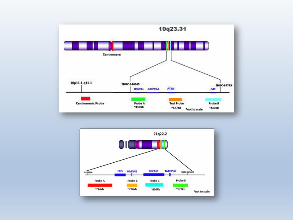

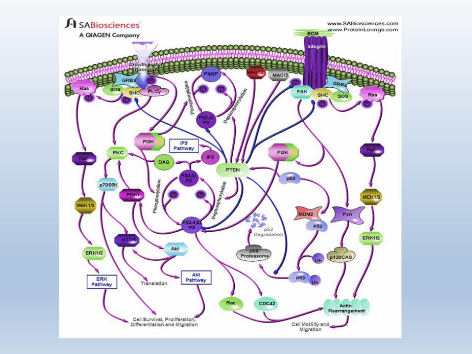
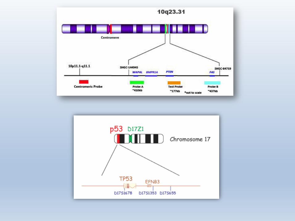
PTEN
PTEN
• The PTEN (phosphatase and tensin) gene encodes a phosphatase which counteracts the PI3K/Akt signaling pathway, one of the most critical cancer-promoting pathways identified to date. It is involved in the regulation of DNA repair, genomic instability, stem cell self-renewal, cellular senescence, and cell migration (metastasis).

PTEN
• PTEN loss is associated with upgrading of prostate cancer from biopsy to radical prostatectomy.
• Studies published , correlate PTEN deletion with poor clinical outcome.
• In addition, it has been observed that the frequency and type of PTEN deletion is correlated to disease progression and early biochemical recurrence.
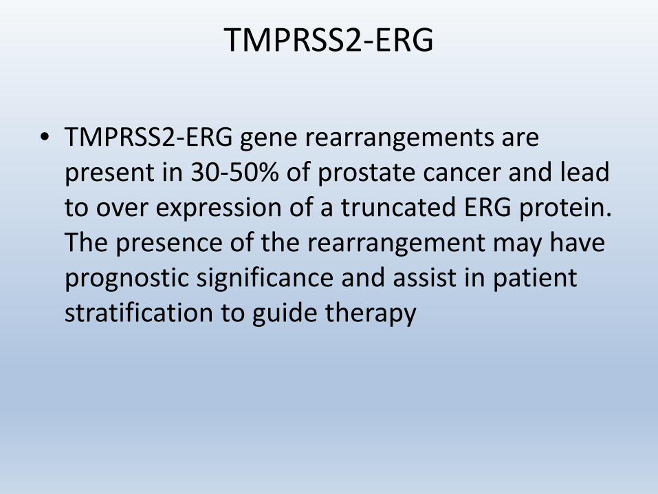
TMPRSS2-ERG
• TMPRSS2-ERG gene rearrangements are present in 30-50% of prostate cancer and lead to over expression of a truncated ERG protein. The presence of the rearrangement may have prognostic significance and assist in patient stratification to guide therapy
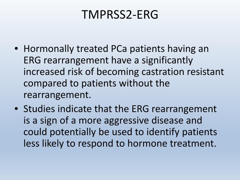
TMPRSS2-ERG
• Hormonally treated PCa patients having an ERG rearrangement have a significantly increased risk of becoming castration resistant compared to patients without the rearrangement.
• Studies indicate that the ERG rearrangement is a sign of a more aggressive disease and could potentially be used to identify patients less likely to respond to hormone treatment.
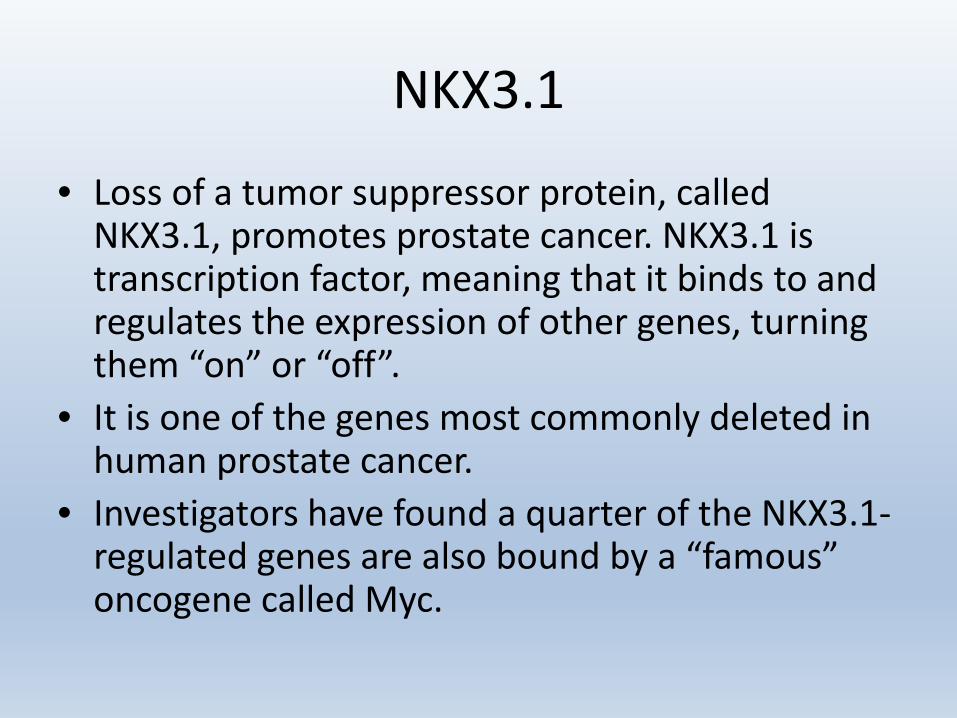
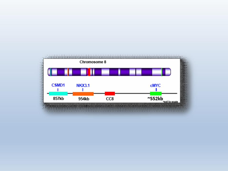
NKX3.1
• Loss of a tumor suppressor protein, called NKX3.1, promotes prostate cancer. NKX3.1 is transcription factor, meaning that it binds to and regulates the expression of other genes, turning them “on” or “off”.
• It is one of the genes most commonly deleted in human prostate cancer.
• Investigators have found a quarter of the NKX3.1-regulated genes are also bound by a “famous” oncogene called Myc.

• Myc is also a transcription factor.• As prostate cancer progresses, NKX3.1 levels
decrease and Myc levels increase.• NKX binds and represses the genes while Myc
binds and activates them.• Myc is the ‘accelerator’ and NKX3.1 is the
‘brake’ on cancer growth.
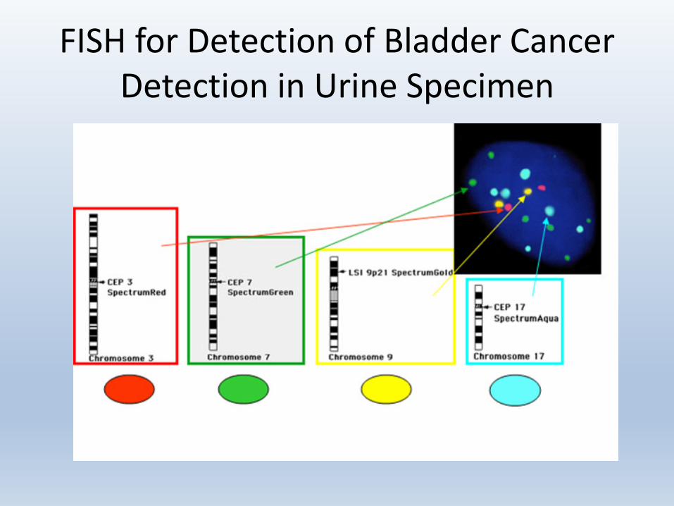
FISH for Detection of Bladder Cancer Detection in Urine Specimen

FISH for Detection of Bladder Cancer in Urine Specimen
• A mixture of CP3, CIP7, CEP17 and LSIp16 probes, each labeled with a different fluorochrome, is used to analyze DNA within cells and to enumerate chromosomes 3, 7, 17, and detect the 9p21 locus deletion.
• A positive result is consistent with a diagnosis of bladder cancer or bladder cancer recurrence, either in the bladder or in another site within the urinary system.
• A negative result is suggestive of the absence of bladder cancer but does not rule it out.

URO-GEN DX™
Examines three major mechanisms of carcinogenesis in bladder cancer:• PTEN: Loss of tumor suppressor gene• P53: Loss of tumor suppressor gene• FGFR3: Gene deletion/translocation
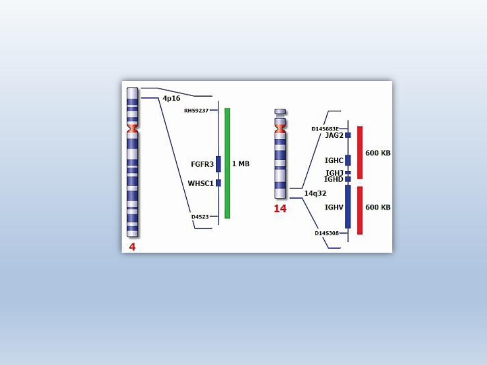

FGFR3
• FGFR3 is an epithelial growth factor found in Chromosome 4p16.3. Its presence, absence, or mutation has prognostic implications in tumor behavior. FGFR3 is translocated from the cytoplasm to the nucleus.
• Non-muscle invasive bladder cancers (NMI-BCs) represent 75% of bladder cancers upon presentation.
• Mutations in the FGFR3 oncogene are common in NMI-BCs and are associated with a lower change of progression to muscle-invasive disease. FGFR3 mutations are equally prevalent in primary and recurrent tumors (63%).
• The FGFR3 assay is used to detect lower stage/grade bladder cancers which have been difficult to detect with other biomarkers.
p53
• The p53 gene is tumor suppressor gene found on chromosome 17 and its product, the p53 protein, is responsible for the death of DNA damaged cells.
• Cells lacking p53 fail to undergo apoptosis (cell death) in response to agents that damage DNA, including radiation and many of the drugs used in cancer chemotherapy.
• This failure to undergo apoptosis in response to DNA damage contributes to the resistance of many tumors to chemotherapy.
• In addition loss of p53 appears to interfere with apoptosis induced by other stimuli, such as growth factor deprivation and oxygen deprivation.
• These effects of p53 inactivation on cell survival are thought to account for the high frequency of p53 mutations in tumors.

LIQUID BIOPSY
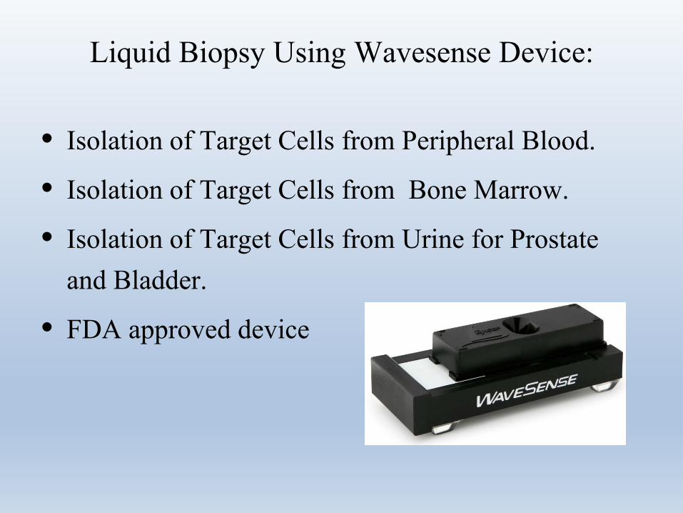
Liquid Biopsy Using Wavesense Device:
• Isolation of Target Cells from Peripheral Blood.
• Isolation of Target Cells from Bone Marrow.
• Isolation of Target Cells from Urine for Prostate and Bladder.
• FDA approved device
Liquid Biopsy for Prostate Cancer
• Non-invasive, liquid alternative to biopsy that uses only urine collected after digital prostate massage.
• Comprehensive and actionable results
• Using FISH for detection of prostate cancer
• Reduces the need for unnecessary biopsies and overall cost to the health system
• Enhances the patient’s prostate cancer risk assessment

This testing is recommended for:• Any patient with increase in PSA and/or
prostatitis, considered to need a prostate biopsy by his Urologist using current standard practice.
• Patients with no prior diagnosis of prostate cancer.
• Patients not receiving treatment influencing PSA level.
• Patients who have not received a prostate massage within 24 hours of sample collection.
• Patients who have not ejaculated within 24 hours of sample collection.

Not recommended for:• Patients with prior diagnosis of prostate
cancer. • Patients receiving treatment influencing PSA
level. • Patients receiving any cancer related therapy. • Patients who have received a prostatectomy.

This testing is recommended for:• Any patient with increase in PSA and/or
prostatitis, considered to need a prostate biopsy by his Urologist using current standard practice.
• Patients with no prior diagnosis of prostate cancer.
• Patients not receiving treatment influencing PSA level.
• Patients who have not received a prostate massage within 24 hours of sample collection.
• Patients who have not ejaculated within 24 hours of sample collection.

Confidential & Proprietary
15339 Barranca ParkwayIrvine, CA 92618
800.807.7760www.wavesense.net
Ms. Stephanie LashbrookSenior Cytogenetic TechnologistHematoGenixTinley Park, IL.
Clinical Validation for EpiSep® Hybridization Slidewith +CD138 Plasma Cell Enrichment
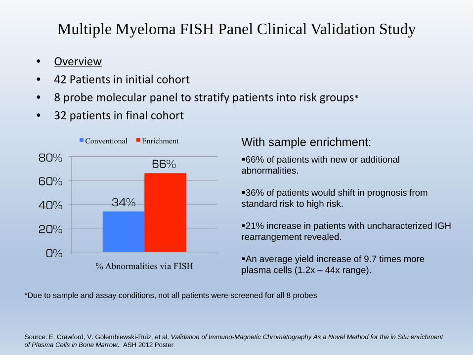
• Overview• 42 Patients in initial cohort• 8 probe molecular panel to stratify patients into risk groups*
• 32 patients in final cohort
Multiple Myeloma FISH Panel Clinical Validation Study
*Due to sample and assay conditions, not all patients were screened for all 8 probes
66% of patients with new or additional abnormalities.
36% of patients would shift in prognosis from standard risk to high risk.
21% increase in patients with uncharacterized IGH rearrangement revealed.
An average yield increase of 9.7 times more plasma cells (1.2x – 44x range).
With sample enrichment:
Source: E. Crawford, V. Golembiewski-Ruiz, et al. Validation of Immuno-Magnetic Chromatography As a Novel Method for the in Situ enrichment of Plasma Cells in Bone Marrow. ASH 2012 Poster

INDIVIDUALIZED GENOMIC STUDIES
Individual Cancer ProfilesIndividual Cancer Profiles (ICP) simplify the laboratory testing process and provide key diagnostic, prognostic, and predictive information you need for up-to-date and
fully informed decision-making.
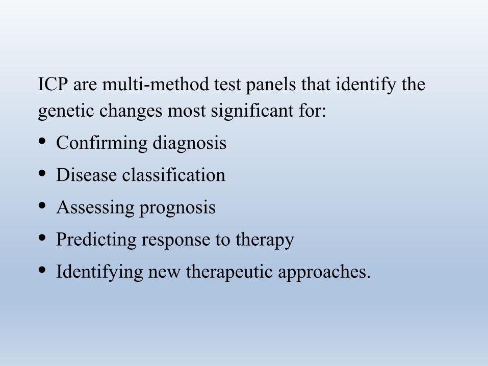
ICP are multi-method test panels that identify the genetic changes most significant for:• Confirming diagnosis• Disease classification• Assessing prognosis• Predicting response to therapy• Identifying new therapeutic approaches.
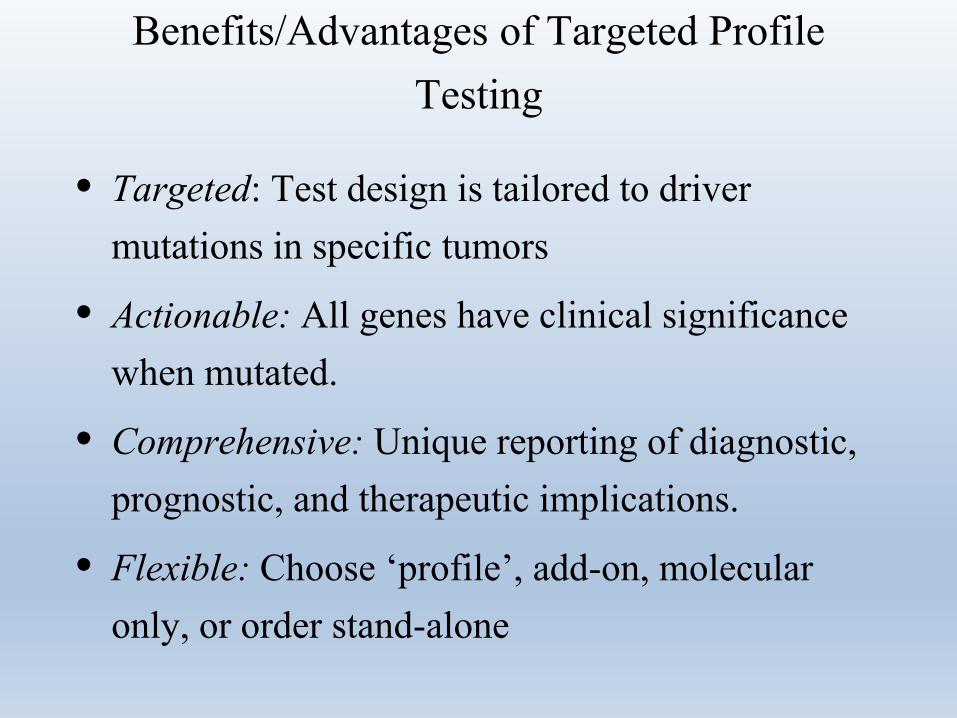
Benefits/Advantages of Targeted Profile Testing
• Targeted: Test design is tailored to driver mutations in specific tumors
• Actionable: All genes have clinical significance when mutated.
• Comprehensive: Unique reporting of diagnostic, prognostic, and therapeutic implications.
• Flexible: Choose ‘profile’, add-on, molecular only, or order stand-alone
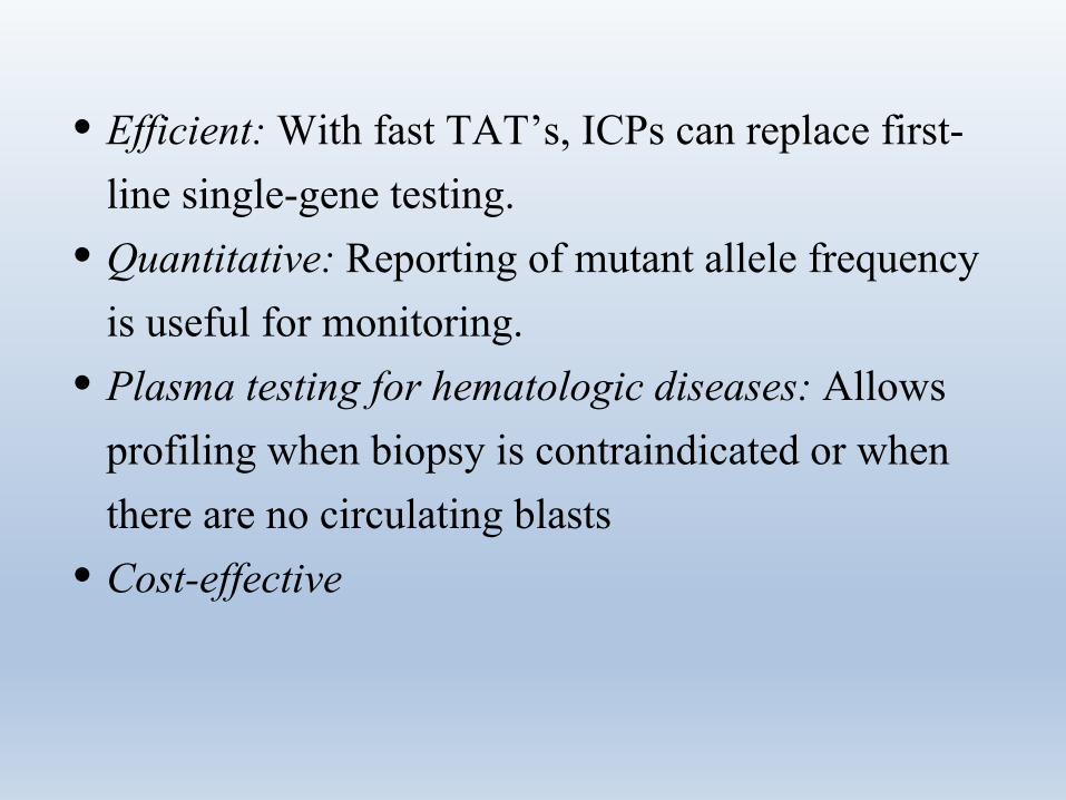
• Efficient: With fast TAT’s, ICPs can replace first-line single-gene testing.
• Quantitative: Reporting of mutant allele frequency is useful for monitoring.
• Plasma testing for hematologic diseases: Allows profiling when biopsy is contraindicated or when there are no circulating blasts
• Cost-effective
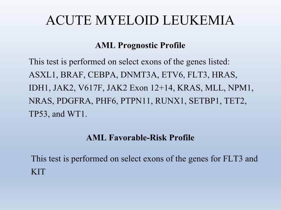
ACUTE MYELOID LEUKEMIA
AML Favorable-Risk Profile
This test is performed on select exons of the genes for FLT3 and KIT
AML Prognostic Profile
This test is performed on select exons of the genes listed: ASXL1, BRAF, CEBPA, DNMT3A, ETV6, FLT3, HRAS, IDH1, JAK2, V617F, JAK2 Exon 12+14, KRAS, MLL, NPM1, NRAS, PDGFRA, PHF6, PTPN11, RUNX1, SETBP1, TET2, TP53, and WT1.
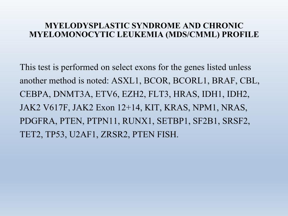
MYELODYSPLASTIC SYNDROME AND CHRONIC MYELOMONOCYTIC LEUKEMIA (MDS/CMML) PROFILE
This test is performed on select exons for the genes listed unless another method is noted: ASXL1, BCOR, BCORL1, BRAF, CBL, CEBPA, DNMT3A, ETV6, EZH2, FLT3, HRAS, IDH1, IDH2, JAK2 V617F, JAK2 Exon 12+14, KIT, KRAS, NPM1, NRAS, PDGFRA, PTEN, PTPN11, RUNX1, SETBP1, SF2B1, SRSF2, TET2, TP53, U2AF1, ZRSR2, PTEN FISH.
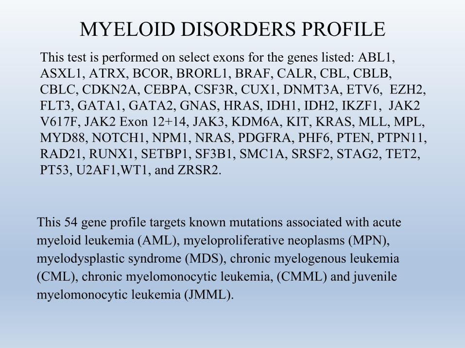
MYELOID DISORDERS PROFILEThis test is performed on select exons for the genes listed: ABL1, ASXL1, ATRX, BCOR, BRORL1, BRAF, CALR, CBL, CBLB, CBLC, CDKN2A, CEBPA, CSF3R, CUX1, DNMT3A, ETV6, EZH2, FLT3, GATA1, GATA2, GNAS, HRAS, IDH1, IDH2, IKZF1, JAK2 V617F, JAK2 Exon 12+14, JAK3, KDM6A, KIT, KRAS, MLL, MPL, MYD88, NOTCH1, NPM1, NRAS, PDGFRA, PHF6, PTEN, PTPN11, RAD21, RUNX1, SETBP1, SF3B1, SMC1A, SRSF2, STAG2, TET2, PT53, U2AF1,WT1, and ZRSR2.
This 54 gene profile targets known mutations associated with acute myeloid leukemia (AML), myeloproliferative neoplasms (MPN), myelodysplastic syndrome (MDS), chronic myelogenous leukemia (CML), chronic myelomonocytic leukemia, (CMML) and juvenile myelomonocytic leukemia (JMML).
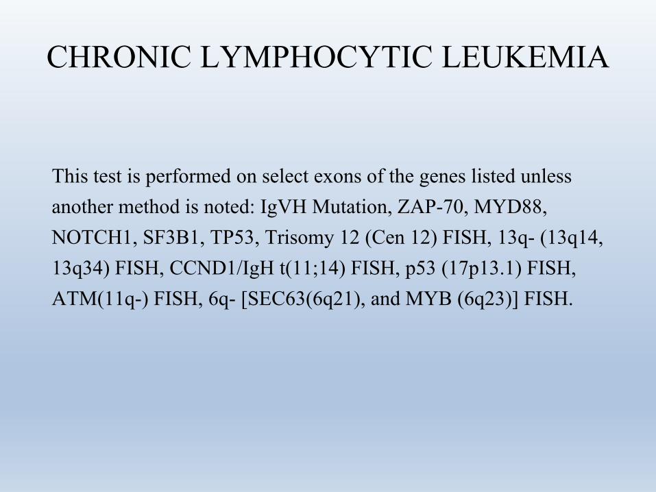
CHRONIC LYMPHOCYTIC LEUKEMIA
This test is performed on select exons of the genes listed unless another method is noted: IgVH Mutation, ZAP-70, MYD88, NOTCH1, SF3B1, TP53, Trisomy 12 (Cen 12) FISH, 13q- (13q14, 13q34) FISH, CCND1/IgH t(11;14) FISH, p53 (17p13.1) FISH, ATM(11q-) FISH, 6q- [SEC63(6q21), and MYB (6q23)] FISH.
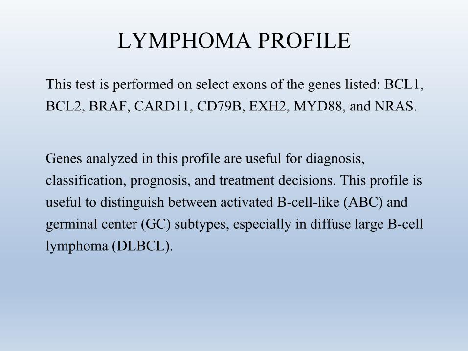
LYMPHOMA PROFILE
This test is performed on select exons of the genes listed: BCL1, BCL2, BRAF, CARD11, CD79B, EXH2, MYD88, and NRAS.
Genes analyzed in this profile are useful for diagnosis, classification, prognosis, and treatment decisions. This profile is useful to distinguish between activated B-cell-like (ABC) and germinal center (GC) subtypes, especially in diffuse large B-cell lymphoma (DLBCL).
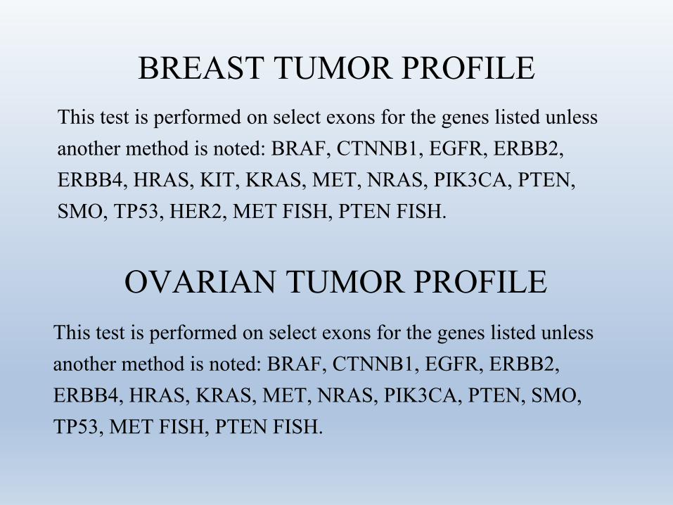
BREAST TUMOR PROFILEThis test is performed on select exons for the genes listed unless another method is noted: BRAF, CTNNB1, EGFR, ERBB2, ERBB4, HRAS, KIT, KRAS, MET, NRAS, PIK3CA, PTEN, SMO, TP53, HER2, MET FISH, PTEN FISH.
OVARIAN TUMOR PROFILEThis test is performed on select exons for the genes listed unless another method is noted: BRAF, CTNNB1, EGFR, ERBB2, ERBB4, HRAS, KRAS, MET, NRAS, PIK3CA, PTEN, SMO, TP53, MET FISH, PTEN FISH.
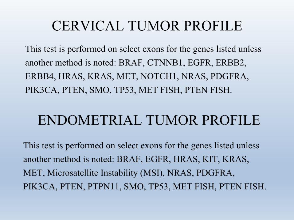
CERVICAL TUMOR PROFILEThis test is performed on select exons for the genes listed unless another method is noted: BRAF, CTNNB1, EGFR, ERBB2, ERBB4, HRAS, KRAS, MET, NOTCH1, NRAS, PDGFRA, PIK3CA, PTEN, SMO, TP53, MET FISH, PTEN FISH.
ENDOMETRIAL TUMOR PROFILEThis test is performed on select exons for the genes listed unless another method is noted: BRAF, EGFR, HRAS, KIT, KRAS, MET, Microsatellite Instability (MSI), NRAS, PDGFRA, PIK3CA, PTEN, PTPN11, SMO, TP53, MET FISH, PTEN FISH.
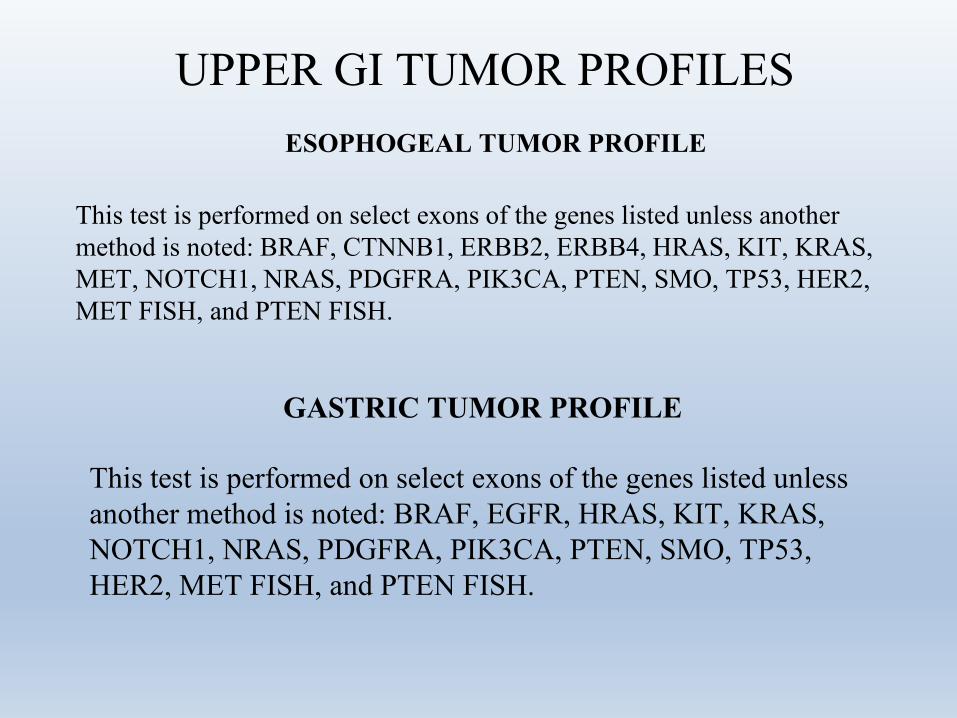
UPPER GI TUMOR PROFILESESOPHOGEAL TUMOR PROFILE
This test is performed on select exons of the genes listed unless another method is noted: BRAF, CTNNB1, ERBB2, ERBB4, HRAS, KIT, KRAS, MET, NOTCH1, NRAS, PDGFRA, PIK3CA, PTEN, SMO, TP53, HER2, MET FISH, and PTEN FISH.
GASTRIC TUMOR PROFILE
This test is performed on select exons of the genes listed unless another method is noted: BRAF, EGFR, HRAS, KIT, KRAS, NOTCH1, NRAS, PDGFRA, PIK3CA, PTEN, SMO, TP53, HER2, MET FISH, and PTEN FISH.
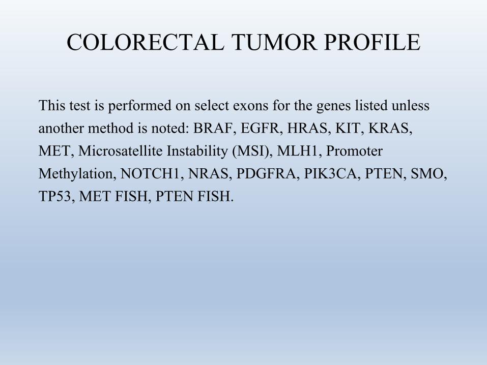
COLORECTAL TUMOR PROFILE
This test is performed on select exons for the genes listed unless another method is noted: BRAF, EGFR, HRAS, KIT, KRAS, MET, Microsatellite Instability (MSI), MLH1, Promoter Methylation, NOTCH1, NRAS, PDGFRA, PIK3CA, PTEN, SMO, TP53, MET FISH, PTEN FISH.
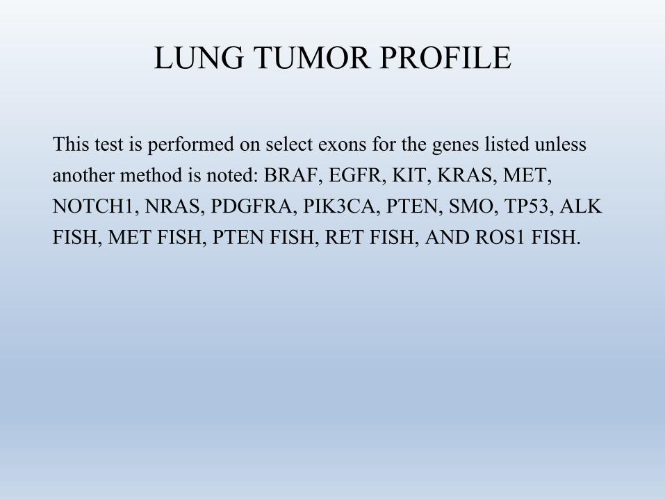
LUNG TUMOR PROFILE
This test is performed on select exons for the genes listed unless another method is noted: BRAF, EGFR, KIT, KRAS, MET, NOTCH1, NRAS, PDGFRA, PIK3CA, PTEN, SMO, TP53, ALK FISH, MET FISH, PTEN FISH, RET FISH, AND ROS1 FISH.
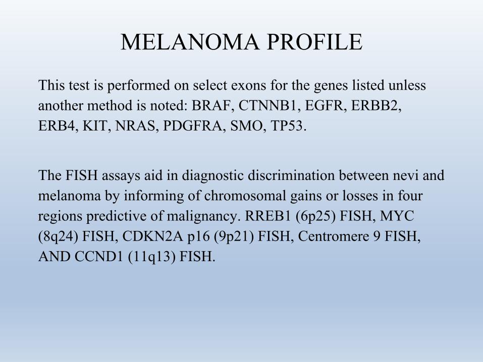
MELANOMA PROFILE
This test is performed on select exons for the genes listed unless another method is noted: BRAF, CTNNB1, EGFR, ERBB2, ERB4, KIT, NRAS, PDGFRA, SMO, TP53.
The FISH assays aid in diagnostic discrimination between nevi and melanoma by informing of chromosomal gains or losses in four regions predictive of malignancy. RREB1 (6p25) FISH, MYC (8q24) FISH, CDKN2A p16 (9p21) FISH, Centromere 9 FISH, AND CCND1 (11q13) FISH.
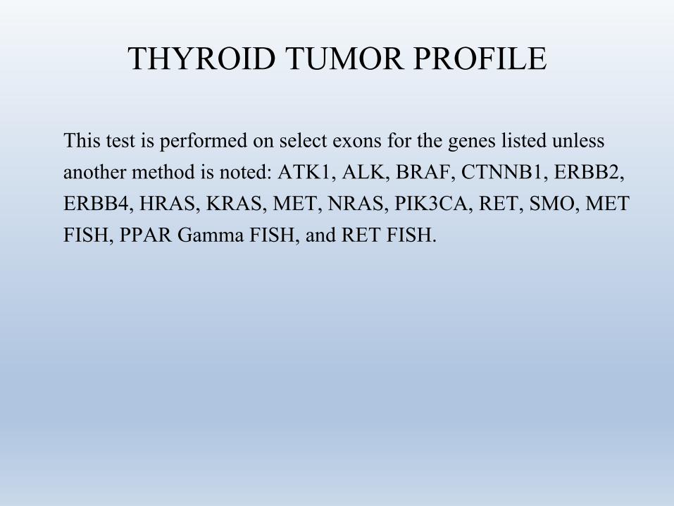
THYROID TUMOR PROFILE
This test is performed on select exons for the genes listed unless another method is noted: ATK1, ALK, BRAF, CTNNB1, ERBB2, ERBB4, HRAS, KRAS, MET, NRAS, PIK3CA, RET, SMO, MET FISH, PPAR Gamma FISH, and RET FISH.
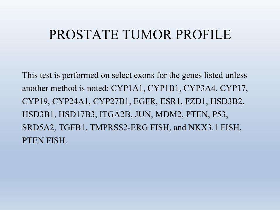
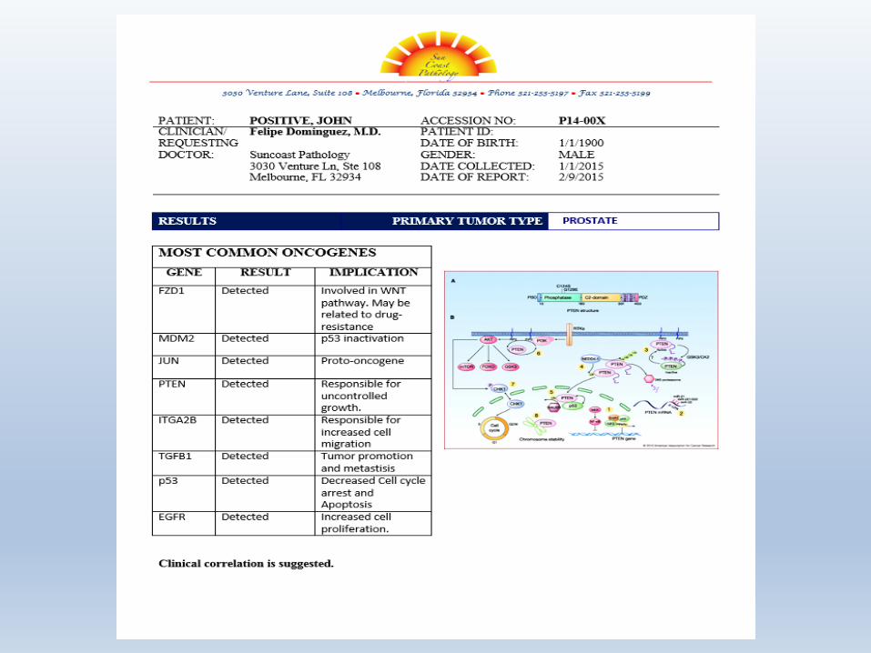
PROSTATE TUMOR PROFILE
This test is performed on select exons for the genes listed unless another method is noted: CYP1A1, CYP1B1, CYP3A4, CYP17, CYP19, CYP24A1, CYP27B1, EGFR, ESR1, FZD1, HSD3B2, HSD3B1, HSD17B3, ITGA2B, JUN, MDM2, PTEN, P53, SRD5A2, TGFB1, TMPRSS2-ERG FISH, and NKX3.1 FISH, PTEN FISH.
BLADDER TUMOR PROFILE
This test is performed on select exons for the genes listed unless another method is noted: BIRC5, CDC25B, COL4A1, FABP4, KPNA2, MBNL2, MSN, COL18A1, COL4A3BP, NEK1, SKAP2, UBE2C
MECHANISMS OF CANCER PROFILE
This test is performed on select exons for the genes listed: ABL1, AKT1, AKT2, APC, BAX, BCAR1, BCL2, BCL2L1, BCL2L11, BID, BRAF, CASP8, CASP9, CCND1, CCND2, CCND3, CCNE1, CDC42, CDH1, CDK2, CDK4, CDKN1A, CDKN1B, CDKN2A, CDKN2B, COL1A1, CRK, CTNNB1, CYCS, DVL1, E2F1, EGFR, ELK1, ERBB2, FADD, FAS, FASLG, FGF2, FN1, FOS, FYN, FZD1, GAPDH, GRB2, GSK3B, GUSB, HGF, HPRT1, HRAS, IGF1, IGF1R, ITGA2B, ITGAV, ITGB1, ITGB3, JUN, KDR, KIT, KRAS, LEF1, MAP2K1, MAP3K5, MAPK1, MAPK14, MAPK3, MAPK8, MAX, MDM2, MYC, NFKB1, NFKB2, NFKBIA, NRAS, PIK3CA, PIK3R1, PTEN, PTK2, PTK2B, RAC1, RAF1, RB1, RELA, RHOA, SHC1, SMAD4, SOS1, SPP1, SRC, TCF3, TGFB1, TGFBR1, TGFBR2, TP53, VEGFA, WNT1

Thank you for attention