prognostic factors for the outcome of nonfunctioning
TRANSCRIPT

https://doi.org/10.1530/ERC-19-0372https://erc.bioscientifica.com © 2020 Society for Endocrinology
Printed in Great BritainPublished by Bioscientifica Ltd.
27:6Endocrine-Related Cancer
S M Sadowski, C R C Pieterman et al.
Prognosis in MEN1-related NF-pNETs
R145–R161
-19-0372
REVIEW
Prognostic factors for the outcome of nonfunctioning pancreatic neuroendocrine tumors in MEN1: a systematic review of literature
S M Sadowski1,*, C R C Pieterman2,3,*, N D Perrier2, F Triponez4 and G D Valk3
1Endocrine Surgery, Surgical Oncology Program, National Cancer Institute, NIH, Bethesda, Maryland, USA2Department of Surgical Oncology, Section of Surgical Endocrinology, University of Texas MD Anderson Cancer Center, Houston, Texas, USA3Department of Endocrine Oncology, University Medical Center Utrecht, Utrecht, The Netherlands4Thoracic and Endocrine Surgery and Faculty of Medicine, University Hospitals of Geneva, Geneva, Switzerland
Correspondence should be addressed to S M Sadowski: [email protected]
*(S M Sadowski and C R C Pieterman contributed equally to this work)
Abstract
Metastatic duodenopancreatic neuro-endocrine tumors (dpNETs) are the most important disease-related cause of death in patients with multiple endocrine neoplasia type 1 (MEN1). Nonfunctioning pNETs (NF-pNETs) are highly prevalent in MEN1 and clinically heterogeneous. Therefore, management is controversial. Data on prognostic factors for risk stratification are limited. This systematic review aims to establish the current state of evidence regarding prognostic factors in MEN1-related NF-pNETs. We systematically searched four databases for studies assessing prognostic value of any factor on NF-pNET progression, development of distant metastases, and/or overall survival. In- and exclusion, critical appraisal and data-extraction were performed independently by two authors according to pre-defined criteria. Thirteen studies (370 unique patients) were included. Prognostic factors investigated were tumor size, timing of surgical resection, WHO grade, methylation, p27/p18 expression by immunohistochemistry (IHC), ARX/PDX1 IHC and alternative lengthening of telomeres. Results were complemented with evidence from studies in MEN1-related pNET for which data could not be separately extracted for NF-pNET and data from sporadic NF-pNET. We found that the most important prognostic factors used in clinical decision making in MEN1-related NF-pNETs are tumor size and grade. NF-pNETs <2 cm may be managed with watchful waiting, while surgical resection is advised for NF-pNETs ≥2 cm. Grade 2 NF-pNETs should be considered high risk. The most promising and MEN1-relevant avenues of prognostic research are multi-analyte circulating biomarkers, tissue-based molecular factors and imaging-based prognostication. Multi-institutional collaboration between clinical, translation and basic scientists with uniform data and biospecimen collection in prospective cohorts should advance the field.
6
Key Words
f MEN1
f prognostic factors
f nonfunctioning pancreatic neuroendocrine tumors
f systematic review
f survival
f metastases
27
Endocrine-Related Cancer (2020) 27, R145–R161
Downloaded from Bioscientifica.com at 01/08/2022 05:02:04PMvia free access
Printed in Great BritainPublished by Bioscientifica Ltd.https://doi.org/10.1530/ERC-19-0372
https://erc.bioscientifica.com © 2020 Society for Endocrinology
R146S M Sadowski, C R C Pieterman et al.
Prognosis in MEN1-related NF-pNETs
27:6Endocrine-Related Cancer
Introduction
Multiple endocrine neoplasia type 1 (MEN1) is a rare hereditary endocrine tumor syndrome caused by germline pathogenic variants in the MEN1 tumor suppressor gene encoding for the menin protein (Chandrasekharappa et al. 1997, Lemmens et al. 1997). During the course of life, carriers of a germline mutation in the MEN1 gene will acquire somatic mutations inactivating the healthy copy of the gene leading to hyperplasia and tumor formation in multiple endocrine and non-endocrine tissues. Primary affected organs are the parathyroid (presenting feature in 90% of the cases), the neuroendocrine pancreas and duodenum, and the pituitary.
Duodenopancreatic neuroendocrine tumors (dpNETs) are highly prevalent in MEN1 (Triponez et al. 2006a, de Laat et al. 2016) and distant metastases are the most important MEN1-related cause of death (Goudet et al. 2010). Of the dpNETs encountered in MEN1, nonfunctioning (NF) tumors are the most frequent, with a prevalence of 50% at the age of 50 (Triponez et al. 2006a) and up to 42% in patients <21 years (Triponez et al. 2006a, Machens et al. 2007, Goncalves et al. 2014, Goudet et al. 2015, Manoharan et al. 2017, Vannucci et al. 2018). There is currently no agreement on interventions and timing thereof in MEN1-related NF-pNETs. Because of pre-symptomatic genetic testing and subsequent surveillance, NF-pNETs in patients with MEN1 are diagnosed more often, at an earlier age and at an earlier stage and their management presents a challenge to patients and physicians. The only curative treatment is surgical resection, which is associated with significant morbidity (Nell et al. 2016), and new NF-pNETs will invariably occur in any remnant pancreas tissue left behind. Recently, multiple retrospective cohorts have reported on the indolent course of most small (<2 cm) NF-pNETs (Triponez et al. 2006a,b, Pieterman et al. 2017, van Treijen et al. 2018). However, subgroups of small NF-pNETs with faster growth are identified, and even small NF-pNETs can metastasize despite seemingly reassuring characteristics (Pieterman et al. 2017). Reliable estimation of prognosis in MEN1-related NF-pNETs is important to inform management decisions in these patients. We therefore systemically reviewed and critically appraised the present literature on prognostic factors for the outcome of NF-pNETs in patients with MEN1. In a comprehensive narrative review, Lee et al. recently provided a general overview of prognostic factors in pancreatic neuroendocrine tumors (Lee et al. 2019). We further aim to compare prognostic factors originating from evidence in sporadic (NF-) pNETs to evidence in
MEN1 and comment on the factors that have not been investigated in MEN1.
Methods
Search strategies
The electronic databases PubMed/MEDLINE, Embase.com, Cochrane Library: CENTRAL and the Cochrane Database of Systematic Reviews, and Web of Science: Core Collection were searched in May and June/July 2019 by a biomedical librarian. Two searches were conducted using a combination of keywords and controlled vocabulary terms for each concept of interest (e.g., ‘multiple endocrine neoplasia type 1’, MEN1, ‘nonfunctioning pancreatic neuroendocrine tumor’, pancreatic tumor, neuroendocrine tumor). The complete search string is documented in Supplementary Material 1 (see section on supplementary materials given at the end of this article). The first search (May) was more focused, including ‘nonfunctioning’ as a search term. A second, broader search (June/July) that did not specify the type of pancreatic neuroendocrine tumor was later completed to ensure that all relevant literature on neuroendocrine pancreatic tumors and MEN1 were retrieved. Search results were limited to those published in Dutch, English, French, and German from 2001 to 2019. The 2001 cut-off point was chosen to represent the era in which pre-symptomatic genetic testing for an MEN1 mutation is possible and guidelines are in place for recommended surveillance.
Study selection
Original studies, systematic reviews, and meta-analyses assessing the prognostic value of any factor on NF-pNET progression, development of distant metastases, and/or overall survival (OS) were eligible for inclusion. Progression could be either growth of existing tumors or development of lymph node or distant metastases. Studies that considered the development of new pNETs to be progression were also included. Studies including both sporadic and MEN1-related NF-pNETs or both functioning and NF-pNETs were eligible if it was possible to extract data for MEN1-related NF-pNETs separately. To minimize selection bias, studies with five or fewer patients with MEN1-related NF-pNET were excluded. All identified articles were independently screened on title and abstract by two authors (S M S and C R C P). Thereafter, independent full-text reviews of potentially relevant studies were performed, and studies were selected
Downloaded from Bioscientifica.com at 01/08/2022 05:02:04PMvia free access

https://erc.bioscientifica.com © 2020 Society for Endocrinology
Printed in Great BritainPublished by Bioscientifica Ltd.https://doi.org/10.1530/ERC-19-0372
R147S M Sadowski, C R C Pieterman et al.
Prognosis in MEN1-related NF-pNETs
27:6Endocrine-Related Cancer
if eligibility criteria were fulfilled (S M S and C R C P). Authors resolved any disagreements by consensus and, when unsuccessful, with the help of a third and fourth reviewer (G D V and F T). Reasons for exclusion at full-text screening were recorded. All included articles were cross-referenced for additional relevant articles.
Risk of bias assessment
Included articles were critically appraised using a modified Quality Assessment in Prognostic Studies (QUIPS) tool (Supplementary Material 2) (Hayden et al. 2006, 2013). Articles were judged on five important domains: study participation, study attrition, prognostic factor measurement, outcome measurement, and statistical analysis and reporting. Critical appraisal was performed independently by two authors (S M S and C R C P), and afterwards, consensus was reached for final decisions. To avoid bias, S M S and F T performed the critical appraisal of the paper for which C R C P was first author.
Data extraction
Study and patient characteristics were retrieved from the included articles. Data were extracted independently by
two authors (S M S and C R C P) as to the study population, baseline characteristics, distribution and measurement of the prognostic factor and outcome, statistical analysis used, and the prognostic value of the investigated factor(s) according to a predefined data-extracting sheet designed by the authors (Supplementary Material 3).
Results
Retrievals and inclusion
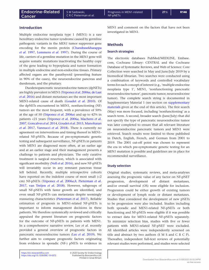
A total of 7024 citations were retrieved from the literature searches (Fig. 1). Of these, 5159 were duplicate citations. A total of 1865 citations were screened. After title and abstract screening, 1643 citations were deemed irrelevant (inter-rater agreement good, Cohen kappa 0.74). Of a total of 222 citations, the full texts were reviewed, after which 209 citations were excluded (inter-rater agreement good, Cohen kappa 0.78). Ultimately, only 13 papers could be included in the risk of bias assessment (Table 1) (Bartsch et al. 2005, Triponez et al. 2006a,b, Sakurai et al. 2007, Davi et al. 2011, D’Souza et al. 2014, Partelli et al. 2016, Conemans et al. 2017a, 2018a,b, Pieterman et al. 2017, Nell et al. 2018, Cejas et al. 2019). A reference search
Figure 1Preferred Reporting Items for Systematic Reviews and Meta-Analyses (PRISMA) flow diagram for identified studies.
Downloaded from Bioscientifica.com at 01/08/2022 05:02:04PMvia free access

Printed in Great BritainPublished by Bioscientifica Ltd.https://doi.org/10.1530/ERC-19-0372
https://erc.bioscientifica.com © 2020 Society for Endocrinology
R148S M Sadowski, C R C Pieterman et al.
Prognosis in MEN1-related NF-pNETs
27:6Endocrine-Related Cancer
performed on these 13 papers did not yield additional papers to be included.
Overview of included studies
We included 13 retrospective studies (from 2 nationwide multicenter cohorts, 1 multi-institutional cohort and 4 single-center cohorts) encompassing 370 unique patients since the same patients were described in multiple studies (Bartsch et al. 2005, Triponez et al. 2006a,b, Sakurai et al. 2007, Davi et al. 2011, D’Souza et al. 2014, Partelli et al. 2016, Pieterman et al. 2017, Conemans et al. 2017a, 2018a,b, Nell et al. 2018, Cejas et al. 2019). A summary of the characteristics and outcomes of the included studies can be viewed in Table 2, more detailed information is available in Supplementary Tables 1 and 2. The studies were of predominantly European origin. Most (9/13) were multi-center studies. With exception of one study (Partelli et al. 2016) follow-up was less than 10 years in all, ranging from 2 to 7 years. Six of the multicenter studies were from the DutchMEN study group (DMSG) and included in part the same patient population (Conemans et al. 2017a, 2018a,b, Pieterman et al. 2017, Nell et al. 2018, Cejas et al. 2019).
More specifically, the three papers from Conemans et al. investigated different factors in the same cohort of surgically resected NF-pNETs, and this cohort was also used by Cejas et al. The paper of Nell et al. on surgery in MEN1-related NF-pNETs includes the same patients as Pieterman et al. and also the same surgical cohort as the paper by Conemans et al. The two included papers from the Groupe d’étude des Tumeurs Endocrines (GTE) (Triponez et al. 2006a,b), a collaborative endocrine tumor research group from France and Belgium, also in part reported on the same population. Specifically, the 65 patients with NF-pNETs <2 cm described by Triponez et al. (2006b) were also included in the previous study on 108 patients with MEN1 and isolated NF-pNETs (Triponez et al. 2006a). Most studies were not specifically designed as prognostic studies.
Outcomes of included studies
Prognostic value of clinical factors: tumor size and size criteria for surgical interventionFour studies investigated tumor size as a prognostic factor for NF-pNETs in MEN1 (Table 2) (Bartsch et al. 2005, Triponez et al. 2006a, Sakurai et al. 2007, Davi et al. 2011).
Table 1 Risk of bias for included studies assessing the prognostic factors in MEN1.
Author, year, ref
Study participationa
Study attritionb
Prognostic factor measurementc
Outcome measurementc
Statistical analysis and reportingd
Overall risk of bias
(Bartsch et al. 2005) + + − − − High(Cejas et al. 2019) + ? + − + Moderate(Conemans et al. 2018a)DNA methylation profiling
+ ? + + + Low
(Conemans et al. 2018b)p27Kip1 and p18Ink4c
+ ? + + − Moderate
(Conemans et al. 2017a)WHO grade
+ ? + + + Low
(Davi et al. 2011) + + − + + Moderate(D’Souza et al. 2014) − ? + + + Moderate(Nell et al. 2018) + ? + + + Low(Partelli et al. 2016) + ? + DM: +
PFS: − + Moderate
(Pieterman et al. 2017) + + + + + Low(Sakurai et al. 2007) + ? − − − High(Triponez et al. 2006b)Is surgery beneficial ≤2 cm
+ − + − + High
(Triponez et al. 2006a)Epidemiology data on 108
MEN1 NF-pNET
+
?
Tumor size −Surgery +
−
+
High
Symbols: + low risk of bias; − high risk of bias; ? unclear. DM, distant metastases; PFS, progression-free survival.aIn study participation, we judged the percentage of the population with MEN1-related NF-pNETs, whether the study population truly represents MEN1 patients as diagnosed according to the guidelines, the sample frame and recruitment, description of source population, and baseline characteristics. bStudy attrition assessed loss to follow-up and whether this could have biased the relationship between prognostic factor and outcome. cFor prognostic factor and outcome measurement, we assessed whether the measurement was clearly described, if the measurement was valid (according to predefined criteria), and whether the measurement was performed according to the same procedure in all participants. dFor statistical analysis, we assessed whether this was adequately described, appropriate, and if there was no selective reporting. See also Supplementary Material 2.
Downloaded from Bioscientifica.com at 01/08/2022 05:02:04PMvia free access

https://erc.bioscientifica.com © 2020 Society for Endocrinology
Printed in Great BritainPublished by Bioscientifica Ltd.https://doi.org/10.1530/ERC-19-0372
R149S M Sadowski, C R C Pieterman et al.
Prognosis in MEN1-related NF-pNETs
27:6Endocrine-Related Cancer
Table 2 Characteristics and outcomes of included studies.
Author, year, country
Study design and follow-up Study population
Prognostic factors analyzed
Outcome measurement Results: prognostic value
(Bartsch et al. 2005)
Germany
Retrospective single center
f/u 3.6 y
n = 26 MEN1 + pancreatic surgery
n = 9 NF-pNET
Tumor size Metastatic potential
NF-pNET: No correlation between size and metastatic potential (P > 0.5)
(Cejas et al. 2019)
USA, The Netherlands
Retrospective multicenter
f/u median 2 y
n = 61 MEN1 + pancreatic surgery
n = 47 NF-pNET
(1) ARX and PDX1(2) ALT status
Distant metastases
Liver relapses (n = 9) only in ARX+ or ARX-/PDX1- cases
HR for distant recurrence in MEN1 NF-pNET 7.1 for ARX+ (P = 0.03) compared to PDX1+ cases
For all cases (sporadic/MEN1) only ALT and ARX+/double negative were independently associated with occurrence of distant relapse
(Conemans et al. 2018b)
The Netherlands
Retrospective multicenter
f/u median 5.8 y
n = 61 MEN1 + pancreatic surgery
n = 46 NF-pNET
IHC expression of p27kip1 and p18ink4c
LM No significant association between p27kip1 and p18ink4c IHC and clinical and pathological characteristics
(Conemans et al. 2018a)
The Netherlands
Retrospective multicenter
f/u median 5.8 y
n = 61 MEN1 + pancreatic surgery
n = 47 NF-pNET
CMI LM Higher CMI in NF-pNETs with LM (P = 0.013)
(Conemans et al. 2017a)
The Netherlands
Retrospective multicenter
f/u mean 6.6 y
n = 69 MEN1 + pancreatic surgery
n = 53 NF-pNET
(1) Tumor size(2) Mitotic index(3) KI-67(4) WHO grade
LM Tumor grade based on KI-67 or combination of KI-67 and mitotic index, not significantly associated with LM
Based on mitotic index, grade significantly associated with LM:
KM survival data 5 y (P = 0.000): ≤2 cm: 100% free of LM >2 cm Grade 1: 90% free of
LM >2 cm Grade 2: 40% free of
LM (Davi et al.
2011)Italy
Prospective single center cohort, retrospective analysis
f/u N/Aa
n = 31 MEN1 + dpNETn = 16 NF-pNET n = 8 ww n = 8 surgery
Tumor size Metastases For patients with NF-pNET who underwent surgery:
No correlation between tumor size and metastases (P = 0.21).
NF-pNET <2 cm: 0% metastasesFor patients conservatively
treated:n = 8 stable, no metastases
after median 2 y (1–10)(D’Souza et al.
2014)USA
Retrospective single center
f/u mean 6.6 y
n = 11 MEN1 NF-PNET Existing or new lesions
Tumor growth
Growth rate differs significantly between existing and new lesions (P = 0.01)
(Nell et al. 2018)
The Netherlands
Retrospectivemulticentermedian f/u ww: 7.2 y surgery: 4.5 y
n = 152 MEN1 NF-pNET
n = 99 ww n = 53 surgery
Surgery versus ww
Metastasis-free survival
Propensity Score-adjusted HR (ww = 1): Surgery 0.73 (95% CI 0.25–2.11)
Surgery <2 cm: 2.04 (95% CI 0.31–13.59)
Surgery 2-3 cm: 1.38 (95% CI 0.09–20.31)
Surgery >3 cm N/A>3 cm: 5/6 (83%) managed with
ww developed LM vs 6/16 (38%) who underwent surgery
(Continued)
Downloaded from Bioscientifica.com at 01/08/2022 05:02:04PMvia free access

Printed in Great BritainPublished by Bioscientifica Ltd.https://doi.org/10.1530/ERC-19-0372
https://erc.bioscientifica.com © 2020 Society for Endocrinology
R150S M Sadowski, C R C Pieterman et al.
Prognosis in MEN1-related NF-pNETs
27:6Endocrine-Related Cancer
Author, year, country
Study design and follow-up Study population
Prognostic factors analyzed
Outcome measurement Results: prognostic value
(Partelli et al. 2016)
Italy, Germany, UK
Retrospective multicenter
median f/u: ww: 9.1 y Surgery: 10.6 y
n = 60 MEN1 NF-pNET <2 cm
n = 33 ww n = 27 surgery
Surgery versus wwDecision at initial
diagnosis
Distant metastases
PFS
PFS not different between ww and surgery (P = 0.2)
Development of new metastases (P = 1), pNET-related death (P = 0.9), and tumor enlargement during f/u (P = 0.2) not different between ww and surgery
(Pieterman et al. 2017)
The Netherlands
Retrospective longitudinal
multicenterf/u median 5y
n = 99 MEN1 + NF-pNET <2 cm (n = 115 tumors)
GenotypeAgeHypergastrinemia Existing/new tumorBaseline size gender
Growth rate (mm/y)
Overall (n = 115) no association prognostic factors and growth rate
No difference in age, gender, genotype, hypergastrinemia, new tumors and baseline size between progressive and stable tumors
Stratified analysis of progressive tumors: tumors with germline missense mutations faster growth (P = 0.09). Other factors not significant.
(Sakurai et al. 2007)
Japan
Retrospective single center
f/u mean 6.5y
n = 14 MEN1 and NF-pNET
Tumor size Metastases n = 5/6 (83%) >35 mm newly developed tumors or metastases
n = 1/8 (13%) <35 mm newly developed tumors
(Triponez et al. 2006a)
France and Belgium
Retrospective multicenter
f/u mean 4.3 y
n = 108 MEN1 NF-pNET
Tumor sizeSurgery
OSMetastases
Larger tumor size associated with metastases (P < 0.01)
0–30 mm better survival compared to >30 mm (P < 0.01)
no difference between <10 mm and 10–30 mm (P = 0.31)
Survival worse in non-curative surgery (P < 0.01)
Survival not different between curative surgery vs ww (P = 0.15)
(Triponez et al. 2006b)
France and Belgium
Retrospective multicenter
mean f/u: ww: 3.3y mean 6.7 y
n = 65 MEN1 NF-pNET ≤2 cm
n = 50 ww n = 15 surgery
Surgery vs ww OSDFS
No significant difference in progression and death between surgery and ww
Overall life expectancy in patients with NF-pNET <2 cm not different than n = 229 MEN1 patients without any dpNET (P = 0.33)
More detailed information on study characteristics and outcomes can be found in Supplementary Tables 1 and 2.aNot separately reported for NF-pNET.ALT, alternative lengthening of telomeres; CMI, cumulative methylation index; DFS, disease-free survival; dpNET, duodenopancreatic neuroendocrine tumor; f/u, follow-up; HR, hazard ratio; IHC, immunohistochemistry; LM, liver metastases; MEN1, multiple endocrine neoplasia type 1; N/A, not available; NF-pNET, non-functional pancreatic neuroendocrine tumor; OS, overall survival; PFS, progression-free survival; pNET, pancreatic neuroendocrine tumor; WHO, World Health Organization; ww, watchful waiting; y, year.
Table 2 Continued.
Downloaded from Bioscientifica.com at 01/08/2022 05:02:04PMvia free access

https://erc.bioscientifica.com © 2020 Society for Endocrinology
Printed in Great BritainPublished by Bioscientifica Ltd.https://doi.org/10.1530/ERC-19-0372
R151S M Sadowski, C R C Pieterman et al.
Prognosis in MEN1-related NF-pNETs
27:6Endocrine-Related Cancer
The development of metastases was the primary endpoint in three, and development of new lesions in one (Sakurai et al. 2007). Studies were at moderate-to-high risk of bias (Table 1), as attributable risks could often not be calculated. Three studies compared surgical resection of NF-pNETs with a watchful waiting strategy based on tumor size (Table 2) (Triponez et al. 2006b, Partelli et al. 2016, Nell et al. 2018). Overall survival and/or disease-, metastases- or progression-free survival were the primary endpoints.
Two studies reported that tumor size was not associated with metastases. Bartsch et al. (n = 9, median follow-up 3.6 years), did not find a correlation between tumors size and metastatic potential. This study did however have a small sample size, short follow-up and high risk of bias (Bartsch et al. 2005). Davi et al. (n = 16, follow-up not available for NF-pNET, moderate risk of bias) report no correlation between tumor size and metastases (lymph node (ln) + distant); however, only in those that underwent surgical resection (n = 8). When looking at the entire study population no metastases were seen in patients with tumors <2 cm (Davi et al. 2011).
In contrast, Triponez et al. (n = 108, follow-up 4.3 years after pNET diagnosis, high risk of bias) found larger tumor size to be correlated with risk of metastases (ln + distant) and worse survival (Triponez et al. 2006a). Sakurai et al. (n = 14, follow-up 6.5 years, high risk of bias) found a tumor size >35 mm to be associated with more newly developed tumors (Sakurai et al. 2007).
Three studies compared surgical resection with watchful waiting in patients with NF-pNETs.
Triponez et al. compared surgical resection (n = 15, follow-up 6.7 years) with watchful waiting (n = 50, follow-up 3.3 years) in patients with NF-pNETs ≤2 cm (Triponez et al. 2006b). This study has a high risk of bias. There was no significant difference in progression of disease and deaths between the two groups. Overall life expectancy in patients with NF-pNET <2 cm was not different than that of 229 MEN1 patients in the registry without any dpNET (P = 0.33) (Triponez et al. 2006b).
Partelli et al. compared surgical resection with watchful waiting in n = 60 patients with NF-pNETs <2 cm, with patients analyzed as intention to treat (Partelli et al. 2016). Risk of bias was moderate. Progression-free survival (PFS) was defined as development of metastases, growth of existing tumors, or development of new tumors. The development of new metastases (P = 1), pNET-related death (P = 0.9), and tumor enlargement during follow-up (P = 0.2) were not different between watchful waiting (median follow-up 9.1 years) and surgery (median follow-up 10.6 years). Overall survival of the entire cohort was 98% at 5
and 10 years, and PFS at 5, 10, and 15 years was 63, 39, and 10%, respectively. There was no statistical difference between watchful waiting and surgical intervention (P = 0.2) (Partelli et al. 2016).
The study by Nell et al. (low risk of bias) comparing surgical resection of NF-pNETs with watchful waiting from the DutchMEN study group (DMSG), had the largest sample size (n = 152) (Nell et al. 2018). Fifty-three patients underwent surgery with a median follow-up of 4.5 years, and 99 underwent watchful waiting for a median follow-up of 7.2 years. Using a propensity score analysis to correct for differences between both groups, surgery for NF-pNETs was found not to be associated with a significantly lower risk of liver metastases or death (adjusted HR = 0.73 (0.25–2.11)). Adjusted HR after stratification by size were <2 cm = 2.04 (0.31–13.59) and 2–3 cm = 1.38 (0.09–20.31). The subgroup >3 cm was too small for time varying analysis; however, 5/6 (83%) patients with NF-pNETs >3 cm managed by watchful waiting developed liver metastases or died compared with 6/16 (38%) patients who underwent surgical intervention (Nell et al. 2018).
Although there is overall a significant risk of bias, results from studies looking at prognostic value of size compared with the results of studies that compare watchful waiting with surgical resection based on size-criteria show that risk of metastases and (disease-related) death is low in MEN1-related pNETs <2 cm.
Prognostic value of tissue-based markersFour studies were included that investigated the prognostic value of tissue-based markers, in all studies these were assessed by pathological examination of surgically resected MEN1-related pNETs (Conemans et al. 2017a, 2018a,b, Cejas et al. 2019) (Table 2). The studies by Conemans et al. had a low risk of bias, by Cejas et al. moderate (Table 1). All of these studies report on the same MEN1 patient population (from the DMSG). Development of liver metastases was the primary endpoint and occurred in 17%, mostly metachronous.
When assessing the prognostic value of the World Health Organization (WHO) grade in MEN1-related NF-pNETs, higher WHO grade based on mitotic index was associated with a higher risk of liver metastases in tumors >2 cm (5-year liver metastases free survival 90% for grade 1 tumors and 40% for grade 2 tumors; log rank P = 0.000). WHO grade based on Ki-67 labeling index (LI) or combined mitotic index and Ki-67 LI was not associated with liver metastases (Conemans et al. 2017a).
Downloaded from Bioscientifica.com at 01/08/2022 05:02:04PMvia free access

Printed in Great BritainPublished by Bioscientifica Ltd.https://doi.org/10.1530/ERC-19-0372
https://erc.bioscientifica.com © 2020 Society for Endocrinology
R152S M Sadowski, C R C Pieterman et al.
Prognosis in MEN1-related NF-pNETs
27:6Endocrine-Related Cancer
Cejas et al. investigated the prognostic value of NF-pNET subsets based on their resemblance to islet alpha and beta cells (Cejas et al. 2019). They confirmed that A (resembling alpha cells) and B (resembling beta cells) type tumors expressed transcription factors (TFs) ARX and PDX1, respectively (these TFs can be assessed on tumor specimen by immunohistochemistry (IHC)). They subsequently assessed the prognostic value of tumor type (ARX+, PDX1+, double positive (DP) or double negative (DN)) and alternative lengthening of telomeres (ALT) for the occurrence of distant relapses in resected NF-pNETs. They found that ARX and PDX1 IHC status significantly correlated with occurrence of liver metastases. Liver metastases were only seen in ARX+ or DN cases, not PDX1+ or DP cases. When comparing ARX+ with PDX1+ cases, HR for relapse was 7.09 (95% CI 1.72–42.86) for ARX+ cases. ALT positivity was only seen in ARX+ or DN tumors but not in PDX1+/DN tumors. ALT positivity significantly correlated with relapse rate.
Although the studies examining expression of p27Kip1/p18Inkc4c (Conemans et al. 2018b) and DNA methylation (Conemans et al. 2018a) in MEN1-related pNETs did not have a primary prognostic aim, they did include prognostic data. No significant association between p27Kip1 and p18Inkc4c expression and clinical and pathological characteristics was seen (Conemans et al. 2018b). NF-pNETs with synchronous or metachronous liver metastases had a higher (1036 vs 869, P = 0.013) cumulative methylation index (defined as the sum of methylation percentages of the promotors of the 56 investigated tumor suppression genes) (Conemans et al. 2018a).
Based on these studies, we conclude that in patients with MEN1 undergoing resection of an NF-pNET, grade by mitotic index can be used to identify patients at higher risk for future development of liver metastases. In addition, ARX/PDX1 IHC and ALT status seem to be potential powerful prognostic indicators. Additional prospective studies must follow to determine feasibility in the clinical setting. Assessing p27Kip1 and p18Inkc4c alone to determine the future risk of developing liver metastases is not useful. DNA methylation status might be of interest as a prognostic biomarker; however, additional data are necessary.
Prognostic factors associated with tumor growthTwo studies aimed to assess growth rate/natural course of NF-pNETs <2 cm in patients with MEN1 (Table 2) (D’Souza et al. 2014, Pieterman et al. 2017). In the first study, a population-based study with low risk of bias, the natural course of 115 NF-pNETs <2 cm from 99 patients
is described (Pieterman et al. 2017), with a median follow-up of 5 years after the first imaging. Indication for watchful waiting or intervention was determined by the treating physician/team. Tumor growth was assessed on MRI/CT using linear mixed-model analysis and genotype, age, gender, hypergastrinemia, existing versus new tumor, and baseline tumor size were all assessed for influence on growth rate. Growth rate was 0.4 mm/y. Thirty percent of the tumors was progressive (growth rate 1.6 mm/y), while 70% remained stable without identifiable growth. Genotype was a significant modifier of growth in the subgroup of progressive tumors, with tumors with germline missense mutations demonstrating faster growth. Other factors did not influence growth rate in the subgroup of progressive tumors, and none of the factors distinguished between progressive and stable tumors. D’souza et al. (moderate risk of bias) reported the natural course of 18 NF-pNETs <2 cm in 11 patients with MEN1 assessed by Endoscopic Ultrasound (EUS) (D’Souza et al. 2014) during a mean follow-up of 6.5 years. They report significantly different growth rates for existing lesions (1.32 mm/year) compared to newly diagnosed lesions (3 mm/year). We suspect this finding to be caused by selection bias and do not consider this an important modifier of growth.
Discussion
This systematic review summarizes prognostic factors in MEN1-related NF-pNETs, based on 13 studies including n = 370 unique patients since the same patients were described in multiple studies. Results show that tumor size (using 2 cm as cut-off) and WHO grade are prognostic factors that can be used in clinical practice, while ARX/PDX1 IHC status and ALT are potential novel prognostic biomarkers.
Prognostic data from studies in MEN1-related pNETs for which data cannot be separately extracted for NF-pNET can be used as supporting evidence and to identify prognostic factors that might be applied to all NF-pNETs as well (overview provided in Supplementary Table 3). These studies corroborate the increased risk of distant metastases in pNETs >2 cm (Vinault et al. 2018). In addition, they show that despite numerous efforts, no definitive genotype-phenotype correlation has been identified in MEN1-related pNETs mainly due to lack of validation of reported associations, and therefore, we currently do not recommend basing management decisions on a specific genotype (Thevenon et al. 2013, Bartsch et al. 2014, Giudici et al. 2017, Christakis et al. 2018). A biological
Downloaded from Bioscientifica.com at 01/08/2022 05:02:04PMvia free access

https://erc.bioscientifica.com © 2020 Society for Endocrinology
Printed in Great BritainPublished by Bioscientifica Ltd.https://doi.org/10.1530/ERC-19-0372
R153S M Sadowski, C R C Pieterman et al.
Prognosis in MEN1-related NF-pNETs
27:6Endocrine-Related Cancer
reason for the lack of validated genotype-phenotype correlations may be that menin does not have intrinsic enzymatic activity and is involved in multiple cellular processes (most importantly epigenetic regulation of gene transcription) through interaction with other proteins (Iyer & Agarwal 2018). It might therefore also be of value to investigate if variants in genes coding for menin-interacting proteins might modify the phenotype, such as been suggested in a publication showing that patients with CDKN1B V109G polymorphism had more aggressive tumors (Circelli et al. 2015). For patients with multifocal pNETs, imaging-based prognostication is appealing as it is non-invasive and can be repeated over time. Two small retrospective studies in MEN1 indicate that FDG-avidity (FGD-avidity predicted more aggressive disease) and SUVmax (lower SUVmax associated with decreased median PFS) might be of prognostic value (Lastoria et al. 2016, Kornaczewski Jackson et al. 2017). It is interesting to note that one study observed higher estrogen exposure to be associated with smaller pNETs (Qiu et al. 2017). Although this study had significant risk of bias because only a small selected subgroup of the patients could be used in this analysis, this certainly is an area of interest, given that menin is known to interact with the estrogen receptor (Dreijerink et al. 2006) and that several studies show male sex to be an adverse prognostic factor (Conemans et al. 2017b, Vinault et al. 2018).
As MEN1 is also one of the most important driver genes in sporadic pNETs (Jiao et al. 2011, Scarpa et al. 2017), evidence gained from sporadic pNETs might be applied to MEN1-related pNETs as well. Indeed, cumulative methylation index was found not to be statistically different between MEN1-related and sporadic NF-PNETs (Conemans et al. 2018a), the prognostic value of ARX/PDX1 IHC was found to be similar in MEN1-related and sporadic nf-PNETs (Cejas et al. 2019) and mRNA expression analysis has revealed that a subgroup of sporadic pNETs clustered with MEN1-related pNETs, while others clustered alone (Keutgen et al. 2018). All this lends credence to the fact that at least a subgroup of sporadic (NF)-pNETs - those with somatic MEN1 mutations? - is biologically comparable to MEN1. However, apart from the fact that over 50% of sporadic pNETs are not MEN1-mutated, there are important clinical differences that can influence use and value of prognostic factors. Patients with MEN1 are younger at diagnosis, have multifocal tumors, are diagnosed in an earlier stage due to surveillance and often have other concomitant primary neuroendocrine and non-neuroendocrine tumors. This necessitates validation of evidence from sporadic pNETs
in MEN1 before this can be applied in practice. Table 3 provides a comparison of the prognostic data in sporadic and MEN1-related (NF-)pNETs.
With regards to tumor size in sporadic NF-pNETs, overall, increased tumor size is associated with reduced DFS, with <2 cm a good cutoff for watchful waiting (Lee et al. 2019). A recent large single center retrospective study and a Systematic Review in 540 sporadic NF-pNET revealed low risk of metastases when managing tumors <2 cm with watchful waiting (Partelli et al. 2017, 2019). This is reinforced in a multi-institutional retrospective study of 210 resected NF-pNETs with tumors ≤2 cm. They report a high surgical morbidity rate of 14.3% (n = 30), and found the presence of biliary or pancreatic duct dilatation, and WHO grade 2–3 to be independently associated with recurrence. Thus, they advocate surgery for NF-pNET <2 cm with those features, and a wait-and-see policy in the remaining patients (Sallinen et al. 2017). This is in line with evidence from MEN1-related NF-pNETs.
The North American Neuroendocrine Tumor Society (NANETS) consensus states that, based on a review of retrospective studies, tumors <1 cm have low risk of metastases and should be followed by watchful waiting, however, that tumors between 1 and 2 cm should be managed in an individualized manner (according to risk factors) (Howe et al. 2020).
As in MEN1-related NF-pNETs, retrospective studies of sporadic NF-pNETs <2 cm managed with a watchful waiting strategy show that most do not exhibit meaningful growth during follow-up and no distant metastases were observed (Sallinen et al. 2017, Choi et al. 2018). Median follow-up was less than 5 years in all of these studies. One study did not identify predictors of tumor growth among patient (sex, age) or tumor characteristics (localization, cystic, size) (Gaujoux et al. 2013), while another study found hypervascularity to be associated with less risk of growth as other factors (sex, age, size, location, other tumor characteristics) were not associated with growth (Choi et al. 2018). One study found growth to be associated with grade 2 or grade 3 tumors (Jung et al. 2015). As in MEN1, exact relation between tumor growth rate and outcome in localized disease is unknown in sporadic NF-pNETs as no data on this subject is available. Time from diagnosis to surgical intervention might indicate whether a tumor is growing rapidly, however no data are available on this subject in MEN1.
Overall, in sporadic pNETs, WHO grade and Ki-67 are one of the most important prognostic factors for overall survival and disease-specific survival (DSS) as well as recurrence-free survival (RFS), OS and DSS after surgical
Downloaded from Bioscientifica.com at 01/08/2022 05:02:04PMvia free access

Printed in Great BritainPublished by Bioscientifica Ltd.https://doi.org/10.1530/ERC-19-0372
https://erc.bioscientifica.com © 2020 Society for Endocrinology
R154S M Sadowski, C R C Pieterman et al.
Prognosis in MEN1-related NF-pNETs
27:6Endocrine-Related Cancer
resection (Lee et al. 2019). Recent large retrospective multi-center studies focusing specifically on NF-pNETs have confirmed the important prognostic value of Ki-67 and WHO grade on recurrence (defined as either local or distant recurrence) (Genc et al. 2018a, Zaidi et al. 2019), also in NF-pNET <2 cm (Sallinen et al. 2018). Although most studies follow the cut-off for Ki-67 as set by the WHO (3%), some advocate for 5% as cut-off between G1 and G2 tumors (Lopez-Aguiar et al. 2018). Other studies (also including functioning tumors) show that subdividing
low-grade Ki-67 into <1% vs 1-2.99% might improve prognostic classification (Lopez-Aguiar et al. 2018) or even that Ki-67 has a more linear relation with recurrence and should be viewed more as a continuous than categorical variable (Gao et al. 2018). A different cut-off might improve prognostic value of Ki-67 in MEN1-related NF-pNETs, and may have been the reason no association with outcome could be identified by Conemans et al. (2017a).
Looking at histopathological prognostic markers in sporadic pNETs, a systematic review not surprisingly
Table 3 Overview of prognostic factors with evidence in both MEN1-related and sporadic NF-pNET.
Prognostic factor Evidence in MEN1-related NF-pNETs Evidence in sporadic NF-pNETs
Tumor size Tumor size correlates with risk of metastases, (Triponez et al. 2006a, Sakurai et al. 2007) with low risk for tumors <2 cm (Triponez et al. 2006b, Partelli et al. 2016, Conemans et al. 2017a, Nell et al. 2018)
Increased tumor size associated with reduced DFS, with <2 cm a good cutoff for observation (Lee et al. 2019). Low risk of metastases when observing tumors <2 cm (Partelli et al. 2017, 2019), especially when no bile duct involvement (Sallinen et al. 2018).
WHO grade/Ki-67 In tumors >2 cm, higher WHO grade as defined by mitotic index associated with a higher risk of LM (Conemans et al. 2017a).
Tumor grade based on Ki-67 or combination of Ki-67 and mitotic index not associated with development of LM (Conemans et al. 2017a).
Higher WHO grade/Ki-67 labeling index is one of the most prominent factors associated with worse DFS, DSS and OS (Lee et al. 2019).
DAXX/ATRX and/or ALT ALT positivity associated with distant relapses (Cejas et al. 2019).
ALT and loss of DAXX/ATRX are associated with decreased DFS (Marinoni et al. 2014, Pipinikas et al. 2015, Kim et al. 2017, Singhi et al. 2017, Chou et al. 2018, Roy et al. 2018, Cives et al. 2019), ATRX loss is associated with poorer OS (Chou et al. 2018) and DAXX/ATRX loss is associated with shorted DSS (Marinoni et al. 2014).
ALT associated with distant metastases in NF-pNETs <3 cm (Pea et al. 2020).
In metastatic pNETs, ALT and DAXX/ATRX loss associated with improved OS (Jiao et al. 2011, Dogeas et al. 2014, Kim et al. 2017).
PDX1/ARX Distant metastases only seen in ARX positive or ARX and PDX1 negative tumors (Cejas et al. 2019).
Distant metastases almost exclusively seen in ARX positive or ARX and PDX1 negative tumors (Cejas et al. 2019).
Tumor growth Growth rate of NF-pNETs <2 cm 0.4-3 mm/year (D’Souza et al. 2014, Pieterman et al. 2017).
No clinical factor distinguishes between progressive and stable tumors.
In progressive tumors, tumors with germline missense mutation grow faster (Pieterman et al. 2017).
Most sporadic NF-pNETs <2 cm do not exhibit meaningful growth during observation (Sallinen et al. 2017, Choi et al. 2018).
Hypervascularity was found to be associated with less growth (Choi et al. 2018). Growth was found to be associated with grade 2 or grade 3 tumors (Jung et al. 2015).
Imaging-related characteristics
Lower SUVmax on 68Gallium-dotatate PET associated with decreased PFS in pNET (Lastoria et al. 2016).
FDG-avidity of pNET associated with more aggressive disease (Ki-67 ≥5%) (Kornaczewski Jackson et al. 2017).
Imaging factors associated with worse DFS/OS include: tumoral hypo-enhancement/vascularity, presence of main pancreatic duct involvement, presence of irregular tumor margins (Lee et al. 2019).
Higher uptake on 18F-FDG PET correlates with poorer OS and with advancing classification/grade (Rinzivillo et al. 2018).
ALT, alternative lengthening of telomeres; DFS, disease-free survival; DSS, disease-specific survival; FDG, fluorodeoxyglucose; LM, liver metastases; NF, non-functioning; OS, overall survival; PD-1, programmed cell death protein 1; PFS, progression-free survival; pNET, pancreatic neuroendocrine tumors; SUV, standardized uptake value; TAM, tumor-associated macrophages; WHO, World Health Organization.
Downloaded from Bioscientifica.com at 01/08/2022 05:02:04PMvia free access

https://erc.bioscientifica.com © 2020 Society for Endocrinology
Printed in Great BritainPublished by Bioscientifica Ltd.https://doi.org/10.1530/ERC-19-0372
R155S M Sadowski, C R C Pieterman et al.
Prognosis in MEN1-related NF-pNETs
27:6Endocrine-Related Cancer
found lymph node metastases to be strongly associated with increased risk of recurrence and OS (Tao et al. 2017, Lee et al. 2019). This factor has been widely studied in resections for sporadic pNETs as well as in sporadic gastrinoma/functioning duodenopancreatic NETs. There are unfortunately no studies specifically looking at lymph node dissections or lymph node ratio as prognostic factors for outcome in NF-pNETs in MEN1. In MEN1, functioning and NF-pNETs often co-exist and it is difficult to attribute lymph node metastases to their primary tumor, which poses a challenge to prognostic research. Also, prognostic value of lymph node metastases from gastrinoma might be different from that in NF-pNETs. Additionally, presence of perineural or vascular invasion are predictors of tumor recurrence or metastases (Ge et al. 2017, Lee et al. 2019). Invasion into adjacent organs represents a high risk of recurrence (HR of 1.65 (95% CI, 1.03–2.65; P = 0.038)) (Merath et al. 2018) and R1 resections are associated with shorter DFS (Lee et al. 2019).
Several retrospective cohort studies have assessed the prognostic value of DAXX/ATRX loss and/or ALT positivity in sporadic surgically resected pNETs (nonfunctioning 75–100%) (Jiao et al. 2011, Dogeas et al. 2014, Marinoni et al. 2014, Pipinikas et al. 2015, Kim et al. 2017, Park et al. 2017, Singhi et al. 2017, Chou et al. 2018, Pea et al. 2020, Roy et al. 2018, Cives et al. 2019, Uemura et al. 2019). All but one study (Park et al. 2017) found that ALT and/or DAXX/ATRX loss (IHC) was associated with decreased relapse-, recurrence- or progression-free survival (Marinoni et al. 2014, Pipinikas et al. 2015, Kim et al. 2017, Singhi et al. 2017, Chou et al. 2018, Roy et al. 2018, Cives et al. 2019). One study (Chou et al. 2018) also found ATRX loss to be associated with poorer OS and in another study (Marinoni et al. 2014) DAXX/ATRX loss was associated with shorter DSS. In small (<3 cm) NF-pNETs ALT was found to be associated with the occurrence of distant metastases (Pea et al. 2020). Intriguingly, in metastatic pNETs, ALT and DAXX/ATRX loss have found to be associated with improved OS (Jiao et al. 2011, Dogeas et al. 2014, Kim et al. 2017). As in MEN1, in sporadic NF-pNETs expression of TFs ARX and PDX1 as surrogate markers for alpha or beta cell resemblance, was shown to be associated with metastases (Cejas et al. 2019). Importantly, distant metastases almost exclusively occurred in tumors that were ARX+ or negative for both transcription factors.
As in MEN1-related NF-pNETS, hypermethylation is also a frequent event in sporadic NF-pNETs (Conemans et al. 2018a, Tirosh et al. 2019), although methylation patterns are different between MEN1-related and sporadic NF-pNETs (Tirosh et al. 2019). No data exist regarding
prognostic value of DNA methylation patterns in sporadic NF-pNETs. Further study of methylation patterns and specific genes targeted may provide not only novel therapeutic targets but might also lead to novel tissue-based prognostic biomarkers.
Fine needle aspiration cytology (FNAC) can provide prognostic tissue-based information prior to intervention. Ki-67 can be determined pre-operatively on FNAC specimen, although this has only been assessed specifically for NF-pNETs in two small cohort studies. In the first prospective cohort study of n = 30, concordance between EUS FNAC grade and final post-surgical grade was 83% (Larghi et al. 2012). In the second retrospective cohort study (n = 36), concordance was 73%, with discordant results particularly in intermediate grade tumors (5/8) (Cui et al. 2020). Other studies have also reported the inaccuracy of cytology grading for intermediate or grade 2 tumors (Boutsen et al. 2018, Hackeng et al. 2020). Importantly, ALT (by telomere FISH) and DAXX/ATRX and ARX (by IHC) can also be determined on FNAC specimen (VandenBussche et al. 2017, Hackeng et al. 2020). No data are available on the prognostic value of EUS-FNAC-based markers in patients who are followed with a watchful waiting strategy. Although EUS-FNAC-based prognostication can be valuable to inform management decisions prior to intervention, challenges in MEN1 arise due to multiplicity of tumors and need for repeated assessment.
In recent years, more data have become available on prognostic value of imaging-related factors beyond classic stage-associated information. Factors associated with worse DFS/OS in sporadic pNETs include tumoral hypo-enhancement/vascularity, the presence of main pancreatic duct involvement, as well as the presence of irregular tumor margins (Lee et al. 2019). Additionally, on functional imaging, higher uptake/ SUVmax on 18F-FDG PET correlates with a poorer OS and correlates closely with advancing classification/grade in sporadic pNETs (Rinzivillo et al. 2018), as does low SUVmax on 68-Ga-DOTATATE scans (Lee & Kim 2019, Lee et al. 2019). This complements evidence regarding prognostic value of functional imaging in MEN1-related NF-pNETs, as discussed above (Lastoria et al. 2016, Kornaczewski Jackson et al. 2017).
Several novel biomarkers classes are currently under investigation in sporadic pNETs, for none of which data are available in MEN1-related pNETs.
MicroRNAs are one of these novel biomarker classes, and their role in NETs has been recently reviewed (Malczewska et al. 2018). In tissue-based retrospective
Downloaded from Bioscientifica.com at 01/08/2022 05:02:04PMvia free access

Printed in Great BritainPublished by Bioscientifica Ltd.https://doi.org/10.1530/ERC-19-0372
https://erc.bioscientifica.com © 2020 Society for Endocrinology
R156S M Sadowski, C R C Pieterman et al.
Prognosis in MEN1-related NF-pNETs
27:6Endocrine-Related Cancer
studies (comprising of both NF and functioning pNETs, >90% sporadic) miR-21 was found to be associated with metastasized disease (Roldo et al. 2006) and worse PFS/OS (Grolmusz et al. 2018), miR-210 was found to be associated with metastatic disease (Thorns et al. 2014), miR-196a with decreased DFS/OS (Lee et al. 2015) and miR-3653 with development of metastatic disease following surgical resection (Gill et al. 2019).
The recently developed NETest (Wren Laboratories, Branford, CT, USA), a multi-transcript RNA-based molecular signature for PCR-based blood analysis, has shown promising results in the detection of sporadic NETs (Modlin et al. 2013, 2014). Genç et al. demonstrated that this multigene blood test could effectively detect pNET recurrence after surgical resection (test performed after recurrence occurred in a cohort of NF (83%) and functioning (17%) pNETs) (Genc et al. 2018b). A recent meta-analysis shows an accuracy of 90.2–93.6% as a marker of natural history of NET (not pNET or NF-pNET specific) (Öberg et al. 2020). Therefore, the NETest seems an accurate biomarker suitable for clinical use in NET disease management (Öberg et al. 2020). However, large validation studies with long-term follow-up are now needed. Given the aforementioned characteristics of MEN1, such as multiple co-occurring NETs, this applies especially to patients with MEN1.
There is very little data on circulating tumors cells (CTC) in pNETs. Work by Khan et al. has shown that CTC can be detected in 21% of metastatic pNET and that the presence of CTC is correlated with a worse prognosis; however, this was determined in a cohort of metastatic NETs of all sites, not solely pancreatic NETs (Khan et al. 2011, 2013). Given the low mutational burden in pNET, use of circulating tumor DNA (ctDNA) as prognostic biomarker will be challenging. One study demonstrated ctDNA could be identified in patients with metastatic pNET, however no prognostic data are available to date (Boons et al. 2018). A few retrospective studies have investigated the immune environment of pNETs (most NF but also including functioning tumors) and found a correlation between tumor-associated macrophages and adverse outcome (Pyonteck et al. 2012, Wei et al. 2014, Cai et al. 2019). Also in two other studies PD-1 expression by tumor mononuclear cells was associated with metastases (Sampedro-Nunez et al. 2018) and PD-1 expression by intra-epithelial T-cells was associated with worse outcome (Takahashi et al. 2018). Another small study did not find a correlation between tumor infiltrating lymphocytes and postoperative hepatic recurrence (Sato et al. 2014). Markers of inflammatory
response in peripheral blood have also been investigated for their prognostic value and a higher neutrophil-to-lymphocyte ratio is found to be associated with decreased OS and PFS (Zhou et al. 2018, Panni et al. 2019).
Our systematic review underscores the paucity of dedicated prognostic research in MEN1-related NF-pNETs. There are only very few well described non-selected cohorts with sufficient follow-up data available leading to the same patients described in multiple studies. To enable meaningful prognostic research in MEN1-related (NF)-pNETs collaboration between institutions and research groups and standardized collection of data and biospecimen is essential. This allows for sufficient sample size for predictive modeling as well as providing cohorts for validation of findings. To advance knowledge and make optimal use of data generated in sporadic NF-pNETs while still appropriately validating in MEN1, future prognostic studies might include germline MEN1 mutated, somatic MEN1 mutated and wild-type tumors and perform stratified analysis to identify differential performance of prognostic factors. In addition, novel prognostic factors identified in sporadic NF-pNETs can be validated in MEN1 cohorts. In MEN1, the most actionable time-point for prognostic information is at diagnosis and during surveillance of an NF-pNET because this informs the decision when to intervene. Due to increasing incidental diagnosis, this time-point becomes more important in sporadic NF-pNETs as well, and knowledge from MEN1 might be extrapolated to sporadic NF-pNETs after proper validation. As there is no adjuvant therapy available for MEN1-related NF-pNETs, prognostic information at the time of surgical resection currently only informs on surveillance strategies. Patients identified as high risk may be good candidates for adjuvant therapy trials or biomarker discovery. It is important to realize when designing prognostic research, that in patients with MEN1, pancreatic ‘recurrence’ after resection represents novel primaries and should be recognized as such.
This is the first review systematically summarizing the literature on prognostic factors in MEN1-related NF-pNETs. Due to stringent inclusion criteria as well as limiting inclusion to papers published from 2001 onward, we ensure applicability of the results to present-day patients with MEN1-related pNETs.
A number of limitations should be discussed. We were not able to conduct a meta-analysis due to study heterogeneity. With only 370 unique patients, results are based on a small population. Follow-up in most studies did not exceeded 10 years, which is short given the indolent nature of tumors diagnosed in young patients. Although
Downloaded from Bioscientifica.com at 01/08/2022 05:02:04PMvia free access

https://erc.bioscientifica.com © 2020 Society for Endocrinology
Printed in Great BritainPublished by Bioscientifica Ltd.https://doi.org/10.1530/ERC-19-0372
R157S M Sadowski, C R C Pieterman et al.
Prognosis in MEN1-related NF-pNETs
27:6Endocrine-Related Cancer
we only included studies published from 2001 onward, inclusion periods in the included studies were long and also included patients evaluated before 2001, given their retrospective nature.
Conclusion
Based on our systematic review of prognostic factors in MEN1-related NF-pNETs, combined with evidence from sporadic NF-pNETs and MEN1-related pNETs in general, we conclude that the most important prognostic factors to be used in clinical decision making in MEN1-related NF-pNETs are currently tumor size and grade. Based on the available evidence, NF-pNETs <2 cm may be managed with watchful waiting, while surgical resection is advised for NF-pNETs ≥2 cm. Grade 2 NF-pNETs should be considered high risk. Management decisions should be made in a multi-disciplinary team and patients with MEN1 should be treated by knowledgeable experts. We also conclude that currently available prognostic factors are insufficient for precise individual prognostication and have room for improvement. In all likelihood further stratification of risk will come from genetic and molecular factors refining or perhaps even replacing currently used clinical risk assessment. The most promising and MEN1-relevant avenues of prognostic research are multi-analyte circulating biomarkers, tissue-based molecular factors and imaging-based prognostication. Multi-institutional collaboration between clinical, translation and basic scientists with uniform data and biospecimen collection in prospective cohorts should advance the field.
Supplementary materialsThis is linked to the online version of the paper at https://doi.org/10.1530/ERC-19-0372.
Declaration of interestThe authors declare that there is no conflict of interest that could be perceived as prejudicing the impartiality of this review.
FundingThe study was funded in part by the intramural research program of the National Cancer Institute (S M Sadowski).
AcknowledgmentsThe authors thank Alicia A Livinski from the National Institutes of Health Library for her assistance with development of the search strategy, literature searching, citation management, and full-text retrieval. They appreciate the assistance of Sunita K Agarwal and Ronald de Krijger in the development of the prognostic factor measurement criteria for the modified QUIPS tool.
ReferencesBartsch DK, Fendrich V, Langer P, Celik I, Kann PH & Rothmund M 2005
Outcome of duodenopancreatic resections in patients with multiple endocrine neoplasia type 1. Annals of Surgery 242 757–764, discussion 764. (https://doi.org/10.1097/01.sla.0000189549.51913.d8)
Bartsch DK, Slater EP, Albers M, Knoop R, Chaloupka B, Lopez CL, Fendrich V, Kann PH & Waldmann J 2014 Higher risk of aggressive pancreatic neuroendocrine tumors in MEN1 patients with MEN1 mutations affecting the CHES1 interacting menin domain. Journal of Clinical Endocrinology and Metabolism 99 E2387–E2391. (https://doi.org/10.1210/jc.2013-4432)
Boons G, Vandamme T, Peeters M, Beyens M, Driessen A, Janssens K, Zwaenepoel K, Roeyen G, Van Camp G & Op De Beeck K 2018 Cell-free DNA from metastatic pancreatic neuroendocrine tumor patients contains tumor-specific mutations and copy number variations. Frontiers in Oncology 8 467. (https://doi.org/10.3389/fonc.2018.00467)
Boutsen L, Jouret-Mourin A, Borbath I, Van Maanen A & Weynand B 2018 Accuracy of pancreatic neuroendocrine tumour grading by endoscopic ultrasound-guided fine needle aspiration: analysis of a large cohort and perspectives for improvement. Neuroendocrinology 106 158–166. (https://doi.org/10.1159/000477213)
Cai L, Michelakos T, Deshpande V, Arora KS, Yamada T, Ting DT, Taylor MS, Castillo CF, Warshaw AL, Lillemoe KD, et al. 2019 Role of tumor-associated macrophages in the clinical course of pancreatic neuroendocrine tumors (PanNETs). Clinical Cancer Research 25 2644–2655. (https://doi.org/10.1158/1078-0432.CCR-18-1401)
Cejas P, Drier Y, Dreijerink KMA, Brosens LAA, Deshpande V, Epstein CB, Conemans EB, Morsink FHM, Graham MK, Valk GD, et al. 2019 Enhancer signatures stratify and predict outcomes of non-functional pancreatic neuroendocrine tumors. Nature Medicine 25 1260–1265. (https://doi.org/10.1038/s41591-019-0493-4)
Chandrasekharappa SC, Guru SC, Manickam P, Olufemi SE, Collins FS, Emmert-Buck MR, Debelenko LV, Zhuang Z, Lubensky IA, Liotta LA, et al. 1997 Positional cloning of the gene for multiple endocrine neoplasia-type 1. Science 276 404–407. (https://doi.org/10.1126/science.276.5311.404)
Choi JH, Choi YH, Kang J, Paik WH, Lee SH, Ryu JK & Kim YT 2018 Natural history of small pancreatic lesions suspected to be nonfunctioning pancreatic neuroendocrine tumors. Pancreas 47 1357–1363. (https://doi.org/10.1097/MPA.0000000000001187)
Chou A, Itchins M, De Reuver PR, Arena J, Clarkson A, Sheen A, Sioson L, Cheung V, Perren A, Nahm C, et al. 2018 ATRX loss is an independent predictor of poor survival in pancreatic neuroendocrine tumors. Human Pathology 82 249–257. (https://doi.org/10.1016/j.humpath.2018.07.032)
Christakis I, Qiu W, Hyde SM, Cote GJ, Grubbs EG, Perrier ND & Lee JE 2018 Genotype-phenotype pancreatic neuroendocrine tumor relationship in multiple endocrine neoplasia type 1 patients: a 23-year experience at a single institution. Surgery 163 212–217. (https://doi.org/10.1016/j.surg.2017.04.044)
Circelli L, Ramundo V, Marotta V, Sciammarella C, Marciello F, Del Prete M, Sabatino L, Pasquali D, Izzo F, Scala S, et al. 2015 Prognostic role of the CDNK1B V109G polymorphism in multiple endocrine neoplasia type 1. Journal of Cellular and Molecular Medicine 19 1735–1741. (https://doi.org/10.1111/jcmm.12552)
Cives M, Partelli S, Palmirotta R, Lovero D, Mandriani B, Quaresmini D, Pelle E, Andreasi V, Castelli P, Strosberg J, et al. 2019 DAXX mutations as potential genomic markers of malignant evolution in small nonfunctioning pancreatic neuroendocrine tumors. Scientific Reports 9 18614. (https://doi.org/10.1038/s41598-019-55156-0)
Conemans EB, Brosens LAA, Raicu-Ionita GM, Pieterman CRC, De Herder WW, Dekkers OM, Hermus AR, Van Der Horst-Schrivers AN, Bisschop PH, Havekes B, et al. 2017a Prognostic value of WHO grade in pancreatic neuro-endocrine tumors in multiple endocrine
Downloaded from Bioscientifica.com at 01/08/2022 05:02:04PMvia free access

Printed in Great BritainPublished by Bioscientifica Ltd.https://doi.org/10.1530/ERC-19-0372
https://erc.bioscientifica.com © 2020 Society for Endocrinology
R158S M Sadowski, C R C Pieterman et al.
Prognosis in MEN1-related NF-pNETs
27:6Endocrine-Related Cancer
neoplasia type 1: results from the DutchMEN1 study group. Pancreatology 17 766–772. (https://doi.org/10.1016/j.pan.2017.07.196)
Conemans EB, Nell S, Pieterman CRC, De Herder WW, Dekkers OM, Hermus AR, Van Der Horst-Schrivers AN, Bisschop PH, Havekes B, Drent ML, et al. 2017b Prognostic factors for survival of MEN1 patients with duodenopancreatic tumors metastatic to the liver: results from the DMSG. Endocrine Practice 23 641–648. (https://doi.org/10.4158/EP161639.OR)
Conemans EB, Lodewijk L, Moelans CB, Offerhaus GJA, Pieterman CRC, Morsink FH, Dekkers OM, De Herder WW, Hermus AR, Van Der Horst-Schrivers AN, et al. 2018a DNA methylation profiling in MEN1-related pancreatic neuroendocrine tumors reveals a potential epigenetic target for treatment. European Journal of Endocrinology 179 153–160. (https://doi.org/10.1530/EJE-18-0195)
Conemans EB, Raicu-Ionita GM, Pieterman CRC, Dreijerink KMA, Dekkers OM, Hermus AR, De Herder WW, Drent ML, Van Der Horst-Schrivers ANA, Havekes B, et al. 2018b Expression of p27(Kip1) and p18(Ink4c) in human multiple endocrine neoplasia type 1-related pancreatic neuroendocrine tumors. Journal of Endocrinological Investigation 41 655–661. (https://doi.org/10.1007/s40618-017-0783-y)
Cui Y, Khanna LG, Saqi A, Crapanzano JP, Mitchell JM, Sethi A, Gonda TA, Kluger MD, Schrope BA, Allendorf J, et al. 2020 The role of endoscopic ultrasound-guided Ki67 in the management of non-functioning pancreatic neuroendocrine tumors. Clinical Endoscopy 53 213–220. (https://doi.org/10.5946/ce.2019.068)
Davi MV, Boninsegna L, Dalle Carbonare L, Toaiari M, Capelli P, Scarpa A, Francia G & Falconi M 2011 Presentation and outcome of pancreaticoduodenal endocrine tumors in multiple endocrine neoplasia type 1 syndrome. Neuroendocrinology 94 58–65. (https://doi.org/10.1159/000326164)
De Laat JM, Van Der Luijt RB, Pieterman CR, Oostveen MP, Hermus AR, Dekkers OM, De Herder WW, Van Der Horst-Schrivers AN, Drent ML, Bisschop PH, et al. 2016 MEN1 redefined, a clinical comparison of mutation-positive and mutation-negative patients. BMC Medicine 14 182. (https://doi.org/10.1186/s12916-016-0708-1)
Dogeas E, Karagkounis G, Heaphy CM, Hirose K, Pawlik TM, Wolfgang CL, Meeker A, Hruban RH, Cameron JL & Choti MA 2014 Alternative lengthening of telomeres predicts site of origin in neuroendocrine tumor liver metastases. Journal of the American College of Surgeons 218 628–635. (https://doi.org/10.1016/j.jamcollsurg.2014.01.001)
Dreijerink KMA, Mulder KW, Winkler GS, Höppener JWM, Lips CJM & Timmers HTM 2006 Menin links estrogen receptor activation to histone H3K4 trimethylation. Cancer Research 66 4929–4935. (https://doi.org/10.1158/0008-5472.CAN-05-4461)
D’Souza SL, Elmunzer BJ & Scheiman JM 2014 Long-term follow-up of asymptomatic pancreatic neuroendocrine tumors in multiple endocrine neoplasia type I syndrome. Journal of Clinical Gastroenterology 48 458–461. (https://doi.org/10.1097/MCG.0000000000000062)
Gao H, Liu L, Wang W, Xu H, Jin K, Wu C, Qi Z, Zhang S, Liu C, Xu J, et al. 2018 Novel recurrence risk stratification of resected pancreatic neuroendocrine tumor. Cancer Letters 412 188–193. (https://doi.org/10.1016/j.canlet.2017.10.036)
Gaujoux S, Partelli S, Maire F, D’Onofrio M, Larroque B, Tamburrino D, Sauvanet A, Falconi M & Ruszniewski P 2013 Observational study of natural history of small sporadic nonfunctioning pancreatic neuroendocrine tumors. Journal of Clinical Endocrinology and Metabolism 98 4784–4789. (https://doi.org/10.1210/jc.2013-2604)
Ge W, Zhou D, Xu S, Wang W & Zheng S 2017 Surveillance and comparison of surgical prognosis for asymptomatic and symptomatic non-functioning pancreatic neuroendocrine tumors. International Journal of Surgery 39 127–134. (https://doi.org/10.1016/j.ijsu.2017.01.088)
Genc CG, Jilesen AP, Partelli S, Falconi M, Muffatti F, Van Kemenade FJ, Van Eeden S, Verheij J, Van Dieren S, Van Eijck CHJ, et al. 2018a A
new scoring system to predict recurrent disease in grade 1 and 2 nonfunctional pancreatic neuroendocrine tumors. Annals of Surgery 267 1148–1154. (https://doi.org/10.1097/SLA.0000000000002123)
Genc CG, Jilesen APJ, Nieveen Van Dijkum EJM, Klumpen HJ, Van Eijck CHJ, Drozdov I, Malczewska A, Kidd M & Modlin I 2018b Measurement of circulating transcript levels (NETest) to detect disease recurrence and improve follow-up after curative surgical resection of well-differentiated pancreatic neuroendocrine tumors. Journal of Surgical Oncology 118 37–48. (https://doi.org/10.1002/jso.25129)
Gill P, Kim E, Chua TC, Clifton-Bligh RJ, Nahm CB, Mittal A, Gill AJ & Samra JS 2019 MiRNA-3653 is a potential tissue biomarker for increased metastatic risk in pancreatic neuroendocrine tumours. Endocrine Pathology 30 128–133. (https://doi.org/10.1007/s12022-019-9570-y)
Giudici F, Cavalli T, Giusti F, Gronchi G, Batignani G, Tonelli F & Brandi ML 2017 Natural history of MEN1 GEP-NET: single-center experience after a long follow-up. World Journal of Surgery 41 2312–2323. (https://doi.org/10.1007/s00268-017-4019-2)
Goncalves TD, Toledo RA, Sekiya T, Matuguma SE, Maluf Filho F, Rocha MS, Siqueira SA, Glezer A, Bronstein MD, Pereira MA, et al. 2014. Penetrance of functioning and nonfunctioning pancreatic neuroendocrine tumors in multiple endocrine neoplasia type 1 in the second decade of life. Journal of Clinical Endocrinology and Metabolism 99 E89–E96. (https://doi.org/10.1210/jc.2013-1768)
Goudet P, Murat A, Binquet C, Cardot-Bauters C, Costa A, Ruszniewski P, Niccoli P, Menegaux F, Chabrier G, Borson-Chazot F, et al. 2010 Risk factors and causes of death in MEN1 disease. A GTE (Groupe d’Etude des Tumeurs Endocrines) cohort study among 758 patients. World Journal of Surgery 34 249–255. (https://doi.org/10.1007/s00268-009-0290-1)
Goudet P, Dalac A, Le Bras M, Cardot-Bauters C, Niccoli P, Levy-Bohbot N, Du Boullay H, Bertagna X, Ruszniewski P, Borson-Chazot F, et al. 2015 MEN1 disease occurring before 21 years old: a 160-patient cohort study from the Groupe d’etude des Tumeurs Endocrines. Journal of Clinical Endocrinology and Metabolism 100 1568–1577. (https://doi.org/10.1210/jc.2014-3659)
Grolmusz VK, Kovesdi A, Borks K, Igaz P & Patocs A 2018 Prognostic relevance of proliferation-related miRNAs in pancreatic neuroendocrine neoplasms. European Journal of Endocrinology 179 219–228. (https://doi.org/10.1530/EJE-18-0305)
Hackeng WM, Morsink FHM, Moons LMG, Heaphy CM, Offerhaus GJA, Dreijerink KMA & Brosens LAA 2020 Assessment of ARX expression, a novel biomarker for metastatic risk in pancreatic neuroendocrine tumors, in endoscopic ultrasound fine-needle aspiration. Diagnostic Cytopathology 48 308–315. (https://doi.org/10.1002/dc.24368)
Hayden JA, Cote P & Bombardier C 2006 Evaluation of the quality of prognosis studies in systematic reviews. Annals of Internal Medicine 144 427–437. (https://doi.org/10.7326/0003-4819-144-6-200603210-00010)
Hayden JA, Van Der Windt DA, Cartwright JL, Cote P & Bombardier C 2013 Assessing bias in studies of prognostic factors. Annals of Internal Medicine 158 280–286. (https://doi.org/10.7326/0003-4819-158-4-201302190-00009)
Howe JR, Merchant NB, Conrad C, Keutgen XM, Hallet J, Drebin JA, Minter RM, Lairmore TC, Tseng JF, Zeh HJ, et al. 2020 The North American Neuroendocrine Tumor Society consensus paper on the surgical management of pancreatic neuroendocrine tumors. Pancreas 49 1–33. (https://doi.org/10.1097/MPA.0000000000001454)
Iyer S & Agarwal SK 2018 Epigenetic regulation in the tumorigenesis of MEN1-associated endocrine cell types. Journal of Molecular Endocrinology 61 R13–R24. (https://doi.org/10.1530/JME-18-0050)
Jiao Y, Shi C, Edil BH, De Wilde RF, Klimstra DS, Maitra A, Schulick RD, Tang LH, Wolfgang CL, Choti MA, et al. 2011 DAXX/ATRX, MEN1, and mTOR pathway genes are frequently altered in pancreatic neuroendocrine tumors. Science 331 1199–1203. (https://doi.org/10.1126/science.1200609)
Downloaded from Bioscientifica.com at 01/08/2022 05:02:04PMvia free access

https://erc.bioscientifica.com © 2020 Society for Endocrinology
Printed in Great BritainPublished by Bioscientifica Ltd.https://doi.org/10.1530/ERC-19-0372
R159S M Sadowski, C R C Pieterman et al.
Prognosis in MEN1-related NF-pNETs
27:6Endocrine-Related Cancer
Jung JG, Lee KT, Woo YS, Lee JK, Lee KH, Jang KT & Rhee JC 2015 Behavior of small, asymptomatic, nonfunctioning pancreatic neuroendocrine tumors (NF-PNETs). Medicine 94 e983. (https://doi.org/10.1097/MD.0000000000000983)
Keutgen XM, Kumar S, Gara SK, Boufraqech M, Agarwal S, Hruban RH, Nilubol N, Quezado M, Finney R, Cam M, et al. 2018 Transcriptional alterations in hereditary and sporadic nonfunctioning pancreatic neuroendocrine tumors according to genotype. Cancer 124 636–647. (https://doi.org/10.1002/cncr.31057)
Khan S, Krenning EP, Van Essen M, Kam BL, Teunissen JJ & Kwekkeboom DJ 2011 Quality of life in 265 patients with gastroenteropancreatic or bronchial neuroendocrine tumors treated with [177Lu-DOTA0,Tyr3]octreotate. Journal of Nuclear Medicine 52 1361–1368. (https://doi.org/10.2967/jnumed.111.087932)
Khan MS, Kirkwood A, Tsigani T, Garcia-Hernandez J, Hartley JA, Caplin ME & Meyer T 2013 Circulating tumor cells as prognostic markers in neuroendocrine tumors. Journal of Clinical Oncology 31 365–372. (https://doi.org/10.1200/JCO.2012.44.2905)
Kim JY, Brosnan-Cashman JA, An S, Kim SJ, Song KB, Kim MS, Kim MJ, Hwang DW, Meeker AK, Yu E, et al. 2017 Alternative lengthening of telomeres in primary pancreatic neuroendocrine tumors is associated with aggressive clinical behavior and poor survival. Clinical Cancer Research 23 1598–1606. (https://doi.org/10.1158/1078-0432.CCR-16-1147)
Kornaczewski Jackson ER, Pointon OP, Bohmer R & Burgess JR 2017 Utility of FDG-PET imaging for risk stratification of pancreatic neuroendocrine tumors in MEN1. Journal of Clinical Endocrinology and Metabolism 102 1926–1933. (https://doi.org/10.1210/jc.2016-3865)
Larghi A, Capurso G, Carnuccio A, Ricci R, Alfieri S, Galasso D, Lugli F, Bianchi A, Panzuto F, De Marinis L, et al. 2012 Ki-67 grading of nonfunctioning pancreatic neuroendocrine tumors on histologic samples obtained by EUS-guided fine-needle tissue acquisition: a prospective study. Gastrointestinal Endoscopy 76 570–577. (https://doi.org/10.1016/j.gie.2012.04.477)
Lastoria S, Marciello F, Faggiano A, Aloj L, Caraco C, Aurilio M, D’Ambrosio L, Di Gennaro F, Ramundo V, Camera L, et al. 2016 Role of (68)Ga-DOTATATE PET/CT in patients with multiple endocrine neoplasia type 1 (MEN1). Endocrine 52 488–494. (https://doi.org/10.1007/s12020-015-0702-y)
Lee DY & Kim YI 2019 Prognostic value of maximum standardized uptake value in 68Ga-somatostatin receptor positron emission tomography for neuroendocrine tumors: a systematic review and meta-analysis. Clinical Nuclear Medicine 44 777–783. (https://doi.org/10.1097/RLU.0000000000002694)
Lee YS, Kim H, Kim HW, Lee JC, Paik KH, Kang J, Kim J, Yoon YS, Han HS, Sohn I, et al. 2015 High expression of microRNA-196a indicates poor prognosis in resected pancreatic neuroendocrine tumor. Medicine 94 e2224. (https://doi.org/10.1097/MD.0000000000002224)
Lee L, Ito T & Jensen RT 2019 Prognostic and predictive factors on overall survival and surgical outcomes in pancreatic neuroendocrine tumors: recent advances and controversies. Expert Review of Anticancer Therapy 19 1029–1050. (https://doi.org/10.1080/14737140.2019.1693893)
Lemmens I, Van De Ven WJ, Kas K, Zhang CX, Giraud S, Wautot V, Buisson N, De Witte K, Salandre J, Lenoir G, et al. 1997 Identification of the multiple endocrine neoplasia type 1 (MEN1) gene. The European Consortium on MEN1. Human Molecular Genetics 6 1177–1183. (https://doi.org/10.1093/hmg/6.7.1177)
Lopez-Aguiar AG, Ethun CG, Postlewait LM, Zhelnin K, Krasinskas A, El-Rayes BF, Russell MC, Sarmiento JM, Kooby DA, Staley CA, et al. 2018 Redefining the Ki-67 index stratification for low-grade pancreatic neuroendocrine tumors: improving its prognostic value for recurrence of disease. Annals of Surgical Oncology 25 290–298. (https://doi.org/10.1245/s10434-017-6140-8)
Machens A, Schaaf L, Karges W, Frank-Raue K, Bartsch DK, Rothmund M, Schneyer U, Goretzki P, Raue F & Dralle H 2007 Age-related penetrance of endocrine tumours in multiple endocrine neoplasia type 1 (MEN1): a multicentre study of 258 gene carriers. Clinical Endocrinology 67 613–622. (https://doi.org/10.1111/j.1365-2265.2007.02934.x)
Malczewska A, Kidd M, Matar S, Kos-Kudla B & Modlin IM 2018 A comprehensive assessment of the role of miRNAs as biomarkers in gastroenteropancreatic neuroendocrine tumors. Neuroendocrinology 107 73–90. (https://doi.org/10.1159/000487326)
Manoharan J, Raue F, Lopez CL, Albers MB, Bollmann C, Fendrich V, Slater EP & Bartsch DK 2017 Is routine screening of young asymptomatic MEN1 patients necessary? World Journal of Surgery 41 2026–2032. (https://doi.org/10.1007/s00268-017-3992-9)
Marinoni I, Kurrer AS, Vassella E, Dettmer M, Rudolph T, Banz V, Hunger F, Pasquinelli S, Speel EJ & Perren A 2014 Loss of DAXX and ATRX are associated with chromosome instability and reduced survival of patients with pancreatic neuroendocrine tumors. Gastroenterology 146 453.e5–460.e5. (https://doi.org/10.1053/j.gastro.2013.10.020)
Merath K, Bagante F, Beal EW, Lopez-Aguiar AG, Poultsides G, Makris E, Rocha F, Kanji Z, Weber S, Fisher A, et al. 2018 Nomogram predicting the risk of recurrence after curative-intent resection of primary non-metastatic gastrointestinal neuroendocrine tumors: an analysis of the U.S. Neuroendocrine Tumor study group. Journal of Surgical Oncology 117 868–878. (https://doi.org/10.1002/jso.24985)
Modlin IM, Drozdov I & Kidd M 2013 The identification of gut neuroendocrine tumor disease by multiple synchronous transcript analysis in blood. PLoS ONE 8 e63364. (https://doi.org/10.1371/journal.pone.0063364)
Modlin IM, Drozdov I, Alaimo D, Callahan S, Teixiera N, Bodei L & Kidd M 2014 A multianalyte PCR blood test outperforms single analyte ELISAs (chromogranin A, pancreastatin, neurokinin A) for neuroendocrine tumor detection. Endocrine-Related Cancer 21 615–628. (https://doi.org/10.1530/ERC-14-0190)
Nell S, Borel Rinkes IHM, Verkooijen HM, Bonsing BA, Van Eijck CH, Van Goor H, De Kleine RHJ, Kazemier G, Nieveen Van Dijkum EJ, Dejong CHC, et al. 2016 Early and late complications after surgery for MEN1-related nonfunctioning pancreatic neuroendocrine tumors. Annals of Surgery 267 352–356. (https://doi.org/10.1097/SLA.0000000000002050)
Nell S, Verkooijen HM, Pieterman CRC, De Herder WW, Hermus AR, Dekkers OM, Van Der Horst-Schrivers AN, Drent ML, Bisschop PH, Havekes B, et al. 2018 Management of MEN1 related nonfunctioning pancreatic NETs: a shifting paradigm: results from the DutchMEN1 study group. Annals of Surgery 267 1155–1160. (https://doi.org/10.1097/SLA.0000000000002183)
Öberg K, Califano A, Strosberg JR, Ma S, Pape U, Bodei L, Kaltsas G, Toumpanakis C, Goldenring JR, Frilling A, et al. 2020 A meta-analysis of the accuracy of a neuroendocrine tumor mRNA genomic biomarker (NETest) in blood. Annals of Oncology 31 202–212. (https://doi.org/10.1016/j.annonc.2019.11.003)
Panni RZ, Lopez-Aguiar AG, Liu J, Poultsides GA, Rocha FG, Hawkins WG, Strasberg SM, Trikalinos NA, Maithel S, Fields RC, et al. 2019 Association of preoperative monocyte-to-lymphocyte and neutrophil-to-lymphocyte ratio with recurrence-free and overall survival after resection of pancreatic neuroendocrine tumors (US-NETSG). Journal of Surgical Oncology 120 632–638. (https://doi.org/10.1002/jso.25629)
Park JK, Paik WH, Lee K, Ryu JK, Lee SH & Kim YT 2017 DAXX/ATRX and MEN1 genes are strong prognostic markers in pancreatic neuroendocrine tumors. Oncotarget 8 49796–49806. (https://doi.org/10.18632/oncotarget.17964)
Partelli S, Tamburrino D, Lopez C, Albers M, Milanetto AC, Pasquali C, Manzoni M, Toumpanakis C, Fusai G, Bartsch D, et al. 2016 Active surveillance versus surgery of nonfunctioning pancreatic
Downloaded from Bioscientifica.com at 01/08/2022 05:02:04PMvia free access
Printed in Great BritainPublished by Bioscientifica Ltd.https://doi.org/10.1530/ERC-19-0372
https://erc.bioscientifica.com © 2020 Society for Endocrinology
R160S M Sadowski, C R C Pieterman et al.
Prognosis in MEN1-related NF-pNETs
27:6Endocrine-Related Cancer
neuroendocrine neoplasms ≤2 cm in MEN1 patients. Neuroendocrinology 103 779–786. (https://doi.org/10.1159/000443613)
Partelli S, Cirocchi R, Crippa S, Cardinali L, Fendrich V, Bartsch DK & Falconi M 2017 Systematic review of active surveillance versus surgical management of asymptomatic small non-functioning pancreatic neuroendocrine neoplasms. British Journal of Surgery 104 34–41. (https://doi.org/10.1002/bjs.10312)
Partelli S, Mazza M, Andreasi V, Muffatti F, Crippa S, Tamburrino D & Falconi M 2019 Management of small asymptomatic nonfunctioning pancreatic neuroendocrine tumors: limitations to apply guidelines into real life. Surgery 166 157–163. (https://doi.org/10.1016/j.surg.2019.04.003)
Pea A, Yu J, Marchionni L, Noe M, Luchini C, Pulvirenti A, De Wilde RF, Brosens LA, Rezaee N, Javed A, et al. 2020 Genetic analysis of small well-differentiated pancreatic neuroendocrine tumors identifies subgroups with differing risks of liver metastases. Annals of Surgery 271 566–573. (https://doi.org/10.1097/SLA.0000000000003022)
Pieterman CRC, De Laat JM, Twisk JWR, Van Leeuwaarde RS, De Herder WW, Dreijerink KMA, Hermus ARMM, Dekkers OM, Van Der Horst-Schrivers ANA, Drent ML, et al. 2017 Long-term natural course of small nonfunctional pancreatic neuroendocrine tumors in MEN1-results from the Dutch MEN1 study group. Journal of Clinical Endocrinology and Metabolism 102 3795–3805. (https://doi.org/10.1210/jc.2017-00372)
Pipinikas CP, Dibra H, Karpathakis A, Feber A, Novelli M, Oukrif D, Fusai G, Valente R, Caplin M, Meyer T, et al. 2015 Epigenetic dysregulation and poorer prognosis in DAXX-deficient pancreatic neuroendocrine tumours. Endocrine-Related Cancer 22 L13–L18. (https://doi.org/10.1530/ERC-15-0108)
Pyonteck SM, Gadea BB, Wang HW, Gocheva V, Hunter KE, Tang LH & Joyce JA 2012 Deficiency of the macrophage growth factor CSF-1 disrupts pancreatic neuroendocrine tumor development. Oncogene 31 1459–1467. (https://doi.org/10.1038/onc.2011.337)
Qiu W, Christakis I, Stewart AA, Vodopivec DM, Silva-Figueroa A, Chen H, Woodard TL, Halperin DM, Lee JE, Yao JC, et al. 2017 Is estrogen exposure a protective factor for pancreatic neuroendocrine tumours in female patients with multiple endocrine neoplasia syndrome type 1? Clinical Endocrinology 86 791–797. (https://doi.org/10.1111/cen.13324)
Rinzivillo M, Partelli S, Prosperi D, Capurso G, Pizzichini P, Iannicelli E, Merola E, Muffatti F, Scopinaro F, Schillaci O, et al. 2018 Clinical usefulness of (18)F-fluorodeoxyglucose positron emission tomography in the diagnostic algorithm of advanced entero-pancreatic neuroendocrine neoplasms. Oncologist 23 186–192. (https://doi.org/10.1634/theoncologist.2017-0278)
Roldo C, Missiaglia E, Hagan JP, Falconi M, Capelli P, Bersani S, Calin GA, Volinia S, Liu CG, Scarpa A, et al. 2006 MicroRNA expression abnormalities in pancreatic endocrine and acinar tumors are associated with distinctive pathologic features and clinical behavior. Journal of Clinical Oncology 24 4677–4684. (https://doi.org/10.1200/JCO.2005.05.5194)
Roy S, Laframboise WA, Liu TC, Cao D, Luvison A, Miller C, Lyons MA, O’Sullivan RJ, Zureikat AH, Hogg ME, et al. 2018 Loss of chromatin-remodeling proteins and/or CDKN2A associates with metastasis of pancreatic neuroendocrine tumors and reduced patient survival times. Gastroenterology 154 2060.e8–2063.e8. (https://doi.org/10.1053/j.gastro.2018.02.026)
Sakurai A, Katai M, Yamashita K, Mori J, Fukushima Y & Hashizume K 2007 Long-term follow-up of patients with multiple endocrine neoplasia type 1. Endocrine Journal 54 295–302. (https://doi.org/10.1507/endocrj.k06-147)
Sallinen V, Le Large TY, Galeev S, Kovalenko Z, Tieftrunk E, Araujo R, Ceyhan GO & Gaujoux S 2017 Surveillance strategy for small asymptomatic non-functional pancreatic neuroendocrine tumors – a systematic review and meta-analysis. HPB 19 310–320. (https://doi.org/10.1016/j.hpb.2016.12.010)
Sallinen VJ, Le Large TYS, Tieftrunk E, Galeev S, Kovalenko Z, Haugvik SP, Antila A, Franklin O, Martinez-Moneo E, Robinson SM, et al. 2018 Prognosis of sporadic resected small (≤2 cm) nonfunctional pancreatic neuroendocrine tumors – a multi-institutional study. HPB 20 251–259. (https://doi.org/10.1016/j.hpb.2017.08.034)
Sampedro-Nunez M, Serrano-Somavilla A, Adrados M, Cameselle-Teijeiro JM, Blanco-Carrera C, Cabezas-Agricola JM, Martinez-Hernandez R, Martin-Perez E, Munoz De Nova JL, Diaz JÁ, et al. 2018 Analysis of expression of the PD-1/PD-L1 immune checkpoint system and its prognostic impact in gastroenteropancreatic neuroendocrine tumors. Scientific Reports 8 17812. (https://doi.org/10.1038/s41598-018-36129-1)
Sato S, Tsuchikawa T, Nakamura T, Sato N, Tamoto E, Okamura K, Shichinohe T & Hirano S 2014 Impact of the tumor microenvironment in predicting postoperative hepatic recurrence of pancreatic neuroendocrine tumors. Oncology Reports 32 2753–2759. (https://doi.org/10.3892/or.2014.3530)
Scarpa A, Chang DK, Nones K, Corbo V, Patch AM, Bailey P, Lawlor RT, Johns AL, Miller DK, Mafficini A, et al. 2017 Whole-genome landscape of pancreatic neuroendocrine tumours. Nature 543 65–71. (https://doi.org/10.1038/nature21063)
Singhi AD, Liu TC, Roncaioli JL, Cao D, Zeh HJ, Zureikat AH, Tsung A, Marsh JW, Lee KK, Hogg ME, et al. 2017 Alternative lengthening of telomeres and loss of DAXX/ATRX expression predicts metastatic disease and poor survival in patients with pancreatic neuroendocrine tumors. Clinical Cancer Research 23 600–609. (https://doi.org/10.1158/1078-0432.CCR-16-1113)
Takahashi D, Kojima M, Suzuki T, Sugimoto M, Kobayashi S, Takahashi S, Konishi M, Gotohda N, Ikeda M, Nakatsura T, et al. 2018 Profiling the tumour immune microenvironment in pancreatic neuroendocrine neoplasms with multispectral imaging indicates distinct subpopulation characteristics concordant with WHO 2017 classification. Scientific Reports 8 13166. (https://doi.org/10.1038/s41598-018-31383-9)
Tao L, Xiu D, Sadula A, Ye C, Chen Q, Wang H, Zhang Z, Zhang L, Tao M & Yuan C 2017 Surgical resection of primary tumor improves survival of pancreatic neuroendocrine tumor with liver metastases. Oncotarget 8 79785–79792. (https://doi.org/10.18632/oncotarget.19523)
Thevenon J, Bourredjem A, Faivre L, Cardot-Bauters C, Calender A, Murat A, Giraud S, Niccoli P, Odou MF, Borson-Chazot F, et al. 2013 Higher risk of death among MEN1 patients with mutations in the JunD interacting domain: a Groupe d’etude des Tumeurs Endocrines (GTE) cohort study. Human Molecular Genetics 22 1940–1948. (https://doi.org/10.1093/hmg/ddt039)
Thorns C, Schurmann C, Gebauer N, Wallaschofski H, Kumpers C, Bernard V, Feller AC, Keck T, Habermann JK, Begum N, et al. 2014 Global microRNA profiling of pancreatic neuroendocrine neoplasias. Anticancer Research 34 2249–2254.
Tirosh A, Mukherjee S, Lack J, Gara SK, Wang S, Quezado MM, Keutgen XM, Wu X, Cam M, Kumar S, et al. 2019 Distinct genome-wide methylation patterns in sporadic and hereditary nonfunctioning pancreatic neuroendocrine tumors. Cancer 125 1247–1257. (https://doi.org/10.1002/cncr.31930)
Triponez F, Dosseh D, Goudet P, Cougard P, Bauters C, Murat A, Cadiot G, Niccoli-Sire P, Chayvialle JA, Calender A, et al. 2006a Epidemiology data on 108 MEN 1 patients from the GTE with isolated nonfunctioning tumors of the pancreas. Annals of Surgery 243 265–272. (https://doi.org/10.1097/01.sla.0000197715.96762.68)
Triponez F, Goudet P, Dosseh D, Cougard P, Bauters C, Murat A, Cadiot G, Niccoli-Sire P, Calender A, Proye CA, et al. 2006b Is surgery beneficial for MEN1 patients with small (< or = 2 cm), nonfunctioning pancreaticoduodenal endocrine tumor? An analysis of 65 patients from the GTE. World Journal of Surgery 30 654–662; discussion 663–664. (https://doi.org/10.1007/s00268-005-0354-9)
Downloaded from Bioscientifica.com at 01/08/2022 05:02:04PMvia free access

https://erc.bioscientifica.com © 2020 Society for Endocrinology
Printed in Great BritainPublished by Bioscientifica Ltd.https://doi.org/10.1530/ERC-19-0372
R161S M Sadowski, C R C Pieterman et al.
Prognosis in MEN1-related NF-pNETs
27:6Endocrine-Related Cancer
Uemura J, Okano K, Oshima M, Suto H, Ando Y, Kumamoto K, Kadota K, Ichihara S, Kokudo Y, Maeba T, et al. 2019 Immunohistochemically detected expression of ATRX, TSC2, and PTEN predicts clinical outcomes in patients with grade 1 and 2 pancreatic neuroendocrine tumors. Annals of Surgery [epub]. (https://doi.org/10.1097/SLA.0000000000003624)
Van Treijen MJC, Van Beek DJ, Van Leeuwaarde RS, Vriens MR & Valk GD 2018 Diagnosing nonfunctional pancreatic NETs in MEN1: the evidence base. Journal of the Endocrine Society 2 1067–1088. (https://doi.org/10.1210/js.2018-00087)
Vandenbussche CJ, Allison DB, Graham MK, Charu V, Lennon AM, Wolfgang CL, Hruban RH & Heaphy CM 2017 Alternative lengthening of telomeres and ATRX/DAXX loss can be reliably detected in FNAs of pancreatic neuroendocrine tumors. Cancer Cytopathology 125 544–551. (https://doi.org/10.1002/cncy.21857)
Vannucci L, Marini F, Giusti F, Ciuffi S, Tonelli F & Brandi ML 2018 MEN1 in children and adolescents: data from patients of a regional referral center for hereditary endocrine tumors. Endocrine 59 438–448. (https://doi.org/10.1007/s12020-017-1322-5)
Vinault S, Mariet AS, Le Bras M, Mirallie E, Cardot-Bauters C, Pattou F, Ruszniewski P, Sauvanet A, Chanson P, Baudin E, et al. 2018
Metastatic potential and survival of duodenal and pancreatic tumors in multiple endocrine neoplasia Type 1: a GTE and AFCE cohort study (Groupe d’étude des Tumeurs Endocrines and Association Francophone de Chirurgie Endocrinienne). Annals of Surgery [epub]. (https://doi.org/10.1097/SLA.0000000000003162)
Wei IH, Harmon CM, Arcerito M, Cheng DF, Minter RM & Simeone DM 2014 Tumor-associated macrophages are a useful biomarker to predict recurrence after surgical resection of nonfunctional pancreatic neuroendocrine tumors. Annals of Surgery 260 1088–1094. (https://doi.org/10.1097/SLA.0000000000000262)
Zaidi MY, Lopez-Aguiar AG, Switchenko JM, Lipscomb J, Andreasi V, Partelli S, Gamboa AC, Lee RM, Poultsides GA, Dillhoff M, et al. 2019 A novel validated recurrence risk score to guide a pragmatic surveillance strategy after resection of pancreatic neuroendocrine tumors: an International Study of 1006 patients. Annals of Surgery 270 422–433. (https://doi.org/10.1097/SLA.0000000000003461)
Zhou Y, Li D, Lin Y, Yu M, Lu X, Jian Z, Na N & Hou B 2018 Pretreatment hematologic markers as prognostic predictors of gastroenteropancreatic neuroendocrine tumors: a systematic review and meta-analysis. OncoTargets and Therapy 11 2489–2496. (https://doi.org/10.2147/OTT.S152657)
Received in final form 18 March 2020Accepted 31 March 2020Accepted Manuscript published online 31 March 2020
Downloaded from Bioscientifica.com at 01/08/2022 05:02:04PMvia free access