advances in classical hodgkin lymphoma biology: new ... · advances in classical hodgkin lymphoma...
TRANSCRIPT
2
Advances in Classical Hodgkin Lymphoma Biology: New Prognostic Factors and Outcome Prediction Using Gene Expression Signatures
Beatriz Sánchez-Espiridión, Juan F. García and Margarita Sánchez-Beato Spanish National Cancer Research Centre (CNIO) & M.D.
Anderson Cancer Center Madrid, Spain
1. Introduction
Transcriptional analysis of cancer is a powerful and increasingly useful tool in biomedical research. Many studies are revealing transcriptional patterns using gene expression profiling (GEP) analyses, increasing our knowledge of cancer pathogenesis, identifying signatures related to prognosis and revealing the variation in responses to therapy.
Gene-expression signatures have been identified for the most common types of non-Hodgkin lymphomas. These studies have demonstrated the ability of these technologies to identify pathogenic mechanisms, new molecular targets and biological processes involved in lymphomagenesis (Margalit, Somech et al. 2005). Thus, in the last decade molecular subtypes of diffuse large B-cell, namely germinal center B-cell and activated B-cell-like types, have been identified, each of which has their particular prognostic and therapeutic implications. Likewise, GEP studies have identified relevant molecular characteristics in follicular lymphomas (Alizadeh, Eisen et al. 2000; Alizadeh, Ross et al. 2001), primary mediastinal large B-cell lymphomas (Rosenwald, Wright et al. 2003), Burkitt lymphomas (Dave, Fu et al. 2006; Hummel, Bentink et al. 2006) or mantle cell lymphomas (Rosenwald, Wright et al. 2003). Specific therapeutic targets are likely to emerge from these insights into the molecular pathogenesis of the different lymphomas.
Regarding Hodgkin lymphoma (HL), GEP has provided vital clues and new insights into its pathogenesis (Devilard, Bertucci et al. 2002). More recently, GEP has also identified specific gene patterns related to tumor aggressivity and/or sensitivity to therapy (Sanchez-Aguilera, Montalban et al. 2006; Chetaille, Bertucci et al. 2009; Steidl, Lee et al. 2010).
DNA microarray assays require well-preserved RNA, which is usually extracted from frozen tissue, so this technology is not adequate for clinical applications. However, new strategies for translating this information into clinical practice are currently being investigated, through the identification of smaller gene signatures and validation of them for clinical practice using simple, robust, and conventional assays such as quantitative real time PCR (qRT-PCR) (Sanchez-Espiridion, Sanchez-Aguilera et al. 2009; Sanchez-Espiridion, Montalban et al. 2010).
Hodgkin's Lymphoma 30
In this chapter we review recent advances in the understanding of HL biology, new data and improvements in the clinical management of patients in the future, from the application of high-throughput molecular analyses, including gene and microRNA (miRNA) expression, immunohistochemistry and others.
2. Hodgkin lymphoma biology
HL is currently classified as two distinct disease entities, nodular lymphocyte-predominant HL (NLPHL) and classical HL (cHL), which differ in their clinical presentation, age distribution and prognosis. From a biological point of view, NLPHL has been defined as a different disease entity, characterized by a distinct gene-expression signature similar to indolent B-cell non-Hodgkin lymphoma (NHL)(Brune, Tiacci et al. 2008).
cHL represents a distinctive model of histological complexity, with a minor population of the characteristic Hodgkin and Reed-Sternberg (HRS) tumor cells diluted in a reactive inflammatory background composed of non-neoplastic B- and T-cells, macrophages, eosinophils, neutrophils and plasma cells. This microenvironment is very probably essential for HRS cell survival, as indicated by the difficulty of growing HRS cells in culture or in immunodeficient mice (for a review see (Herreros, Sanchez-Aguilera et al. 2008)).
Also, HRS cells are latently infected by Epstein-Barr virus (EBV) in 40-60% of patients, contributing to cHL pathogenesis (Khan 2006; Kapatai and Murray 2007).
The complex relationship between the HRS cells and their microenvironment is only partially understood, although important, if fragmentary, advances are being made. Essentially, this microenvironment represents an ineffective TH2-type immune response, in which a large number of chemokines and cytokines are involved (Skinnider and Mak 2002). HRS cells attract many cells into the lymphoma tissue, resulting in an inflammatory microenvironment that probably promotes the survival of HRS cells and helps them to escape attack from cytotoxic T or NK cells. A better understanding of these essential cellular interactions may inspire novel approaches for a targeted therapy of this malignancy.
2.1 HRS cells
The tumoral HRS cells are unique in the extent to which they have lost the characteristic B-cell–associated gene expression pattern. Deregulation of transcription factor networks plays a key role in this reprogramming process. These HRS cells show strong constitutive activity of the NF-kappaB transcription factors. Multiple mechanisms probably contribute to this deregulated activation, including signaling through particular receptors and genetic lesions, thus identifying NF-kappaB inhibition as an interesting therapeutic approach to this lymphoid malignancy (Kuppers 2009).
2.2 Hodgkin lymphoma microenvironment
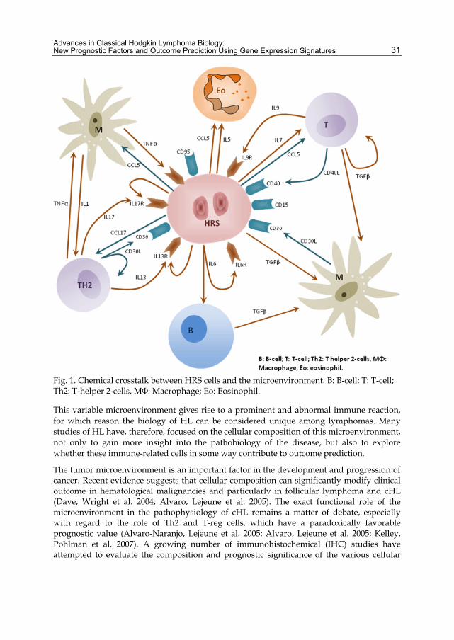
There are various lines of evidence suggesting an active role of this “microenvironment” in tumor biology through bidirectional signaling between the HRS and inflammatory cells that promotes proliferation and survival of neoplastic cells (Kuppers 2009). A complex network of cytokines and cell-contact-mediated interactions between tumor and inflammatory cells are thought to be involved and may rescue HRS cells from the proapoptotic state arising from their characteristic BCR deficiency by providing alternative survival signals (Figure 1).
Advances in Classical Hodgkin Lymphoma Biology: New Prognostic Factors and Outcome Prediction Using Gene Expression Signatures 31
Fig. 1. Chemical crosstalk between HRS cells and the microenvironment. B: B-cell; T: T-cell; Th2: T-helper 2-cells, MΦ: Macrophage; Eo: Eosinophil.
This variable microenvironment gives rise to a prominent and abnormal immune reaction, for which reason the biology of HL can be considered unique among lymphomas. Many studies of HL have, therefore, focused on the cellular composition of this microenvironment, not only to gain more insight into the pathobiology of the disease, but also to explore whether these immune-related cells in some way contribute to outcome prediction.
The tumor microenvironment is an important factor in the development and progression of cancer. Recent evidence suggests that cellular composition can significantly modify clinical outcome in hematological malignancies and particularly in follicular lymphoma and cHL (Dave, Wright et al. 2004; Alvaro, Lejeune et al. 2005). The exact functional role of the microenvironment in the pathophysiology of cHL remains a matter of debate, especially with regard to the role of Th2 and T-reg cells, which have a paradoxically favorable prognostic value (Alvaro-Naranjo, Lejeune et al. 2005; Alvaro, Lejeune et al. 2005; Kelley, Pohlman et al. 2007). A growing number of immunohistochemical (IHC) studies have attempted to evaluate the composition and prognostic significance of the various cellular
Hodgkin's Lymphoma 32
components, with particular interest in tumor-infiltrating lymphocytes. More recently, GEP studies based on DNA microarrays have demonstrated their ability to define more accurately the interaction pathways of HRS cells with nonmalignant reactive and stromal cells in cHL lymphoma tissues (see below). Therefore, a complete understanding of cHL, requires that both cellular components be considered.
3. Prognostic markers: Limitations and new challenges
3.1 The conventional international prognostic score: Current limitations
cHL is assumed to be a curable tumor, but a substantial proportion of patients with advanced disease do not respond favorably to the current standard chemotherapy regimens based on adriamycin (Canellos, Anderson et al. 1992). Historically, outcome in cHL patients has been predicted using standard clinical variables such as bulk disease, patient age, number of nodal sites and erythrocyte sedimentation rate. The most widely used and reproducible prognostic score is based on clinical and analytical parameters that make up the International Prognostic Score (IPS). This consists of seven clinical variables, including serum albumin less than 4 g/dl, hemoglobin less than 10.5 g/dl, male gender, age 45 or above, stage IV disease, white blood cell count at least 15,000/mm3, and absolute lymphocyte count less than 600/mm3.
This clinical measure, although widely accepted, still does not accurately identify, at diagnosis, a significant fraction of patients with very poor prognosis (Gobbi, Zinzani et al. 2001) and it´s not applicable to patients with early-stage HL (Hasenclever and Diehl 1998). Additionally, other study noted that the IPS score had no significant prognostic influence in modern series of patients and that only age and stage IV disease remained significant in multivariate analyses (Sanchez-Espiridion, Montalban et al. 2010). A re-evaluation of the IPS, together with the identification of new biological prognostic factors in larger populations of patients, is necessary to improve patients stratification into clearly defined risk groups for which risk-adapted therapeutic strategies are suitable.
3.2 Risk stratification and new approaches to clinical guidance: Biological prognostic factors and markers in cHL
HL is generally a curable tumor; so, research currently focuses on identifying patients with a low probability of cure who might benefit from more novel and/or intensive treatment strategies, and those with a better prognosis who are suitable for less toxic therapies. To establish a more rational risk-adapted treatment strategy the identification of truly high-risk populations requires supplementation with molecular markers. In this context, biological factors may serve as prognostic markers and provide novel targets for cHL therapy. Moreover, the addition of biological markers to the already recognized clinical prognostic factors (including those of the IPS) may improve patient risk stratification.
This chapter summarizes new insights into cHL biology in a clinical context and the role that new technologies have played in identifying biological markers with future prognostic and therapeutic applications.
Many markers have recently been identified as prognostic factors in HL, including surface receptors, intracellular proteins, cytokines, and genetic abnormalities (amplifications,
Advances in Classical Hodgkin Lymphoma Biology: New Prognostic Factors and Outcome Prediction Using Gene Expression Signatures 33
deletions, epigenetic silencing), or alterations in miRNA in HRS cells and surrounding inflammatory cells. Technical advances such as microarray-based gene and miRNA expression profiling, RT-PCR platforms, comparative genomic hybridization (CGH), SNP arrays, microdissection and IHC studies have led to new discoveries (see below). Their integration with the classical parameters will further improve the understanding of the disease and the management of patients.
Gene expression profiling studies in cHL
It has been difficult to identify biological markers for several reasons, including the scarcity of large and confirmatory prospective trials, the lack of reproducibility and feasibility of assays and the negligible improvement upon already used clinical risk factors (IPS or others). The characteristic histological heterogeneity and scarcity of tumoral cells have also hindered molecular studies in HL. However, several studies have analyzed GEP in whole tissue sections and identified specific transcriptional patterns in the tumoral cells and the non-tumoral microenvironment. Additionally, other studies in HL have demonstrated differential gene expression patterns between HRS cells and normal mature B-cells, providing vital clues for understanding the pathogenesis of the disease (Kuppers, Klein et al. 2003).
DNA microarrays are currently the best developed and most widely used high-throughput molecular technique for identifying biological markers that rely on array-based gene expression analyses using whole-tissue sections. IHC staining has been used as a validation tool, but the technique has inherent limitations, such as the poor reproducibility of the results generated.
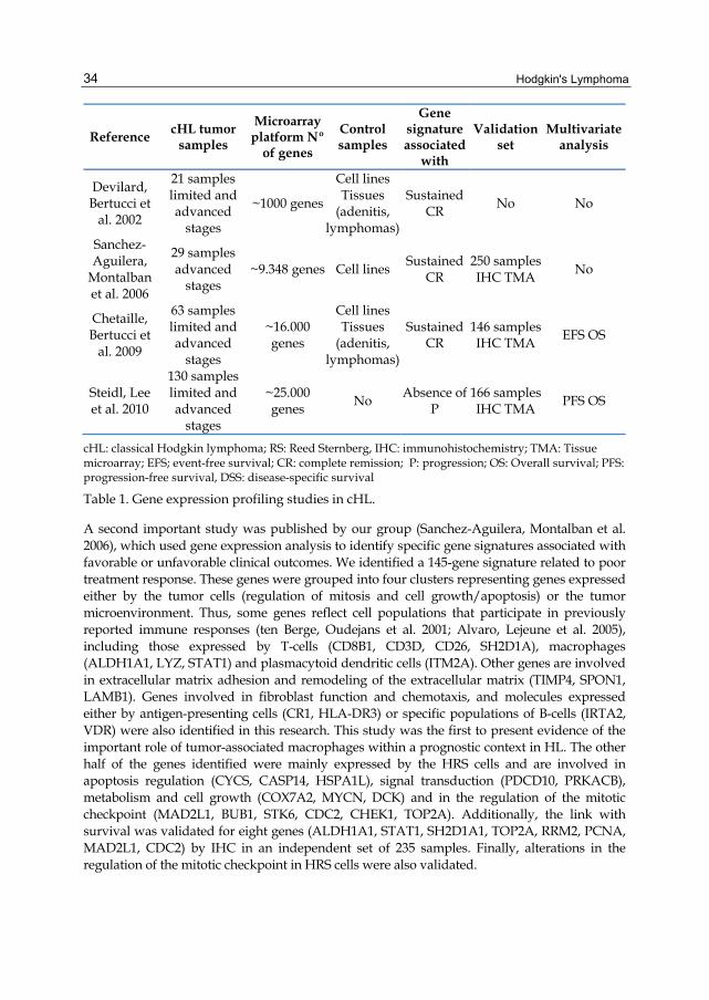
Gene expression studies in cHL initially focused on elucidating HRS cell-specific genes and the biological mechanisms underlying cHL pathogenesis (Kuppers, Klein et al. 2003). Subsequently, the relationship between cell microenvironment and HRS cells with clinical outcome was investigated (Devilard, Bertucci et al. 2002; Sanchez-Aguilera, Montalban et al. 2006; Chetaille, Bertucci et al. 2009) (Table 1). These studies reported prognostic signatures related to tumor HRS and microenvironment cells, identifying genes that, although not completely overlapping, did suggest the involvement of the same cellular subpopulations (macrophages, B-cells, T-cells) as those affecting clinical outcome (for a recent review (Bertucci, Chetaille et al. 2011)).
The first study to suggest the existence of correlations between gene expression profiles, mainly microenvironment-related genes, and prognosis was that of Devilard et al. (Devilard, Bertucci et al. 2002). They measured the mRNA expression levels of around 1,000 genes in 34 benign and malignant lymphoid samples including 21 cHL tissue samples and identified three molecular groups of HL. Samples from patients with bad outcomes clustered together, whereas the two other groups contained most of the good outcome cases. These good outcome cases overexpressed genes involved in apoptosis induction and cell signaling (including cytokines), while the bad outcome samples were characterized by upregulation of genes involved in fibroblast activation, angiogenesis, extracellular matrix remodeling, cell proliferation and downregulation of tumor suppressor genes. However, the number of genes and samples analyzed was small, thus preventing any robust conclusions being drawn.
Hodgkin's Lymphoma 34
Reference cHL tumor
samples
Microarray platform Nº
of genes
Control samples
Gene signature associated
with
Validation set
Multivariate analysis
Devilard, Bertucci et
al. 2002
21 samples limited and advanced
stages
~1000 genes
Cell lines Tissues
(adenitis, lymphomas)
Sustained CR
No No
Sanchez-Aguilera,
Montalban et al. 2006
29 samples advanced
stages ~9.348 genes Cell lines
Sustained CR
250 samples IHC TMA
No
Chetaille, Bertucci et
al. 2009
63 samples limited and advanced
stages
~16.000 genes
Cell lines Tissues
(adenitis, lymphomas)
Sustained CR
146 samples IHC TMA
EFS OS
Steidl, Lee et al. 2010
130 samples limited and advanced
stages
~25.000 genes
No Absence of
P 166 samples IHC TMA
PFS OS
cHL: classical Hodgkin lymphoma; RS: Reed Sternberg, IHC: immunohistochemistry; TMA: Tissue microarray; EFS; event-free survival; CR: complete remission; P: progression; OS: Overall survival; PFS: progression-free survival, DSS: disease-specific survival
Table 1. Gene expression profiling studies in cHL.
A second important study was published by our group (Sanchez-Aguilera, Montalban et al. 2006), which used gene expression analysis to identify specific gene signatures associated with favorable or unfavorable clinical outcomes. We identified a 145-gene signature related to poor treatment response. These genes were grouped into four clusters representing genes expressed either by the tumor cells (regulation of mitosis and cell growth/apoptosis) or the tumor microenvironment. Thus, some genes reflect cell populations that participate in previously reported immune responses (ten Berge, Oudejans et al. 2001; Alvaro, Lejeune et al. 2005), including those expressed by T-cells (CD8B1, CD3D, CD26, SH2D1A), macrophages (ALDH1A1, LYZ, STAT1) and plasmacytoid dendritic cells (ITM2A). Other genes are involved in extracellular matrix adhesion and remodeling of the extracellular matrix (TIMP4, SPON1, LAMB1). Genes involved in fibroblast function and chemotaxis, and molecules expressed either by antigen-presenting cells (CR1, HLA-DR3) or specific populations of B-cells (IRTA2, VDR) were also identified in this research. This study was the first to present evidence of the important role of tumor-associated macrophages within a prognostic context in HL. The other half of the genes identified were mainly expressed by the HRS cells and are involved in apoptosis regulation (CYCS, CASP14, HSPA1L), signal transduction (PDCD10, PRKACB), metabolism and cell growth (COX7A2, MYCN, DCK) and in the regulation of the mitotic checkpoint (MAD2L1, BUB1, STK6, CDC2, CHEK1, TOP2A). Additionally, the link with survival was validated for eight genes (ALDH1A1, STAT1, SH2D1A1, TOP2A, RRM2, PCNA, MAD2L1, CDC2) by IHC in an independent set of 235 samples. Finally, alterations in the regulation of the mitotic checkpoint in HRS cells were also validated.
Advances in Classical Hodgkin Lymphoma Biology: New Prognostic Factors and Outcome Prediction Using Gene Expression Signatures 35
In another study, Chetaille et al. (Chetaille, Bertucci et al. 2009) analyzed gene expression data from a set of 63 cHL tissue samples with both early and advanced stages, including control tissues (lymphadenitis) and cHL cell lines and cHL samples from patients. Available clinical data allowed the comparison of profiles from 41 cases with favorable outcome and 21 with unfavorable outcome, identifying a set of 450 genes associated with survival. The authors found that expression of genes associated with B-cells and apoptosis was associated with good prognosis, whereas genes involved in stroma remodeling were correlated with poor prognosis. An independent set of 146 samples was analyzed by IHC, enabling the prognostic significance of some of the identified markers including FOXP3, CD20 and TOP2A to be validated. These proved to have a remarkably strong prognostic value for BCL11A in the series. Additionally, a gene signature associated with the EBV status of samples was identified. This seems to be characteristic of the Th1 antiviral immune response, thus providing new clues about the differences between EBV+ and EBV– cases. The influence of EBV infections on HL and the knowledge obtained from GEP is discussed in more detail below.
Finally, a study performed by Steidl et al. (Steidl, Lee et al. 2010) reported the results of a retrospective study using IHC and GEP. Initial analyses of RNA isolated from 130 frozen specimens identified a GEP significantly associated with primary treatment failure that included increased gene signatures related to tumor-infiltrating macrophages, angiogenic cells, adipocytes, HRS cells, with overexpression of matrix metallopeptidases (like MMP1) and underexpression of gene signatures related to germinal center B-cells. A validation study using IHC showed that low percentages of CD68+ macrophages (< 5 percent) correlated with higher progression-free survival (PFS), with a 100 percent disease-specific survival (DSS) rate observed for patients with limited-stage disease (stages IA and IIA) and absence of tumor-infiltrating macrophages.
These data are consistent with a previous observation made by Chetaille et al. (Chetaille, Bertucci et al. 2009), which showed reduced presence of background small B-cells (≤ 10 percent) and overexpression of MMP1 associated with shorter survival. Interestingly, in multivariate analyses, CD68+ macrophages remained an independent adverse prognostic factor for DSS, outperforming the IPS.
RT-PCR models
It is now feasible to apply multigenic predictive molecular tests in a routine setting by using alternative techniques, like quantitative RT-PCR. An assay for patients with advanced cHL has recently been described that incorporates a limited number of genes and pathways from both tumor and microenvironment cell components, designed for routine formalin-fixed paraffin-embedded (FFPE) samples (Sanchez-Espiridion, Montalban et al. 2010). Thirty genes were chosen on the basis of the findings of two previous studies (Sanchez-Aguilera, Montalban et al. 2006; Sanchez-Espiridion, Sanchez-Aguilera et al. 2009) and their deployment in 282 FFPE samples enabled a prognostic model to be developed. The model included 11 genes from four functional pathways (apoptosis, cell cycle regulation, macrophage activation and interferon regulatory factor-IRF4). This 4-cluster/11-gene signature was included in a final algorithm (defined as molecular risk score, MRS) that was able to identify subgroups of patients with different probabilities of treatment failure.
Hodgkin's Lymphoma 36
One of the well established clinical variables (stage IV) was also included in the score, thus combining the main molecular characteristics of the treatment response-related tumors and tumor burden estimates into a single scoring system. The multivariate Cox model indicated that most patients with stage IV cHL and with a high MRS (≥ 0.3) will have a very poor outcome, with a 5-year FFS probability of 24.3 percent and overall survival (OS) probability of 76.3 percent (Sanchez-Espiridion, Montalban et al. 2010). This assay represented another interesting, although preliminary, attempt to translate gene profiling results into routine practice, providing a tool for further exploring and refining the already available biological and clinical prognostic markers.
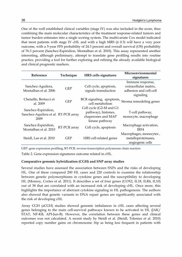
Reference Technique HRS cells signatures Microenvironmental
signatures
Sanchez-Aguilera, Montalban et al. 2006
GEP Cell cycle, apoptosis, signals transduction
Immune response, extracellular matrix,
adhesion and cell-cell signaling
Chetaille, Bertucci et al. 2009
GEP BCR signaling, apoptosis,
cell metabolism Stroma remodeling genes
Sanchez-Espiridion, Sanchez-Aguilera et al.
2009 RT-PCR array
Cell cycle (G2-M and G1 pathway), histones,
chaperones and MAP kinase pathway
T-cell pathway, monocyte, macrophage
Sanchez-Espiridion, Montalban et al. 2010
RT-PCR array Cell cycle, apoptosis Macrophage activation,
IRF4
Steidl, Lee et al. 2010 GEP HRS cell-related genes Macrophages, monocytes ,
metalloproteinases, angiogenic cells
GEP: gene expression profiling; RT-PCR: reverse-transcription polymerase chain reaction
Table 2. Gene expression signatures outcome related in cHL.
Comparative genomic hybridization (CGH) and SNP array studies
Several studies have assessed the association between SNPs and the risks of developing HL. One of these compared 200 HL cases and 220 controls to examine the relationship between genetic polymorphisms in cytokine genes and the susceptibility to developing HL (Monroy, Cortes et al. 2011). It describes a set of four genes (COX2, IL18, ILR4, IL10) out of 38 that are correlated with an increased risk of developing cHL. Once more, this highlights the importance of aberrant cytokine signaling in HL pathogenesis. The authors also showed that genetic variants in DNA repair genes are significantly associated with the risk of developing cHL
Array CGH (aCGH) studies showed genomic imbalances in cHL cases affecting several genes belonging to the main cell-survival pathways known to be activated in HL (JAK/ STAT, NF-KB, API-Jun-B). However, the correlation between these genes and clinical outcomes was not calculated. A recent study by Steidl et al. (Steidl, Telenius et al. 2010) reported copy number gains on chromosome 16p as being less frequent in patients with

Advances in Classical Hodgkin Lymphoma Biology: New Prognostic Factors and Outcome Prediction Using Gene Expression Signatures 37
primary refractory disease and strongly associated with prognosis after ABVD chemotherapy. These results could be related to the presence of the multidrug resistant gene ABCC1 located in this chromosomal region (16p13.11). In addition, frequent losses on 4q27 (IL2/IL21 genes) and 17p12, and gains on 19q13.3 (BCL3/RELB) have also been described as being associated with ABVD resistance (Slovak, Bedell et al. 2011).
Immunohistochemical markers and gene expression studies
Some of the markers identified by GEP have been identified as being of potential use, leading to their clinical application by IHC techniques, especially those associated with the complex reactive inflammatory infiltrate.
BCL2. The relationship between BCL2 expression and patient outcome in HL is a controversial issue with studies reporting BCL2 expression to be independently associated with reduced FFS in addition to other clinical prognostic variables (age over 45 years, stage IV disease, low albumin and elevated lactate dehydrogenase) (Rassidakis, Medeiros et al. 2002), whereas others have not demonstrated the same relationship (Montalban, Garcia et al. 2004).
p21 and p53. Similarly to BCL2, other markers like p21 and p53 have been associated with treatment outcomes (Sup, Alemany et al. 2005).
MMP11 is a marker of matrix metallopeptidases expressed by tumor-associated macrophages, found to be associated with treatment failure by GEP and IHC (Steidl, Lee et al. 2010) with good discriminatory power with respect to patient outcome. More than 1% MMP11 staining by IHC predicted reduced PFS survival in both univariate and multivariate analyses.
CD20 encodes a B-lymphocyte cell-surface antigen and has been evaluated by IHC in several studies (Chetaille, Bertucci et al. 2009; Steidl, Lee et al. 2010), which revealed a positive association of an increased number of tumor-infiltrating CD20+ cells with both prolonged PFS and DFS. Similar findings, in which high numbers of CD20+ cells present in the inflammatory background were correlated with improved event-free survival (EFS) and OS were reported by Chetaille (Chetaille, Bertucci et al. 2009). No correlation with the number of CD20+ HRS cells and patient outcome was found in Steidl’s study (Steidl, Lee et al. 2010) or another one of 598 patients (Rassidakis, Medeiros et al. 2002). However, a small study performed by Portlock et al. (Portlock, Donnelly et al. 2004) in a set of 248 samples showed that both time to treatment and OS were significantly lower in patients treated with ABVD with CD20+ cells.
Tzankov et al. found that the presence of more than 10 percent of CD20+ HRS cells was associated with worse patient outcomes in patients treated from 1974 to 1980 whereas there was no effect on FFS in patients treated from 1981 to 1999 (Tzankov, Krugmann et al. 2003). Thus, the prognostic relevance of CD20 in HL might depend on other factors, such as the therapeutic modality and the criteria used to define CD20 positivity. As is the case with many other identified markers, these results need further validation in prospective studies to determine their reproducibility.
CD68 is a macrophage marker encoding a transmembrane glycoprotein that is strongly expressed by human monocytes and tissue macrophages. It is a member of the
Hodgkin's Lymphoma 38
lysosomal/endosomal-associated membrane glycoprotein (LAMP) family. In the aforementioned study (Sinha, Adhikari et al. 2001) the initial finding of a tumor infiltrating macrophage gene signature associated with treatment failure prompted an additional IHC validation of CD68 expression relation with clinical outcome. In univariate analysis, the presence of a large number of infiltrating CD68+ cells was correlated with reduced PFS and DFS. Remarkably, this marker is better than the IPS in predicting DFS in multivariate analysis (p=0.003 vs. p=0.030) and as such is currently a matter under further investigation in many studies that are attempting to validate its predictive value (Kamper, Bendix et al. 2011).
Similar findings correlating the presence of tumor-infiltrating macrophages with adverse patient outcomes have also been described in follicular lymphoma (Dave, Wright et al. 2004; Farinha, Masoudi et al. 2005), suggesting that interactions between the malignant lymphoma cells and the microenvironment are critical to the pathogenesis and progression of these diseases.
Despite these results, the use of CD68 alone as a marker is currently a matter of interest, with several studies having produced contradictory results (Azambuja, Natkunam et al. 2011). Nevertheless, there is no doubt about their prognostic value in cHL.
CD163 is a monocyte/macrophage-specific protein in the scavenger receptor cysteine-rich superfamily that appears to be involved in anti-inflammatory functions believed to be predominantly associated with M2 (macrophages). This marker has been found to be correlated with outcome in several studies in a similar manner to CD68 (Steidl, Lee et al. 2010; Kamper, Bendix et al. 2011). However, it is considered to be more specific and thus more accurate for macrophage identification than CD68.
T-cells. The presence of T-cells in the inflammatory infiltrate also seems to influence patient outcomes and several studies have examined the ratio of regulatory T-cells (CD4+, CD25+ and FOXP3 expression) and cytotoxic T-cells (TIA-1+ or granzyme B expression) in relationship to OS and DFS rates (Alvaro, Lejeune et al. 2005). The ratio of regulatory FOXP3+ T-cells to cytotoxic T/NK (natural killer cells) with granzyme B expression independently predicts patient survival.
Other markers. A variety of other prognostic markers have been described in HL. Increased serum levels of soluble CD30 (Zanotti, Trolese et al. 2002), IL-10, BAFF, TNFα, TARC and VEGF have all been associated with adverse patient outcomes (Casasnovas, Mounier et al. 2007). In several studies, combining the CD30 level with the IPS gave improved predictive values (Ma, Ding et al. 2009; Renna, Mocciaro et al. 2009).
3.3 Influence of EBV
Finally, it is essential to mention studies concerning the role of EBV infection in cHL. Before the use of GEP by DNA arrays, only a few differences had been found between the EBV + and EBV- microenvironments, and these had not been comprehensively characterized.
GEP studies looking for EBV-induced alterations showed that Epstein-Barr nuclear antigen 1 (EBNA1) upregulated the expression of a chemokine (CCL20) in EBV+ cHL cells, ultimately leading to increased chemotaxis of T-regulatory cells (Baumforth, Birgersdotter et
Advances in Classical Hodgkin Lymphoma Biology: New Prognostic Factors and Outcome Prediction Using Gene Expression Signatures 39
al. 2008). Additionally, Chetaille et al. (Chetaille, Bertucci et al. 2009) demonstrated that EBV+ and EBV- cHL cases can be clearly separated from each other by a robust gene signature involving innate immunity and antiviral responses in EBV+ cases. Thus, EBV+ cases overexpressed antiviral genes such as NS1BP, PLSCR1 and OAS, together with TLR8 receptor and MDA5 helicase, which are both, involved in the recognition of viruses and mediate innate immunity against viral infection. The molecular profile of EBV+ tumors (overexpression of IFNG, CXCL9, CXL10, and CXCL11/ITAC) also provided evidence of intratumoral Th1 activity in EBV+ cHL cases, which might be orchestrated by IFNG. Nonetheless, EBV+ patients did not have a better outcome, suggesting that the intratumoral immune reaction described is inadequate for eliminating tumor cells.
ITK (IL-2–inducible T-cell kinase) deficiency, a novel primary immunodeficiency disease characterized by severe EBV associated immune dysregulation commonly progresses to cHL with a variable treatment response (Rezaei, Hedayat et al. 2011). Additionally, three cases EBV-positive HL associated to this deficiency were subsequently found to harbor a homozygous nonsense mutation in the ITK gene (Stepensky, Weintraub et al. 2011). Additionally, the study of Steidl et al. (Steidl, Lee et al. 2010) showed that CD68 and CD163 markers were both associated with latent EBV infection of the malignant cells, thus highlighting the potential prognostic implications of EBV infection. Despite these results, the impact of EBV infection on clinical outcome remains unclear; more studies are needed to elucidate its exact role in cHL patients.
4. miRNAs: A new approach to elucidating the biology of Hodgkin’s lymphoma
Several studies have demonstrated the role of miRNA deregulation in cancer pathogenesis and treatment response in several tumor models. Others confirm that miRNAs are good biomarkers for cancer diagnosis and prognosis, including hematological malignancies. miRNAs are small, noncoding RNAs that regulate the expression of multiple mRNAs by binding to the 3’ untranslated region (UTR) of their target genes. miRNAs are the best characterized members of a family of noncoding RNAs (ncRNAs) that are now the object of intense investigation. They function by targeting mRNA and inducing its degradation or inhibiting its translation. A number of studies suggest that miRNAs may regulate around 30% of the genome. As a consequence they are implicated in almost every cellular process, and, in fact miRNA expression has been shown to be tissue-specific, playing key roles in the control of the various biological processes such as differentiation, proliferation and others, all of which are involved in cancer pathogenesis. The deregulated expression of miRNA in tumoral samples compared with their normal counterparts, and their frequent genomic location in fragile chromosomal sites, also points to their role in the development of cancer. In fact, specific miRNA signatures have been identified for some tumor types, and they are thought to have tumor suppressor or oncogenic properties (for which reason they are termed “oncomirs”), as well as metastasis regulatory functions. miRNAs are encoded by intronic or intergenic regions. They are first transcribed to pri-miRNA, then processed to pre-miRNA by the Drosha complex, and subsequently exported to the cytoplasm, where the RNase Dicer cleavages them to generate a double strand molecule of 21-25 nucleotides. One of the two chains is then joined to the RNA-induced silencing complex (RISC), which
Hodgkin's Lymphoma 40
interacts with Argonaute proteins and binds to 3’UTR on target mRNAs, inducing their degradation and/or blocking translation (Esquela-Kerscher and Slack 2006).
Altered miRNA expression has a role in hematopoietic malignancies. One of the first pieces of evidence of the role of miRNAs in cancer came from a study of B-cell chronic lymphocytic leukemia where miR-15 and miR-16 were downregulated due to hemi- or homozygous chromosomal deletion at 13q14 (Calin, Dumitru et al. 2002). As mentioned before, several miRNA loci reside on chromosomal fragile sites, which are frequently altered in cancer. These include the miR-15a/16 cluster, which is also deleted in pituitary adenomas, miR-143 and miR-145, which are located in the 5q33 that is deleted in lung cancer (Calin and Croce 2006), and the miR-17-92 cluster, which is frequently amplified in B-cell lymphomas (Ota, Tagawa et al. 2004; He, Thomson et al. 2005) and lung cancers (Hayashita, Osada et al. 2005). Expression of specific miRNAs is implicated in B-cell and hematological differentiation, and regulates germinal center formation (Kluiver, Kroesen et al. 2006; Thai, Calado et al. 2007; Zhou, Wang et al. 2007). It is possible that miRNA losses and gains have a significant role in MCL by regulating CCND1 mRNA expression (Chen, Bemis et al. 2008). Another study in MCL showed that the most essential pathways and genes in MCL pathogenesis are potentially targeted simultaneously by multiple miRNAs, suggesting that transcriptional regulation by miRNAs in MCL is the result of the concurrent deregulation of multiple miRNAs with related targets (Di Lisio, Gomez-Lopez et al. 2010).
Furthermore, they may constitute markers of differentiation stage, malignant transformation, sensitivity or resistance to specific drugs. Their capacity as prognostic markers has also been demonstrated in lymphomas. Montes-Moreno et al. have developed a model based on miRNA expression, valid for FFPE samples, that uses RT-PCR to predict OS and PFS in chemoimmunotherapy-treated DLBCL patients, improving the prediction just based on clinical variables (Montes-Moreno, Martinez et al. 2011).
Some attempts have been made to elucidate specific miRNA signatures for the characteristic HRS cells and their microenvironment. Preliminary work showed that miRNA losses and gains may explain some of the biological and clinical features of different lymphoma types, and recent analyses of miRNA expression in cHL-derived cell-lines and tumors have also demonstrated deregulated expression of some miRNAs in this malignancy. Overexpression of BIC/miR-155 has been found in cHL cell lines and samples by qRT-PCR analysis (Kluiver, Poppema et al. 2005) but not in other NHL types, except some PMBL and DLBCL samples. miRNA profiling of cHL-derived cell lines performed with qRT-PCR and microarrays has revealed specifically expressed miRNAs that include miR-17-92 cluster members, miR-16, miR-21, miR-24 and miR-155 (Gibcus, Tan et al. 2009) and a significant downregulation of miR-150 in cHL. The authors also demonstrated the targeting of IKBKE by miR-155, among others, in cHL cell lines; thus, miR-155 expression in HL might represent an attempt to reduce NFkappaB activity in this lymphoma. In primary tumors, comparison of miRNA profiles of microdissected HRS cells from cHL patients, four common cell lines (HDLM2, L540, KMH2 and L1236) and CD77+ germinal center B-cells (used as normal counterparts) yielded a distinct cHL signature of 12 overexpressed and three underexpressed miRNAs (Van Vlierberghe, De Weer et al. 2009) including some of the previously described miRNAs, including miR-21 and miR-155. Additionally, a 25-specific miRNA signature differentiating cHL and reactive lymphoid nodes was presented by
Advances in Classical Hodgkin Lymphoma Biology: New Prognostic Factors and Outcome Prediction Using Gene Expression Signatures 41
Navarro et al. (Navarro, Gaya et al. 2008). When studying cell lines, some of the miRNAs in the samples and cell lines were found to be different, suggesting that this could be due to the microenvironment or to the immortalization process of the cell lines. They also identified some miRNAs (miR-96, miR-128a, miR-128b) whose expression was related to the presence of EBV, although no prognostic implication of the identified specific EBV-related miRNAs was demonstrated.
These results imply that a small subset of miRNAs may define tumor entities better than microarray expression data from thousands of messenger RNAs, and suggest that miRNAs may play an important role in the biology of cHL, and may be useful in the development of therapies targeting miRNAs in tumor cells.
Although cHL-specific miRNA signatures have been proposed, their potential prognostic role remains unclear. There are few observations that link miRNA deregulation and clinical characteristics of the patients. Overexpression of miR-328 has been found in advanced cHL stages (III-IV stages) (Navarro, Gaya et al. 2008), and lower miR-138 levels. However, the potential prognostic role of miRNA signatures in cHL has not yet been sufficiently well investigated in larger series of patients.
Taken together, these findings suggest that miRNAs may play significant roles in HL pathogenesis, and could help to explain its biology, being additionally useful for patient risk stratification and development of new therapeutic approaches. Thus, the relevance of miRNA expression in cHL and its putative value for outcome prediction is worthy of further investigation.
Recently, our group has identified specific profiles from tumor cells and their non-tumoral microenvironment by miRNA expression studies. Initial analyses suggest that clinical outcome can be predicted by models that integrate miRNA signatures. These preliminary findings suggest a possible role for miR-21 and miR-30d as potential targets to overcome treatment resistance in cHL (Sánchez-Espiridión B, Figueroa V et al. 2011).
5. Perspectives
An increasing body of knowledge in cHL pathogenesis, and the complex relationship between the tumoral HRS cells and the microenvironment has been built up from gene and miRNA profiling studies. The correlations that these studies identify, spotlighting tumor-infiltrating macrophages in classic HL, reinforce the critical roles of microenvironmental and immunomodulatory interactions between RS and non-tumor cells, stromal, cytokine, and membrane molecules in disease pathobiology and treatment responsiveness. As we have seen in this chapter, these studies have led to remarkable discoveries such as the relevance of B-cells and macrophages in patient outcome, and to the identification of several new prognostic markers. All these results, including the optimization of new technologies such as RT-PCR in FFPE tissues and miRNA profiling, are clinically promising. However, further validation in large and independent series of patients is needed for them to be included as part of clinical routine. There is no doubt that their integration with results generated by other modern high-throughput molecular analyses (such as proteomics) will further improve our understanding of the disease and the patient management.
Hodgkin's Lymphoma 42
6. Acknowledgments
We would like to acknowledge all members of the former CNIO’s Lymphoma Group. We also would thank AM Martin for their thoughtful discussion and help with the figure. This work was supported by grant PI10/00621 from FIS, Spain.
7. References
Alizadeh, A. A., M. B. Eisen, et al. (2000). "Distinct types of diffuse large B-cell lymphoma identified by gene expression profiling." Nature 403(6769): 503-11.
Alizadeh, A. A., D. T. Ross, et al. (2001). "Towards a novel classification of human malignancies based on gene expression patterns." J Pathol 195(1): 41-52.
Alvaro-Naranjo, T., M. Lejeune, et al. (2005). "Tumor-infiltrating cells as a prognostic factor in Hodgkin's lymphoma: a quantitative tissue microarray study in a large retrospective cohort of 267 patients." Leuk Lymphoma 46(11): 1581-91.
Alvaro, T., M. Lejeune, et al. (2005). "Outcome in Hodgkin's lymphoma can be predicted from the presence of accompanying cytotoxic and regulatory T cells." Clin Cancer Res 11(4): 1467-73.
Azambuja, D., Y. Natkunam, et al. (2011). "Lack of association of tumor-associated macrophages with clinical outcome in patients with classical Hodgkin's lymphoma." Ann Oncol.
Baumforth, K. R., A. Birgersdotter, et al. (2008). "Expression of the Epstein-Barr virus-encoded Epstein-Barr virus nuclear antigen 1 in Hodgkin's lymphoma cells mediates Up-regulation of CCL20 and the migration of regulatory T cells." Am J Pathol 173(1): 195-204.
Bertucci, F., B. Chetaille, et al. (2011). "Gene Expression Profiling for In Silico Microdissection of Hodgkin's Lymphoma Microenvironment and Identification of Prognostic Features." Adv Hematol 2011: 485310.
Brune, V., E. Tiacci, et al. (2008). "Origin and pathogenesis of nodular lymphocyte-predominant Hodgkin lymphoma as revealed by global gene expression analysis." J Exp Med 205(10): 2251-68.
Calin, G. A. and C. M. Croce (2006). "Genomics of chronic lymphocytic leukemia microRNAs as new players with clinical significance." Semin Oncol 33(2): 167-73.
Calin, G. A., C. D. Dumitru, et al. (2002). "Frequent deletions and down-regulation of micro- RNA genes miR15 and miR16 at 13q14 in chronic lymphocytic leukemia." Proc Natl Acad Sci U S A 99(24): 15524-9.
Canellos, G. P., J. R. Anderson, et al. (1992). "Chemotherapy of advanced Hodgkin's disease with MOPP, ABVD, or MOPP alternating with ABVD." N Engl J Med 327(21): 1478-84.
Casasnovas, R. O., N. Mounier, et al. (2007). "Plasma cytokine and soluble receptor signature predicts outcome of patients with classical Hodgkin's lymphoma: a study from the Groupe d'Etude des Lymphomes de l'Adulte." J Clin Oncol 25(13): 1732-40.
Chen, R. W., L. T. Bemis, et al. (2008). "Truncation in CCND1 mRNA alters miR-16-1 regulation in mantle cell lymphoma." Blood 112(3): 822-9.
Advances in Classical Hodgkin Lymphoma Biology: New Prognostic Factors and Outcome Prediction Using Gene Expression Signatures 43
Chetaille, B., F. Bertucci, et al. (2009). "Molecular profiling of classical Hodgkin lymphoma tissues uncovers variations in the tumor microenvironment and correlations with EBV infection and outcome." Blood 113(12): 2765-3775.
Dave, S. S., K. Fu, et al. (2006). "Molecular diagnosis of Burkitt's lymphoma." N Engl J Med 354(23): 2431-42.
Dave, S. S., G. Wright, et al. (2004). "Prediction of survival in follicular lymphoma based on molecular features of tumor-infiltrating immune cells." N Engl J Med 351(21): 2159-69.
Devilard, E., F. Bertucci, et al. (2002). "Gene expression profiling defines molecular subtypes of classical Hodgkin's disease." Oncogene 21(19): 3095-102.
Di Lisio, L., G. Gomez-Lopez, et al. (2010). "Mantle cell lymphoma: transcriptional regulation by microRNAs." Leukemia 24(7): 1335-42.
Esquela-Kerscher, A. and F. J. Slack (2006). "Oncomirs - microRNAs with a role in cancer." Nat Rev Cancer 6(4): 259-69.
Farinha, P., H. Masoudi, et al. (2005). "Analysis of multiple biomarkers shows that lymphoma-associated macrophage (LAM) content is an independent predictor of survival in follicular lymphoma (FL)." Blood 106(6): 2169-74.
Gibcus, J. H., L. P. Tan, et al. (2009). "Hodgkin lymphoma cell lines are characterized by a specific miRNA expression profile." Neoplasia 11(2): 167-76.
Gobbi, P. G., P. L. Zinzani, et al. (2001). "Comparison of prognostic models in patients with advanced Hodgkin disease. Promising results from integration of the best three systems." Cancer 91(8): 1467-78.
Hasenclever, D. and V. Diehl (1998). "A prognostic score for advanced Hodgkin's disease. International Prognostic Factors Project on Advanced Hodgkin's Disease." N Engl J Med 339(21): 1506-14.
Hayashita, Y., H. Osada, et al. (2005). "A polycistronic microRNA cluster, miR-17-92, is overexpressed in human lung cancers and enhances cell proliferation." Cancer Res 65(21): 9628-32.
He, L., J. M. Thomson, et al. (2005). "A microRNA polycistron as a potential human oncogene." Nature 435(7043): 828-33.
Herreros, B., A. Sanchez-Aguilera, et al. (2008). "Lymphoma microenvironment: culprit or innocent?" Leukemia 22(1): 49-58.
Hummel, M., S. Bentink, et al. (2006). "A biologic definition of Burkitt's lymphoma from transcriptional and genomic profiling." N Engl J Med 354(23): 2419-30.
Kamper, P., K. Bendix, et al. (2011). "Tumor-infiltrating macrophages correlate with adverse prognosis and Epstein-Barr virus status in classical Hodgkin's lymphoma." Haematologica 96(2): 269-76.
Kapatai, G. and P. Murray (2007). "Contribution of the Epstein Barr virus to the molecular pathogenesis of Hodgkin lymphoma." J Clin Pathol 60(12): 1342-9.
Kelley, T. W., B. Pohlman, et al. (2007). "The ratio of FOXP3+ regulatory T cells to granzyme B+ cytotoxic T/NK cells predicts prognosis in classical Hodgkin lymphoma and is independent of bcl-2 and MAL expression." Am J Clin Pathol 128(6): 958-65.
Khan, G. (2006). "Epstein-Barr virus, cytokines, and inflammation: a cocktail for the pathogenesis of Hodgkin's lymphoma?" Exp Hematol 34(4): 399-406.
Hodgkin's Lymphoma 44
Kluiver, J., B. J. Kroesen, et al. (2006). "The role of microRNAs in normal hematopoiesis and hematopoietic malignancies." Leukemia 20(11): 1931-6.
Kluiver, J., S. Poppema, et al. (2005). "BIC and miR-155 are highly expressed in Hodgkin, primary mediastinal and diffuse large B cell lymphomas." J Pathol 207(2): 243-9.
Kuppers, R. (2009). "The biology of Hodgkin's lymphoma." Nat Rev Cancer 9(1): 15-27. Kuppers, R., U. Klein, et al. (2003). "Identification of Hodgkin and Reed-Sternberg cell-
specific genes by gene expression profiling." J Clin Invest 111(4): 529-37. Ma, M. M., J. W. Ding, et al. (2009). "Odd-even width effect on persistent current in zigzag
hexagonal graphene rings." Nanoscale 1(3): 387-90. Margalit, O., R. Somech, et al. (2005). "Microarray-based gene expression profiling of
hematologic malignancies: basic concepts and clinical applications." Blood Rev 19(4): 223-34.
Monroy, C. M., A. C. Cortes, et al. (2011). "Hodgkin disease risk: role of genetic polymorphisms and gene-gene interactions in inflammation pathway genes." Mol Carcinog 50(1): 36-46.
Montalban, C., J. F. Garcia, et al. (2004). "Influence of biologic markers on the outcome of Hodgkin's lymphoma: a study by the Spanish Hodgkin's Lymphoma Study Group." J Clin Oncol 22(9): 1664-73.
Montes-Moreno, S., N. Martinez, et al. (2011). "miRNA expression in diffuse large B-cell lymphoma treated with chemoimmunotherapy." Blood 118(4): 1034-40.
Navarro, A., A. Gaya, et al. (2008). "MicroRNA expression profiling in classic Hodgkin lymphoma." Blood 111(5): 2825-32.
Ota, A., H. Tagawa, et al. (2004). "Identification and characterization of a novel gene, C13orf25, as a target for 13q31-q32 amplification in malignant lymphoma." Cancer Res 64(9): 3087-95.
Portlock, C. S., G. B. Donnelly, et al. (2004). "Adverse prognostic significance of CD20 positive Reed-Sternberg cells in classical Hodgkin's disease." Br J Haematol 125(6): 701-8.
Rassidakis, G. Z., L. J. Medeiros, et al. (2002). "BCL-2 expression in Hodgkin and Reed-Sternberg cells of classical Hodgkin disease predicts a poorer prognosis in patients treated with ABVD or equivalent regimens." Blood 100(12): 3935-41.
Rassidakis, G. Z., L. J. Medeiros, et al. (2002). "CD20 expression in Hodgkin and Reed-Sternberg cells of classical Hodgkin's disease: associations with presenting features and clinical outcome." J Clin Oncol 20(5): 1278-87.
Renna, S., F. Mocciaro, et al. (2009). "Is splenectomy a treatment option for aseptic abscesses in patients with Crohn's disease?" Eur J Gastroenterol Hepatol 21(11): 1314-6.
Rezaei, N., M. Hedayat, et al. (2011). "Primary immunodeficiency diseases associated with increased susceptibility to viral infections and malignancies." J Allergy Clin Immunol 127(6): 1329-41 e2; quiz 1342-3.
Rosenwald, A., G. Wright, et al. (2003). "Molecular diagnosis of primary mediastinal B cell lymphoma identifies a clinically favorable subgroup of diffuse large B cell lymphoma related to Hodgkin lymphoma." J Exp Med 198(6): 851-62.

Advances in Classical Hodgkin Lymphoma Biology: New Prognostic Factors and Outcome Prediction Using Gene Expression Signatures 45
Rosenwald, A., G. Wright, et al. (2003). "The proliferation gene expression signature is a quantitative integrator of oncogenic events that predicts survival in mantle cell lymphoma." Cancer Cell 3(2): 185-97.
Sanchez-Aguilera, A., C. Montalban, et al. (2006). "Tumor microenvironment and mitotic checkpoint are key factors in the outcome of classic Hodgkin lymphoma." Blood 108(2): 662-8.
Sánchez-Espiridión B, Figueroa V, et al. (2011). "MicroRNAS in advanced classical Hodgkin lymphoma:signatures with prognostic significance " Ann Oncol 22((Suppl 4)): iv179-iv182.
Sanchez-Espiridion, B., C. Montalban, et al. (2010). "A molecular risk score based on 4 functional pathways for advanced classical Hodgkin lymphoma." Blood 116(8): e12-7.
Sanchez-Espiridion, B., A. Sanchez-Aguilera, et al. (2009). "A TaqMan low-density array to predict outcome in advanced Hodgkin's lymphoma using paraffin-embedded samples." Clin Cancer Res 15(4): 1367-75.
Sinha, N., N. Adhikari, et al. (2001). "Effect of endosulfan during fetal gonadal differentiation on spermatogenesis in rats." Environ Toxicol Pharmacol 10(1-2): 29-32.
Skinnider, B. F. and T. W. Mak (2002). "The role of cytokines in classical Hodgkin lymphoma." Blood 99(12): 4283-97.
Slovak, M. L., V. Bedell, et al. (2011). "Molecular karyotypes of Hodgkin and Reed-Sternberg cells at disease onset reveal distinct copy number alterations in chemosensitive versus refractory Hodgkin lymphoma." Clin Cancer Res 17(10): 3443-54.
Steidl, C., T. Lee, et al. (2010). "Tumor-associated macrophages and survival in classic Hodgkin's lymphoma." N Engl J Med 362(10): 875-85.
Steidl, C., A. Telenius, et al. (2010). "Genome-wide copy number analysis of Hodgkin Reed-Sternberg cells identifies recurrent imbalances with correlations to treatment outcome." Blood 116(3): 418-27.
Stepensky, P., M. Weintraub, et al. (2011). "IL-2-inducible T-cell kinase deficiency: clinical presentation and therapeutic approach." Haematologica 96(3): 472-6.
Sup, S. J., C. A. Alemany, et al. (2005). "Expression of bcl-2 in classical Hodgkin's lymphoma: an independent predictor of poor outcome." J Clin Oncol 23(16): 3773-9.
ten Berge, R. L., J. J. Oudejans, et al. (2001). "Percentage of activated cytotoxic T-lymphocytes in anaplastic large cell lymphoma and Hodgkin's disease: an independent biological prognostic marker." Leukemia 15(3): 458-64.
Thai, T. H., D. P. Calado, et al. (2007). "Regulation of the germinal center response by microRNA-155." Science 316(5824): 604-8.
Tzankov, A., J. Krugmann, et al. (2003). "Prognostic significance of CD20 expression in classical Hodgkin lymphoma: a clinicopathological study of 119 cases." Clin Cancer Res 9(4): 1381-6.
Van Vlierberghe, P., A. De Weer, et al. (2009). "Comparison of miRNA profiles of microdissected Hodgkin/Reed-Sternberg cells and Hodgkin cell lines versus CD77+ B-cells reveals a distinct subset of differentially expressed miRNAs." Br J Haematol 147(5): 686-90.

Hodgkin's Lymphoma 46
Zanotti, R., A. Trolese, et al. (2002). "Serum levels of soluble CD30 improve International Prognostic Score in predicting the outcome of advanced Hodgkin's lymphoma." Ann Oncol 13(12): 1908-14.
Zhou, B., S. Wang, et al. (2007). "miR-150, a microRNA expressed in mature B and T cells, blocks early B cell development when expressed prematurely." Proc Natl Acad Sci U S A 104(17): 7080-5.