progastrin expression in mammalian pancreas
TRANSCRIPT

Proc. Nadl. Acad. Sci. USAVol. 87, pp. 298-302, January 1990Biochemistry
Progastrin expression in mammalian pancreas(biosynthesis/gastrinoma/peptide hormone/radioimmunoassay)
LINDA BARDRAM*, LINDA HILSTEDt, AND JENS F. REHFELDttUniversity Departments of *Surgical Gastroenterology and tClinical Chemistry, Rigshospitalet, DK-2100 Copenhagen, Denmark
Communicated by Diter von Wettstein, August 31, 1989 (receivedfor review May 11, 1989)
ABSTRACT Expression and processing of progastrin wereexamined in fetal, neonatal, and adult pancreatic tissue fromfive mammalian species (cat, dog, man, pig, and rat). A libraryof sensitive, sequence-specific immunoassays for progastrinand its products was used to monitor extractions and chroma-tography before and after cleavage with processing-like en-zymes. The results showed that progastrin and its products areexpressed in the pancreas of all species in total concentrationsvarying from 0.3 to 58.9 pmol/g of tissue (medians). Thedegree of processing was age- and species-dependent. In com-parison with adult pancreatic tissue the fetal or neonatalpancreas processed a higher fraction to bioactive, C-terminallyamidated gastrin. Nevertheless, the pancreatic processing wasalways less complete than that of the adult antral mucosa. Themoderate level of expression and the attenuated processing inthe adult pancreas contribute to explain previous failures todetect gastrin in normal pancreatic tissue. Our results indicatethat gastrin-producing tumors in the pancreas are not ectopic,but arise from cells that normally express the gastrin gene.
Gastrin stimulates the secretion of gastric acid and growth ofthe fundic mucosa (1). Accordingly, gastrin-producing tu-mors (gastrinomas) induce hypersecretion of acid with en-suing duodenal ulcer disease and fundic mucosal hypertro-phy, i.e., the Zollinger-Ellison syndrome. By far most gas-trinomas originate in the pancreas, although gastrin normallyis synthesized in the antroduodenal mucosa. Gastrinomas arenevertheless a frequent type of endocrine tumor in the humanpancreas (2, 3) and have been found also in dogs (4, 5).The pancreatic origin of gastrinomas has been an enigma.
Insulinomas, glucagonomas, somatostatinomas, pancreatic-polypeptide tumors (PPomas), and vasoactive intestinal poly-peptide tumors (VIPomas) all have cellular counterparts thatexpress the corresponding hormone in the normal pancreas. Incontrast, attempts to detect gastrin in the normal pancreashave so far either failed (6-10) or resulted in the misidentifi-cation of somatostatin cells as gastrin cells (11-13). The onlyreproducible exception has been the apparently transientgastrin synthesis in the fetal and neonatal rat pancreas (14-18).
Deduction of the sequence of mammalian preprogastrins(19-21) has paved the way for development of radioimmu-noassays for progastrin and its processing intermediates(22-25). Such radioimmunoassays have proved useful forstudies ofgastrin biosynthesis in tissues known to express thegastrin gene (24-29). The observation of expression butattenuated processing of progastrin in the normal pituitary(29) suggested that progastrin might be expressed also in thenormal pancreas, and hence explain the gastrinoma enigma(30). This hypothesis has now been examined.
NOMENCLATUREMammalian preprogastrins comprise 101-104 amino acidresidues (19-21). They have an N-terminal signal sequence,
a spacer sequence, the sequence containing the major bio-active forms of gastrin (i.e., gastrin-34 and gastrin-17), andfinally a C-terminal flanking peptide. The decisive amino acidsequence during the biosynthetic maturation is shown in Fig.1. In the present study we have distinguished three majorcategories of preprogastrin products: (i) progastrins, whichare defined as products extended beyond glycine at the Cterminus; (ii) glycine-extended intermediates, which are fur-ther processed progastrins that constitute the immediateprecursors of the mature, bioactive gastrins; (iii) amidatedgastrins (the bioactive gastrins), in which the C-terminalphenylalanine residue is amidated using glycine as the amidedonor. Each category contains a number of peptides ofdifferent chain length. For instance, the amidated gastrinscomprise component I, gastrin-34, and gastrin-17 [in bothtyrosine-sulfated and nonsulfated forms (Fig. 1)].
MATERIALS AND METHODSTissue Sampling. Pancreatic tissue was collected from five
mammalian species. First, an extensive collection was car-ried out in rats at ages that allowed us to follow the ontoge-netic development. We also collected antral, duodenal, jeju-nal, and ileal rat tissues in order to compare pancreaticgastrin expression with that of the gastrointestinal tract. Inaddition, muscle tissue was sampled for control. Wistar ratswere reared with their mothers in temperature- and light-controlled cages. They were allowed to suckle ad libitum andhad free access to food and water. When 21 days old, theyoung rats were separated from their mothers to separatecages with free access to food and water. Rats aged 14, 21,30, and 100 days were fasted overnight with free access towater. Pups 7 days old, pups 4 days old, and newborn ratswere separated from their mothers on the morning of thedissection day. Fetuses at 19 days of gestation were obtainedfrom anaesthetized animals by hysterotomy. Before dissec-tion, the newborn rats were decapitated whereas the olderrats were anaesthetized with pentobarbital (60 mg/kg, intra-peritoneally). A midline incision was made and the pancreas,duodenum (whole duodenal wall), and gastric antrum (cor-responding to the distal third of the stomach) were excised.The antrum and duodenum were then opened, and, afterremoval of the luminal content, the tissue was immediatelyfrozen in liquid N2. For each age, 20 rats were dissected, andthe tissue was divided into five pools of four pieces each.
After the results of the rat study were obtained, we contin-ued the collection of pancreatic tissue from newborn (i.e., <3hr postpartum) or fetal and adult cats, mongrel dogs, and pigsin numbers shown in Table 1. The pancreas was removed fromappropriately anaesthetized animals after midline incision andthe tissues were immediately frozen in liquid N2. Humanpancreatic tissue was obtained from human fetuses in connec-tion with abortion induced on sociomedical indications. Theages of the fetuses ranged from 15 to 23 weeks. Moreover,adult pancreatic tissue was obtained from human organ do-
fTo whom reprint requests should be addressed.
298
The publication costs of this article were defrayed in part by page chargepayment. This article must therefore be hereby marked "advertisement"in accordance with 18 U.S.C. §1734 solely to indicate this fact.

Proc. Natl. Acad. Sci. USA 87 (1990) 299
101PREPROGASTRIN [S_ ,
f-Trp-Met-Asp-Phe-Gly-Arg-Arg-Ser-
LIttrypsin-likeI endopeptidase
PROGASTRINS
(Ab. 3208 after trypsui-carboxypeptidase B.)ccarboxypeptidase B
like exopeptidase
GLYCINE-EXTENDED
INTERMEDIATES
(Ab. 3208)
CARBOXYAMIDATEDGASTRINS
(Ab. 2604)
~~GlyGly
-Gly
CONH2CONH2CONH2
-Trp-Met-Asp-Phe-Gly
t peptidylglycine a-amida-I ting monooxygenase
-Trp-Met-Asp-Phe-NH2 _ 'Active site'
FIG. 1. Mammalian preprogastrin and core steps of its maturation. Bioactive gastrins require a C-terminal Trp-Met-Asp-Phe-NH2 sequence,which can be obtained by cleavage with a trypsin-like endopeptidase, a carboxypeptidase B-like exopeptidase, and finally peptidylglycinea-amidating monooxygenase. The maturation process can be monitored, as indicated, by antibodies (Ab.) 2604 (which bind only C-terminallyamidated gastrins), Ab. 3208 (which bind only glycine-extended gastrins), and by Ab. 3208 after trypsin and carboxypeptidase B cleavage (furtherC-terminally extended progastrins). G17, gastrin-17.
nors, from whom corpus and cauda pancreatis was used astransplant by the segmental duct-occlusion method, leavingparts of the caput pancreatis for our examination. The use ofhuman tissue was approved by the local ethics committee.
Extraction of Tissue. While frozen the tissue was cut intopieces weighing a few milligrams, immersed in boiling water (5ml/g of tissue, pH 6.5) for 20 min, homogenized, and centri-fuged at 10,000 x g for 30 min. The supernatant was decanted(the neutral extract) and the pellet was reextracted by additionof0.5 M CH3COOH (5 ml/g of tissue), rehomogenized, left for20 min at room temperature, and then centrifuged as above.Radioimmunoassays. The extracts were assayed for pro-
gastrins, glycine-extended intermediates, and amidated gas-trins (Fig. 1) by two types of radioimmunoassay. For mea-surement of glycine-extended gastrins we used antiserum3208, which was raised in rabbit 3208 against a tetradecapep-tide corresponding to sequence 5-17 of human gastrin-17extended with glycine (22). Gastrin-(5-17)-Gly was monoio-dinated by mild chloramine-T treatment and purified onAE-cellulose (21). Gastrin-(5-17)-Gly (custom-synthesizedby Cambridge Research Biochemicals, Harston, U.K.) wasalso used as standard. Antiserum 3208 crossreacts <0.2%with amidated gastrin-17 (22). The antiserum recognizesgastrin-34-Gly with 80% the potency with which it recognizesgastrin-17-Gly (27). For measurement of amidated gastrinswe used antiserum 2604, raised in rabbit 2604 against humangastrin-(2-17) (31). Human gastrin-17 was monoiodinated bya mild chloramine-T technique (32). For standard we usednonsulfated human gastrin-17 (Imperial Chemical Indus-tries). Antiserum 2604 crossreacts <0.2% with gastrin-(5-17)-Gly compared to gastrin-17 (22). It binds sulfated andnonsulfated gastrin with equimolar potency. It also bindsgastrin-34 and gastrin-17 with almost equimolar potency (33).For human pancreatic tissue we also used antiserum 8017
in control measurements. Antiserum 8017 was raised againstthe N-terminal tridecapeptide sequence of human gastrin-17and is specific for human gastrin (34). Used in combinationwith tryptic cleavage, it recognizes unprocessed or partlyprocessed human progastrin (23).The detection limits for measurement of progastrins, gly-
cine-extended gastrins, and amidated gastrins in solutionhave been detailed elsewhere (22, 23, 31, 34). For measure-
ment in tissue extracts, the detection limit is 0.1 pmol/g (wetweight) with our methods of extraction.Enzymatic Treatment. Determination of gastrins extended
beyond the C-terminal glycine residue [gastrin-Gly-Arg-Arg(-R)] was made by measurement ofglycine-extended gastrinsbefore and after sequential treatment of the extracts withtrypsin (LS 0003741, Worthington) and carboxypeptidase B(batch 103 233, Boehringer Mannheim), as described (refs. 22and 27 and Fig. 1). To evaluate the size of these precursors,fractions from gel filtration chromatography of undigestedextract were assayed before and after treatment with trypsinand carboxypeptidase B (27).Chromatography. Extracts were applied to 10 x 1000 mm
columns of Sephadex G-50 superfine (Pharmacia) and elutedwith 0.125 M NH4 HCO3 at a flow rate of 4 ml/hr. Fractionsof 1.0 ml were collected. The columns were calibrated with1251-labeled albumin and 22NaCl for indication of void andtotal volume, respectively, with synthetic human gastrin-17,gastrin-17-Gly, and gastrin-34 (Cambridge Research Bio-chemicals), and with natural amidated gastrins and glycine-extended gastrins as well as progastrins from antral extractsof some of the corresponding animal species. To ensureidentity of pancreatic gastrin, extracts of feline pancreaticand antral mucosal tissue were characterized by anion-exchange chromatography on calibrated 10 x 150 mm What-man AE-41 cellulose columns, using a linear gradient from0.05 to 0.20M NH4HCO3 (pH 8.2, 300 ml). The flow rate was33 ml/hr and fractions of 2.5 ml were collected. Finally,samples of feline pancreatic heptadecapeptide-like gastrinisolated first by gel filtration chromatography were charac-terized by reverse-phase HPLC on a calibrated 4 x 240 mmC4 Vydac column, using a gradient from 0.1% CF3COOH inwater to 0.06% CF3COOH in CH3CN (as indicated in Fig. 5)at room temperature. The flow rate was 1 ml/min andfractions of 0.5 ml were collected.
RESULTSIn all species examined the pancreatic tissue containedprogastrin, glycine-extended intermediates, and amidated(i.e., bioactive) gastrins (Table 1). Generally, the fetal and
Biochemistry: Bardram et A
300 Biochemistry: Bardram et al.
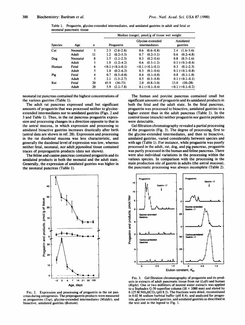
Table 1. Progastrin, glycine-extended intermediates, and amidated gastrins in adult and fetal orneonatal pancreatic tissue
Median (range), pmol/g of tissue wet weight
Glycine-extended AmidatedSpecies Age n Progastrin intermediates gastrins
Cat Neonatal 3 2.3 (2.0-2.8) 0.6 (0.6-0.8) 2.4 (1.6-3.6)Adult 11 1.2 (0.2-3.3) 0.7 (0.2-3.1) 0.6 (0.2-4.8)
Dog Neonatal 8 1.5 (1.1-2.3) 0.3 (0.2-0.6) 0.8 (0.3-1.6)Adult 3 1.9 (1.2-4.2) 0.6 (0.3-1.2) 0.1 (<0.1-0.4)
Human Fetal 6 <0.1 (<0.1-0.1) <0.1 (<0.1-0.1) 0.3 (0.1-2.3)Adult 5 1.0 (0.2-6.2) 0.3 (0.1-0.6) 0.1 (<0.1-0.8)
Pig Fetal 4 0.7 (0.5-0.8) 0.4 (0.1-0.8) 0.8 (0.1-1.8)Adult 5 2.1 (1.3-2.7) 0.5 (0.3-0.8) 0.1 (<0.1-0.1)
Rat Fetal 20 43.9 (34-73) 2.0 (0.8-3.0) 13.0 (10-28)Adult 20 5.9 (2.1-7.8) 0.1 (<0.1-0.4) <0.1 (<0.1-0.2)
neonatal rat pancreas contained the highest concentrations ofthe various gastrins (Table 1).The adult rat pancreas expressed small but significant
amounts of progastrin that was processed neither to glycine-extended intermediates nor to amidated gastrins (Figs. 2 and3 and Table 1). Thus, in the rat pancreas progastrin expres-sion and processing changes in a direction opposite to that inthe antral mucosa, in which expression and processing toamidated bioactive gastrins increases drastically after birth(antral data are shown in ref. 28). Expression and processingin the rat duodenal mucosa was less characteristic, andgenerally the duodenal level of expression was low, whereasneither fetal, neonatal, nor adult jejunoileal tissue containedtraces of preprogastrin products (data not shown).The feline and canine pancreas contained progastrin and its
amidated products in both the neonatal and the adult state.Generally, the expression of amidated gastrins was higher inthe neonatal pancreas (Table 1).
The human and porcine pancreas contained small butsignificant amounts of progastrin and its amidated products inboth the fetal and the adult state. In the fetal pancreas,ptogastrin was processed to bioactive, amidated gastrins to ahigher extent than in the adult pancreas (Table 1). In thecontrol tissue (muscle) neither progastrin nor gastrin peptideswere detectable.Gel filtration chromatography revealed a partial processing
of the progastrin (Fig. 3). The degree of processing, first tothe glycine-extended intermediates, and then to bioactive,amidated gastrins, varied considerably between species andwith age (Table 1). For instance, while progastrin was poorlyprocessed in the adult, rat, dog, and pig pancreas, progastrinwas partly processed in the human and feline pancreas. Therewere also individual variations in the processing within thevarious species. In comparison with the processing in themain production site of gastrin in adults (the antral mucosa),the pancreatic processing was always incomplete (Table 2).
15
10 .
:z- 5
E
0L05
~0
I..
CLC
coQ_
a.
-2 0 4 7 14 21 30 100
Age, days
FIG.' 2. Expression and processing of progastrin in the rat pan-
creas during ontogenesis. The preprogastrin products were measuredas progastrins (Top), glycine-extended intermediates (Middle), andbioactive, amidated gastrins (Bottom).
0 0.5 1.0 0
Elution constant, Kav0.5 1.0
FIG. 3. Gel filtration chromatography of progastrin and its prod-ucts in extracts of adult pancreatic tissue from rat (Left) and human(Right). One or two milliliters of neutral water extracts was appliedto a Sephadex G-50 superfine column (10 x 1000 mm) and eluted by0.125 M NH4HCO3 (pH 8.2). The fractions were dried, reconstitutedin 0.02 M sodium barbital buffer (pH 8.4), and analyzed for progas-trin, glycine-extended gastrins, and amidated gastrins as described inthe text and in the legend to Fig. 1.
Progastrins
Gastrin- 17
Glycine-extendedGastrins
a---xo . 1
CarboxyamidatedGastrns
5
O . _ .
ProgastrinsGastrin- 17
125
oJGiycne-extendedGastrin- 17
25
0
Carboxyamidate50 Gastrin- 17
25
0 V.
Proc. Natl. Acad. Sci. USA 87 (1990)
L

Proc. Natl. Acad. Sci. USA 87 (1990) 301
Table 2. Progastrin, glycine-extended intermediates, and amidated gastrins in adult pancreatic and antralmucosal tissue
Median (range), pmol/g of tissue wet weight
Glycine-extended Amidated gastrins,Species Tissue n Progastrin intermediates Amidated gastrins % of total*Human Pancreas 5 1.0 (0.2-6.2) 0.3 (0.1-0.6) 0.1 (<0.1-0.8) 7.3
Antrum 11 130 (37-281) 39 (7-80) 2500 (580-5700) 93.7Pig Pancreas 5 2.1 (1.3-2.7) 0.5 (0.3-0.8) 0.1 (<0.1-0.1) 3.7
Antrum 7 160 (66-297) 152 (93-446) 3400 (1300-6100) 91.6Rat Pancreas 20 5.9 (2.1-7.8) 0.1 (<0.1-0.4) <0.1 (<0.1-0.2) <1.5
Antrum 20 53 (22-108) 112 (94-319) 1240 (1000-2200) 88.3
*Percent of total progastrin product.
The identity of the bioactive pancreatic gastrin as a hep-tadecapeptide corresponding to sulfated gastrin-17 from an-tral tissue was ensured by identical elution in three differentchromatographic systems fractionating according to size,charge, and hydrophobicity (Figs. 3-5).
DISCUSSIONThis study has shown that the normal mammalian pancreasexpresses the gastrin gene at peptide level. Since expressionwas detectable in all the five species examined, it is appar-ently a general phenomenon.Except for fetal and neonatal rats (Fig. 2), the pancreatic
expression is lower than that of the antrum (Fig. 2 and Table2). It is, however, possible that a low level of expression hasphysiological significance. Hence, the concentration of lo-cally released gastrin may be sufficient to affect the growthof neighboring pancreatic cells, as illustrated by the islet cellhyperplasia (35-37) and hyperinsulinemia (38) accompanyinghypergastrinemia in man. In this context, the completesulfation of pancreatic gastrin (refs. 16 and 39 and Fig. 4)seems expedient, since the pancreozymic effect of gastrin isincreased by sulfation (40, 41). In fetal and neonatal pancre-atic tissues of all five species, progastrin was processed toamidated bioactive gastrin to a higher degree than in adultpancreatic tissue (Table 1). This observation supports the
Gastrin- 17
(ns) (s)( 150 0.3
E0.
.7 1005 ,& ,-- 0.2 0~100 -
0) Z
V 50 , - 0.1E --
0 0
0 50 100
Elution volume, ml
FIG. 4. Ion-exchange chromatography of amidated gastrins fromfeline pancreatic tissue. A pooled sample of heptadecapeptide gastrinisolated first by gel filtration chromatography was applied to acalibrated AE-41 cellulose column (10 x 150 mm) and eluted at 20°Cin fractions of 2.5 ml with a gradient (---) from 0.05 to 0.2 MNH4HCO3. Elution was monitored by a radioimmunoassay specificfor amidated gastrin (o) using antiserum 2604, which binds sulfated(s) and nonsulfated (ns) gastrins equally.
idea of a trophic role of pancreatic gastrin in the early stagesof development.
In the neonatal rat pancreas, it has been shown that thegastrin gene is expressed in cells having an ultrastructuralsimilarity to the antral gastrin cells (14). The pancreatic gastrincells are disseminated in both exocrine tissue and islets (14, 42,43). Such distribution also supports the hypothesis of local,paracrine effects of pancreatic gastrin. Early suggestionsabout gastrin synthesis in the delta cells of the pancreatic islets(11, 13) have not been confirmed with highly specific antisera(8, 9, 14). Instead, it is now well known that the delta cellsproduce somatostatin (2, 37). In the adult cat, dog, and pig, afew gastrin cells of a structure similar to those of the neonatalrat pancreas have been seen scattered in the exocrine pancreas(ref. 44 and F. Sundler, personal communication). In thehuman pancreas, the cellular origin of progastrin and gastrinremains to be determined. On balance, however, it looks as ifthe gastrin gene is expressed in a few endocrine cells through-out the mammalian pancreas, perhaps including a subfractionof the beta cells (45).Due to the preferential occurrence of gastrinomas in the
pancreas, much attention has been given to the possibleexpression of gastrin in the normal pancreas. However,previous studies have either been unable to detect gastrin inthe adult pancreas (6-10, 14-17) or (as mentioned) mistak-enly suggested that somatostatin cells produce gastrin (11,13). There are several factors that may explain the discrep-
L-
U)CU0)
E
:L
150oj
100
50k
0
80
60
z0
40 I0
20
0
0 20 40 60
Time, min
FIG. 5. Reverse-phase HPLC of amidated gastrins from felinepancreatic tissue. A pooled sample of heptadecapeptide gastrinisolated first by gel filtration chromatography was applied to a
calibrated C4 Vydac column (4 x 240 mm) and eluted at 20°C infractions of 0.5 ml (per 0.5 min) with a gradient ( ---) from 0.1%CF3COOH in water to 0.06% CF3COOH in CH3CN. After thefractions were dried and reconstituted in the initial volume of assaybuffer, elution was monitored by radioimmunoassay (e) as in Fig. 4.
Gastrin- 1 7(S)
r -1
III
'I.,I
1,II
I
I,I
IIL-
0 0 0 0 0 s j
Biochemistry: Bardram et al.

302 Biochemistry: Bardram et al.
ancy between earlier results (6-10) and the present data.First, the state of the tissue and the methods of extraction arecrucial. Autopsy material cannot be used, because postmor-tem degradation is extensive in pancreas. Since the pancreasis so rich in proteolytic enzymes, complete denaturation ofthe enzymes during extraction is also necessary. On the otherhand, acid/ethanol, used to extract insulin, glucagon, andsomatostatin, cannot be used for pancreatic gastrins, whichdissolve poorly in ethanol. Instead, careful boiling in waterand homogenization are essential.
Second, the sensitivity and specificity of the assays used todetect gastrin expression are crucial. Gastrin bioassays (6),mRNA detection methods such as Northern and dot blots (17,45, 46), and some radioimmunoassays are too insensitive todetect the low levels of expression in all but the neonatal ratpancreas (17). Moreover, polyclonal gastrin antisera raisedagainst antral extracts contain somatostatin antibodies thatmay give misleading immunocytochemical results (11, 13).Finally, the fact that much of the expressed polypeptide isprogastrin, especially in the adult pancreas, explains whypreviously used bioassays and radioimmunoassays are lessuseful, since they detect only the mature, amidated gastrins,which are present in only minute amounts. Thus, onlysensitive progastrin assays (22, 23) can detect the expressionin adult rat, pig, dog, and human pancreas (Table 1).
Incomplete, attenuated processing of progastrin has alsobeen found in normal pituitary corticotrophs (29). We do notknow the biological significance of such expression (47).Silent expression may, however, reach significance by trans-formation of normal cells to tumor cells in which prohor-mones are processed to mature, bioactive peptides (48).Hence, the present results suggest that the expression ofgastrin in pancreatic gastrinomas does not develop by ectopicdedifferentiation of transcription mechanisms, but rather byacceleration of already existing translational and posttrans-lational processing mechanisms.
The skillful technical assistance of Susanne Hummelgaard, AliceLieth, and Bente Rotb0ll is gratefully acknowledged. We thank thehead of faculty animal house, Dr. H. J. Skovgaard, and his staff forexpert assistance. The study was supported by grants from theDanish Medical Research Council, the Danish Cancer Union, and theEinar Willumsen Foundation.
1. Gregory, R. A. (1988) in Advances ofMetabolic Disorders, ed.Mutt, V. (Academic, San Diego), Vol. 11, pp. 163-198.
2. Creutzfeldt, W. (1977) in The Diabetic Pancreas, eds. Volk,B. W. & Wellmann, K. F. (Plenum, New York), pp. 551-590.
3. Jacobsen, O., Bardram, L. & Rehfeld, J. F. (1986) Scand. J.Clin. Lab. Invest. 46, 423-427.
4. Straus, E., Johnson, G. F. & Yalow, R. S. (1977) Gastroen-terology 72, 380-381.
5. Happd, R. P., Gaag, I. v. d., Lamers, C. B. H. W., Tooren-brug, J. V., Rehfeld, J. F. & Larsson, L.-I. (1980) Vet. Pathol.17, 177-186.
6. Hallenbeck, G. A., Code, C. F. & McIlrath, D. C. (1963)Gastroenterology 44, 627-630.
7. Creutzfeldt, W., Arnold, R., Creutzfeldt, C., Feurle, G. &Ketterer, H. (1971) Eur. J. Clin. Invest. 1, 461-479.
8. Lotstra, F., Loo, W. v. d. & Gepts, W. (1974) Diabetology 1,291-302.
9. Track, N. S., Creutzfeldt, C., Litzenberger, J., Neuhoff, C.,Arnold, R. & Creutzfeldt, W. (1979) Digestion 19, 292-306.
10. Andersen, B. N., Abramovich, D., Brand, S. J., Petersen, B.& Rehfeld, J. F. (1985) Regul. Pept. 10, 329-338.
11. Lomsky, R., Langr, F. & Vortel, V. (1969) Nature (London)223, 618-619.
12. Greider, M. H. & McGuigan, J. E. (1971) Diabetes 20, 389-396.
13. Erlandsen, S. L., Hegre, 0. D., Parsons, J. A., McEvoy,R. C. & Elde, R. P. (1976) J. Histochem. Cytochem. 24,883-897.
14. Larsson, L.-I., Rehfeld, J. F., Sundler, F. & Hakanson, R.(1976) Nature (London) 262, 609-610.
15. Majumdar, A. D. N. & Rehfeld, J. F. (1983) Digestion 27,165-173.
16. Brand, S. J., Andersen, B. N. & Rehfeld, J. F. (1984) Nature(London) 309, 456-458.
17. Brand, S. J. & Fuller, P. J. (1988) J. Biol. Chem. 263, 5342-5347.
18. Braaten, J. T., Greider, M. H., McGuigan, J. E. & Mintz,D. H. (1976) Endocrinology 79, 1049-1053.
19. Yoo, 0. J., Powell, C. T. & Agarwal, K. L. (1982) Proc. Natl.Acad. Sci. USA 79, 1049-1053.
20. Boel, E., Vuust, J., Norris, F., Norris, K., Wind, A., Rehfeld,J. F. & Marcker, K. A. (1984) Proc. Natl. Acad. Sci. USA 80,2866-2869.
21. Fuller, P., Stone, D. L. & Brand, S. J. (1987) Mol. Endocrinol.1, 306-311.
22. Hilsted, L. & Rehfeld, J. F. (1986) Anal. Biochem. 152, 119-126.
23. Bardram, L. & Rehfeld, J. F. (1988) Anal. Biochem. 175,537-543.
24. Pauwels, S., Desmond, H., Dimaline, R. & Dockray, G. J.(1986) J. Clin. Invest. 77, 376-381.
25. Sugano, K., Aponte, G. W. & Yamada, T. (1985) J. Biol.Chem. 260, 11724-11729.
26. Del Valle, J., Sugano, K. & Yamada, T. (1987) Gastroenter-ology 92, 1908-1912.
27. Hilsted, L. & Rehfeld, J. F. (1987) J. Biol. Chem. 262, 16953-16957.
28. Hilsted, L., Bardram, L. & Rehfeld, J. F. (1988) Biochem. J.255, 397-402.
29. Rehfeld, J. F. (1986) J. Biol. Chem. 261, 5841-5847.30. Rehfeld, J. F., Bardram, L., Cantor, P., Hilsted, L. &
Mogensen, N. (1986) in Highlights on Endocrinology, Proceed-ings of the First International European Congress on Endocri-nology, eds. Christiansen, C. & Riis, B. J. (Norhaven Bogtryk-keri, Viborg, Denmark), pp. 251-257.
31. Rehfeld, J. F., Stadil, F. & Rubin, B. (1972) Scand. J. Clin.Lab. Invest. 30, 221-232.
32. Stadil, F. & Rehfeld, J. F. (1972) Scand. J. Clin. Lab. Invest.30, 361-368.
33. Rehfeld, J. F., de Magistris, L. & Andersen, B. N. (1981)Regul. Pept. 2, 333-334.
34. Bardram, L. & Rehfeld, J. F. (1989) Scand. J. Clin. Lab.Invest. 49, 173-182.
35. Larsson, L.-I., Ljungberg, O., Sundler, F., Hakansson, R.,Svensson, S. O., Rehfeld, J. F., Stadil, F. & Holst, J. J. (1973)Virchows Arch. A 306, 305-314.
36. Creutzfeldt, W., Arnold, R., Creutzfeldt, C. & Track, N. (1976)Hum. Pathol. 6, 47-76.
37. Larsson, L.-I. (1977) Virchows Arch. A 376, 209-219.38. Rehfeld, J. F. (1976) J. Clin. Invest. 58, 41-49.39. Cantor, P., Andersen, B. N. & Rehfeld, J. F. (1986) FEBS Lett.
195, 272-274.40. Dockray, G. J. (1973) Q. J. Exp. Physiol. 58, 163-169.41. Jensen, S. L., Rehfeld, J. F., Holst, J. J., Fahrenkrug, J.,
Nielsen, 0. V. & Schaffalitzky de Muckadell, 0. B. (1980) Am.J. Physiol. 238, E186-E192.
42. Onolfo, J. P. & Lehy, T. (1987) Anat. Rec. 218, 416-425.43. Fujii, S. (1979) Arch. Histol. Jpn. 42, 467-479.44. Rehfeld, J. F., Larsson, L.-I., Goltermann, N., Schwartz,
T. W., Holst, J. J., Jensen, S. L. & Morley, J. S. (1980) Nature(London) 284, 33-39.
45. Brand, S. J. & Wang, T. (1988) J. Biol. Chem. 263, 16597-16602.
46. Powell, C. T., Ney, C., Aran, P. & Agarwal, K. (1985) NucleicAcids Res. 13, 7299-7305.
47. Rehfeld, J. F. (1988) J. Mol. Endocrinol. 1, 87-94.48. Rehfeld, J. F., Lindholm, J., Andersen, B. N., Bardram, L.,
Cantor, P., Fenger, M. & Ludecke, D. K. (1987) N. Engl. J.Med. 316, 1244-1247.
Proc. Natl. Acad. Sci. USA 87 (1990)