production and characterization of exopolysaccharides (eps ... · production and characterization...
TRANSCRIPT
Braz. Arch. Biol. Technol. v.54 n.2: pp. 259-265, Mar/Apr 2011
259
Vol.54, n. 2: pp. 259-265, March-April 2011 ISSN 1516-8913 Printed in Brazil
BRAZILIAN ARCHIVES OF BIOLOGY AND TECHNOLOGY
A N I N T E R N A T I O N A L J O U R N A L
Production and Characterization of Exopolysaccharides (EPS) from Biofilm Forming Marine Bacterium Muthusamy Ashok kumar1*, Kanapathi Thangavel Kasirajan Anandapandian2 and Karuppiah Parthiban3 1Department of Biotechnology; Karunya University; Coimbatore - India. 2Department of Medical Microbiology; Al-Tahadi University; Sirte - Libya. 3Department of Microbiology; H.K.R.H College; Uthamapalayam - India
ABSTRACT Marine water was used for the biofilm forming bacterial isolation. A biofilm forming device was made by using PVC pipes with sterile glass slides. Five morphologically different bacteria were isolated from the scrapping of glass slides from the device after a month period of time. The efficiency of EPS production was checked for all the isolates. Using scanning electron microscope (SEM), the morphology of microbial cells and colonies was studied. The maximum EPS was produced by the strain B3, which was analyzed and characterized by thin layer chromatography (TLC) and Fourier transform infrared spectroscopy (FTIR) spectra. B3 EPS displayed a strong absorption band of –OH at 3415.31 cm-1 and COOH at 1631.48 cm-1 showing it to be polysaccharide. Key words: Biofilm, Marine Bacteria, EPS, SEM, TLC and FTIR
*Author for correspondence: [email protected]
INTRODUCTION Bacteria can adhere to natural or artificial surfaces and form sessile multicellular communities known as biofilms (Dalton and March, 1998). Bacterial colonization on abiotic materials such as suspended particles, metal surfaces and concrete or on biotic surfaces was thought to be one of the microbial survival strategies because it provides microorganisms with important advantages, including, (i) increased access to nutrients; (ii) protection against toxins and antibiotics; (iii) maintenance of extracellular enzyme activities and (iv) shelter from predation (Dang and Lovell, 2000). Although the attachment is initially reversible, it becomes stronger and irreversible, with time (Dang and Lovell, 2000). Bacteria seem
to initiate biofilm development in response to specific environmental cues, such as nutrient availability (Kotler et al., 1993). Recent advances in confocal scanning laser microscopy have allowed the visual inspection of fully hydrated biofilms, with a concomitant radical shift in how biofilm architecture is viewed and studied (Lawrence and Korber, 1991). Exopolysaccharides (EPS) are important in the attachment of bacteria to substrata and thus development of biofilms (Costerton et al., 1987). Exopolysaccharides are excreted from multiple bacterial species, which make biofilms a good source for screening exopolysaccharides producing bacteria (Davey and O’Toole, 2000). During the process of colonization on particular surfaces, bacteria produces extracellular polymeric
Ashok kumar, M. et al.
Braz. Arch. Biol. Technol. v.54 n.2: pp. 259-265, Mar/Apr 2011
260
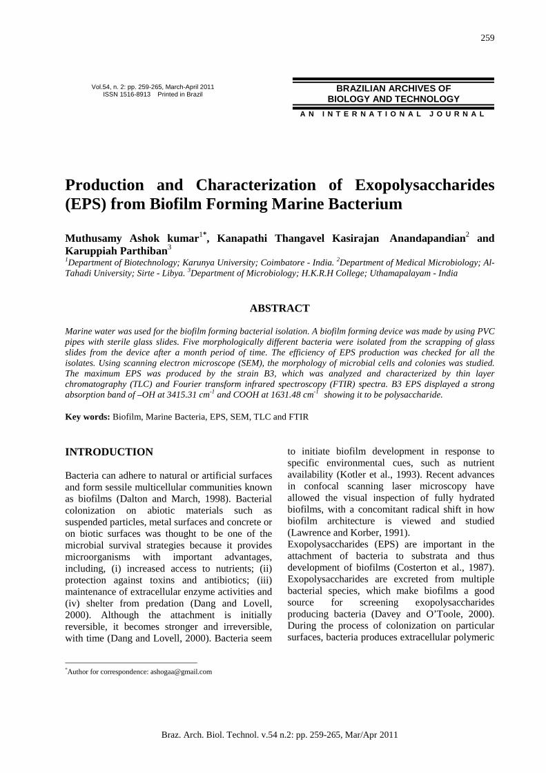
substance (Geesey and white, 1990), which construct the biofilm matrix. These polymeric substances mainly comprise of exopolysaccharide (40-95%), protein (1-60%), nucleic acids (1-10%) and lipids (1-40%) (Davey and O’Toole, 2000, Flemming and Wingender, 2001). Microbial cells generally contain various polysaccharide structures contributing to their shape and rigidity. Capsule EPS are produced mainly during the log phase of bacterial growth and slilme EPS produced during the stationary phase (Plante and Shriver.1998).The structure of polysaccharide is relatively simple, comprising of homo polysaccharides (Polymer containing one type of sugar) or hetropolysaccharides (containing more than two types of monosaccharide units). These extracellular materials (polysaccharides, Lipids, glycoproteins and lipopolysaccharides) can be used as stabilizers, gelling agents, adhesives, thickening agents, emulsifying agents, flocculants and flushing agents (Becker et al., 1998).The present work aimed to study the EPS production potential of bacteria isolated from marine environment and also chemical investigation of exopolysaccharides by TLC and FTIR. MATERIALS AND METHODS Sea water The formation of biofilm was studied in sea water. The sea water samples were collected in a plastic container from a coastal area of Tuticorin in South India following the method of Lehtola et al., (2002). Biofilm formation Biofilm development was studied with polyvinyl chloride (PVC) pipe which was 7 cm as length and 4 cm in width. The glass slide (25 mm in width, 75 mm in height and 1mm in thickness) were used for the biofilm formation. The glass slides were pre-cleaned with 1N HCL and treated with sodium hypochlorite solution of 10 mg/L for 24 hours and rinsed with sterile distilled water before the experimental setup. Then the slides were placed in PVC chamber at 21oC and covered by aluminium foil. Sea water was pumped at flow velocity of 10 to 20 drops/minute in the PVC chambers. Ten slides were placed in the PVC chamber with regular distance for the formation of biofilm (Hong et al., 2002). The sea water passed through
the PVC chamber with inserted glass slide for a month for bacterial adherence (Lehtola et al., 2002). Scanning Electron Microscope Analysis The microbial biofilm on glass slides were studied using Scanning Electron Microscope (SEM). Isolation of attached bacteria The surfaces of the glass slides were carefully removed from the device, scrapped and suspended in 10ml of filter sterilized aged sea water using a cell scrapper (Corning) and scrapped materials were serially diluted. After a series of dilutions, 100µl of diluents was spread on Zobell marine agar medium plates (peptone 5g, yeast extract 1g, ferrous phosphate 0.01g, agar 15g, distilled water 250 ml, aged sea water 750 ml, pH 7.2).The plates were incubated at 25oC for one week and bacterial colonies showing different morphological characteristics were transferred onto fresh Zobell agar medium plates (Indraneel et al., 1999). EPS production Five different morphologically strains (B1,B2,B3,B4 and B5) were isolated and they were grown on YMG agar medium (glucose 10g, yeast extract 3g, malt extract 3g,peptone 5g, distilled water 500 ml, aged sea water 500 ml). The pre-inoculam was prepared in YMG broth by incubating at 25oC for 24 hours and 200µl of this culture broth was inoculated into 50ml of YMG broth and incubated at 25oC for 5 days at 120 rpm. The contents were centrifuged (10,000xg for 20 minutes) and the culture supernatant was mixed with three volumes of isopropanol or ethanol slowly along the side wall of the conical flask and allowed to stand at 4oC for 20 minutes for precipitation of exopolysaccharides. The weight of the precipitated EPS was measured after drying at 80o C for three days (Hong et al., 2002). EPS characterization The EPS pellets 2 mg was mixed with 200 milligram of dry potassium bromide (KBr), then the mixture was pressed into a 16 mm diameter mold and used for IR spectroscopy for the detection of C=O bonds and – OH bonds (Mancuso Nichols et al., 2004). Monosaccharide composition of the EPS was analyzed by TLC (Schaal, 1985). The samples were prepared by the method of Staneck and
Production and Characterization of Exopolysaccharides
Braz. Arch. Biol. Technol. v.54 n.2: pp. 259-265, Mar/Apr 2011
261
Roberts (1974). EPS samples (50mg) were hydrolyzed with 4ml of 2 N sulphuric acid at 1000 C for 2 h and were neutralized by adding 0.5g of Ba (OH)2. After centrifugation (17,000xg, 4o C, 10 minutes), the supernatant was filtered through
0.22 µm membrane filter, concentrated by evaporating under reduced pressure, and placed on TLC plates. Glucose was used as standard. The amount of total carbohydrate was measured by phenol-sulfuric acid method (Dubois et al., 1956).
Figure 1 - Biofilm formation device.
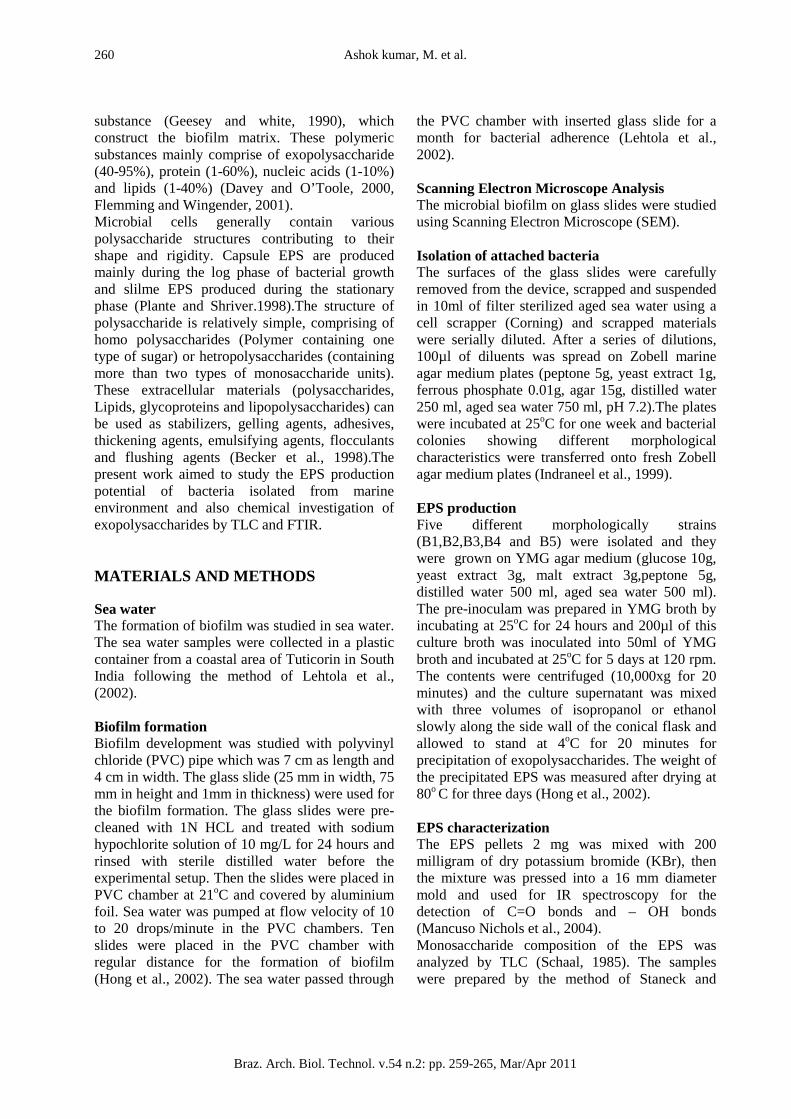
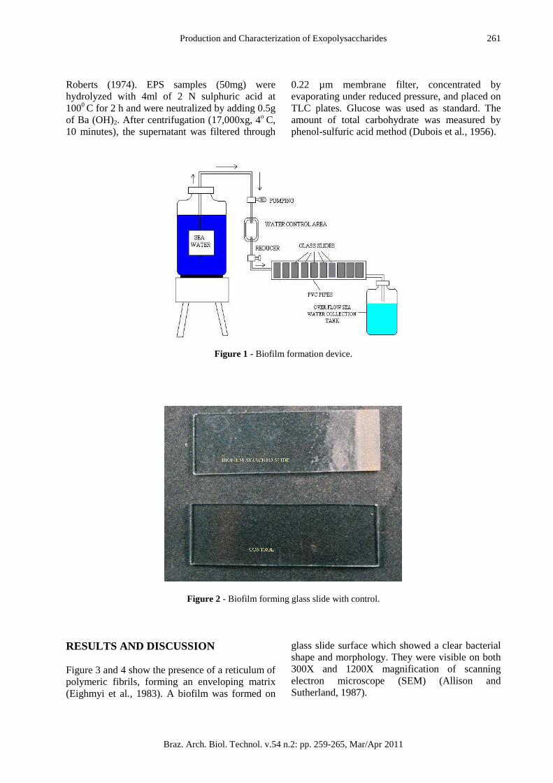
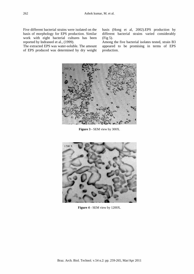
Figure 2 - Biofilm forming glass slide with control. RESULTS AND DISCUSSION Figure 3 and 4 show the presence of a reticulum of polymeric fibrils, forming an enveloping matrix (Eighmyi et al., 1983). A biofilm was formed on
glass slide surface which showed a clear bacterial shape and morphology. They were visible on both 300X and 1200X magnification of scanning electron microscope (SEM) (Allison and Sutherland, 1987).
Ashok kumar, M. et al.
Braz. Arch. Biol. Technol. v.54 n.2: pp. 259-265, Mar/Apr 2011
262
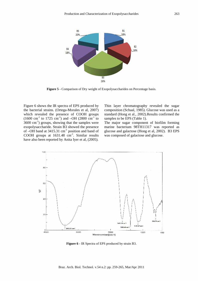
Five different bacterial strains were isolated on the basis of morphology for EPS production. Similar work with eight bacterial cultures has been reported by Indraneel et al., (1999). The extracted EPS was water-soluble. The amount of EPS produced was determined by dry weight
basis (Hong et al, 2002).EPS production by different bacterial strains varied considerably (Fig 5). Among the five bacterial isolates tested, strain B3 appeared to be promising in terms of EPS production.
Figure 3 - SEM view by 300X.
Figure 4 - SEM view by 1200X.
Production and Characterization of Exopolysaccharides
Braz. Arch. Biol. Technol. v.54 n.2: pp. 259-265, Mar/Apr 2011
263
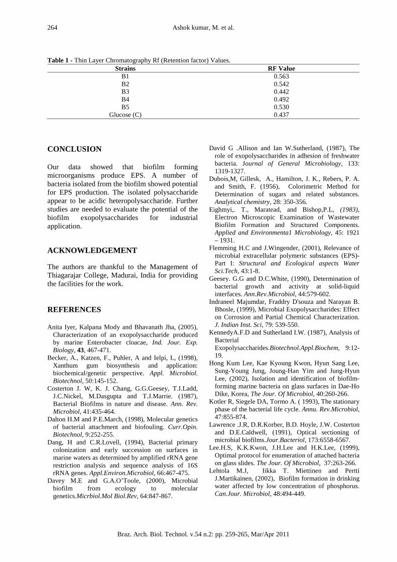
Figure 5 - Comparison of Dry weight of Exopolysaccharides on Percentage basis. Figure 6 shows the IR spectra of EPS produced by the bacterial strains. (Ortega-Morales et al, 2007) which revealed the presence of COOH groups (1600 cm-1 to 1725 cm-1) and –OH (2800 cm-1 to 3600 cm-1) groups, showing that the samples were exopolysaccharide. Strain B3 showed the presence of –OH band at 3415.31 cm-1 position and band of COOH groups at 1631.48 cm-1. Similar results have also been reported by Anita Iyer et al, (2005).
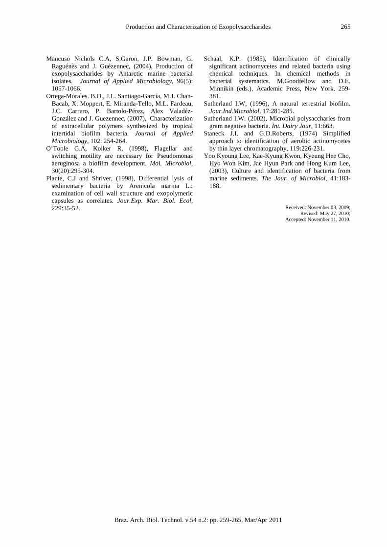
Thin layer chromatography revealed the sugar composition (Schaal, 1985). Glucose was used as a standard (Hong et al., 2002).Results confirmed the samples to be EPS (Table 1). The major sugar component of biofilm forming marine bacterium 98TH11317 was reported as glucose and galactose (Hong et al, 2002). B3 EPS was composed of galactose and glucose.
Figure 6 - IR Spectra of EPS produced by strain B3.
Ashok kumar, M. et al.
Braz. Arch. Biol. Technol. v.54 n.2: pp. 259-265, Mar/Apr 2011
264
Table 1 - Thin Layer Chromatography Rf (Retention factor) Values. Strains RF Value
B1 0.563 B2 0.542 B3 0.442 B4 0.492 B5 0.530
Glucose (C) 0.437 CONCLUSION Our data showed that biofilm forming microorganisms produce EPS. A number of bacteria isolated from the biofilm showed potential for EPS production. The isolated polysaccharide appear to be acidic heteropolysaccharide. Further studies are needed to evaluate the potential of the biofilm exopolysaccharides for industrial application. ACKNOWLEDGEMENT The authors are thankful to the Management of Thiagarajar College, Madurai, India for providing the facilities for the work. REFERENCES Anita Iyer, Kalpana Mody and Bhavanath Jha, (2005),
Characterization of an exopolysaccharide produced by marine Enterobacter cloacae, Ind. Jour. Exp. Biology, 43, 467-471.
Becker, A., Katzen, F., Puhler, A and Ielpi, L, (1998), Xanthum gum biosynthesis and application: biochemical/genetic perspective. Appl. Microbiol. Biotechnol, 50:145-152.
Costerton J. W, K. J. Chang, G.G.Geesey, T.I.Ladd, J.C.Nickel, M.Dasgupta and T.J.Marrie. (1987), Bacterial Biofilms in nature and disease. Ann. Rev. Microbiol, 41:435-464.
Dalton H.M and P.E.March, (1998), Molecular genetics of bacterial attachment and biofouling. Curr.Opin. Biotechnol, 9:252-255.
Dang, H and C.R.Lovell, (1994), Bacterial primary colonization and early succession on surfaces in marine waters as determined by amplified rRNA gene restriction analysis and sequence analysis of 16S rRNA genes. Appl.Environ.Microbiol, 66:467-475.
Davey M.E and G.A.O’Toole, (2000), Microbial biofilm from ecology to molecular genetics.Micrbiol.Mol Biol.Rev, 64:847-867.
David G .Allison and Ian W.Sutherland, (1987), The role of exopolysaccharides in adhesion of freshwater bacteria. Journal of General Microbiology, 133: 1319-1327.
Dubois,M, Gillesk, A., Hamilton, J. K., Rebers, P. A. and Smith, F. (1956), Colorimetric Method for Determination of sugars and related substances. Analytical chemistry, 28: 350-356.
Eighmyi,. T., Maratead, and Bishop,P.L, (1983), Electron Microscopic Examination of Wastewater Biofilm Formation and Structured Components. Applied and Environmenta1 Microbiology, 45: 1921 – 1931.
Flemming H.C and J.Wingender, (2001), Relevance of microbial extracellular polymeric substances (EPS)-Part I: Structural and Ecological aspects Water Sci.Tech, 43:1-8.
Geesey. G.G and D.C.White, (1990), Determination of bacterial growth and activity at solid-liquid interfaces. Ann.Rev.Microbiol, 44:579-602.
Indraneel Majumdar, Fraddry D'souza and Narayan B. Bhosle, (1999), Microbial Exopolysaccharides: Effect on Corrosion and Partial Chemical Characterization. J. Indian Inst. Sci, 79: 539-550.
KennedyA.F.D and Sutherland I.W. (1987), Analysis of Bacterial Exopolysaccharides.Biotechnol.Appl.Biochem, 9:12-19.
Hong Kum Lee, Kae Kyoung Kwon, Hyun Sang Lee, Sung-Young Jung, Joung-Han Yim and Jung-Hyun Lee, (2002), Isolation and identification of biofilm-forming marine bacteria on glass surfaces in Dae-Ho Dike, Korea, The Jour. Of Microbiol, 40:260-266.
Kotler R, Siegele DA, Tormo A. ( 1993), The stationary phase of the bacterial life cycle. Annu. Rev.Microbiol, 47:855-874.
Lawrence .J.R, D.R.Korber, B.D. Hoyle, J.W. Costerton and D.E.Caldwell, (1991), Optical sectioning of microbial biofilms.Jour.Bacteriol, 173:6558-6567.
Lee.H.S, K.K.Kwon, J.H.Lee and H.K.Lee, (1999), Optimal protocol for enumeration of attached bacteria on glass slides. The Jour. Of Microbiol, 37:263-266.
Lehtola M.J, Iikka T. Miettinen and Pertti J.Martikainen, (2002), Biofilm formation in drinking water affected by low concentration of phosphorus. Can.Jour. Microbiol, 48:494-449.
Production and Characterization of Exopolysaccharides
Braz. Arch. Biol. Technol. v.54 n.2: pp. 259-265, Mar/Apr 2011
265
Mancuso Nichols C.A, S.Garon, J.P. Bowman, G. Raguénès and J. Guézennec, (2004), Production of exopolysaccharides by Antarctic marine bacterial isolates. Journal of Applied Microbiology, 96(5): 1057-1066.
Ortega-Morales. B.O., J.L. Santiago-García, M.J. Chan-Bacab, X. Moppert, E. Miranda-Tello, M.L. Fardeau, J.C. Carrero, P. Bartolo-Pérez, Alex Valadéz-González and J. Guezennec, (2007), Characterization of extracellular polymers synthesized by tropical intertidal biofilm bacteria. Journal of Applied Microbiology, 102: 254-264.
O’Toole G.A, Kolker R, (1998), Flagellar and switching motility are necessary for Pseudomonas aeruginosa a biofilm development. Mol. Microbiol, 30(20):295-304.
Plante, C.J and Shriver, (1998), Differential lysis of sedimentary bacteria by Arenicola marina L.: examination of cell wall structure and exopolymeric capsules as correlates. Jour.Exp. Mar. Biol. Ecol, 229:35-52.
Schaal, K.P. (1985), Identification of clinically significant actinomycetes and related bacteria using chemical techniques. In chemical methods in bacterial systematics. M.Goodfellow and D.E. Minnikin (eds.), Academic Press, New York. 259-381.
Sutherland I.W, (1996), A natural terrestrial biofilm. Jour.Ind.Microbiol, 17:281-285.
Sutherland I.W. (2002), Microbial polysaccharies from gram negative bacteria. Int. Dairy Jour, 11:663.
Staneck J.I. and G.D.Roberts, (1974) Simplified approach to identification of aerobic actinomycetes by thin layer chromatography, 119:226-231.
Yoo Kyoung Lee, Kae-Kyung Kwon, Kyeung Hee Cho, Hyo Won Kim, Jae Hyun Park and Hong Kum Lee, (2003), Culture and identification of bacteria from marine sediments. The Jour. of Microbiol, 41:183-188.
Received: November 03, 2009; Revised: May 27, 2010;
Accepted: November 11, 2010.