proceedings - suranaree university of technology
TRANSCRIPT

Proceedings
The 4th PIM International Conference (Online Conference) “Globalization Revisited: Building Organization Resilience
with Digital Transformation”
March 3, 2021 Panyapiwat Institute of Management
Chaengwatthana Rd., Nonthaburi, Thailand
Organized by
Panyapiwat Institute of Management American International University-Bangladesh Asian Institute of Technology & Management Chuo University Kansai University Moscow State Institute of International Relations Nanjing Tech University Pujiang Institute Shantou University Shibaura Institute of Technology Waseda University The Association of Council of IT Deans (CITT)
ISBN (e-book) 978-616-7851-13-6
The 4th
PIM International Conference
March 3, 2021
1
The 4th PIM International Conference “Globalization Revisited: Building Organization Resilience
with Digital Transformation” (Online Conference)
March 3, 2021 Panyapiwat Institute of Management
Chaeng Watthana Rd., Nonthaburi, Thailand
----------------------------------------------------
The 4th PIM International Conference are the annual academic conferences, organized by Panyapiwat Institute of Management (PIM). The purposes consist of providing a forum presentation of research papers, conducted by faculty staff and students from various institution and for sharing of findings and knowledge among researchers and professionals in many areas. In addition, participants will be provided significant suggestions from the qualified experts. This conference will be beneficial for research improvement in the future and for the creation of cooperative networks among the related academic disciplines. The main topic of the conference is “Globalization Revisited: Building Organization Resilience with Digital Transformation
Expected Outcomes: 1. Academics, professionals, faculty staff, researchers, and students have the forum
to present their research papers, to exchange and share their knowledge, experiences and ideas, and to hear suggestions from qualified experts.
2. The creation of co-operative networks among academics in the same or related disciplines.
Participants: Researchers, academics, and interested individuals from Thailand and overseas. The
expected numbers of participants are between 200-300 people.
Conference formats: 1. Research 2. Academic
The 4th
PIM International Conference
March 3, 2021
2
The conference welcomes papers on the following disciplines: - Social Science and Humanities - Engineering and Technology All accepted papers will be published online in the Conference Proceedings.
Types of Presentation: - Online VDO Presentation - E-Poster Presentation
Languages for Presentation: - English - Chinese
Manuscript preparation: The manuscript must be prepared based on the following template which is
available at: https://conference.pim.ac.th/zh/template/
Registration and Paper Submission: - Registration can be done via: https://conference.pim.ac.th/zh - The manuscript must be saved in document format (Microsoft Word version
2007) and PDF format.
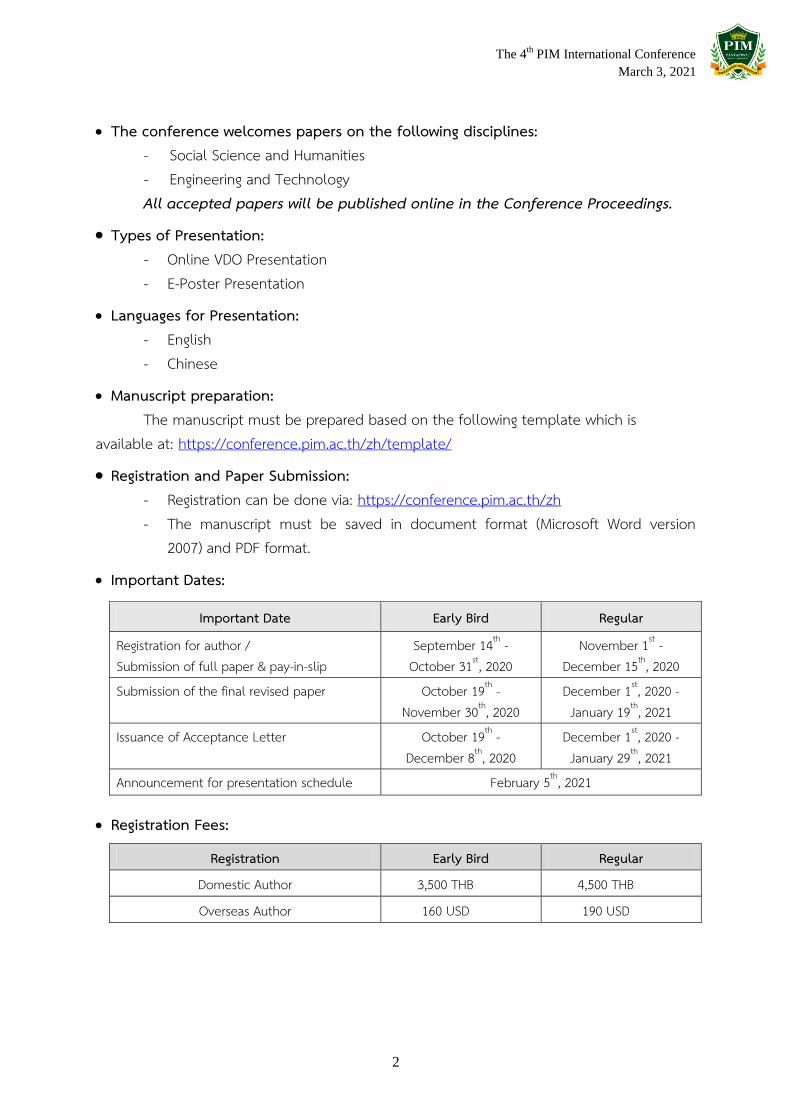
Important Dates:
Important Date Early Bird Regular
Registration for author / Submission of full paper & pay-in-slip
September 14th - October 31st, 2020
November 1st - December 15th, 2020
Submission of the final revised paper October 19th - November 30th, 2020
December 1st, 2020 - January 19th, 2021
Issuance of Acceptance Letter October 19th - December 8th, 2020
December 1st, 2020 - January 29th, 2021
Announcement for presentation schedule February 5th, 2021
Registration Fees:
Registration Early Bird Regular
Domestic Author 3,500 THB 4,500 THB
Overseas Author 160 USD 190 USD

The 4th
PIM International Conference
March 3, 2021
3
Contact information:
Ms. Paweena Kimsuvan or Ms. Kittiya Leartkitjanuwat Tel. +662855-1107, +662855-0940 Email : [email protected] Conference Website https://conference.pim.ac.th/
------------------------------------------------------------------------------------------

The 4th PIM International Conference
March 3, 2021
Table of Contents
Page
The 4th PIM International Conferences 2021 1
Conference Program 4
List of Articles 5
Detail of Articles A - Agriculture 24 B - Business 34 C - Communications 241 D - Economics 251 E - Education 349 F - Engineering and Technology 441 G - Library Arts 562 H - Management 597 I - Social Sciences and Humanities 715
Appendixes Announcement of Administrative Committee of The 4th PIM International
Conferences A1
Co-host A11 Peer Reviewers A13

The 4th PIM International Conference March 3, 2021
5
List of Articles

The 4th PIM International Conference March 3, 2021
14
F - Engineering and Technology
No. Title Name - Surname Department Page
1 A HYBRID APPROACH OF SENTIMENT BASED ALGORITHM FOR COMPUTERIZED OPINION ANALYSIS IN THE CONTEXT OF PUBLIC UNIVERSITY ADMISSION SYSTEM IN BANGLADESH
Tanzila Nasrin Tazin Jannatul Ferdous Prionti Das Farjana Yeasmin Md. Abdullah-Al-Jubair Ng Giap Weng
American International University-Bangladesh
441
2 AN IOT BASED FIRE ALARMING AND RESCUE ASSIST SYSTEM FOR HOUSEHOLD & INDUSTRY USING GSM MODULE
Md. Amin Ahmed Chowdhury Md. Ar Rafi Sarker Obida Bin Alauddin Rezwanur Rahman Md. Abdullah-Al-Jubair Ng Giap Weng
American International University- Bangladesh
Universiti Malaysia Sabah, Malaysia
450
3 BRAIN FUNCTION ANALYSIS OF COMPUTER-BASED AND PAPER-BASED TESTS USING EEG: A PILOT STUDY
Thanate Angsuwatanakul Naruemol Inthaphuk Jamie A. O’Reilly Shaik Abdul Malik
Mohamed Ismail Chittra Chantragatrawi Thapanee Khemanuwong
College of Biomedical Engineering, Rangsit University, Thailand
School of Educational Studies, Universiti Sains Malaysia, Malaysia
School of Creative Educational Management, Panyapiwat Institute of Management, Thailand
Faculty of Liberal Arts, King Mongkut’s Institute of Technology Ladkrabang, Thailand
464
4 CARBON EMISSIONS OF INTERLOCKING CEMENT-STABILIZED COMPRESSED EARTH BRICK HOUSES IN DHUNGENTAR, NEPAL
Prakash Dulal Pravakar Pradhan Lena Sthapit Rabindra Raj Giri
Department of Civil Engineering, Asian Institute of Technology & Management (AITM), Khumaltar, Lalitpur, Nepal
480
5 DEVELOPMENT OF COMPUTER APPLICATION FOR GHS-BASED CHEMICAL HAZARD COMMUNICATION
Sarisak Soontornchai School of Health Science, Sukhothai Thammathirat Open University
494

The 4th PIM International Conference March 3, 2021
15
No. Title Name - Surname Department Page
6 DIRTY DATA CLEANING FOR THE BETTERMENT OF ONLINE BUSINESSES
Md. Ismail Hossen Md. Nazmul Hossain Md. Armanur Rahman
Department of Computer Science, American International University-Bangladesh (AIUB), Dhaka 1212, Bangladesh
Department of Computer Science, Multimedia University, Malaysia
503
7 INTERACTIVE LEARNING MEDIA FOR KIDS Arnab Basak Md. Abdullah-Al-Jubair Wasif Zaman Rafid Shahriar Md. Mridul Mallik Ng Giap Weng
Faculty of Science & Technology, American International University-Bangladesh
Faculty of Computer and Informatics, Universiti Malaysia Sabah
510
8 METHOD FOR CLASSIFICATION OF PERMANENT TOOTH CLASSES FROM PHOTOGRAPHS
Jessada Tanthanuch Chaninart Yord-in Akanat Wetayawanich Sakdinee Rattana
Suranaree University of Technology
523
9 STATISTICAL DISTRIBUTIONS APPEARING IN MEDICAL ULTRASOUND IMAGES
Mangkon Damnet Akanat Wetayawanich Jessada Tanthanuch
Suranaree University of Technology
536
10 立体绿化城市环境绩效评价指标体系研究 THE INDEX SYSTEM OF ENVIRONMENTAL PERFORMANCE EVALUATION OF THREE-DIMENSIONAL GREENING CITY
Jing Li Ersi Liu Anshou Yao
Chinese Graduate School, Panyapiwat Institute of Management
548
F Engineering and Technology
The 4th
PIM International Conference
March 3, 2021
523
METHOD FOR CLASSIFICATION OF PERMANENT TOOTH CLASSES
FROM PHOTOGRAPHS
Jessada Tanthanuch1, Chaninart Yord-in
2, Akanat Wetayawanich
3, Sakdinee Rattana
4,*
1,3,4 School of Mathematics, Institute of Science,
Suranaree University of Technology, Nakhon Ratchasima, Thailand 2 School of Biomedical Innovation Engineering, Institute of Engineering,
Suranaree University of Technology, Nakhon Ratchasima, Thailand *Corresponding author, E-mail: [email protected]
Abstract
In this work the system of methods in digital image processing was proposed to classify teeth
in the photographs of the model of permanent teeth. The system consisted of segmentation the dental
arch by colors, and classification of the individual teeth by image registration. Image registration was
the major technique chosen by discussion from former works on the photographs of oral cavity. The
images of teeth were obtained from the ‘sim kit’ model (teeth model) in different angles of tilt and
pan. The method could classify teeth correctly up to 8 teeth out of 14 teeth. The best view to take the
photographs was suggested by the result to be the extremal elevation angles and the model pointing
directly to the image sensor.
Keywords: Digital image processing, Image segmentation, Image registration, Dental photography,
Teeth type classification
Introduction
Diagnosing oral health diseases usually is done by experts from dentistry area, for the sake of
accuracy. However, the accessibility problems such as too few experts and difficult travelling to
healthcare facilities, which often happen in rural areas, decrease the chance for patients to see doctor
and get cured on time.
Digital image processing is a study of applying mathematical and computational methods on
digital images to gain information. Some of its application are medical image analysis (Tanthanuch,
Kaptsov, and Meleshko, 2019), and traffic management (Hasan et al., 2014). When combining with
other technology like artificial intelligence, it helps automate systems, which means decrease human
workload.
This work is the first part of developing an automated system which can be used to analyze
dental images (various types of images taken from oral cavity). The colored photographs are taken
from different views of permanent teeth model. The teeth in the photographs are to be classified into
one of four types of teeth: incisors, canines, premolars, and molars. Techniques in digital image
processing are considered into helping automate the system of analyzing the images (the photographs)
and classifying the teeth. If the system yields high enough accuracy, it can be used to increase the
speed and accuracy of the artificial intelligence in the part of learning dental images, and develop the
system which can detect disease traces in photographs.
This paper is organized as follows: first, in the literature review, the model of human teeth
and the teeth types are presented, the imaging methods in dentistry are gathered and the related works
on dental images are reviewed. Choosing the main method - the image registration - is also discussed.
Research

The 4th
PIM International Conference
March 3, 2021
524
Then in the methodology, the view setting and the image acquisition are shown. The image
segmentation by color components and morphological operations is then employed to decrease
computational complexity. The image registration is then applied and the results are discussed.
Research Objective
To find an efficient method in digital image processing that can classify teeth in the
photographs of permanent teeth model.
Literature Review
1. Human teeth and their types
1.1 Teeth – general information
Teeth are an organ / a structure located in a mouth and is the first part of digestive
system. Teeth break down food particles and help pronounce words when one speaks. Usually,
humans have 2 sets of teeth: primary teeth and permanent teeth. Primary teeth are found in children
approximately from the age 6 months to 12 years old. Permanent teeth or complete permanent
dentition is found in people age from approximately 12 years old.
1.2 Permanent teeth
The set of permanent teeth consists of 32 teeth, and these teeth can be classified into 4
types, as follows:
1) Incisors are at the front of the dental arch. There are 8 teeth; 4 uppers and 4 lowers.
They are thin and flat. They cut food from the big piece into a smaller one that can be taken in the
mouth.
2) Canines are next to incisors. There are 4 teeth; 2 uppers and 2 lowers. They are
sharp, spike-like shape. They tear and hold food piece.
3) Premolars or bicuspids are next to canines. There are 8 teeth; 4 uppers and 4 lowers.
They are bigger than incisors and canines. They tear and crunch food particles.
4) Molars, including wisdom teeth or third molars, are next to premolars and at the
deepest part of the dental arch. They are 12 teeth; 6 uppers and 6 lowers. They are the biggest teeth.
They are with flat faces which crunch food particles.

The 4th
PIM International Conference
March 3, 2021
525
Picture 1: Permanent teeth diagram
2. Imaging in dentistry
Defined by American Dental Association, dentistry, also known as dental medicine and oral
medicine, is a branch of medicine that consists of the study, diagnosis, prevention, and treatment of
diseases, disorders, and conditions of the oral cavity, commonly in the dentition but also the oral
mucosa, and of adjacent and related structures and tissues, particularly in the maxillofacial (jaw and
facial) area.
In the past three decades it benefits much from helping technologies, especially imaging
methods. Shah, Bansal, and Logani (2014) gathered the imaging methods used in dentistry. Table 1
shows the imaging methods they gathered and examples. It also includes some methods not
mentioned in their paper.
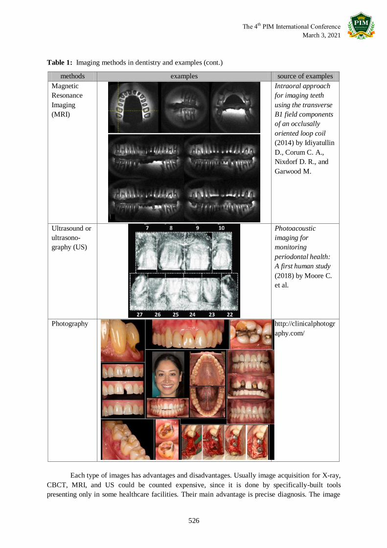
Table 1: Imaging methods in dentistry and examples
methods examples source of examples
X-ray
https://www.hawkins
familydental.com/bl
og/understanding-
dental-x-rays/
Cone Beam
Computed
Tomography
(CBCT)
(3D)
https://johannlochner
.com/treatments-
offered/3d-x-rays-
cbct/
upper
teeth
lower
teeth
Types of teeth by color incisor
canine premolar molar (possible) third molar or wisdom tooth
The 4th
PIM International Conference
March 3, 2021
526
Table 1: Imaging methods in dentistry and examples (cont.)
methods examples source of examples
Magnetic
Resonance
Imaging
(MRI)
Intraoral approach
for imaging teeth
using the transverse
B1 field components
of an occlusally
oriented loop coil
(2014) by Idiyatullin
D., Corum C. A.,
Nixdorf D. R., and
Garwood M.
Ultrasound or
ultrasono-
graphy (US)
Photoacoustic
imaging for
monitoring
periodontal health:
A first human study
(2018) by Moore C.
et al.
Photography
http://clinicalphotogr
aphy.com/
Each type of images has advantages and disadvantages. Usually image acquisition for X-ray,
CBCT, MRI, and US could be counted expensive, since it is done by specifically-built tools
presenting only in some healthcare facilities. Their main advantage is precise diagnosis. The image
The 4th
PIM International Conference
March 3, 2021
527
acquisition of photography is the cheapest, but with the limitation of views, especially to the inside of
teeth and jawbones.
3. Tasks in medical image analysis
In the survey paper of Litjens et al. (2017), the medical image analysis tasks in which
technologies are employed to help decrease human workload can be grouped as follows:
3.1 Classification
Examples of classification in medical image analysis are image/exam classification, in
which the goal is to tell if the disease is present or not, and object/lesion classification, in which the
objects in the images are to be classified into types.
3.2 Detection
Detecting objects in images such as organs, regions, lesion, consists of 2 tasks:
localization of the object and object identification.
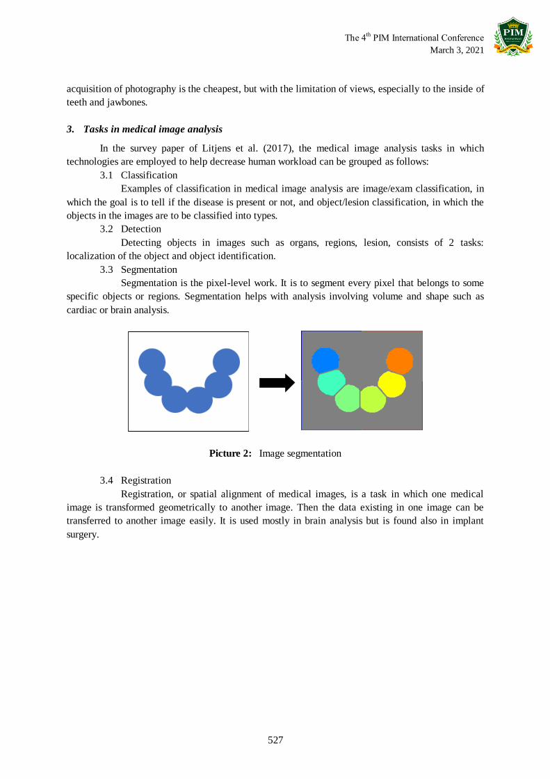
3.3 Segmentation
Segmentation is the pixel-level work. It is to segment every pixel that belongs to some
specific objects or regions. Segmentation helps with analysis involving volume and shape such as
cardiac or brain analysis.
Picture 2: Image segmentation
3.4 Registration
Registration, or spatial alignment of medical images, is a task in which one medical
image is transformed geometrically to another image. Then the data existing in one image can be
transferred to another image easily. It is used mostly in brain analysis but is found also in implant
surgery.

The 4th
PIM International Conference
March 3, 2021
528
Picture 3: Examples of geometric transformations
Picture 4: Image registration
3.5 Other tasks
The other tasks include learning image database, image generation, and matching images
with reports.
4. Related works
In medical image analysis, there are many types of images since the purposes of analysis are
vary and the image sensors are different. It is so with the dental images. The types of dental images
include the x-ray images, the CT scanned images, the 3D meshes, and the photographs, with the x-ray
images being the most used since they show the inner parts of teeth. The methods from digital image
processing are chosen by considering the type of the images and the purpose of analysis. The
following are some works involving with dental image analysis and teeth classification:
affine transformation
nonlinear transformation
moving / reference image
template image
aligning
parameters of
transformation
adjusting
similarity
checking
maximum similarity /
maximum number of cycles
The 4th
PIM International Conference
March 3, 2021
529
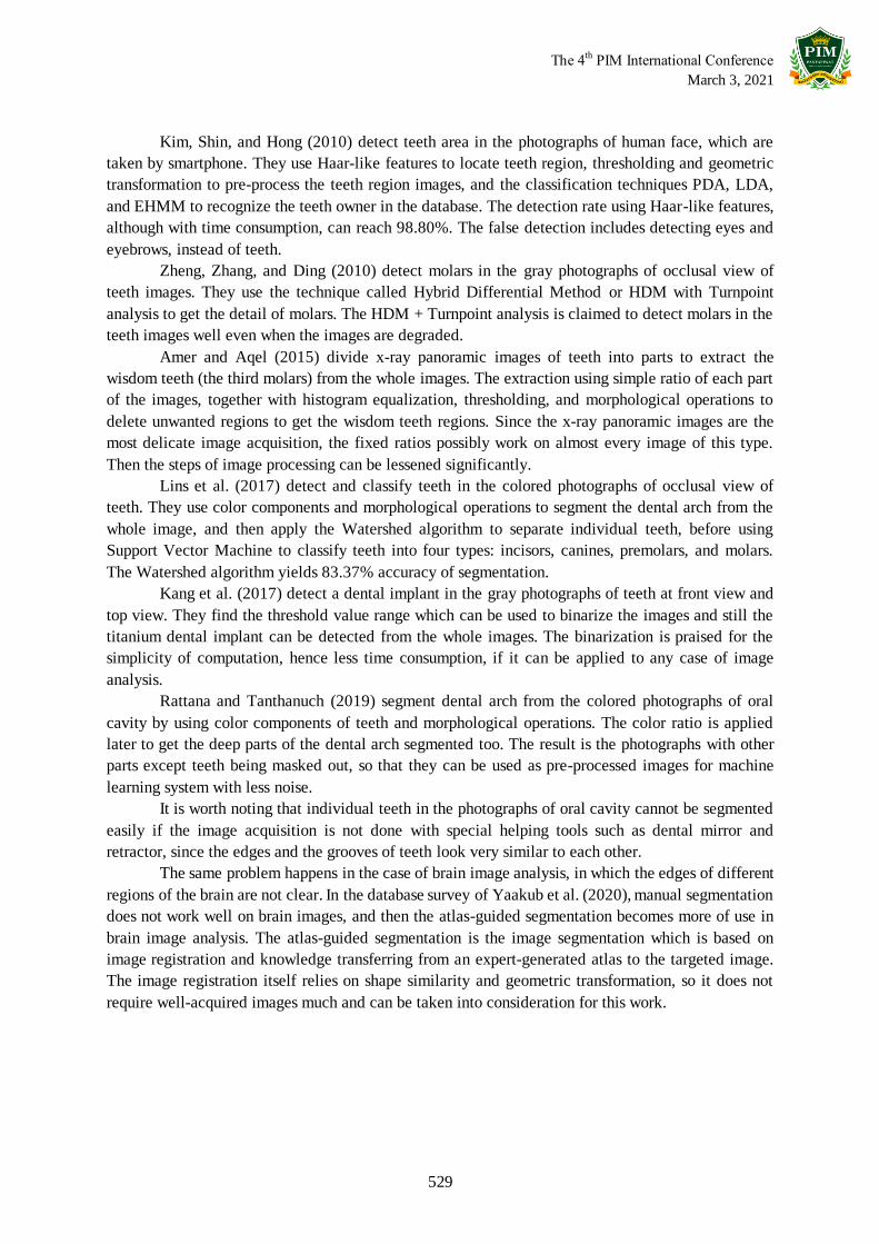
Kim, Shin, and Hong (2010) detect teeth area in the photographs of human face, which are
taken by smartphone. They use Haar-like features to locate teeth region, thresholding and geometric
transformation to pre-process the teeth region images, and the classification techniques PDA, LDA,
and EHMM to recognize the teeth owner in the database. The detection rate using Haar-like features,
although with time consumption, can reach 98.80%. The false detection includes detecting eyes and
eyebrows, instead of teeth.
Zheng, Zhang, and Ding (2010) detect molars in the gray photographs of occlusal view of
teeth images. They use the technique called Hybrid Differential Method or HDM with Turnpoint
analysis to get the detail of molars. The HDM + Turnpoint analysis is claimed to detect molars in the
teeth images well even when the images are degraded.
Amer and Aqel (2015) divide x-ray panoramic images of teeth into parts to extract the
wisdom teeth (the third molars) from the whole images. The extraction using simple ratio of each part
of the images, together with histogram equalization, thresholding, and morphological operations to
delete unwanted regions to get the wisdom teeth regions. Since the x-ray panoramic images are the
most delicate image acquisition, the fixed ratios possibly work on almost every image of this type.
Then the steps of image processing can be lessened significantly.
Lins et al. (2017) detect and classify teeth in the colored photographs of occlusal view of
teeth. They use color components and morphological operations to segment the dental arch from the
whole image, and then apply the Watershed algorithm to separate individual teeth, before using
Support Vector Machine to classify teeth into four types: incisors, canines, premolars, and molars.
The Watershed algorithm yields 83.37% accuracy of segmentation.
Kang et al. (2017) detect a dental implant in the gray photographs of teeth at front view and
top view. They find the threshold value range which can be used to binarize the images and still the
titanium dental implant can be detected from the whole images. The binarization is praised for the
simplicity of computation, hence less time consumption, if it can be applied to any case of image
analysis.
Rattana and Tanthanuch (2019) segment dental arch from the colored photographs of oral
cavity by using color components of teeth and morphological operations. The color ratio is applied
later to get the deep parts of the dental arch segmented too. The result is the photographs with other
parts except teeth being masked out, so that they can be used as pre-processed images for machine
learning system with less noise.
It is worth noting that individual teeth in the photographs of oral cavity cannot be segmented
easily if the image acquisition is not done with special helping tools such as dental mirror and
retractor, since the edges and the grooves of teeth look very similar to each other.
The same problem happens in the case of brain image analysis, in which the edges of different
regions of the brain are not clear. In the database survey of Yaakub et al. (2020), manual segmentation
does not work well on brain images, and then the atlas-guided segmentation becomes more of use in
brain image analysis. The atlas-guided segmentation is the image segmentation which is based on
image registration and knowledge transferring from an expert-generated atlas to the targeted image.
The image registration itself relies on shape similarity and geometric transformation, so it does not
require well-acquired images much and can be taken into consideration for this work.

The 4th
PIM International Conference
March 3, 2021
530
Methodology
1. The images and the image acquisition
In this work, the colored photographs of permanent teeth model are considered into use. A
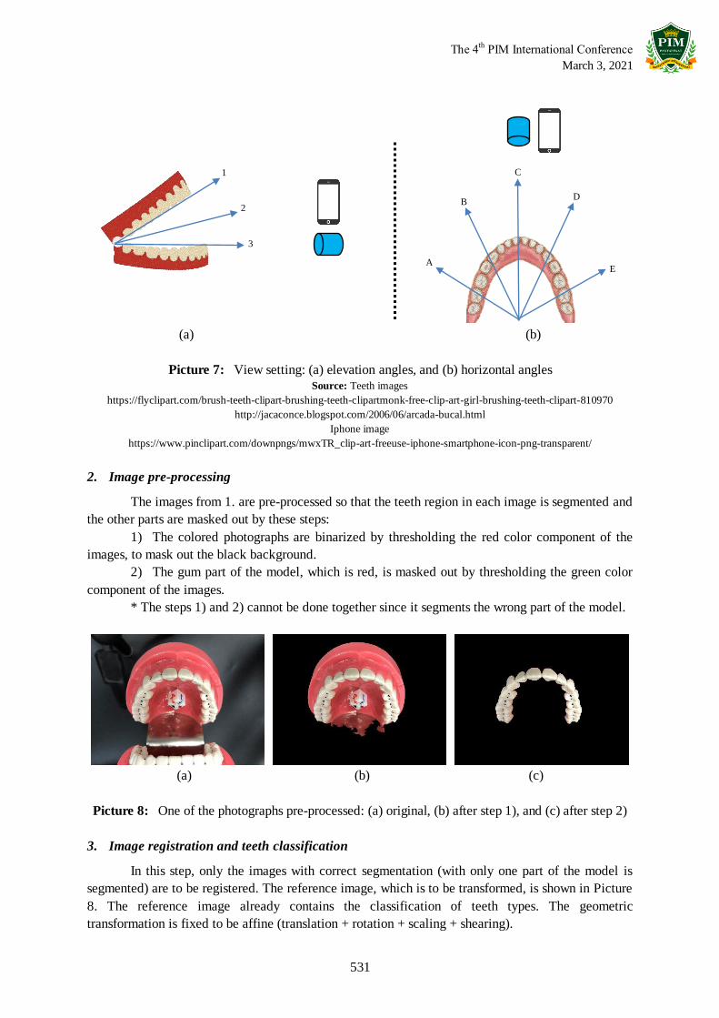
model consists of 2 parts: the upper teeth and the lower teeth. Both parts are photographed by 15
different views; 3 different elevation angles x 5 different horizontal angles, so there are 30
photographs taken from the model. The setting is shown in Picture 5 and some examples of the
photographs are shown in Picture 6.
Picture 5: Setting of image acquisition
Picture 6: Examples of the model being photographed by different views
The 4th
PIM International Conference
March 3, 2021
531
(a) (b)
Picture 7: View setting: (a) elevation angles, and (b) horizontal angles Source: Teeth images
https://flyclipart.com/brush-teeth-clipart-brushing-teeth-clipartmonk-free-clip-art-girl-brushing-teeth-clipart-810970
http://jacaconce.blogspot.com/2006/06/arcada-bucal.html
Iphone image
https://www.pinclipart.com/downpngs/mwxTR_clip-art-freeuse-iphone-smartphone-icon-png-transparent/
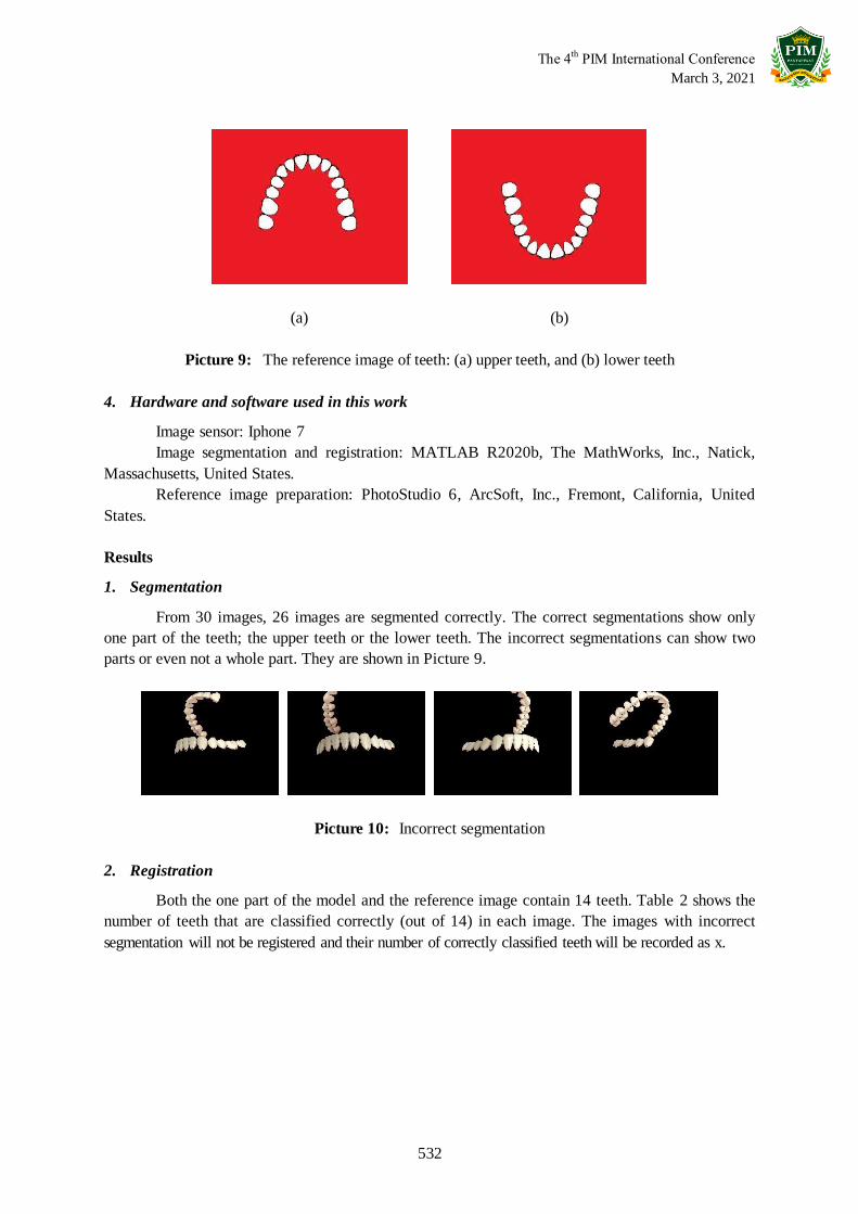
2. Image pre-processing
The images from 1. are pre-processed so that the teeth region in each image is segmented and
the other parts are masked out by these steps:
1) The colored photographs are binarized by thresholding the red color component of the
images, to mask out the black background.
2) The gum part of the model, which is red, is masked out by thresholding the green color
component of the images.
* The steps 1) and 2) cannot be done together since it segments the wrong part of the model.
(a) (b) (c)
Picture 8: One of the photographs pre-processed: (a) original, (b) after step 1), and (c) after step 2)
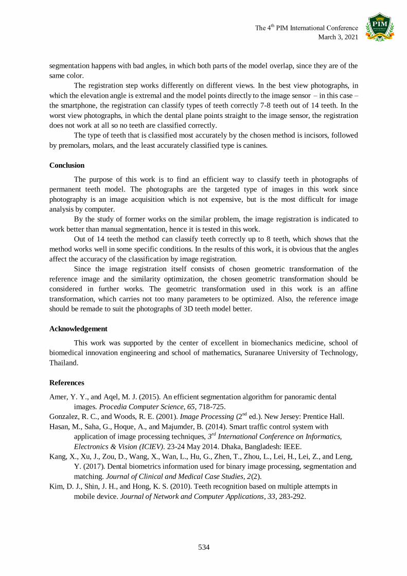
3. Image registration and teeth classification
In this step, only the images with correct segmentation (with only one part of the model is
segmented) are to be registered. The reference image, which is to be transformed, is shown in Picture
8. The reference image already contains the classification of teeth types. The geometric
transformation is fixed to be affine (translation + rotation + scaling + shearing).
C
D B
A E
3
2
1
The 4th
PIM International Conference
March 3, 2021
532
(a) (b)
Picture 9: The reference image of teeth: (a) upper teeth, and (b) lower teeth
4. Hardware and software used in this work
Image sensor: Iphone 7
Image segmentation and registration: MATLAB R2020b, The MathWorks, Inc., Natick,
Massachusetts, United States.
Reference image preparation: PhotoStudio 6, ArcSoft, Inc., Fremont, California, United
States.
Results
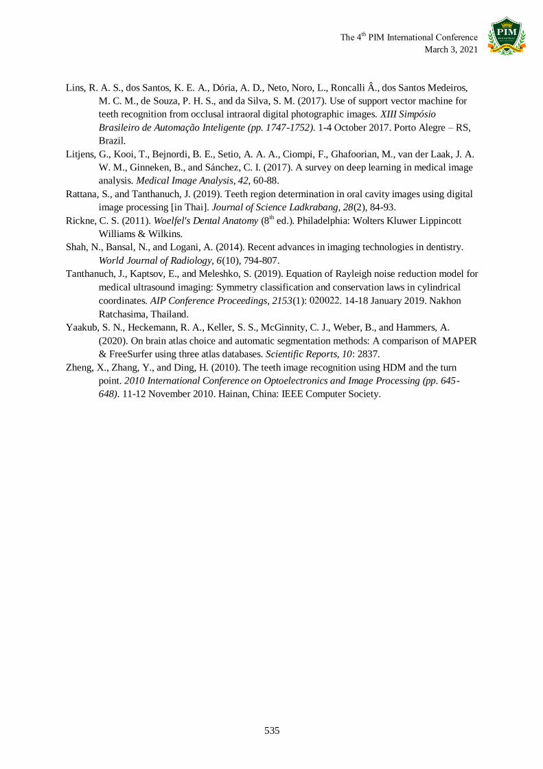
1. Segmentation
From 30 images, 26 images are segmented correctly. The correct segmentations show only
one part of the teeth; the upper teeth or the lower teeth. The incorrect segmentations can show two
parts or even not a whole part. They are shown in Picture 9.
Picture 10: Incorrect segmentation
2. Registration
Both the one part of the model and the reference image contain 14 teeth. Table 2 shows the
number of teeth that are classified correctly (out of 14) in each image. The images with incorrect
segmentation will not be registered and their number of correctly classified teeth will be recorded as x.

The 4th
PIM International Conference
March 3, 2021
533
(a) (b)
Picture 11: The result of image registration: (a) the worst, and (b) the best
Table 2: The number of teeth correctly classified in each image
Elevation angles Horizontal angles
A B C D E
Lower 1 x x 0 x x
Lower 2 1 2 8 3 2
Lower 3 2 6 7 3 4
Upper 1 1 6 8 8 7
Upper 2 2 5 7 2 0
Upper 3 4 1 3 3 0
3. Statistical data
Number of registered photographs: 26
Maximum number of teeth correctly classified in a photograph: 8
Minimum number of teeth correctly classified in a photograph: 0
Total number of teeth to be classified in a photograph: 14
incisors: 4, canines: 2, premolars: 4, molars: 4
Total number of teeth to be classified: 26 x 14 = 364
incisors: 104, canines: 52, premolars: 104, molars: 104
Number of teeth correctly classified: 95
incisors: 46, canines: 8, premolars: 24, molars: 17
Accuracy of the chosen classification method, by type of teeth
incisors: 44.23%, canines: 15.38%, premolars: 23.08%, molars: 16.35%
Discussion
A permanent teeth model is taken into 30 photographs of different views. There are 2 parts of
the model: the upper teeth and the lower teeth. Each part is taken into 15 photographs of different
views. The photographs are segmented until only teeth region is left, and then are registered.
The segmentation steps, which use the color components and the morphological operations,
work successfully on 26 out of 30 photographs. It means that the color components, with the help of
morphological operations, can be used to segment teeth region in the photographs. The incorrect
The 4th
PIM International Conference
March 3, 2021
534
segmentation happens with bad angles, in which both parts of the model overlap, since they are of the
same color.
The registration step works differently on different views. In the best view photographs, in
which the elevation angle is extremal and the model points directly to the image sensor – in this case –
the smartphone, the registration can classify types of teeth correctly 7-8 teeth out of 14 teeth. In the
worst view photographs, in which the dental plane points straight to the image sensor, the registration
does not work at all so no teeth are classified correctly.
The type of teeth that is classified most accurately by the chosen method is incisors, followed
by premolars, molars, and the least accurately classified type is canines.
Conclusion
The purpose of this work is to find an efficient way to classify teeth in photographs of
permanent teeth model. The photographs are the targeted type of images in this work since
photography is an image acquisition which is not expensive, but is the most difficult for image
analysis by computer.
By the study of former works on the similar problem, the image registration is indicated to
work better than manual segmentation, hence it is tested in this work.
Out of 14 teeth the method can classify teeth correctly up to 8 teeth, which shows that the
method works well in some specific conditions. In the results of this work, it is obvious that the angles
affect the accuracy of the classification by image registration.
Since the image registration itself consists of chosen geometric transformation of the
reference image and the similarity optimization, the chosen geometric transformation should be
considered in further works. The geometric transformation used in this work is an affine
transformation, which carries not too many parameters to be optimized. Also, the reference image
should be remade to suit the photographs of 3D teeth model better.
Acknowledgement
This work was supported by the center of excellent in biomechanics medicine, school of
biomedical innovation engineering and school of mathematics, Suranaree University of Technology,
Thailand.
References
Amer, Y. Y., and Aqel, M. J. (2015). An efficient segmentation algorithm for panoramic dental
images. Procedia Computer Science, 65, 718-725.
Gonzalez, R. C., and Woods, R. E. (2001). Image Processing (2nd ed.). New Jersey: Prentice Hall.
Hasan, M., Saha, G., Hoque, A., and Majumder, B. (2014). Smart traffic control system with
application of image processing techniques, 3rd International Conference on Informatics,
Electronics & Vision (ICIEV). 23-24 May 2014. Dhaka, Bangladesh: IEEE.
Kang, X., Xu, J., Zou, D., Wang, X., Wan, L., Hu, G., Zhen, T., Zhou, L., Lei, H., Lei, Z., and Leng,
Y. (2017). Dental biometrics information used for binary image processing, segmentation and
matching. Journal of Clinical and Medical Case Studies, 2(2).
Kim, D. J., Shin, J. H., and Hong, K. S. (2010). Teeth recognition based on multiple attempts in
mobile device. Journal of Network and Computer Applications, 33, 283-292.
The 4th
PIM International Conference
March 3, 2021
535
Lins, R. A. S., dos Santos, K. E. A., Dória, A. D., Neto, Noro, L., Roncalli Â., dos Santos Medeiros,
M. C. M., de Souza, P. H. S., and da Silva, S. M. (2017). Use of support vector machine for
teeth recognition from occlusal intraoral digital photographic images. XIII Simpósio
Brasileiro de Automação Inteligente (pp. 1747-1752). 1-4 October 2017. Porto Alegre – RS,
Brazil.
Litjens, G., Kooi, T., Bejnordi, B. E., Setio, A. A. A., Ciompi, F., Ghafoorian, M., van der Laak, J. A.
W. M., Ginneken, B., and Sánchez, C. I. (2017). A survey on deep learning in medical image
analysis. Medical Image Analysis, 42, 60-88.
Rattana, S., and Tanthanuch, J. (2019). Teeth region determination in oral cavity images using digital
image processing [in Thai]. Journal of Science Ladkrabang, 28(2), 84-93.
Rickne, C. S. (2011). Woelfel's Dental Anatomy (8th ed.). Philadelphia: Wolters Kluwer Lippincott
Williams & Wilkins.
Shah, N., Bansal, N., and Logani, A. (2014). Recent advances in imaging technologies in dentistry.
World Journal of Radiology, 6(10), 794-807.
Tanthanuch, J., Kaptsov, E., and Meleshko, S. (2019). Equation of Rayleigh noise reduction model for
medical ultrasound imaging: Symmetry classification and conservation laws in cylindrical
coordinates. AIP Conference Proceedings, 2153(1): 020022. 14-18 January 2019. Nakhon
Ratchasima, Thailand.
Yaakub, S. N., Heckemann, R. A., Keller, S. S., McGinnity, C. J., Weber, B., and Hammers, A.
(2020). On brain atlas choice and automatic segmentation methods: A comparison of MAPER
& FreeSurfer using three atlas databases. Scientific Reports, 10: 2837.
Zheng, X., Zhang, Y., and Ding, H. (2010). The teeth image recognition using HDM and the turn
point. 2010 International Conference on Optoelectronics and Image Processing (pp. 645-
648). 11-12 November 2010. Hainan, China: IEEE Computer Society.