probing the binding of syzygium-derived α-glucosidase inhibitors with n- and c-terminal human...
TRANSCRIPT

Probing the Binding of Syzygium-Derived α-GlucosidaseInhibitors with N- and C-Terminal Human MaltaseGlucoamylase by Docking and Molecular DynamicsSimulation
Debasish Roy & Vinod Kumar & Kshitish K. Acharya &
Kavitha Thirumurugan
Received: 6 July 2013 /Accepted: 29 August 2013 /Published online: 8 September 2013# Springer Science+Business Media New York 2013
Abstract Human maltase glucoamylase (MGAM) is a potent molecular target for control-ling post prandial glucose surplus in type 2 diabetes. Binding of small molecules fromSyzygium sp. with α-glucosidase inhibitory potential in MGAM has been investigated insilico. Our results suggest that myricetin was the most potent inhibitor with high bindingaffinity for both N- and C-terminals of MGAM. Molecular dynamics revealed that myricetininteracts in its stretched conformation through water-mediated interactions with C-terminalof MGAM and by normal hydrogen bonding with the N-terminal. W1369 of the extended 21amino acid residue helical loop of C-terminal plays a major role in myricetin binding. Owingto its additional sugar sites, overall binding of small molecules favours C-terminal MGAM.
Keywords Molecular docking . MD dynamics simulation . MGAM . Syzygium .
Type 2 diabetes
Introduction
Postprandial hyperglycaemia is one of the major manifestations in type 2 diabetes. Insulinresistance in Type 2 diabetes renders the body unable to utilize existing Insulin to amelioratethe high glucose flux after meals. Due to this impaired removal of glucose from plasma,diabetics maintain a high glucose level in their blood, which is linked to several
Appl Biochem Biotechnol (2014) 172:102–114DOI 10.1007/s12010-013-0497-3
Electronic supplementary material The online version of this article (doi:10.1007/s12010-013-0497-3)contains supplementary material, which is available to authorized users.
D. Roy :V. Kumar : K. Thirumurugan (*)206, Structural Biology Lab, Centre for Biomedical Research, School of Bio Sciences & Technology,VIT University, Vellore 632014 Tamil Nadu, Indiae-mail: [email protected]
V. Kumar : K. K. AcharyaShodhaka Life Sciences Pvt. Ltd, IBAB Biotech Park, Electronic City, Phase 1,Bengaluru (Bangalore) 560100 Karnataka, India

complications [5]. Management of enzymes involved in carbohydrate metabolism therebyoffers a means to control the high glucose level. Human maltase glucoamylase (MGAM)belongs to the glycosyl hydrolase 31 (GH31) family [15, 16] and it catalyses the key endreaction of starch digestion by hydrolysing linear α(1→4) glucose oligomers or dextrins atnon reducing ends and releases glucose in the blood [20]. Modulation of MGAM activityplays a major role in delaying post prandial hyperglycaemia through reciprocal interactionwith glucose homoeostasis [17] and thus aids treatment of type 2 diabetes. Specific inhib-itors towards this target have a very high pharmacological importance, providing possiblemedication in type 2 diabetes [2]. MGAM has two catalytic domains; N-terminal(NtMGAM) and C-terminal (CtMGAM) [9] with 60–70 % sequence similarity in between.Earlier studies report NtMGAM to have a preference for cleaving short α(1→4) oligosac-charide units; in other words it has lower efficiency in binding to longer maltoside residues.On the other hand, CtMGAM prefers longer substrates for its additional sugar subsites in thecatalytic site [22]. Understanding the differential binding of inhibitors towards these twosubunits could provide us with clues on their functional selectivity. Commercial α-glucosidase inhibitors like acarbose (Ac), voglibose, miglitol has different binding potentialsfor CtMGAM and NtMGAM [21]. Although these drugs have been successful over the lasttwo decades, they still continue to present a number of complications [32].
Numerous plants described in the Indian traditional system of medicine have anti-diabeticproperties [8]. Small molecules derived from these natural sources have shown inhibitoryactivity against Human MGAM and yeast α-glucosidase which is several folds higher thancommercial inhibitors. Thiosugars containing inhibitors like salacinol [35], kotanalol [36],derived from Salacia reticulata, a plant commonly found in the southern parts of India andSri Lanka have been extensively studied. Most of the anti-diabetic plants with α-glucosidaseinhibitory potential have been thoroughly reviewed by Benalla and colleagues [4]. Thisprovides a scope for investigation into their active ingredients responsible for inhibiting α-glucosidase with potential drug like candidates for future therapy.
For more than 100 years Syzygium sp., a plant from Myrtaceae family has been in use as atraditional remedy for various disorders [29], its use as an anti-diabetic has been extensivelyreviewed [10]. Earlier studies with crude ethanolic extracts of Syzygium cuminii have beenreported to have hypoglycaemic potential in alloxan induced diabetic rats [24, 25]. Similarreports by Oliveira et al. [18], Texiera et al. [30, 31] and Shinde et al. [26] showed that S.cuminii (L.) skeels have potent alpha glucosidase inhibitory effect and lowers blood glucoselevels significantly. Recent purification studies with ethanolic extract of Syzygium sp. leavesyielded small molecules (mainly flavonoids) having significant α-glucosidase inhibitoryactivity [13, 23].
In the present study, we have explored the binding mechanism of six potent α-glucosidaseinhibitory small molecules derived from Syzygium sp. with both the terminals of HumanMGAM using in silico methods. Evaluation of the mode of interaction of small moleculesderived from Syzygium sp. will elicit our understanding on the specificity of their binding withMGAM and thereby provides a molecular insight into their glucose regulating activity.
Materials and Methods
Ligand Preparation
The molecular structures of small molecules used in this study are given in Fig. 1. Threedimensional co-ordinates of all structures were obtained from the PubChem database and
Appl Biochem Biotechnol (2014) 172:102–114 103

their geometries were further optimized to obtain the most stable conformation by usingUCSF Chimera [19]. Partial atomic charges were computed using the Antechamber program[33] and final energy minimization was carried out using the general amber force field(GAFF) with 10,000 steps, of step size 0.02 Å and a steepest descent method followed by theconjugate gradient method using the same parameters.
Protein Preparation
Crystal structure files of co-crystallized acarbose–NtMGAM (PDB-ID: 2QMJ) and acar-bose–CtMGAM (PDB-ID: 3TOP) used in this study were derived from the Protein DataBank. All crystallographic water molecules were removed, polar hydrogens added andKollmann partial atomic charges were assigned to the protein by ADT 1.5.4 [14] and wereconverted into Autodock readable file format.
Molecular Docking
Docking was performed by Autodock 4.2 [7] and corresponding results were analysed withADT 1.5.4 package [14]. The protein was left rigid and small molecules were made flexiblein the docking run. Torsion angles of all small molecules were set free. Lamarckian geneticalgorithm (LGA) was used for docking by using the following settings: maximum number of25,000,000 energy evaluation, number of generation=27,000, mutation rate=0.02, crossoverrate=0.80, elitism value of 1 and maximum number of iterations=300. The grid size was60 Å×60 Å×60 Å with a spacing of 0.375 Å and was centred on acarbose as in crystalstructure of NtMGAM–acarbose/CtMGAM–acarbose co-crystallized structure. Exactdimensions of the grid centre of NtMGAM are x=−17.151; y=−4.154; z=−22.157 andCtMGAM are: x=−33.096; y=31.586; z=29.883. Fifty independent docking runs wereperformed for each small molecule.
Molecular Dynamics Simulation
Molecular dynamics performed by GROMACS 4.5.6 software was used to understand stableinteractions of protein–ligand complex. Simulations for all residues were carried out by
Fig. 1 Molecular structure of compounds used in the study—(1) morroniside, (2) europetin-3-o-rhamnoside,(3) myricetin-3-o-rhamnoside, (4) apigenin-7-o-glucoside, (5) myricetin, (6) kaempferol, (7) myrigalon-B, (8)phloretin (numbers are read from left to right, sequentially)
104 Appl Biochem Biotechnol (2014) 172:102–114
using AMBER force field. Amber 99sb force field [11] parameters were used for proteinstructure and GAFF [34] parameters were used for small molecules using ACPYPE tool[28]. The docked complexes were simulated in cubic box with SPCE water model to soakthe protein–ligand structures in water. Finally, the system was neutralized by adding sodiumions. The neutralized protein–ligand complexes were energy minimized by using steepestdescent method. After energy minimization, the systems were equilibrated by Positionrestrained molecular dynamics at constant temperature 300 K and a constant pressure of1 atm for about 100 ps. Finally, the equilibrated systems were subjected to MD for about5,000 ps using NPT ensemble.
After bound structure simulation, unbound ligands were simulated in SPCE water modelwith NPT ensemble to obtain short-range energy differences between NtMGAM andCtMGAM complex to determine the stability of the complex.
Results and Discussion
Molecular Docking
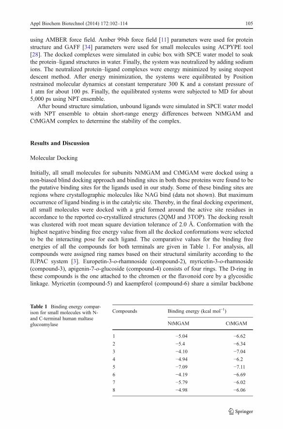
Initially, all small molecules for subunits NtMGAM and CtMGAM were docked using anon-biased blind docking approach and binding sites in both these proteins were found to bethe putative binding sites for the ligands used in our study. Some of these binding sites areregions where crystallographic molecules like NAG bind (data not shown). But maximumoccurrence of ligand binding is in the catalytic site. Thereby, in the final docking experiment,all small molecules were docked with a grid formed around the active site residues inaccordance to the reported co-crystallized structures (2QMJ and 3TOP). The docking resultwas clustered with root mean square deviation tolerance of 2.0 Å. Conformation with thehighest negative binding free energy value from all the docked conformations were selectedto be the interacting pose for each ligand. The comparative values for the binding freeenergies of all the compounds for both terminals are given in Table 1. For analysis, allcompounds were assigned ring names based on their structural similarity according to theIUPAC system [3]. Europetin-3-o-rhamnoside (compound-2), myricetin-3-o-rhamnoside(compound-3), apigenin-7-o-glucoside (compound-4) consists of four rings. The D-ring inthese compounds is the one attached to the chromen or the flavonoid core by a glycosidiclinkage. Myricetin (compound-5) and kaempferol (compound-6) share a similar backbone
Table 1 Binding energy compar-ison for small molecules with N-and C-terminal human maltaseglucoamylase
Compounds Binding energy (kcal mol−1)
NtMGAM CtMGAM
1 −5.04 −6.622 −5.4 −6.343 −4.10 −7.044 −4.94 −6.25 −7.09 −7.116 −4.19 −6.697 −5.79 −6.028 −4.98 −6.06
Appl Biochem Biotechnol (2014) 172:102–114 105

with compound 2, 3 and 4. Morroniside having a chromen backbone (compound-1) hasthree rings. Myrigalon-B (compound-5) and phloretin (compound-6) have two rings eachand the tri-substituted phenyl ring in these two structures is named ring A. The dockedcomplexes were visualized using PyMOL. Ligand bound in the binding site was focussed tovisualize the possible interacting residues (hydrogen bonding, van der Waals interaction andelectrostatic interaction).
Structural Differences in Binding Sites of NtMGAM and CtMGAM
There are two sugar subsites (+1 and −1) for substrate binding in the reported crystalstructure of acarbose–NtMGAM. Amino acids enclosing the +1 subsite are: R526,D542, D203, F450, F575, W406, Y299 and −1 subsite are: D327, D443, H600,W441, I364, D367 and I328 [27]. Both the sugar subsites are predominantly nega-tively charged with polar hydrophilic residues providing an open cavity for smallmolecule and water interactions. As compared to NtMGAM, there are two additionalsubsites in the catalytic domain of CtMGAM; +2 and +3. Amino acid residuesenclosing the −1 sugar subsite are: W1523, D1279, Y1251, W1418, H1584; +1 sugarsubsite is enclosed by: D1526, F1559, W1355, R1510; +2 sugar subsite is enclosedby: D1157 and W1369 and +3 sugar subsite is enclosed by: F1560 and P1159 [22].The presence of an additional 21 residues in CtMGAM accommodates larger sub-strates and better binding for molecules.
Docking of Small Molecules to NtMGAM
Hydrogen bond interaction of all docked small molecules with their amino acid partnershave been noted in Fig. 2a (Supplementary Data 2). The hydrogen bonding threshold wasgiven at 2.2 Å with a bond angle of 120°–180°. Myricetin, europetin-3-o-rhamnoside,myricetin-3-o-rhamnoside and apigenin-7-o-glucoside have similar structural backbones(flavonoid backbone). Myricetin has the least number of torsions (7) as compared toeuropetin-3-o-rhamnoside (11), myricetin-3-o-rhamnoside (11) and apigenin-7-o-glucoside(10) and thereby loses a lesser amount of torsional energy during docking. Therefore,myricetin has the least binding free energy of −7.09 kcal mol−1. Europetin-3-o-rhamnosideis weakly bound to D327 (+1 sugar subsite) and W605 through the O5 of ligand D-ring at adistance of 3.34 Å and O6 of ligand A-ring at 3.2 Å, respectively. O5 is also involved in amoderately strong hydrogen bonding with the catalytic nucleophile D443 at a distance of2.61 Å and thereby forms a bidentate H-bonding. Apigenin-7-o-glucoside forms weakbidentate H-bonding to R526 at a distance of 3.20 Å and to D443 at 3.01 Å via the O10of B-ring. Additionally, it weakly binds to R526 and D443 via the O4 and O8, respectively.On the other hand, morroniside has a chromen backbone with ten torsional regions and hashigher binding affinity as compared to apigenin-7-o-glucoside, owing to greater contribu-tions from the intermolecular forces. This could be explained by its three-ringed structurewhich provides a better orientation for interaction. It forms a moderately strong hydrogenbond with D327 (sugar subsite +1) at the O9 of C-ring at a distance of 2.63 Å. The O10 ofring A interacts with H600 (−1 sugar subsite) by a weak hydrogen bond at a distance of3.32 Å. The glycoside linked D-ring interacts deep in the sugar subsites, whereas the A- andB-ring (pyrano-pyran ring) projections have several non-bonded interactions and a weakhydrogen bonded interaction which is not so deeply embedded in the catalytic core.Myrigalon-B and phloretin have two rings with more than 75 % structural scaffoldingsimilarity. The B-rings of both myrigalon-B and phloretin are buried in the +1 sugar subsite.
106 Appl Biochem Biotechnol (2014) 172:102–114

B-ring is the contributor for H-bonding interactions with positively charged R526, R542,polar M444 and hydrophilic H600.
Fig. 2 a Docked poses of Syzygium-derived small molecules with N-terminal maltase glucoamylase. b Alldocked poses of compounds at the catalytic site of NtMGAM. Labelled residues are the interacting residuesengulfing the sugar sites. The stretch of amino acids in Insert 1 is represented by red cartoon and Insert 2 byblue
Appl Biochem Biotechnol (2014) 172:102–114 107
By superimposing the best binding conformation of all small molecules (Fig. 2b) itcan be shown that in compounds with more than three rings (compounds 2, 3 and 4)the D-ring plays a major role in forming H-bond with residues in the sugar subsite +1and −1 and has a high torsional flexibility to orient inside the active site. This canalso be observed in morroniside with a chromen ring backbone. The A- and B-ringsare slightly overhung and exposed outwards like acarbose in its crystal structure.Compounds with three rings or less orient themselves fairly well in the active sitewith greater binding affinity.
Docking of Small Molecules to CtMGAM
The C-terminal has two additional sugar subsites, +2 and +3, and therefore accommodateslarger substrate molecules [22]. We speculated greater amounts of intermolecular interactionand greater binding affinity due to the presence of additional subsites. The docking resultsconfirm the same. All molecules show apparent higher binding affinity towards CtMGAM.Myricetin has the best binding affinity of −7.11 kcal mol−1. Myricetin interacts with H1584in the −1 sugar subsite at 3.3 Å, D1526 and R1510 of the +1 sugar subsite at 2.78 and 3.0 Å,respectively, by OH substituted at C7, C11 of the ligand A-ring. Kaempferol, having 90 %structural similarity with myricetin, forms moderate H-bonding with H1584, D1279 andT1586 of the −1 subsite through the ligand B-ring. Among the pyrano-pyran ring moleculeswith four rings, europetin-3-o-rhamnoside (11), myricetin-3-o-rhamnoside (11) andapigenin-7-o-glucoside (10) have almost equal number of torsions and thereby loses thesame level of energy in docking. Although myricetin-3-o-rhamnoside has greater number ofH-bonded interactions, it has a higher binding affinity. Owing to its better orientation at thebinding site, chromen backbone containing morroniside has a slightly higher binding affinitythan pyrano-pyran backbone molecules. Morroniside interacts with +1 sugar subsite residuesH1584, T1586 and D1279 by forming H-bonding via substituted OH residues at C9, C11and C16 of D-ring. Myrigalon-B and phloretin has similar nature of binding in consequenceof high structural similarity, both interacting by H-bonding via substituted OH residues of A-ring.
Superimposing the best binding conformations of all small molecules (Fig. 3b) at thebinding site, it can be seen that all small molecules are fairly accommodated. Similar to theN-terminal binding, the D-ring plays a major role in orienting molecules at the binding sitefor compounds with four rings and similarly, the B-ring for compounds with three rings.Amino acid residues of additional +2 sugar subsites contribute to an increase in the bindingaffinity of small molecules with CtMGAM.
MD Simulation of Myricetin with NtMGAM and CtMGAM
The simulated complex structures of NtMGAM–myricetin and CtMGAM–myricetin weresuperimposed to observe the interaction at the binding site (Fig. 4). Simulated structuresshowed varied number of hydrogen bonds in both complexes throughout the simulation(Table 2). This indicated that alpha helix residue W1369 of CtMGAM at the active site helpsstabilize the complex by H-bond interaction, whereas the helical loop formed by additional21 amino acid residues in C-terminal is absent in NtMGAM. In order to verify conservednumber of hydrogen bonds throughout the simulation time, g_hbond tool of Gromacspackage was used. It showed that the average number of hydrogen bonds throughout thesimulation time for NtMGAM–myricetin and CtMGAM–myricetin were 2.2 and 3.4,respectively.
108 Appl Biochem Biotechnol (2014) 172:102–114

A stretched conformation was obtained in as final geometrical conformation of myricetinafter 5 ns molecular dynamics simulation. It forms intramolecular H-bonding between the
Fig. 3 a Docked poses of Syzygium-derived small molecules with C-terminal maltase glucoamylase. b All dockedposes of compounds at the catalytic site of CtMGAM. Labelled residues are the interacting residues engulfing thesugar sites. The stretch of amino acids in the Insert 1 is represented by red cartoon and Insert 2 by blue
Appl Biochem Biotechnol (2014) 172:102–114 109

C5 (OH) hydroxyl and C4 (O) carbonyl oxygen. This intramolecular H-bondingbehaviour of flavonoid compounds at similar positions is consistent with moleculardynamics study of human secretory PLA2-flavonoid [12] complex and these sites arethereby improbable for structural modification (i.e., addition of hydrophobic groups)for better lead design. Intramolecular H-bonding analysis of NtMGAM does not showany water-mediated amino acid interaction, but interacts with peripheral water mole-cules by C7 (OH). The H-bond plot obtained post simulation reveals that threehydrogen bonds are conserved during the entire period. The B-ring is held in thecatalytic −1 sugar subsite by interaction with D327 by forming strong bidentate H-bonding mediated through C11 (OH) and C12 (OH) at distances 1.45 and 1.752 Å,respectively. It also interacts with H600 by H-bonding via C12 (O) at a distance of1.8 Å (Fig. 5b). When compared to the docking conformation, myricetin shiftstowards the interior of the catalytic site, owing to its stretched conformation.
An analysis of the binding of myricetin with CtMGAM shows that the most critical andsignificant difference in the binding free energy is contributed by additional H-bondinginteraction due to a larger catalytic site. The H-bond plot reveals an average of four hydrogenbonds present throughout the course of simulation. Water molecules play a role in the
Fig. 4 Superimposition of simulated CtMGAM (green) with myricetin (red) complex shows hydrogen bondwith Trp1369 (yellow). In NtMGAM (cyan) structure Trp-1369 helical structure sequence was absent near tobinding region
Table 2 Hydrogen bond analysis post simulation
Protein structure Intermolecularhydrogen bonds
Residues involvedin H-bond
Average H-bond numbersin simulation
CtMGAM–myricetin 4 W1369, D1420, K1460, H1584 3.456
NtMGAM–myricetin 3 D327, H600 2.279
110 Appl Biochem Biotechnol (2014) 172:102–114

stabilization of the ligand at the catalytic site which is mediated through Intramolecular H-bonding. D1420 of the −1 sugar subsite mediates in orienting the B-ring of myricetin byforming bidentate H-bonding with C11 (OH) (Fig. 6b) at distances 1.569 and 2.206 Å,additional C12(OH) mediated linkages are also present. W1369 at +2 sugar subsite provides
Fig. 5 a Number of hydrogen bonds present in NtMGAM during the simulation, approx. 2–3 (blue). b MD-simulated conformation of myricetin in the NtMGAM binding site. Intramolecular H-bonding is depicted byblack solid lines
Fig. 6 a Number of hydrogen bonds present in CtMGAM during the simulation, approx. 3–4 (red). b MD-simulated conformation of myricetin in the CtMGAM binding site. Intramolecular H-bonding is depicted byblack solid lines
Appl Biochem Biotechnol (2014) 172:102–114 111

additional H- bonding linkage with C2 (OH) at 2.24 Å. H1584 and K1460 interacts withmyricetin by water-mediated interactions at C13 (OH) and C7 (O).
Potential energy for the complexes was obtained by MD trajectory analysis tool g_energy from Gromacs. NtMGAM–myricetin and CtMGAM–myricetin potential energywere equilibrated at −2.097×10⁶ kJ/mol and −2.461×10⁶ kJ/mol, respectively (Fig. 7).RMSD plot (Fig. 8) for protein backbone NtMGAM and CtMGAM shows an equilibrationof protein structure. NtMGAM–myricetin structure initially took 600 ps for equilibrationbeyond which it shows significantly stabilized average RMSD value of 0.2467 nm in5,000 ps. The CtMGAM–myricetin complex was stabilized after 750 ps with an averageRMSD value of 0.2230 nm in 5,000 ps. This RMSD value indicates a stabilization ofCtMGAM–myricetin in the presence of hydrogen bonds. Linear interaction energy calcula-tion considers short-range energy contribution between bound and unbound states of ligand[1, 6]. Short-range energy interactions such as Coulombic and van der Waals, for bothprotein–ligand complexes and free ligand solvation energy were obtained from the energyfile of simulation. These energies help measure energy differences between bound andunbound ligand and determines the short-range energy contributions for the interaction ofligand in the bound and unbound states. The total short-range energy contribution for
Fig. 7 Potential energy plot of a myricetin–NtMGAM complex. b Myricetin–CtMGAM complex evaluatedfrom 5,000 ps trajectory
Fig. 8 RMSD plot of a NtMGAM–myricetin complex (blue) and protein backbone (black). b CtMGAM–myricetin complex (red) and protein backbone (black)
112 Appl Biochem Biotechnol (2014) 172:102–114
CtMGAM–myricetin is lesser than the NtMGAM–myricetin. Short-range energy valuesshow differences in CtMGAM and NtMGAM stabilization with myricetin (Table 3).
Conclusion
Our findings suggest that flavonoids isolated from Syzygium sp. binds with differentspecificities to the terminals of human maltase glucoamylase. They demonstrate a higherbinding affinity for CtMGAM and a slightly lower binding affinity for NtMGAM. Wespeculate that this is majorly contributed by differences in their short-range energies;water-mediated interactions, H-bonding and thin structural variations between them. Mo-lecular dynamics simulation of the best docked compound myricetin shows stable complexformation with both the terminals in its stretched conformation. The extended 21 aminoacids forming a helical loop near the binding site of CtMGAM provide scope for stableintegration and interaction of molecules by H-bonding and thereby tend to have a higheraffinity for binding. A future aspect of this work is the development of 3D-QSAR model ofmyricetin-based pharmacophores for in vitro and in vivo evaluation.
Acknowledgments We thank the School of Biosciences and Technology, VIT University for providing fundsand infrastructure for this work. We gratefully acknowledge the CDAC supercomputing grid facility, BRAFfor providing cluster computer network for running the simulations.
References
1. Åqvist, J., & Hansson, T. (1996). The Journal of Physical Chemistry, 100, 9512–9521.2. Asano, N. (2003). Glycobiology, 13, 93R–104R.3. Beecher, G. R. (2003). Journal of Nutrition, 133, 3248S–3254S.4. Benalla, W., Bellahcen, S., & Bnouham, M. (2010). Current Diabetes Review, 6, 247–254.5. Ceriello, A. (2005). Diabetes, 54, 1–7.6. Cortés Cabrera, A., & Rueda Pérez, C. (2010) Anales de la Real Academia Nacional de Farmacia, pp. 3.7. Goodsell, D. S., Morris, G. M., & Olson, A. J. (1996). Journal of Molecular Recognition, 9, 1–5.8. Grover, J. K., Yadav, S., & Vats, V. (2002). Journal of Ethnopharmacology, 81, 81–100.9. Gunther, S., Wehrspaun, A., & Heymann, H. (1996). Archives of Biochemistry & Biophysics, 327, 295–
302.10. Helmstadter, A. (2008). Die Pharmazie-An International Journal of Pharmaceutical Sciences, 63, 91–
101.
Table 3 Short-energy data for protein–ligand complex
Proteincomplex
Columbic SRenergy inKJ/mol
vdW SRenergy inKJ/mol
Total averageSR interactionenergy
Ligandsolvation totalSR energy
(Total average SR interactionenergy)−(total ligandsolvation SR energy)
NtMGAM–myricetin
−150±3 −97±3 −247.857±2.5 −236.84 −11.017
CtMGAM–myricetin
−186.35±5 −83.065±1.5 −269.415±5.4 −236.84 −32.575
SR short range, vdW van der Waal
Appl Biochem Biotechnol (2014) 172:102–114 113
11. Hornak, V., Abel, R., Okur, A., Strockbine, B., Roitberg, A., & Simmerling, C. (2006). Proteins, 65, 712–725.
12. Lattig, J., Bohl, M., Fischer, P., Tischer, S., Tietbohl, C., Menschikowski, M., et al. (2007). Journal ofComputer Aided Molecular Design, 21, 473–483.
13. Manaharan, T., Appleton, D., Cheng, H. M., & Palanisamy, U. D. (2012). Food Chemistry, 132, 1802–1807.
14. Morris, G. M., Huey, R., Lindstrom, W., Sanner, M. F., Belew, R. K., Goodsell, D. S., et al. (2009).Journal of Computational Chemistry, 30, 2785–2791.
15. Nichols, B. L., Avery, S., Sen, P., Swallow, D. M., Hahn, D., & Sterchi, E. (2003). Proceedings of theNational Academy of Sciences of the United States of America, 100, 1432–1437.
16. Nichols, B. L., Eldering, J., Avery, S., Hahn, D., Quaroni, A., & Sterchi, E. (1998). Journal of BiologicalChemistry, 273, 3076–3081.
17. Nichols, B. L., Quezada-Calvillo, R., Robayo-Torres, C. C., Ao, Z., Hamaker, B. R., Butte, N. F., et al.(2009). Journal of Nutrition, 139, 684–690.
18. Oliveira, A. C., Endringer, D. C., Amorim, L. A., das Gracas, L. B. M., & Coelho, M. M. (2005). Journalof Ethnopharmacology, 102, 465–469.
19. Pettersen, E. F., Goddard, T. D., Huang, C. C., Couch, G. S., Greenblatt, D. M., Meng, E. C., et al. (2004).Journal of Computational Chemistry, 25, 1605–1612.
20. Quezada-Calvillo, R., Sim, L., Ao, Z., Hamaker, B. R., Quaroni, A., Brayer, G. D., et al. (2008). Journalof Nutrition, 138, 685–692.
21. Ren, L., Cao, X., Geng, P., Bai, F., & Bai, G. (2011). Carbohydrate Research, 346, 2688–2692.22. Ren, L., Qin, X., Cao, X., Wang, L., Bai, F., Bai, G., et al. (2011). Protein & Cell, 2, 827–836.23. Samadder, A., Chakraborty, D., De, A., Bhattacharyya, S. S., Bhadra, K., & Khuda-Bukhsh, A. R. (2011).
European Journal of Pharmaceutical Sciences, 44, 207–217.24. Sharma, S. B., Nasir, A., Prabhu, K. M., & Murthy, P. S. (2006). Journal of Ethnopharmacology, 104,
367–373.25. Sharma, S. B., Nasir, A., Prabhu, K. M., Murthy, P. S., & Dev, G. (2003). Journal of Ethnopharmacology,
85, 201–206.26. Shinde, J., Taldone, T., Barletta, M., Kunaparaju, N., Hu, B., Kumar, S., et al. (2008). Carbohydrate
Research, 343, 1278–1281.27. Sim, L., Quezada-Calvillo, R., Sterchi, E. E., Nichols, B. L., & Rose, D. R. (2008). Journal of Molecular
Biology, 375, 782–792.28. Sousa da Silva, A. W., & Vranken, W. F. (2012). BMC Research Notes, 5, 367.29. Srivastava, S., & Chandra, D. (2013). Journal of the Science of Food and Agriculture, 93(9), 2084–2093.30. Teixeira, C. C., Pinto, L. P., Kessler, F. H., Knijnik, L., Pinto, C. P., Gastaldo, G. J., et al. (1997). Journal
of Ethnopharmacology, 56, 209–213.31. Teixeira, C. C., Weinert, L. S., Barbosa, D. C., Ricken, C., Esteves, J. F., & Fuchs, F. D. (2004). Diabetes
Care, 27, 3019–3020.32. Van de Laar, F.A., Lucassen, P.L., Akkermans, R.P., Van de Lisdonk, E.H., Rutten, G.E., & Van Weel, C.
(2005). Cochrane Database of Systematic Reviews, CD003639.33. Wang, J., Wang, W., Kollman, P. A., & Case, D. A. (2006). Journal of Molecular Graphics and
Modelling, 25, 247–260.34. Wang, J., Wolf, R. M., Caldwell, J. W., Kollman, P. A., & Case, D. A. (2004). Journal of Computational
Chemistry, 25, 1157–1174.35. Yoshikawa, M., Morikawa, T., Matsuda, H., Tanabe, G., & Muraoka, O. (2002). Bioorganic & Medicinal
Chemistry, 10, 1547–1554.36. Yoshikawa, M., Murakami, T., Yashiro, K., & Matsuda, H. (1998). Chemical & pharmaceutical bulletin,
46, 1339.
114 Appl Biochem Biotechnol (2014) 172:102–114