probing cysteine self-assembled monolayers over gold ... · probing cysteine self-assembled...
TRANSCRIPT
Ps
AD
a
ARRAA
KEGSNA
1
drcnisdctmotf[
(
0d
Talanta 93 (2012) 264– 273
Contents lists available at SciVerse ScienceDirect
Talanta
jo u r n al hom epage: www.elsev ier .com/ locate / ta lanta
robing cysteine self-assembled monolayers over gold nanoparticles – Towardselective electrochemical sensors
hmed Galal ∗, Nada F. Atta, Ekram H. El-Adsepartment of Chemistry, Faculty of Science, Cairo University, 12613 Giza, Egypt
r t i c l e i n f o
rticle history:eceived 9 August 2011eceived in revised form 11 February 2012ccepted 15 February 2012vailable online 22 February 2012
eywords:lectrochemical sensorold nanoparticleself-assembly monolayereurotransmittersscorbic acid
a b s t r a c t
Cysteine forms self-assembled monolayers over gold nanoparticles. Based on this knowledge, a novelelectrochemical sensor (Au-Aunano-Cys-SDS) has been constructed by the formation of self-assemblymonolayer (SAM) of cysteine on gold-nanoparticles modified gold electrode (Au-Aunano-Cys) to be uti-lized for determination of dopamine in the presence of sodium dodecyl sulfate (SDS). Electrochemicalinvestigation and characterization of the modified electrode sensor was achieved using cyclic voltam-metry, electrochemical impedance spectroscopy, scanning electron, and atomic force microscopies.Au-Aunano-Cys electrode in the presence of SDS gave comparable high current response to that of thegold nanoparticles modified gold electrode (Au-Aunano). The Au-Aunano-Cys-SDS electrode current signalwas remarkably stable via repeated cycles and long term stability due to the strong Au S bond. Verysmall peak separation, almost zero or 15 mV peak separation was also obtained by repeated cycles indi-cating unusual high reversibility. The oxidation peak current was determined to be linearly dependent onthe dopamine concentration. A resulting calibration curve using square wave voltammetry (SWV) was
−1 −1
obtained over concentration range of 30–100 �mol L and 120–320 �mol L with correlation coeffi-cients of 0.996 and 0.994 and a limit of detection of 16 and 57 nmol L−1, respectively. Using differentialpulse voltammetry (DPV), a highly selective and simultaneous determination of tertiary mixture of ascor-bic acid AA, dopamine, and acetaminophen APAP was explored at this modified electrode. It has beendemonstrated that Au-Aunano-Cys-SDS electrode can be used as a sensor with excellent reproducibility,sensitivity, and long term stability.. Introduction
Catecholamines are a class of neurotransmitters, and theiretection in the human body has been of great interest to neu-oscientists [1]. Dopamine (DA) is one of the most significantatecholamines and belongs to the family of excitatory chemicaleurotransmitters. It plays a very important role in the function-
ng of the central nervous, cardiovascular, renal and hormonalystems, as well as a key role in drug addiction, and Parkinson’sisease [2]. Tremendous consideration has been given in biomedi-al research; to design selective, sensitive and reliable methods forhe direct measurement of dopamine in the presence of interfering
olecules. Electrochemical analysis of binary or tertiary mixturen unmodified electrodes such as glassy carbon (GC), Pt, Au elec-
rodes has limitations because of overlapping voltammetric peaks,ouling due to adsorption of byproducts and high detection limits1–4]. To overcome these problems, various modified electrodes∗ Corresponding author. Tel.: +20 0237825266; fax: +20 0235727556.E-mail addresses: [email protected], [email protected], [email protected]
A. Galal).
039-9140/$ – see front matter © 2012 Elsevier B.V. All rights reserved.oi:10.1016/j.talanta.2012.02.032
© 2012 Elsevier B.V. All rights reserved.
have been constructed. Some examples of modified electrodesinclude Pd and Pt nanoclusters modified poly(3-methylthiophene)(PMT), poly(N-methylpyrrole) (PMPy) and polyfuran film-coatedplatinum (Pt) electrode [1–4], poly(3,4-ethylene dioxythiophene)(PEDOT) modified Pt electrode in the presence of sodium dodecylsulfate (SDS) [5], gold nanoparticles [6–12], self-assembled mono-layer (SAM) [13–32], and gold nanoparticles immobilized on SAMmodified electrodes [33–37]. Self-assembly procedure is precisemodification of the surface structure in nanometer-scale, whichwas employed recently in surface protection, electronics, and fab-rication of sensors and biosensors [18].
SAM which was formed by spontaneous adsorption of self-assembling molecules on metals such as gold, silver, copper, iron,nickel, or platinum [23,30,38,39], was found as an elegant wayto orient as well as address electrically a molecular componentof interest [23]. In particular the self-assembly of organosulfurcompounds on gold surface has been extensively studied [19]. Ithas been shown that organosulfur compounds upon spontaneous
chemisorption on gold surface, lose the hydrogen from the thiolgroup as molecular hydrogen H2 [30] resulting in a strong, cova-lent and thermodynamically favored S Au bond formation [21].The reason for adsorbing thiols on gold as a preferred substrate is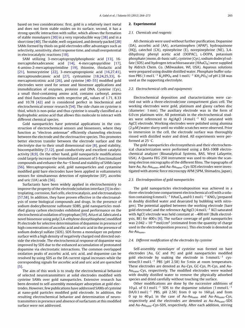
nta 9
basosSsi
m4[mmeiaaaethd
sfbbebacc[msa
it[ysienPhsssidorc[
ocbtarts
DA/0.1 mol L−1 PBS, pH 2.58) from 0 up to 160 �L and from0 up to 40 �L in the case of Au-Aunano and Au-Aunano-Cys,
A. Galal et al. / Tala
ased on two considerations: first, gold is a relatively inert metalnd does not form stable oxides on its surface, second, it has atrong specific interaction with sulfur, which allows the formationf stable monolayers [30] in a very reproducible way [38] and in ahort time [40]. The stable, well-organized, and densely packed [28]AMs formed by thiols on gold electrodes offer advantages such aselectivity, sensitivity, short response time, and small overpotentialn electrocatalytic reactions [18].
SAM utilizing 3-mercaptopropylphosphonic acid [13], 16-ercaptohexadecanoic acid [14], 4-mercaptopyridine [17],
-amino-2-mercaptopyrimidine [19], thionine, thiolactic acid21], homocysteine [22], 3-mercaptopropionic acid [16,27,41],
ercaptoundecanoic acid [27], cysteamine [18,24,25,33], 6-ercaptonicotinic acid [26], and cysteine [40–55] modified gold
lectrodes were used for sensor and biosensor applications andmmobilization of enzymes, proteins and DNA. Cysteine (Cys),
small thiol-containing amino acid, contains carboxyl, aminond thiol functionalities with respective pKa values of 1.71, 8.33nd 10.78 [42] and is considered perfect in biochemical andlectrochemical sensor research [54]. The side chain on cysteine ishiol, which is non-polar and thus cysteine is usually classified as aydrophobic amino acid that allows this molecule to interact withifferent chemical species.
Gold nanoparticles have potential applications in the con-truction of electrochemical sensors and biosensors, where theyunction as “electron antennae” efficiently channeling electronsetween the electrode and the electroactive species. This promotesetter electron transfer between the electrode surface and thelectrolyte due to their small dimensional size [8], good stability,iocompatibility [7,12], good conductivity and excellent catalyticctivity [8,9]. On the other hand, gold nanoparticle modificationsould largely increase the immobilized amount of S-functionalizedompounds and enhance the Au S bond and stability of SAMs layer56]. Mercaptopropionic acid, gold nanoparticles and cystamine
odified gold bare electrodes have been applied in voltammetricensors for simultaneous detection of epinephrine (EP), ascorbicnd uric (UA) acids [56].
Surfactants have been widely applied in electrochemistry tomprove the property of the electrode/solution interface [5] in elec-roplating, corrosion, fuel cells, electrocatalysis, and electroanalysis57,58]. In addition, they have proven effective in the electroanal-sis of some biological compounds and drugs. In the presence ofodium dodecylbenzene sulfonate SDBS, gold nanoparticles mod-fied glassy carbon electrodes exhibited good performance on thelectrochemical oxidation of tryptophan [59]. Atta et al. fabricated aovel biosensor using poly(3,4-ethylene dioxythiophene) modifiedt electrode for selective determination of dopamine in presence ofigh concentrations of ascorbic acid and uric acid in the presence ofodium dodecyl sulfate (SDS). SDS forms a monolayer on polymerurface with a high density of negatively charged end directed out-ide the electrode. The electrochemical response of dopamine wasmproved by SDS due to the enhanced accumulation of protonatedopamine via electrostatic interactions. The common overlappedxidation peaks of ascorbic acid, uric acid, and dopamine can beesolved by using SDS as the DA current signal increases while theorresponding signals for ascorbic acid and uric acid are quenched5].
The aim of this work is to study the electrochemical behaviorf selected neurotransmitters at solid electrodes modified withysteine SAMs over gold nanoparticles. Extensive research haseen devoted to self-assembly monolayer adsorption at gold elec-rodes. However, few publications have addressed SAMs of cysteinet nano-gold particles modified gold electrode. In addition, theesulting electrochemical behavior and determination of neuro-
ransmitters in presence and absence of surfactants at this modifiedurface were not addressed.3 (2012) 264– 273 265
2. Experimental
2.1. Chemicals and reagents
All chemicals were used without further purification. Dopamine(DA), ascorbic acid (AA), acetaminophen (APAP), hydroquinone(HQ), catechol (CA), epinephrine (E), norepinephrine (NE), 3,4-dihydroxy phenyl acetic acid (DOPAC), l-DOPA, potassiumphosphate (mono, di-basic salt), cysteine (Cys), sodium dodecyl sul-fate (SDS) and hydrogen tetrachloroaurate (HAuCl4) were suppliedby Aldrich Chem. Co. (Milwaukee, WI, USA). Aqueous solutionswere prepared using double distilled water. Phosphate buffer solu-tion PBS (1 mol L−1 K2HPO4 and 1 mol L−1 KH2PO4) of pH 2.58 wasused as the supporting electrolyte.
2.2. Electrochemical cells and equipments
Electrochemical deposition and characterization were car-ried out with a three-electrode/one compartment glass cell. Theworking electrodes were gold, platinum and glassy carbon disc(diameter: 1 mm). The auxiliary electrode was in the form of6.0 cm platinum wire. All potentials in the electrochemical stud-ies were referenced to Ag/AgCl (4 mol L−1 KCl saturated withAgCl) electrode. Working electrodes were polished using alumina(2 �M)/water slurry until no visible scratches were observed. Priorto immersion in the cell, the electrode surface was thoroughlyrinsed with distilled water and dried. All experiments were per-formed at 25 ◦C.
The gold nanoparticles electrosynthesis and their electrochem-ical characterization were performed using a BAS-100B electro-chemical analyzer (Bioanalytical Systems, BAS, West Lafayette,USA). A Quanta FEG 250 instrument was used to obtain the scan-ning electron micrographs of the different films. The topographs ofbare Au, Au-Aunano, and Au-Aunano-Cys-SDS electrodes were inves-tigated with atomic force microscopy AFM (SPM, Shimadzu, Japan).
2.3. Electrodeposition of gold nanoparticles
The gold nanoparticles electrodeposition was achieved in athree-electrode/one compartment electrochemical cell with a solu-tion containing 6 mmol L−1 HAuCl4 and 0.1 mol L−1 KNO3 (preparedin doubly distilled water and deaerated by bubbling with nitro-gen). The potential applied between the working electrode (baregold electrode) and the reference Ag/AgCl (4 mol L−1 KCl saturatedwith AgCl) electrode was held constant at −400 mV (Bulk electrol-ysis, BE) for 400 s [6]. The surface coverage of gold nanoparticleswas 2.042 × 10⁻⁶ mol/cm2 (estimated from the quantity of chargeused in the electrodeposition process). This electrode is denoted asAu-Aunano.
2.4. Different modification of the electrodes by cysteine
Self-assembly monolayer of cysteine was formed on bareelectrodes (Au, GC and Pt) and gold nanoparticles modifiedgold electrode by soaking the electrode in 5 mmol L−1 cys-teine/0.1 mol L−1 PBS (pH 2.58) for 5 min at room temperature.These electrodes are denoted as Au-Cys, GC-Cys, Pt-Cys, and Au-Aunano-Cys, respectively. The modified electrodes were washedwith doubly distilled water to remove the physically adsorbedspecies and dried carefully without touching the surface.
Other modifications are done by the successive additions of10 �L of 0.1 mol L−1 SDS to the dopamine solution (1 mmol L−1
respectively and the electrodes are denoted as Au-Aunano-SDSand Au-Aunano-Cys-SDS, respectively. After each addition, stirring
266 A. Galal et al. / Talanta 93 (2012) 264– 273
Ft
te
2
idnramdv
3
3
D(cisfawspiaetcAtggAgCat
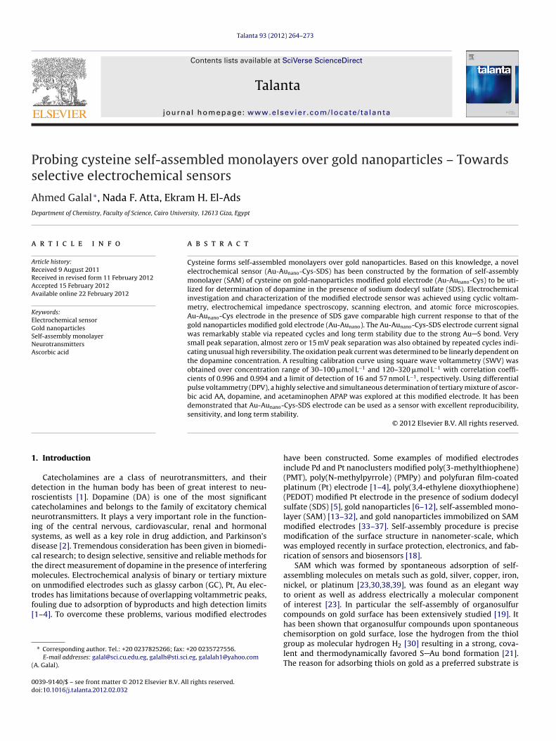
ig. 1. CVs of 1 mmol L−1 DA/0.1 mol L−1 PBS (pH 2.58) at bare Au and Au-Cys elec-rodes, scan rate 50 mV s−1.
akes place for 5 min then holds for 1 min before running thexperiment.
.5. Electrochemical impedance spectroscopy (EIS)
EIS was performed using a Gamry-750 instrument and a lock-n-amplifier that was connected to a personal computer. Theata analysis was provided with the instrument and appliedon-linear least square fitting with Levenberg–Marquardt algo-ithm. All impedance experiments were recorded between 0.1 Hznd 100 kHz with an excitation signal of 10 mV amplitude. Theeasurements were performed under potentiostatic control at
ifferent applied potentials which were decided from the cyclicoltammograms recorded for the modified electrodes.
. Results and discussion
.1. Electrochemistry of DA at cysteine modified gold electrode
Fig. 1 shows the cyclic voltammetric (CV) behavior of 1 mmol L−1
A in 0.1 mol L−1 PBS (pH 2.58) tested at bare gold electrode Ausolid line) and gold modified with cysteine Au-Cys (dash line). It islear that the redox voltammetric peaks of DA at bare gold electrodes quasi-reversible with peak potential separation (�Ep = 448 mV),uggesting slow electron transfer kinetics, presumably due to theouling of the electrode surface by the oxidation products. However,
relatively well-defined redox waves with better reversibility of DAere obtained at Au-Cys electrode. The oxidation peak potential
hifts negatively to 636 mV, and the reduction peak potential shiftsositively to 379 mV (�Ep = 257 mV). The reduction peak current
ncreases significantly from 0.48 �A to 2.1 �A at Au-Cys electrodes compared to the bare Au electrode. The Au-Cys SAM modifiedlectrode affects the mechanistic redox reaction of DA with twoypes of interactions. The first is the interaction between Au andysteine through thiol group of cysteine to form strong covalentu S bond (molecule–substrate interaction). The second interac-
ion is between cysteine on Au and DA molecules through aminoroup of cysteine and hydroxyl-phenol group of DA via hydro-en bond formation (molecule–molecule interaction). The DA onu-Cys electrocatalytic oxidation is due to the formation of a hydro-
en bond between hydroxyl-phenol of DA and the nitrogen in theys-SAM. The hydrogen bond formation has a great impact on thectivation of the hydroxyl-phenol on the benzene ring. As a result,he bond energy between hydrogen and oxygen is weakened andFig. 2. CVs of separation of binary mixture of: 1 mmol L−1 AA and 1 mmol L−1 l-DOPAat bare Au and Au-Cys electrodes, scan rate 50 mV s−1.
the electron transfer is more likely to occur via N· · ·H O bond. Thus,the Cys-SAM system can act as a promoter to increase the rate ofelectron transfer, lowering the overpotential of DA at the modifiedelectrode, and enhancing the reversibility of the redox reaction ofdopamine.
Also, Au-Cys electrode shows excellent stability via repeatedcycles (Supplement 1) which means no fouling of surface byoxidized products of DA as seem on bare gold electrodes. ThusAu-Cys electrode is a chemically modified electrode demonstratesantifouling properties with excellent stability for the oxidation ofneurotransmitters. The same study was done for other compoundssuch as E, NE, DOPAC, l-DOPA, HQ, CA, and AA and the trend wasrepeated.
3.2. Binary and tertiary mixture separation at cysteine SAMmodified gold electrode
Fig. 2 shows CVs for the analysis of a binary mixture of1 mmol L−1 AA, and 1 mmol L−1 l-DOPA in 0.1 mol L−1 PBS (pH 2.58)at bare Au (solid line) and Au-Cys (dash line) electrodes. An unre-solved oxidation peak at 833 mV was observed illustrating that theoxidation peaks of AA, and l-DOPA cannot be separated on thebare Au electrode. However, two resolved peaks were obtainedat Au-Cys electrode at 391 mV and 659 mV for AA and l-DOPA,respectively. This indicates that this sensor can be used for thesimultaneous determination of AA and l-DOPA. Also, the analy-sis of a binary mixture of 1 mmol L−1 AA, and 1 mmol L−1 DA in0.1 mol L−1 PBS (pH 2.58) at bare Au electrode and Au-Cys mod-ified electrode was studied and two well separated signals wereobtained at Au-Cys electrode at 402 mV, and 606 mV for AA, and DA,respectively (figure not shown). In addition, Au-Cys electrode canbe used to separate tertiary mixture of 1 mmol L−1 AA, 1 mmol L−1
DA, and 1 mmol L−1 APAP in 0.1 mol L−1 PBS (pH 2.58) as evidencedby three resolved peaks at 356 mV, 556 mV, and 664 mV for AA, DA,and APAP, respectively (Supplement 2).
3.3. The effect of cysteine SAM on different substrates
Cysteine SAM was formed by spontaneous adsorption of self-assembled molecules on metals such as gold, silver, copper, iron,
nickel, palladium or platinum [23,30,38,60]. Fig. 3(A) shows theCVs of 1 mmol L−1 DA in 0.1 mol L−1 PBS (pH 2.58) at differentelectrodes (Au, Pt, and GC) modified with cysteine. If we comparethese CVs with those obtained at bare electrodes, we concluded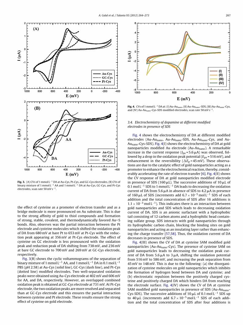
A. Galal et al. / Talanta 93 (2012) 264– 273 267
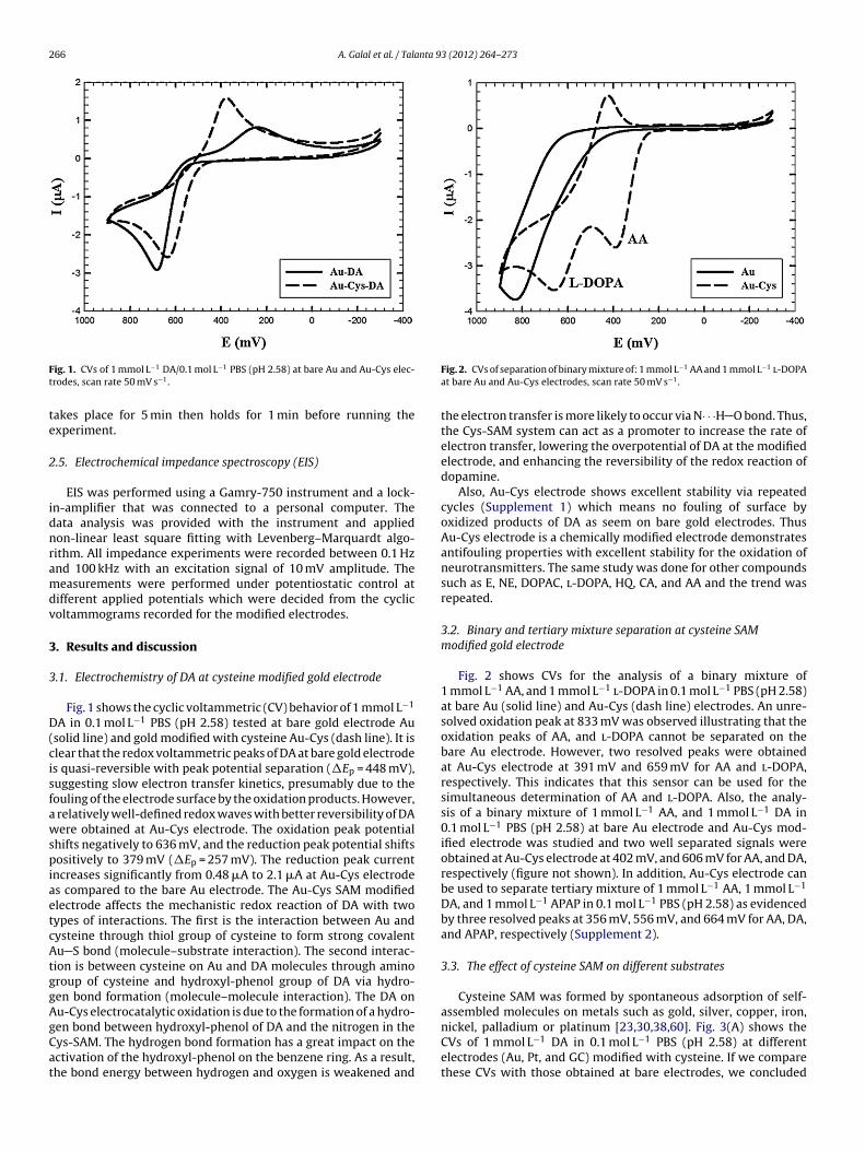
Fig. 3. (A) CVs of 1 mmol L−1 DA at Au-Cys, Pt-Cys, and GC-Cys electrodes, (B) CVs ofbinary mixture of 1 mmol L−1 AA and 1 mmol L−1 DA at Au-Cys, GC-Cys, and Pt-Cyse −1
tbtobeotcpar
bP(pfoetbe
Cys-SDS). The successive additions of 10 �L of 0.1 mol L−1 SDS up
lectrodes, scan rate 50 mV s .
he effect of cysteine as a promoter of electron transfer and as aridge molecule is more pronounced on Au substrate. This is dueo the strong affinity of gold to thiol compounds and formationf strong, stable, covalent, and thermodynamically favored Au Sonds. Also, observes was the partial interaction between the Ptlectrode and cysteine molecules which shifted the oxidation peakf DA from 680 mV at bare Pt to 653 mV at Pt-Cys with the reduc-ion peak appearing at 356 mV at Pt-Cys electrode. The effect ofysteine on GC electrode is less pronounced with the oxidationeak and reduction peak of DA shifting from 738 mV, and 236 mVt bare GC electrode to 700 mV and 269 mV at GC-Cys electrode,espectively.
Fig. 3(B) shows the cyclic voltammograms of the separation ofinary mixture of 1 mmol L−1 AA, and 1 mmol L−1 DA in 0.1 mol L−1
BS (pH 2.58) at Au-Cys (solid line), GC-Cys (dash line), and Pt-Cysdotted line) modified electrodes. Two well-separated oxidationeaks were obtained using Au-Cys electrode at 402 mV and 606 mVor AA, and DA, respectively. However, an overlapped combinedxidation peak is obtained at GC-Cys electrode at 731 mV. At Pt-Cyslectrode, the two oxidation peaks are more resolved and separatedhan at GC-Cys electrode and this ensures the partial interaction
etween cysteine and Pt electrode. These results ensure the strongffect of cysteine on gold electrode.Fig. 4. CVs of 1 mmol L−1 DA at: (I) Au-Aunano, (II) Au-Aunano-SDS, (III) Au-Aunano-Cys,and (IV) Au-Aunano-Cys-SDS modified electrodes, scan rate 50 mV s−1.
3.4. Electrochemistry of dopamine at different modifiedelectrodes in presence of SDS
Fig. 4 shows the electrochemistry of DA at different modifiedelectrodes (Au-Aunano, Au-Aunano-SDS, Au-Aunano-Cys, and Au-Aunano-Cys-SDS). Fig. 4(I) shows the electrochemistry of DA at goldnanoparticles modified Au electrode (Au-Aunano). A remarkableincrease in the current response (Ipa = 5.6 �A) was observed, fol-lowed by a drop in the oxidation peak potential (Epa = 516 mV), andenhancement in the reversibility (�Ep = 45 mV). These observa-tions are due to the catalytic effect of gold nanoparticles acting as apromoter to enhance the electrochemical reaction, thereby, consid-erably accelerating the rate of electron transfer [6]. Fig. 4(II) showsthe CV response of DA at gold nanoparticles modified electrodein presence of SDS (160 �L). The successive additions of 10 �L of0.1 mol L−1 SDS to 1 mmol L−1 DA leads to decreasing the oxidationcurrent of DA from 5.6 �A in absence of SDS to 4.2 �A in presenceof 160 �L of SDS (increments add 6.7 × 10⁻⁵ mol L−1 SDS of eachaddition and the total concentration of SDS after 16 additions is1.1 × 10−3 mol L−1). This indicates there is an interaction betweengold nanoparticles and SDS which leads to decreasing oxidationcurrent of DA. SDS is an anionic surfactant with a hydrophobictail consisting of 12 carbon atoms and a hydrophilic head contain-ing sulfate group. SDS interacts with gold nanoparticles throughits hydrophobic carbon chain, blocking the binding sites on goldnanoparticles and acting as an insulating layer rather than enhanc-ing the charge transfer [57,58]. Thus, the oxidation current of DAdecreases in presence of SDS.
Fig. 4(III) shows the CV of DA at cysteine SAM modified goldnanoparticles (Au-Aunano-Cys). The presence of cysteine SAM ongold nanoparticles leads to decreasing the oxidation peak cur-rent of DA from 5.6 �A to 3 �A, shifting the oxidation potentialfrom 516 mV to 586 mV, and increasing the peak separation from45 mV to 148 mV. This is due to the following: (a) the disorgani-zation of cysteine molecules on gold nanoparticles which inhibitsthe formation of hydrogen bond between DA and cysteine; and(b) electrostatic repulsion between the positively charged cys-teine and positively charged DA which hinders DA from reachingthe electrode surface. Fig. 4(IV) shows the CV of DA at cysteineSAM modified gold nanoparticles in presence of SDS (Au-Aunano-
to 40 �L (increments add 6.7 × 10⁻⁵ mol L−1 SDS of each addi-tion and the total concentration of SDS after four additions is

268 A. Galal et al. / Talanta 93 (2012) 264– 273
Sc
235iSaetcthf
conrttb
brs
3
v(terrrAe
trf(ip
cheme 1. Schematic model of Au-Aunano-Cys-SDS electrode in presence of DAation and AA.
.7 × 10−4 mol L−1) leads to increasing the oxidation current from �A to 4.3 �A, shifting the oxidation potential from 586 mV to04 mV and decreasing the peak separation from 148 mV to 47 mV
n presence of 40 �L SDS. The schematic model of Au-Aunano-Cys-DS electrode is illustrated in Scheme 1. There is an electrostaticttraction between the cationic DA and the anionic SDS whichnhances the diffusion of DA through the positively charged cys-eine SAM. Also there is an interaction between the positivelyharged cysteine and anionic SDS which allows the reorganiza-ion of cysteine molecules on gold nanoparticles. This enhances theydrogen bond formation between DA and cysteine and promotes
aster electron transfer kinetics.When the CVs of DA at Au-Cys and Au-Aunano-Cys electrodes are
ompared, it is concluded that the effect of cysteine on the responsef polycrystalline bare Au is more pronounced than that on goldanoparticles. The effect of adding SDS on gold nanoparticlesesults in enhanced signal response. This allows the reorganiza-ion of cysteine on gold nanoparticles that results in the increase ofhe oxidation current, decrease in the peak separation and henceetter reversibility.
Au-Aunano electrode displays a little higher current responseut Au-Aunano-Cys in presence of SDS gives beside high currentesponse, shows better repeatability and long term stability. Thistudy will focus on Au-Aunano-Cys-SDS electrode.
.5. Stability of the modified electrodes
The stability of the different modified electrodes was studiedia repeated cycles up to 50 cycles. Au-Aunano, Au-Aunano-SDS160 �L), Au-Aunano-Cys, and Au-Aunano-Cys-SDS (40 �L) elec-rodes in 1 mmol L−1 DA/0.1 mol L−1 PBS (pH 2.58) producedxcellent stability as evidenced by noticeable decrease in currentesponse indicating that these modified electrodes have a goodeproducibility and do not suffer from surface fouling during theepetitive voltammetric measurement [5]. In comparison, the bareu electrode demonstrated poor stability relative to the modifiedlectrodes due to surface fouling by cysteine oxidation products.
Very small peak separation (almost zero or 15 mV peak separa-ion) was obtained indicating unusually high reversibility throughepeated cycling (Supplement 3). Comparing the stability of the CVs
or the 1st, 25th, and 50th cycles on the Au-Aunano-Cys electrodefigure not shown), it was observed that Ipa increased from 2.5 �An the 1st cycle up to 3.8 �A in the 25th and 50th cycles. Also, theeak separation decreased from 120 mV in the 1st cycle to ∼0 mV inFig. 5. Long term stability of: (A) Au-Aunano and (B) Au-Aunano-Cys-SDS (40 �L) afterone week, 50 repeated cycles, 50 mV s−1 scan rate.
the 25th and 50th cycles. This indicates that cysteine molecules arereorganized on gold nanoparticles by repeated cycles. This reorga-nization enhances the hydrogen bond formation between DA andcysteine which increases the electron transfer rate. Thus, cysteinemolecules are reorganized on gold nanoparticles by repeated cyclesor by the addition of SDS (instantaneous reorganization).
In addition, the long term stability of Au-Aunano, Au-Aunano-Cysand Au-Aunano-Cys-SDS electrodes was studied for a time periodof up to one week. After each measurement, the electrode wasstored in 0.1 mol L−1 PBS (pH 2.58) in the refrigerator (at 5 ◦C).Fig. 5(A) and (B) shows the repeated cycles after one week of stor-age of Au-Aunano, and Au-Aunano-Cys-SDS electrodes, respectively.In the case of Au-Aunano electrode, Ipa of the 50th cycle decreasedby 26%, and 44% after 3 days and 1 week, respectively. Peak sepa-ration increased to 120 mV, and 180 mV after 3 days and one weekof storage, respectively.
In the case of Au-Aunano-Cys electrode, Ipa of the 50th cycledecreased by 23.6%, and 35.6% after 3 days and 1 week, respec-tively. Peak separation was zero and 15 mV after 3 days and 1 week,respectively. Also, for Au-Aunano-Cys-SDS electrode, Ipa decreasedby 24% and the peak separation was 15, and 60 mV after 3 days
and one week of storage, respectively. These results indicate thatthe presence of SAM of cysteine on gold nanoparticles enhancesthe reversibility and the long term stability of Au-Aunano-Cys and
A. Galal et al. / Talanta 93 (2012) 264– 273 269
Fig. 6. Linear relationship of Ip vs. �1/2 for 1 mmol L−1 DA at (I) Au-Aunano, (II) Au-Aunano-SDS, (III) Au-Aunano-Cys, and (IV) Au-Aunano-Cys-SDS.
Table 1Values of diffusion coefficient of different modified electrodes.
Electrode type Dox (cm2 s−1) Dred (cm2 s−1)
Bare Au 5.89 × 10⁻⁶ 6.97 × 10⁻⁸
Au-Cys 3.66 × 10⁻⁶ 2.79 × 10⁻⁶
Au-Aunano 1.74 × 10⁻⁵ 1.16 × 10⁻⁵
Au-Au -Cys 5.00 × 10⁻⁶ 5.88 × 10⁻⁶
Ab
3
(Tsb
I
wTirionm
calAAdvSpeit
Fig. 7. (A) SWV of 15 mL of 0.1 mol L−1 PBS (pH 2.58) at Au-Aunano-Cys elec-−1
nano
Au-Aunano-SDS 9.88 × 10⁻⁶ 1.01 × 10⁻⁵
Au-Aunano-Cys-SDS (40 �L) 1.06 × 10⁻⁵ 8.92 × 10⁻⁶
u-Aunano-Cys-SDS electrodes due to the formation of strong Au Sond.
.6. Apparent diffusion coefficients
The diffusion coefficients of 1 mmol L−1 DA in 0.1 mol L−1 PBSpH 2.58) at different modified electrodes have been calculated.he relation between anodic peak current (Ipa A) and the diffu-ion coefficient of the electroactive species (D cm2 s−1), is giveny Randles–Sevcik equation:
pa = (2.69 × 10⁵)n3/2AC◦D1/2�1/2
here n is the number of electrons exchanged in oxidation at = 298 K, A is the geometrical electrode area = 7.854 × 10⁻³ cm2, C◦
s the analyte concentration (1 × 10⁻⁶ mol cm−3) and v is the scanate V s−1 [6]. It is important to note the apparent surface area usedn the calculations does not take into account the surface roughnessf gold nanoparticles (the roughness factor of bare gold and goldanoparticles calculated from atomic force microscopy measure-ents are 0.656 and 1.07, respectively).For a diffusion-controlled process, a plot of the anodic peak
urrent values versus the square root of the scan rate results in straight-line relationship. Fig. 6 shows a comparison of theseinear relationships for DA at different modified electrodes: (I)u-Aunano, (II) Au-Aunano-SDS, (III) Au-Aunano-Cys and (IV) Au-unano-Cys-SDS. Also, Table 1 summarizes the Dapp values for DA atifferent modified electrodes. From Table 1 it is shown that the Dapp
alues increase in the following order: Au-Aunano-Cys < Au-Aunano-DS < Au-Aunano-Cys-SDS < Au-Aunano. These results confirm the
revious results that the presence of cysteine SAM on bare Aulectrode enhances the electron transfer rate. The sequence ofncreasing Dapp obtained from scan rate effect is comparable withhat obtained from chronoamperometry study.trode in presence of 40 �L of 0.1 mol L SDS in different concentrations ofDA (30–320 �mol L−1), (B) calibration curve for DA of concentrations from(120 �mol L−1 to 320 �mol L−1) and from (30 �mol L−1 to 100 �mol L−1, the inset).
3.7. Determination of dopamine using Au-Aunano-Cys in presenceof SDS
The voltammetric behavior of DA was examined using squarewave voltammetry (SWV). Fig. 7(A) shows typical SWV of suc-cessive additions of 10 �l of 1 mmol L−1 dopamine to 40 �Lof 0.1 mol L−1 SDS in 15 mL of 0.1 mol L−1 PBS (pH 2.58)with the total concentration of SDS after 4 additions being2.7 × 10−4 mol L−1. Fig. 7(A) shows that by increasing the con-centration of DA, the anodic peak current increased indicatingthe electrochemical response of DA is apparently improvedby SDS addition due to enhanced accumulation of protonatedDA via electrostatic interaction with negatively charged SDSat the Au-Aunano-Cys electrode surface. Fig. 7(B) and the insetshow the calibration curves of the anodic peak current val-ues in the linear range of 30–100 �mol L−1 with the regressionequation of Ip (A) = 1.927 × 10⁻⁸c (�mol L−1) + (−4.586 × 10⁻⁷)and 120–320 �mol L−1 DA with the regression equation of Ip(A) = 1.396 × 10⁻⁸c (�mol L−1) + (2.465 × 10⁻⁷) with correlationcoefficients of 0.996 and 0.994 and detection limits of 16 nmol L−1
and 57 nmol L−1, respectively. The detection limit was calculated
from the equation:DL = 3s
b,

270 A. Galal et al. / Talanta 93 (2012) 264– 273
Table 2Comparison for determination of dopamine at various modified electrodes based in literature reports.
Electrode Compound pH LDR (�M) Sensitivity (�A/�M) LOD (nM) Reference
CPE-Aunano DA 7.4 0.1–6 0.288 5.9 [6]GC-CA-Aunano DA 7.0 0.01–25 9.79 4 [7]ITO-Aunano DA 7.2 0.001–500 NR 0.53.0 [8]
5-HT 0.01–250 NRAu-MPPA DA in presence of 1 mM AA 7.4 0.4–20 0.0475 150 [13]Au-Cys UA 7.0 54–150 0.0099 2000 [53]
AA 140–500 0.0040 11,000Au-Cys HQ 4.8 2–200 0.034 400 [40]Au-CA-Aunano DA in presence of 0.1 mM AA 7.2 NR NR 220 [33]Pt-PEDOT-SDS DA 7.4 0.5–25 NR 61 [5]Au-Au -Cys-SDS DA 2.58 30–100 0.0193 16 This work
L n pasm
wtde
3
acrDecI07e
Fa6a
TE
nano
DR, linear dynamic range; LOD, limit of detection; NR, not reported; CPE, carboercaptopropylphosphonic acid; HQ, hydroquinone.
here s is the standard deviation and b is the slope of the calibra-ion curve. Table 2 shows the comparison for the determination ofopamine at Au-Aunano-Cys-SDS electrode with various modifiedlectrodes based in literature reports.
.8. Effect of solution pH
It is known that buffer solution pH affects protonation of themino acid and the sensitivity of the electrode. The effect ofhanging pH of the supporting electrolyte on the electrochemicalesponse of DA was studied. Fig. 8 shows the CVs of 1 mmol L−1
A in 0.1 mol L−1 PBS of pH 2.58, 7.00, and 9.00 at Au-Aunano-Cyslectrode in presence of 40 �L of 0.1 mol L−1 SDS with the totaloncentration of SDS after 4 additions being 2.7 × 10⁻⁴ mol L−1.
t should be mentioned that cysteine solution was prepared in.1 mol L−1 PBS of the same pH of the DA solution studied (2.58,.00, and 9.00) and SDS was prepared in double distilled water. It isvident that changing pH of the supporting electrolyte altered bothig. 8. CVs of 1 mmol L−1 DA in 0.1 mol L−1 PBS of different pH 2.58, 7.00, and 9.00t Au-Aunano-Cys electrode in presence of 40 �L of 0.1 mol L−1 SDS (increments add.7 × 10−5 mol L−1 SDS of each addition and the total concentration of SDS after 4dditions is 2.7 × 10⁻⁴ mol L−1).
able 3IS fitting data corresponding to Fig. 9(A).
RL1/104 � cm2
RL2/� cm2
Rct/103 � cm2
Cc3/10−5 F cm−2
Rs/102 � cm2
5.268 31.06 2.294 3.603 4.838
te electrode; CA, cysteamine; ITO, indium tin oxide; 5-HT, Serotonin; MPPA, 3-
peak potentials and the peak currents of dopamine. In general, allthe oxidation and reduction peak potentials of DA shifted to lesspositive values upon using PBS of pH 7.00, and 9.00 as support-ing electrolytes when compared to PBS of pH 2.58. The potentialpeaks separation were 47, 133, and 72 mV and the oxidation peakcurrents were 4.34, 2.7, and 5.13 upon using PBS of pH 2.58, 7.00,and 9.00, respectively. These results indicated that the electrocat-alytic oxidation of DA at Au-Aunano-Cys electrode in presence of40 �L of 0.1 mol L−1 SDS is a pH-dependent reaction and protona-tion/deprotonation was taking part in the charge transfer process[58].
3.9. Electrochemical impedance spectroscopy (EIS) studies
EIS is regarded as an effective method to monitor the interfacialproperties of surface-modified electrodes [5,6]. Therefore, EIS wasused to investigate the nature of DA interaction at Au-Aunano-Cys-SDS surface. EIS data were obtained for the modified electrode atac frequency varying between 0.1 Hz and 100 kHz with an appliedpotential (510 mV) in the region corresponding to the electrolyticoxidation of DA in 0.1 mol L−1 PBS (pH 2.58).
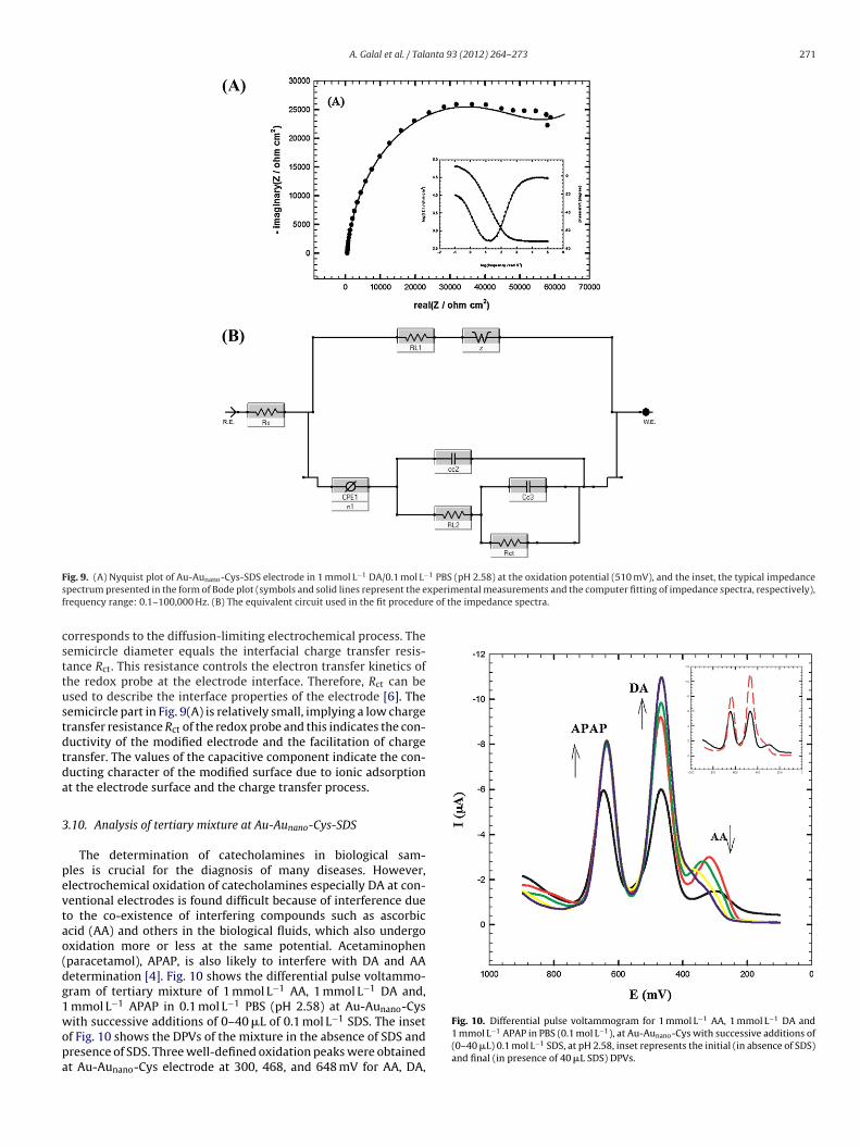
Fig. 9(A) shows a typical impedance spectrum presented in theform of Nyquist plot of DA at Au-Aunano-Cys-SDS electrode and theinset shows the typical impedance spectrum presented in the formof Bode plot. The experimental data were compared to an equiv-alent circuit that used some of the conventional circuit elements,namely; resistance, capacitance, diffusion, and induction elements[4,6]. The equivalent circuit is shown in Fig. 9(B). In this circuit, Rs isthe solution resistance, RL1 is the resistance due to the surfactantfilm on the surface, RL2 is the resistance due to the SAM of cysteine,and Rct is the charge transfer resistance. Capacitors in EIS experi-ments do not behave ideally; instead they act like a constant phaseelement (CPE) [4]. Therefore, CPE1 is a constant phase element andn1 is its corresponding exponent (n is less than one). Cc2 and Cc3represent the capacitance of the double layer. Diffusion can createan impedance known as the Warburg impedance Z. Table 3 lists thebest fitting values calculated from the equivalent circuit (Fig. 9(B))for the impedance data of Fig. 9(A).
As shown in Fig. 9(A), the impedance spectra include a semicir-cle portion and a linear portion; the semicircle part at the higherfrequencies corresponds to the electron-transfer limiting electro-chemical process, and the linear part at the lower frequencies
Cc2/10−8 F cm−2
CPE1/105 Fcm−2
n1 Z104 � s−1/2
8.201 3.191 0.9094 1.926
A. Galal et al. / Talanta 93 (2012) 264– 273 271
F 1 PBS (pH 2.58) at the oxidation potential (510 mV), and the inset, the typical impedances xperimental measurements and the computer fitting of impedance spectra, respectively),f e of the impedance spectra.
csttustdtda
3
pevtao(dg1wopa
ig. 9. (A) Nyquist plot of Au-Aunano-Cys-SDS electrode in 1 mmol L−1 DA/0.1 mol L−
pectrum presented in the form of Bode plot (symbols and solid lines represent the erequency range: 0.1–100,000 Hz. (B) The equivalent circuit used in the fit procedur
orresponds to the diffusion-limiting electrochemical process. Theemicircle diameter equals the interfacial charge transfer resis-ance Rct. This resistance controls the electron transfer kinetics ofhe redox probe at the electrode interface. Therefore, Rct can besed to describe the interface properties of the electrode [6]. Theemicircle part in Fig. 9(A) is relatively small, implying a low chargeransfer resistance Rct of the redox probe and this indicates the con-uctivity of the modified electrode and the facilitation of chargeransfer. The values of the capacitive component indicate the con-ucting character of the modified surface due to ionic adsorptiont the electrode surface and the charge transfer process.
.10. Analysis of tertiary mixture at Au-Aunano-Cys-SDS
The determination of catecholamines in biological sam-les is crucial for the diagnosis of many diseases. However,lectrochemical oxidation of catecholamines especially DA at con-entional electrodes is found difficult because of interference dueo the co-existence of interfering compounds such as ascorbiccid (AA) and others in the biological fluids, which also undergoxidation more or less at the same potential. Acetaminophenparacetamol), APAP, is also likely to interfere with DA and AAetermination [4]. Fig. 10 shows the differential pulse voltammo-ram of tertiary mixture of 1 mmol L−1 AA, 1 mmol L−1 DA and,
mmol L−1 APAP in 0.1 mol L−1 PBS (pH 2.58) at Au-Aunano-Cys
ith successive additions of 0–40 �L of 0.1 mol L−1 SDS. The insetf Fig. 10 shows the DPVs of the mixture in the absence of SDS andresence of SDS. Three well-defined oxidation peaks were obtainedt Au-Aunano-Cys electrode at 300, 468, and 648 mV for AA, DA,
Fig. 10. Differential pulse voltammogram for 1 mmol L−1 AA, 1 mmol L−1 DA and1 mmol L−1 APAP in PBS (0.1 mol L−1), at Au-Aunano-Cys with successive additions of(0–40 �L) 0.1 mol L−1 SDS, at pH 2.58, inset represents the initial (in absence of SDS)and final (in presence of 40 �L SDS) DPVs.

272 A. Galal et al. / Talanta 9
Fig. 11. SEM images of: Au-Aunano-Cys (A), Au-Aunano-Cys-SDS (B) electrodes, andAFM image by contact mode of Au-Aunano-Cys-SDS electrode (C).
3 (2012) 264– 273
and APAP, respectively. Thus Au-Aunano-Cys electrode can be usedfor the simultaneous determination of AA, DA, and APAP in a mix-ture. By the successive additions of 10 �L of 0.1 mol L−1 SDS inthe mixture solution, the oxidation peak current of DA and APAPincreases while the oxidation current of AA decreases until even-tual complete disappearance. It is expected that the addition ofSDS will cause a formation of a surfactant film over Au-Aunano-Cyselectrode. This amiphilic film will align in a way where the headgroups of surfactant molecules face the aqueous medium leavingthe hydrophobic part in contact with each other and away fromthe aqueous medium. The negative charge of the adsorbed sur-factant film as well as the hydrophobic character of the interiorof this film will act to repel hydrophilic AA molecules or its AA−
away from the electrode surface while enhancing the preconcen-tration/accumulation of hydrophobic cations of DA and APAP. Thisconcept is illustrated in Scheme 1 [5]. Au-Aunano-Cys electrodestability for mixture separation in presence of SDS was studiedvia repeated cycling up to 50 cycles with excellent stability beingdemonstrated (Supplement 4). Also, long term stability for sepa-ration of tertiary mixture of AA, DA, and APAP on Au-Aunano-Cyselectrode was studied in presence of SDS. After 3 days and oneweek of storage, Ipa of DA decreased by 14% and 17% and Ipa ofAPAP decreased by 15% and 28%, respectively. In addition, thelong term stability for the separation of the same mixture at Au-Aunano electrode was studied. After 3 days and one week of storage,Ipa of DA decreased by 34% and 44% and Ipa of APAP decreasedby 9% and 33%, respectively. Thus, Au-Aunano-Cys in presence ofSDS gives better stability via repeated cycles and longer termstability not only for DA detection, but also for tertiary mixtureseparation.
3.11. Surface morphology of Au-Aunano-Cys-SDS electrode
The response of an electrochemical sensor was related to thephysical morphology of its surface. Several factors such as sur-face roughness, porosity of films, and inclusion of defects shouldaffect the current response of the electrode. The SEM images ofAu-Aunano-Cys and Au-Aunano-Cys-SDS electrodes are shown inFig. 11(A) and (B), respectively. As observed in the SEM images,gold nanoparticles modified with cysteine SAM, are randomlydistributed on the surface, possessing a dendritic shape and non-homogenous size. In comparison, gold nanoparticles modified withcysteine SAM and further modified with SDS are homogenously dis-tributed on the surface, better dispersed and highly packed. Theinteraction between the anionic SDS and cationic cysteine SAMallows the reorganization and redispersion of gold nanoparticles.A spongy film was observed in Fig. 11(B) which may be due to thesurfactant film on the surface.
Atomic force microscopy is a powerful tool to measure topogra-phy and properties of surfaces. The AFM technique was employedto illustrate the self-assembly process of Au-Aunano-Cys-SDS elec-trode. Fig. 11(C) shows the AFM 3D image of Au-Aunano-Cys-SDSelectrode by the contact mode. Typical AFM 3D images of bare Au,Au-Aunano, and Au-Aunano-Cys-SDS electrodes by the non-contactmode were taken (figures not shown).
4. Conclusions
In this work, the difference in the electrochemical behavior ofSAMs of cysteine at Au and Au-Aunano are compared for the firsttime. In this study, the selective and simultaneous determination
of tertiary mixture of AA, DA, and APAP using Au-Aunano-Cys elec-trode in presence of SDS has been demonstrated. A novel approachfor the utilization of anionic surfactants in electroanalytical appli-cations has been described in this work. The negatively charged SDS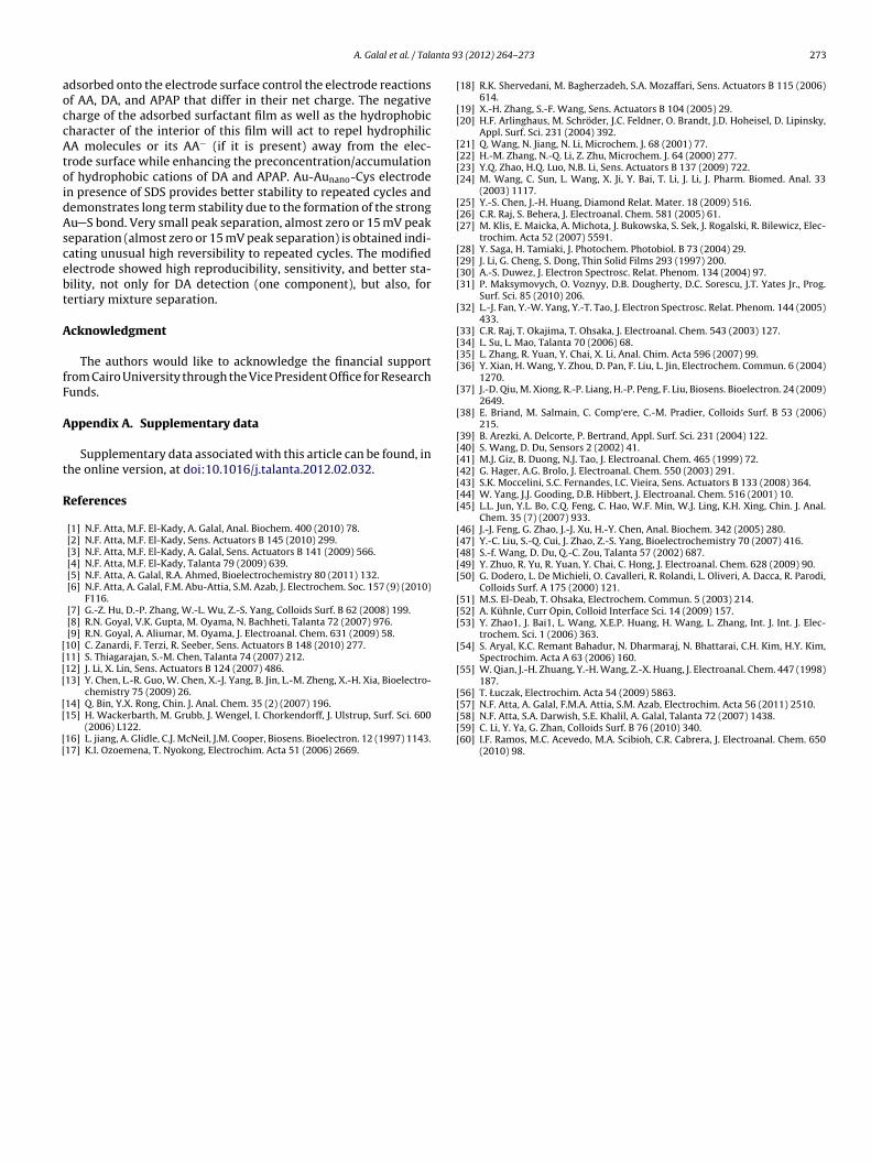
nta 9
aoccAtoidAscebt
A
fF
A
t
R
[[[[
[[
[[
[
[[
[[[[
[[[
[[[[
[
[[[[
[
[
[[[[[[[
[[[[[
[[[
[
[
[
A. Galal et al. / Tala
dsorbed onto the electrode surface control the electrode reactionsf AA, DA, and APAP that differ in their net charge. The negativeharge of the adsorbed surfactant film as well as the hydrophobicharacter of the interior of this film will act to repel hydrophilicA molecules or its AA− (if it is present) away from the elec-
rode surface while enhancing the preconcentration/accumulationf hydrophobic cations of DA and APAP. Au-Aunano-Cys electroden presence of SDS provides better stability to repeated cycles andemonstrates long term stability due to the formation of the strongu S bond. Very small peak separation, almost zero or 15 mV peakeparation (almost zero or 15 mV peak separation) is obtained indi-ating unusual high reversibility to repeated cycles. The modifiedlectrode showed high reproducibility, sensitivity, and better sta-ility, not only for DA detection (one component), but also, forertiary mixture separation.
cknowledgment
The authors would like to acknowledge the financial supportrom Cairo University through the Vice President Office for Researchunds.
ppendix A. Supplementary data
Supplementary data associated with this article can be found, inhe online version, at doi:10.1016/j.talanta.2012.02.032.
eferences
[1] N.F. Atta, M.F. El-Kady, A. Galal, Anal. Biochem. 400 (2010) 78.[2] N.F. Atta, M.F. El-Kady, Sens. Actuators B 145 (2010) 299.[3] N.F. Atta, M.F. El-Kady, A. Galal, Sens. Actuators B 141 (2009) 566.[4] N.F. Atta, M.F. El-Kady, Talanta 79 (2009) 639.[5] N.F. Atta, A. Galal, R.A. Ahmed, Bioelectrochemistry 80 (2011) 132.[6] N.F. Atta, A. Galal, F.M. Abu-Attia, S.M. Azab, J. Electrochem. Soc. 157 (9) (2010)
F116.[7] G.-Z. Hu, D.-P. Zhang, W.-L. Wu, Z.-S. Yang, Colloids Surf. B 62 (2008) 199.[8] R.N. Goyal, V.K. Gupta, M. Oyama, N. Bachheti, Talanta 72 (2007) 976.[9] R.N. Goyal, A. Aliumar, M. Oyama, J. Electroanal. Chem. 631 (2009) 58.10] C. Zanardi, F. Terzi, R. Seeber, Sens. Actuators B 148 (2010) 277.11] S. Thiagarajan, S.-M. Chen, Talanta 74 (2007) 212.12] J. Li, X. Lin, Sens. Actuators B 124 (2007) 486.13] Y. Chen, L.-R. Guo, W. Chen, X.-J. Yang, B. Jin, L.-M. Zheng, X.-H. Xia, Bioelectro-
chemistry 75 (2009) 26.
14] Q. Bin, Y.X. Rong, Chin. J. Anal. Chem. 35 (2) (2007) 196.15] H. Wackerbarth, M. Grubb, J. Wengel, I. Chorkendorff, J. Ulstrup, Surf. Sci. 600(2006) L122.16] L. jiang, A. Glidle, C.J. McNeil, J.M. Cooper, Biosens. Bioelectron. 12 (1997) 1143.17] K.I. Ozoemena, T. Nyokong, Electrochim. Acta 51 (2006) 2669.
[[[[
3 (2012) 264– 273 273
18] R.K. Shervedani, M. Bagherzadeh, S.A. Mozaffari, Sens. Actuators B 115 (2006)614.
19] X.-H. Zhang, S.-F. Wang, Sens. Actuators B 104 (2005) 29.20] H.F. Arlinghaus, M. Schröder, J.C. Feldner, O. Brandt, J.D. Hoheisel, D. Lipinsky,
Appl. Surf. Sci. 231 (2004) 392.21] Q. Wang, N. Jiang, N. Li, Microchem. J. 68 (2001) 77.22] H.-M. Zhang, N.-Q. Li, Z. Zhu, Microchem. J. 64 (2000) 277.23] Y.Q. Zhao, H.Q. Luo, N.B. Li, Sens. Actuators B 137 (2009) 722.24] M. Wang, C. Sun, L. Wang, X. Ji, Y. Bai, T. Li, J. Li, J. Pharm. Biomed. Anal. 33
(2003) 1117.25] Y.-S. Chen, J.-H. Huang, Diamond Relat. Mater. 18 (2009) 516.26] C.R. Raj, S. Behera, J. Electroanal. Chem. 581 (2005) 61.27] M. Klis, E. Maicka, A. Michota, J. Bukowska, S. Sek, J. Rogalski, R. Bilewicz, Elec-
trochim. Acta 52 (2007) 5591.28] Y. Saga, H. Tamiaki, J. Photochem. Photobiol. B 73 (2004) 29.29] J. Li, G. Cheng, S. Dong, Thin Solid Films 293 (1997) 200.30] A.-S. Duwez, J. Electron Spectrosc. Relat. Phenom. 134 (2004) 97.31] P. Maksymovych, O. Voznyy, D.B. Dougherty, D.C. Sorescu, J.T. Yates Jr., Prog.
Surf. Sci. 85 (2010) 206.32] L.-J. Fan, Y.-W. Yang, Y.-T. Tao, J. Electron Spectrosc. Relat. Phenom. 144 (2005)
433.33] C.R. Raj, T. Okajima, T. Ohsaka, J. Electroanal. Chem. 543 (2003) 127.34] L. Su, L. Mao, Talanta 70 (2006) 68.35] L. Zhang, R. Yuan, Y. Chai, X. Li, Anal. Chim. Acta 596 (2007) 99.36] Y. Xian, H. Wang, Y. Zhou, D. Pan, F. Liu, L. Jin, Electrochem. Commun. 6 (2004)
1270.37] J.-D. Qiu, M. Xiong, R.-P. Liang, H.-P. Peng, F. Liu, Biosens. Bioelectron. 24 (2009)
2649.38] E. Briand, M. Salmain, C. Comp‘ere, C.-M. Pradier, Colloids Surf. B 53 (2006)
215.39] B. Arezki, A. Delcorte, P. Bertrand, Appl. Surf. Sci. 231 (2004) 122.40] S. Wang, D. Du, Sensors 2 (2002) 41.41] M.J. Giz, B. Duong, N.J. Tao, J. Electroanal. Chem. 465 (1999) 72.42] G. Hager, A.G. Brolo, J. Electroanal. Chem. 550 (2003) 291.43] S.K. Moccelini, S.C. Fernandes, I.C. Vieira, Sens. Actuators B 133 (2008) 364.44] W. Yang, J.J. Gooding, D.B. Hibbert, J. Electroanal. Chem. 516 (2001) 10.45] L.L. Jun, Y.L. Bo, C.Q. Feng, C. Hao, W.F. Min, W.J. Ling, K.H. Xing, Chin. J. Anal.
Chem. 35 (7) (2007) 933.46] J.-J. Feng, G. Zhao, J.-J. Xu, H.-Y. Chen, Anal. Biochem. 342 (2005) 280.47] Y.-C. Liu, S.-Q. Cui, J. Zhao, Z.-S. Yang, Bioelectrochemistry 70 (2007) 416.48] S.-f. Wang, D. Du, Q.-C. Zou, Talanta 57 (2002) 687.49] Y. Zhuo, R. Yu, R. Yuan, Y. Chai, C. Hong, J. Electroanal. Chem. 628 (2009) 90.50] G. Dodero, L. De Michieli, O. Cavalleri, R. Rolandi, L. Oliveri, A. Dacca, R. Parodi,
Colloids Surf. A 175 (2000) 121.51] M.S. El-Deab, T. Ohsaka, Electrochem. Commun. 5 (2003) 214.52] A. Kühnle, Curr Opin, Colloid Interface Sci. 14 (2009) 157.53] Y. Zhao1, J. Bai1, L. Wang, X.E.P. Huang, H. Wang, L. Zhang, Int. J. Int. J. Elec-
trochem. Sci. 1 (2006) 363.54] S. Aryal, K.C. Remant Bahadur, N. Dharmaraj, N. Bhattarai, C.H. Kim, H.Y. Kim,
Spectrochim. Acta A 63 (2006) 160.55] W. Qian, J.-H. Zhuang, Y.-H. Wang, Z.-X. Huang, J. Electroanal. Chem. 447 (1998)
187.56] T. Łuczak, Electrochim. Acta 54 (2009) 5863.
57] N.F. Atta, A. Galal, F.M.A. Attia, S.M. Azab, Electrochim. Acta 56 (2011) 2510.58] N.F. Atta, S.A. Darwish, S.E. Khalil, A. Galal, Talanta 72 (2007) 1438.59] C. Li, Y. Ya, G. Zhan, Colloids Surf. B 76 (2010) 340.60] I.F. Ramos, M.C. Acevedo, M.A. Scibioh, C.R. Cabrera, J. Electroanal. Chem. 650(2010) 98.