primary teeth chapter 18 - fscjdental.com · primary teeth are present by approximately age 2 ½...
TRANSCRIPT

PrimaryTeethChapter18
DentalAnatomy2016

Primary Teeth - Introduction
• Synonyms – deciduous teeth, baby teeth, temporary teeth, milk teeth.
• There are 20 primary teeth, designated as A thru T in the Universal numbering system.
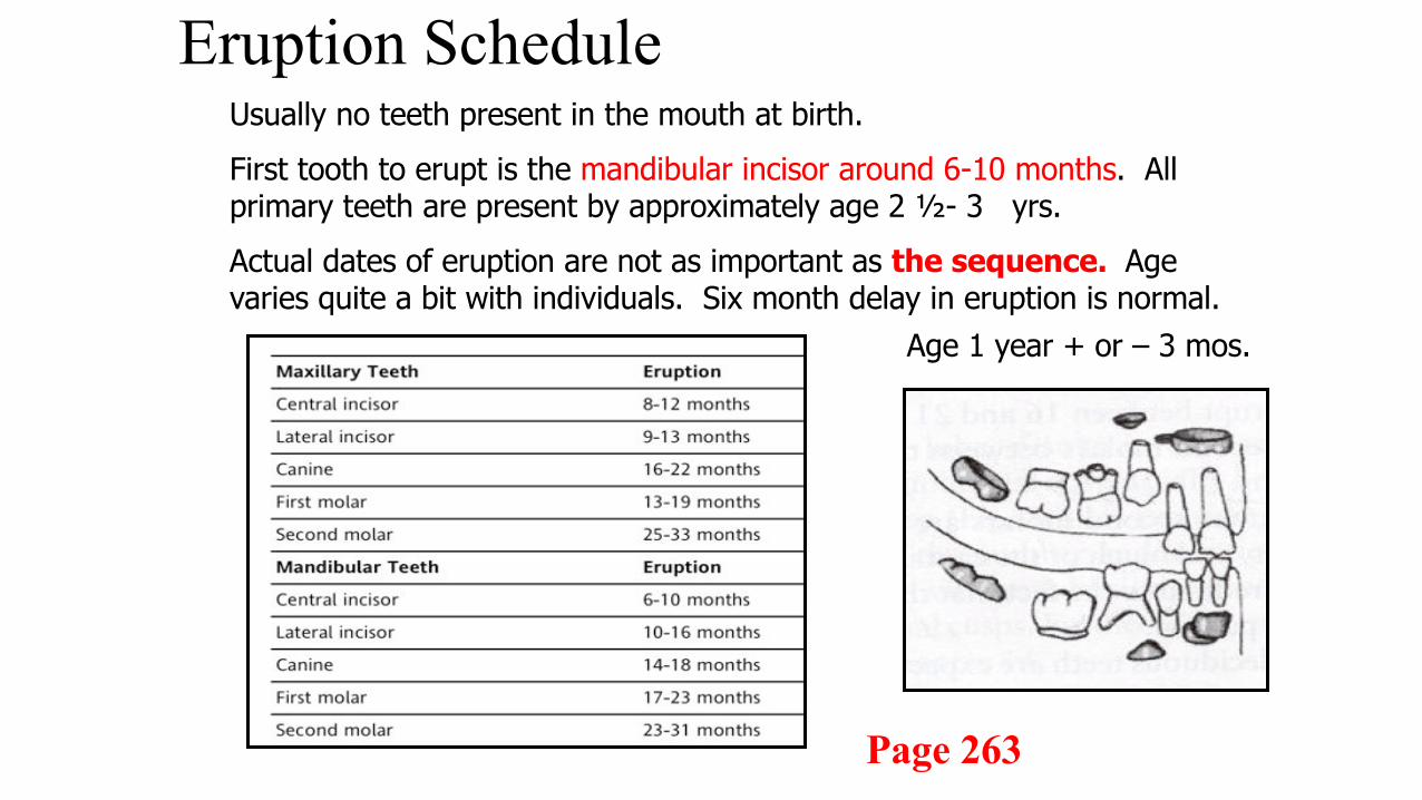
Eruption Schedule
Age 1 year + or – 3 mos.
Usually no teeth present in the mouth at birth.First tooth to erupt is the mandibular incisor around 6-10 months. All primary teeth are present by approximately age 2 ½- 3 yrs.Actual dates of eruption are not as important as the sequence. Age varies quite a bit with individuals. Six month delay in eruption is normal.
Page 263

Primate Spacing
• Primate spacing is natural spacing between the maxillary lateral & canines.
• Also between the mandibular canine and first molar.
• As jaw grows in length additional spacing occurs to allow room for larger permanent teeth.• Leeway spaces result from the
width differential between primary molars and permanent premolars
Erupting permanent teeth follow the resorbing primary root
4
Importance of Deciduous Teeth
• Value cannot be overemphasized to the caregiver. Read page 263 – Clinical considerations for Primary Dentition.
• Efficient chewing and appearance.• Clear speech.• Maintain space for permanent teeth.• Guide proper alignment of permanent teeth.
5

6
Differences With Permanent Teeth
• Primary teeth smaller in crown and root, though deciduous molars are wider than permanent premolars that will replace them.
• Deciduous roots are narrower• Prominent cervical ridge in
deciduous dentition

7
• Buccal and lingual surfaces taper toward occlusal more than permanent teeth; results in narrower occlusal table
• Roots of primary molars are longer and narrower than their permanent counterparts; roots also flare more to accommodate developing permanent crown
• Primary crowns are whiter in color than permanent crowns

8

9
• Pulp chamber large for crown in primary teeth; pulp horns longer as well
• Less dentinal thickness in primary tooth crown due to larger pulp chamber
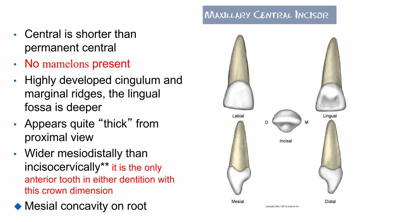
Maxillary Central Incisor
• Central is shorter than permanent central
• No mamelons present• Highly developed cingulum and
marginal ridges, the lingual fossa is deeper
• Appears quite “thick” from proximal view
• Wider mesiodistally than incisocervically** it is the only anterior tooth in either dentition with this crown dimension
u Mesial concavity on root

Maxillary Lateral Incisor
• Crown is smaller than central incisor
• Crown+ root is longer than central
• Longer incisocervically than mesiodistally-exact opposite of the central
• Root longer in appearance than crown compared to central
12
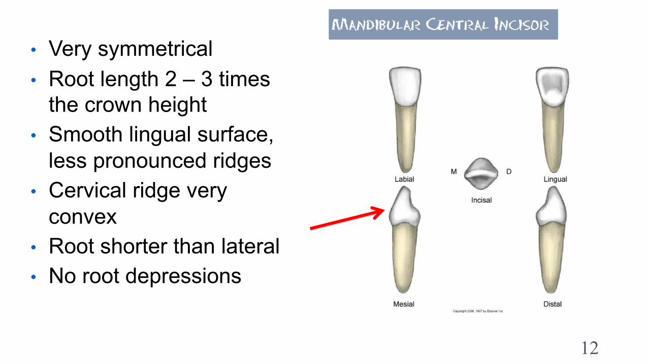
Mandibular Central Incisor
• Very symmetrical• Root length 2 – 3 times
the crown height• Smooth lingual surface,
less pronounced ridges• Cervical ridge very
convex• Root shorter than lateral• No root depressions

Mandibular Lateral Incisor• Wider and longer than central
incisor• Cingulum more developed than
central incisor• Lateral incisor “thicker” than
central• Incisal edge slopes distally• Root longer, narrower and more
tapered than central• From the incisal view, the
crown is NOT as symmetrical as the central
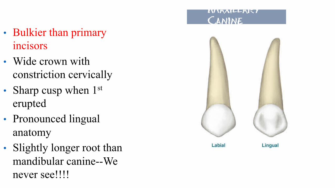
Maxillary Canine
• Bulkier than primary incisors
• Wide crown with constriction cervically
• Sharp cusp when 1st
erupted• Pronounced lingual
anatomy• Slightly longer root than
mandibular canine--We never see!!!!

15
From the incisal aspect, the crown is diamond shaped
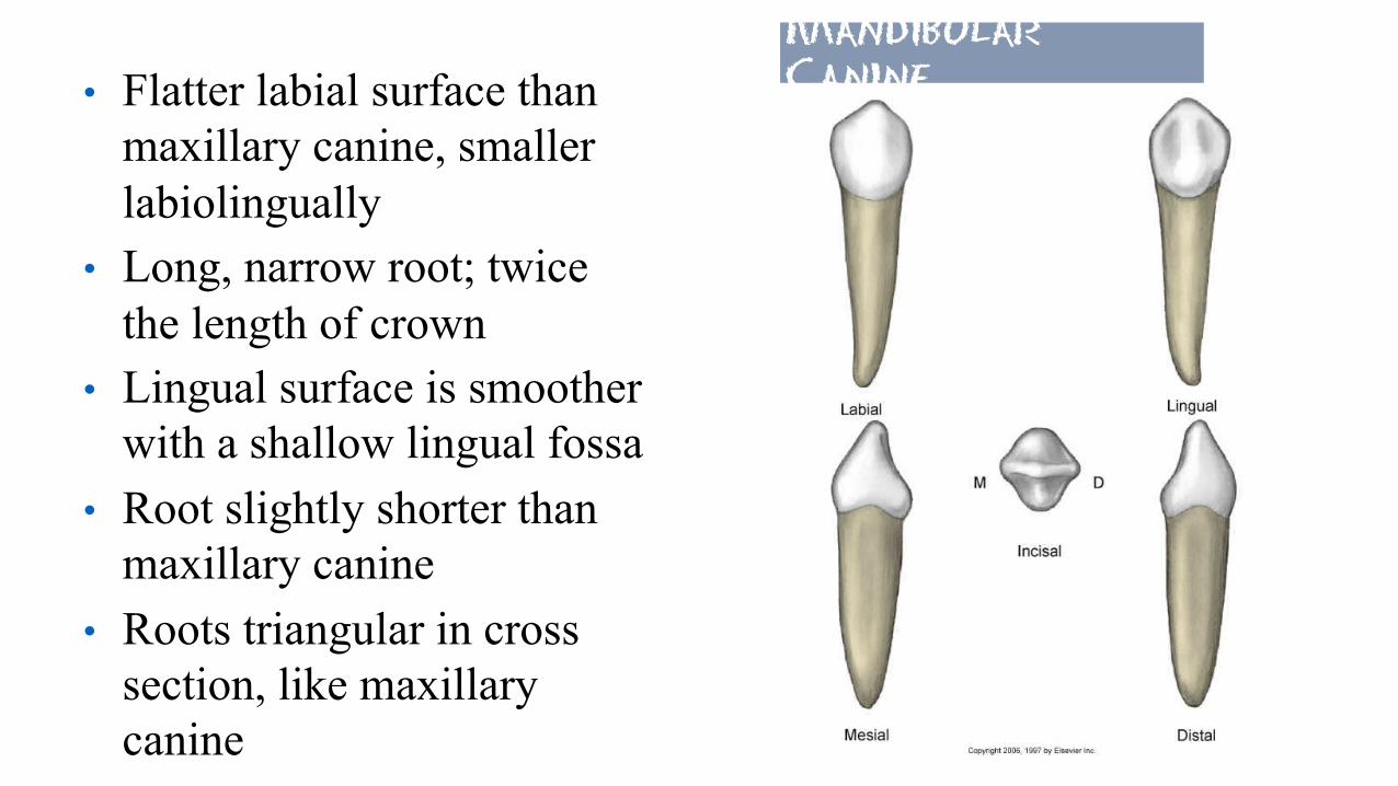
Mandibular Canine• Flatter labial surface than
maxillary canine, smaller labiolingually
• Long, narrow root; twice the length of crown
• Lingual surface is smoother with a shallow lingual fossa
• Root slightly shorter than maxillary canine
• Roots triangular in cross section, like maxillary canine

PrimaryMolars– generalfeatures• Primary molars are replaced by permanent premolars.• None of the primary 1st molars resembles any other tooth in
either dentition. The crown of each 2nd molar resembles the first molars of the permanent dentition that will erupt distal to them.
• A prominent cervical ridge is present on the buccal surface.• Occlusal table of a primary molar is more constricted
buccolingually than a permanent molar.• The occlusal anatomy of cusps is not as pronounced as permanent
molars.• Roots are flared beyond the crown outlines. Space is created
between the roots for the developing permanent premolars.
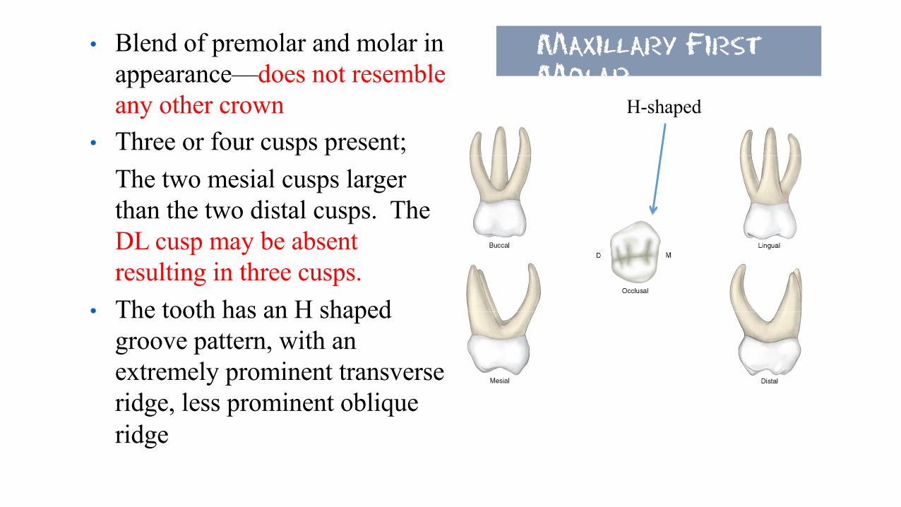
Maxillary First Molar
• Blend of premolar and molar in appearance—does not resemble any other crown
• Three or four cusps present;The two mesial cusps larger than the two distal cusps. The DL cusp may be absent resulting in three cusps.
• The tooth has an H shaped groove pattern, with an extremely prominent transverse ridge, less prominent oblique ridge
H-shaped

• Three roots-- two buccal & one lingual-same number and position as permanent molars.
• Roots long, slender, and flared
• Very short root trunk• Extremely prominent buccal
cervical ridge.
Primary Maxillary 1st molar
Occlusal table converges buccolingually

Maxillary Second Molar
• Larger than the primary 1st
molar• Resembles small permanent
maxillary first molar – has 5 cusps
• Two equal buccal cusps from buccal view with buccal groove
• Three cusps visible from lingual including cusp of Carabelli
• ML cusp larger than MB cusp
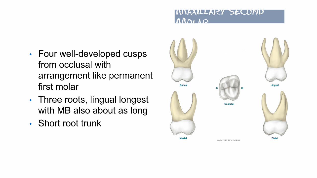
• Four well-developed cusps from occlusal with arrangement like permanent first molar
• Three roots, lingual longest with MB also about as long
• Short root trunk
Maxillary Second Molar

Mandibular First Molar
• Has a crown unlike any other tooth of either dentition
• Straight mesial contour from buccal view and convex distal
• Two distinct buccal cusps with less consistent groove between
• Long, slender, flared roots• Short root trunk• Four cusps with the mesial cusps
larger
Prominent buccal cervical ridge

23
• Mesial marginal ridge well developed (may appear to be a cusp)
• Mesiolingual cusp is long, pointed, and angled in on the occlusal table.
• A transverse ridge passes between the mesiobuccal and mesiolingual cusps.
• Two roots positioned similarly to permanent mandibular molars.
Mandibular First Molar

24
• Two roots, flat and widely flared
• Mesial root has longitudinal developmental groove and two canals
• Distal root shorter and thinner
Mandibular First Molar
25
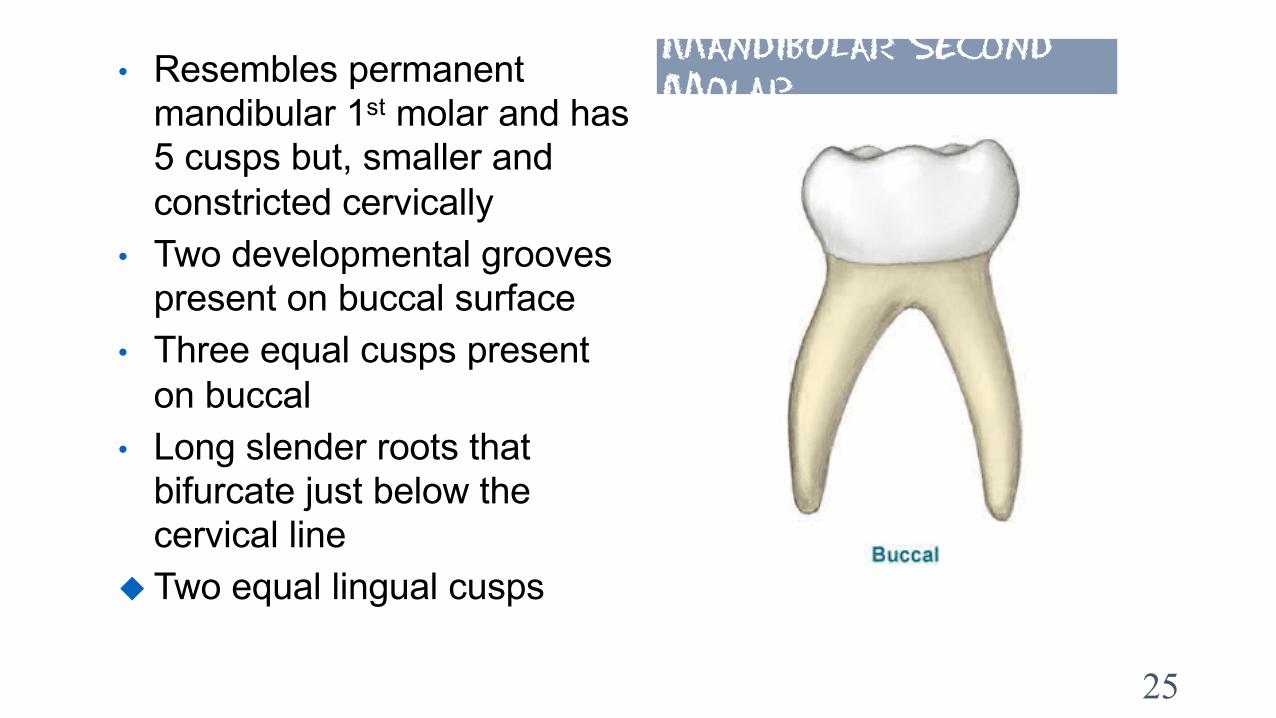
Mandibular Second Molar• Resembles permanent
mandibular 1st molar and has 5 cusps but, smaller and constricted cervically
• Two developmental grooves present on buccal surface
• Three equal cusps present on buccal
• Long slender roots that bifurcate just below the cervical line
u Two equal lingual cusps

• Cervical bulge present mesiobuccally
• Mesial marginal ridge quite high giving cusps a short appearance; ML cusp taller than MB cusp
• Tooth converges lingually from occlusal view
Mandibular Second Molar

• Roots twice as long as crown• Two roots; mesial w/ 2
canals and longitudinal groove & distal w/ one canal and no groove
Mandibular Second Molar
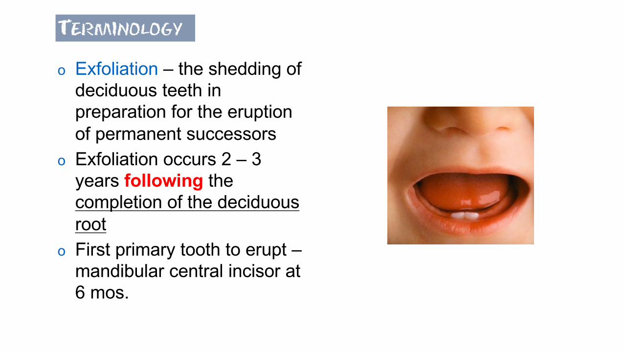
Terminology
o Exfoliation – the shedding of deciduous teeth in preparation for the eruption of permanent successors
o Exfoliation occurs 2 – 3 years following the completion of the deciduous root
o First primary tooth to erupt –mandibular central incisor at 6 mos.

Shedding & Exfoliation
• The permanent tooth develops in lingual position to primary teeth.
• Cells involved in resorption of primary roots:Osteoclasts resorb bone; Odontoclasts resorb dentin.Osteoblasts form bone; Odontoblasts produce dentin.
• Intermittent process of breakdown and build-up of tissue to replace.
• Because process is on-again, off-again it is common for teeth to get loose and tighten up and get loose again.
29

30
MixedDentitionPeriod

31

32
It is common to see extensive wear on incisal edges and occlusal of molars. Bruxismis outgrown, treatment not needed in deciduous teeth.

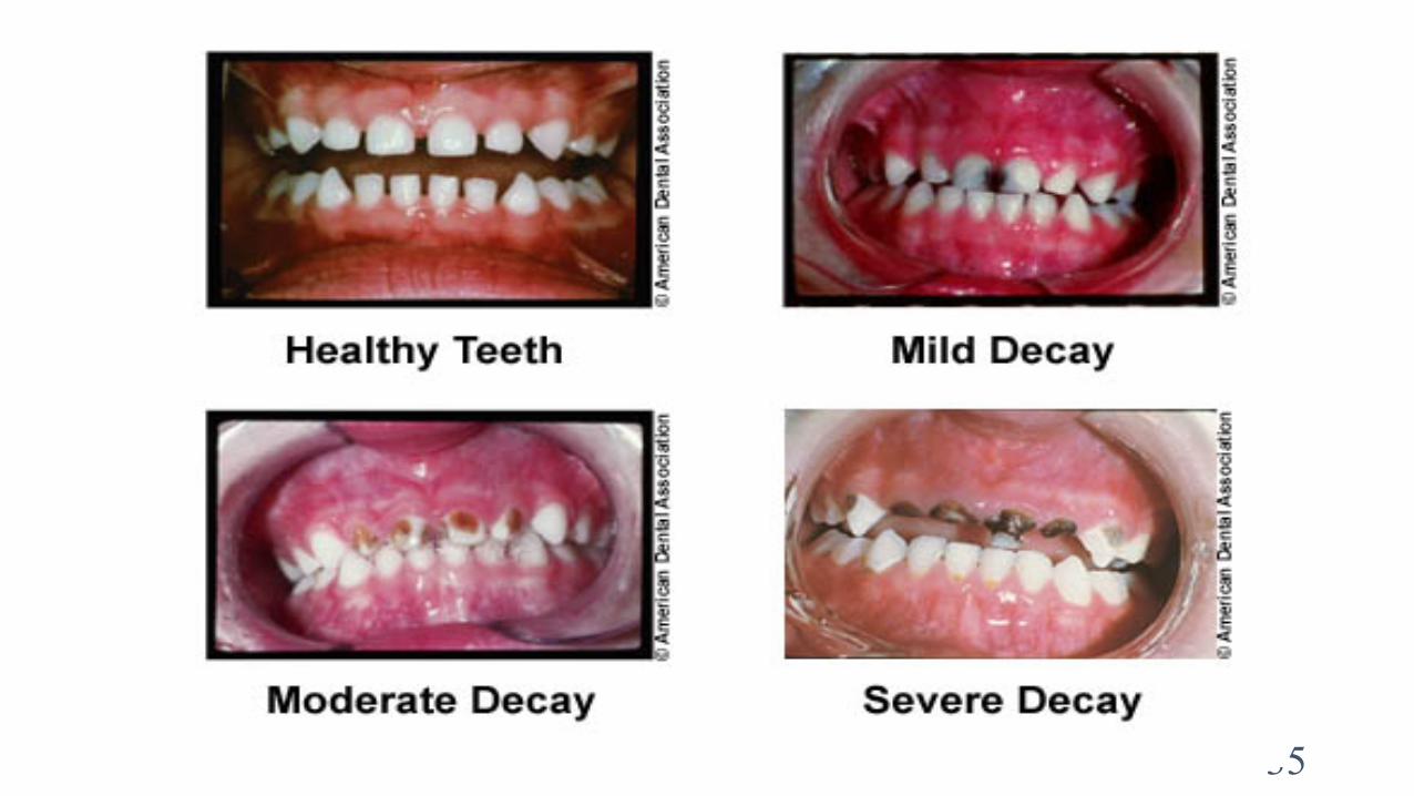
Early Childhood Caries (bottle mouth decay)
• Thought to be caused by feeding bottle at bedtime. Now known to be multi-factorial cause.
• Affects 18% of children. Rapid and rampant decay. • Defined as 1 or more decayed, missing, or filled tooth
surfaces in child younger than 6 years.• Cause is plaque, acid and fermentable carbohydrates.
Direct relationship between the mother and the child’s status. Decay is an infectious and transmissible disease. (Streptococcus mutans)
• Homecare must start early!!!!

• Supervised homecare must begin early, as the first primary teeth erupt into the oral cavity, to prevent premature loss of the primary teeth.
35

Clinical Consideration for Primary Dentition
• Prolonged nighttime use of a baby bottle with cavity-causing beverage or sugar on a pacifier must also be considered in a child patient with extensive acute caries of the primary teeth.

Comparison of Primaryto Permanent• Because the enamel and
dentin are thinner, the risk of endodontic complications is greater for the primary dentition.
• In addition, because the pulp chamber and pulp horns are also larger, there is increased risk of pulpal exposure during cavity preparation.
Both these teeth have interproximal caries