displacement of primary and permanent anterior teeth
TRANSCRIPT

Trauma Lec. 5 Pedodontics Fifth stage
Baghdad College of dentistry
21/11/2018 1
Assist. Prof. Dr. Aseel Haidar
Displacement of Primary and Permanent
Anterior Teeth
(LUXATION)
The displacement of anterior primary and permanent teeth presents a challenge
in diagnosis and treatment for the dentist.
INTRUSION AND EXTRUSION OF TEETH
INTRUSION: It is the most severe form of luxation injury
because it causes severe damage to the periodontal ligament
resulting in a greater incidence of external root resorption. The tooth
may be completely or partially intruded into its socket.
Clinically crown appears shorter. The best approach is to "wait and
watch for the tooth to re-erupt on its own.
Primary Teeth Intrusion by forceful impaction of maxillary anterior primary teeth is a common
occurrence in children during the first 3 years of life, why? Frequent falls and striking of
the teeth on hard objects may force the teeth into the alveolar
process to the extent that the entire clinical crown becomes
buried in bone and soft tissue. Although there is a difference
of opinion regarding treatment of injuries of this type, it is
generally agreed that immediate attention should be given to
soft tissue damage. Intruded primary teeth should be
observed; with few exceptions, no attempt should be made to
reposition them after the accident. Most injuries of this type
occur at an age when it would be difficult to construct a splint
or a retaining appliance to stabilize the repositioned teeth.
Normally, the developing permanent incisor tooth buds lie lingual to the roots of the
primary central incisors. Therefore when an intrusive displacement occurs, the primary
tooth usually remains labial to the developing permanent tooth. If the intruded primary
tooth is found to be in a lingual or encroaching relationship to the developing permanent
tooth, it should be removed. Such a relationship may be confirmed from a lateral
Intruded
Tooth

Trauma Lec. 5 Pedodontics Fifth stage
Baghdad College of dentistry
21/11/2018 2
radiograph of the anterior segment. The examination should be carried out as previously
described, and radiographs should be made to detect evidence of root fracture, fracture of
the alveolar bone, and damage to permanent teeth. However, predicting whether the
permanent successors will show evidence of interrupted growth and development is
impossible unless actual encroachment of their space can be seen radiographically.
Primary anterior teeth intruded as a result of a blow may often re-erupt within 3
to 4 weeks after the injury. These teeth may even retain their vitality and later undergo
normal resorption and be replaced on schedule by their permanent successors. During the
first 6 months after the injury, however, the dentist often observes one or more of the
reactions of the pulp and supporting tissues, the most common of which is pulpal necrosis.
Even after reeruption, a necrotic pulp can be treated if the tooth is sound in the alveolus
and no pathologic root resorption is evident.
Primary teeth that are displaced but not intruded should be repositioned by the
dentist or parent as soon as possible after the accident, to prevent interference with
occlusion. The prognosis for severely loosened primary teeth is poor. Frequently the
teeth remain mobile and undergo rapid root resorption.
The immediate and future prognosis for the pulp is more favorable if root
formation is still incomplete at the time of the accident. Teeth with complete root formation
seemed to undergo resorption more frequently than those with incomplete root formation.
However, when resorption did occur, it was more extensive and progressed more rapidly
in teeth with incomplete root development.
Permanent Teeth Intruded permanent teeth apparently have a poorer prognosis than similarly
injured primary teeth. The tendency for the injury to be followed by rapid root resorption,
pulpal necrosis, or ankylosis is greater.
The treatment:
1) For a permanent tooth with a closed root end,
A) Intruded less than 3 mm, is to allow the tooth to erupt without intervention. If no
movement is evident after 2 to 4 weeks, the tooth may be repositioned either
orthodontically or surgically before ankylosis can take place.
B) If the tooth is intruded 7 mm or more, the tooth is repositioned surgically and
stabilized for 4 to 8 weeks by means of a flexible splint.
In most instances the pulp will become necrotic with intrusive injuries in teeth
with complete root formation. Root canal treatment should be initiated, with calcium
hydroxide as a temporary canal filling material, 2 to 3 weeks after stabilization.
2) The treatment for an intruded permanent tooth with incomplete root formation is:

Trauma Lec. 5 Pedodontics Fifth stage
Baghdad College of dentistry
21/11/2018 3
A) To allow it to erupt spontaneously. If no movement is seen within a few weeks,
orthodontic repositioning should begin.
B) If the tooth is intruded 7 mm or more, the tooth can be repositioned surgically and
stabilized by means of a flexible splint. Endodontic therapy is often required, however, and
the tooth should be monitored closely while a decision on endodontic therapy is pending.
It appears that spontaneous eruption results in the fewest complications in
immature teeth, regardless of the degree of intrusion.
The frequency of pulpal necrosis in teeth with complete root development is
higher than in those with incomplete root development. Teeth with uncomplicated crown
fractures with luxation and crown-fractured teeth with intrusion had a higher incidence of
pulpal necrosis than any other types of concurrent luxation. A concurrent luxation injury
and complete root development are important risk factors of pulpal necrosis with
uncomplicated crown fractures.
It seems that both treatment approaches to the treatment of severely intruded
permanent teeth (early repositioning or waiting for spontaneous re-eruption) have
demonstrated reasonably successful results. However, the affected teeth seem to benefit by
early calcium hydroxide endodontic therapy with either treatment approach. The decision
to reposition mechanically or hope for spontaneous re-eruption of intruded permanent teeth
remains a matter of clinical judgment that may be based on several conditions associated
with the particular case.
Extrusion: It is also called peripheral displacement
or partial avulsion. It is partial displacement of tooth out of its
socket (it appear longer). The extrusive luxation of a
permanent tooth usually results in pulpal necrosis. The
immediate treatment involves the careful repositioning of the
tooth and stabilization.
If mature repositioned teeth do not respond to pulp
vitality tests within 2 to 3 weeks after being repositioned,
endodontic treatment should be undertaken before there is evidence of root resorption,
which often occurs after severe injuries of this type. The need for endodontic intervention
is virtually certain in cases of significant extrusion (more than 2 mm) of mature teeth. With
extruded immature teeth, the clinician should monitor the situation frequently and be
prepared to intervene with endodontic therapy, as described later, if conditions warrant.
Extruded
Tooth

Trauma Lec. 5 Pedodontics Fifth stage
Baghdad College of dentistry
21/11/2018 4
Lateral Luxation: Displacement of tooth in any direction other
than axial.
Clinical Features
1) Tooth is mobile and displaced.
2) Bleeding from gingival crevice.
3) Tooth is tender to percussion and masticatory forces.
Radiographic Features
Widening of PDL space on one side and crushing of lamina dura on other side.
Treatment
1) Administer local anesthesia if forceful positioning is
anticipated.
2) Reposition the tooth in normal position using digital
pressure.
3) Splint the tooth for 2 weeks and if there is a marginal
bone breakdown then splint for 6-8 weeks.
4) Advice soft diet.
5) Follow up period of 1 year.
AVULSION AND REPLANTATION Term used to describe complete displacement of tooth from its
alveolus. It is also called as exarticulation. Maxillary teeth are most
commonly involved.
Clinical Features
Bleeding socket with missing tooth.

Trauma Lec. 5 Pedodontics Fifth stage
Baghdad College of dentistry
21/11/2018 5
Radiographic Features: 1) Empty socket.
2) Associated bone fractures.
3) If the wound is recent then lamina dura is visible
otherwise it is obliterated.
Treatment
1. Replantation.
2. If apical foramen is not closed-endodontic therapy is
delayed till first signs of apical closure are seen.
3. If apical foramen is closed -endodontic therapy is done after 1-2 weeks depending
on type of reimplantation.
Replantation is the technique in which a tooth, usually one in the anterior region, is
reinserted into the alveolus after its loss or displacement by accidental means.
Replantation of permanent teeth continues to be practiced and recommended, however,
because prolonged retention is also achieved in many cases, especially when replantation
occurs soon after the accident.
Importance of reimplantation: 1) The replanted tooth serves as a space maintainer and often guides adjacent teeth into
their proper position in the arch, a function that is important during the transitional
dentition period.
2) The replantation procedure also has psychological value. It gives the unfortunate child
and parents hope for success; even though they are told of the possibility of eventual loss
of the tooth, the early result often appears favorable and softens the emotional blow of the
accident.
The success of the replantation procedure depend on: 1) It is undoubtedly related to the length of time that elapses between the loss of the tooth
and its replacement in the socket.
2) The condition of the tooth and particularly the condition of the periodontal ligament
tissue remaining on the root surface are also important factors that influence the success of
replantation.

Trauma Lec. 5 Pedodontics Fifth stage
Baghdad College of dentistry
21/11/2018 6
Notes:
Many reports indicated that immediate replacement of a permanent tooth
occasionally results in the maintenance of vitality and indefinite retention. However,
replantation should generally be viewed as a temporary measure. Under favorable
conditions, many replanted teeth are retained for 5 or 10 years and a few for a
lifetime. Others, however, fail soon after replantation.
The tooth most commonly avulsed in both the primary and the permanent dentition
is a maxillary central incisor. Most often, an avulsion injury involves only a single
tooth. **Avulsion injuries are three times more frequent in boys than in girls and
occur most commonly in children from 7 to 9 years of age, when permanent incisors
are erupting. **Loosely structured periodontal ligament surrounding the erupting
teeth favors complete avulsion.
The sooner a tooth can be replanted in its socket after avulsion, the better the
prognosis will be for retention without root resorption. The prognosis is therefore
more favorable. Also, if the apical end of the tooth is incompletely developed at the
time of the injury, there is a greater chance of regaining pulp vitality after
replantation. If the apex is closed, the dentist should proceed with a pulpectomy a
few days after the replantation, even if the extraoral time for the tooth was brief.
If a parent calls to report that a tooth has been avulsed, and it can be determined
that the injury is without other oral, neurologic, or higher-priority physical complications,
the dentist may instruct the parent to do the following (primary teeth should not be
replanted):
1. Keep the patient calm.
2. Find the tooth and pick it up by the crown (the white part). Avoid touching the root.
3. If the tooth is dirty, wash it briefly (10 seconds) under cold running water and reposition
it. Try to encourage the patient/parent to replant the tooth. Bite on a handkerchief to hold
the tooth in position.
4. If repositioning is not possible, place the tooth in a suitable storage medium.
5. Seek emergency dental treatment immediately, unless the patient was knocked
unconscious. If the child was unconscious for a period of time, first seek emergency
medical evaluation for a concussion.
Notes:
Hank's balanced salt solution (HBSS): Considered as best transport medium.

Trauma Lec. 5 Pedodontics Fifth stage
Baghdad College of dentistry
21/11/2018 7
Milk: Excellent medium, has a favorable pH and
osmolality. Few virulent microrganisms owing to
Pasteurization. Maintains vitality of periodontal cells
for about 3hours’
Saliva: Allows storage for 2 hours but it is hypotonic.
Saline: Has physiologic pH and is isotonic
Tap water: Considered as bad as dry storage, its
hypotonicity causes cell lysis. It should be used only
if any of the above is not readily available
If the parent cannot or will not replant it, the
tooth must be kept moist during the trip to the dental
office. Allowing the avulsed tooth to dehydrate before
replantation is damaging to a favorable prognosis.
Hanks’ buffered saline, isotonic saline, and pasteurized bovine milk may be the most
favorable known storage media. Although tap water has been a commonly recommended
storage solution (and its use would be preferable to dehydration of the tooth), it is
hypotonic, and its use leads to rapid cell lysis and increased inflammation on replantation.
The patient should receive immediate attention after arriving at the dental office. If the
tooth has not already been replanted, the dentist should make every effort to minimize the
additional time that the tooth is out of the socket.
The patient’s general status should be quickly assessed to confirm that there are no
higher-priority injuries. If an evaluation of the socket area shows no evidence of alveolar
fracture or severe soft-tissue injury, the tooth is intact, and only a few minutes have elapsed
since the injury, the dentist should replant the tooth immediately.
Under the conditions just described, every effort should be directed toward preserving
a viable periodontal ligament. That treatment should be directed at avoiding or minimizing
the resultant inflammation that occurs as a direct result of the two main consequences of
tooth avulsion attachment damage and pulpal infection.
If the tooth was cleanly avulsed, it can probably be replanted without local
anesthetic, and obtaining the initial radiograph can also be delayed until the tooth is
replaced in the socket and held with finger pressure. The minutes saved may contribute to
a more successful replantation. If a clot is present in the socket, it will be displaced as the
tooth is repositioned; the socket walls should not be scraped with an instrument. If the tooth
does not slip back into position with relative ease when finger pressure is used, local
anesthesia and a radiographic evaluation are indicated. Local anesthetic should also be
administered when fractured and displaced alveolar bone must be repositioned before the
tooth is replanted. Soft-tissue suturing may be delayed until the tooth has been replaced in

Trauma Lec. 5 Pedodontics Fifth stage
Baghdad College of dentistry
21/11/2018 8
the socket; however, the suturing should be performed to control hemorrhage before the
tooth is stabilized with a bonded splint. The root canals were hermetically sealed with
gutta-percha, and the teeth were splinted for 1 month. Subsequent microscopic examination
under fluorescent and incandescent light revealed deposition of secondary cementum and
new alveolar bone, which entrapped the periodontal fibers.
The preservation of an intact and viable periodontal ligament is the most important
factor in achieving healing without root resorption. Delicate handling of the tooth, storage
in an appropriate moist environment, quick replantation, and appropriate stabilization are
all important in preserving the periodontal ligament. Undesirable periodontal ligament
reactions may result in replacement resorption (ankylosis) or inflammatory resorption of
the root. Either reaction may cause eventual loss of the tooth unless the resorption can be
controlled. Use of an enamel matrix derivative (Emdogain) has been shown to increase the
incidence of healed periodontal ligament when this gel is applied to the root surface of the
avulsed tooth and/or inserted directly into the alveolar socket before implantation. It
appears to aid in preventing or retarding resorption and ankylosis.
Stabilization of Replanted Teeth
After replantation of a tooth that has been avulsed, a splint is required to
stabilize it during at least the first week of healing. Acceptable splint should meet the
following criteria:
1. It should be easy to fabricate directly in the mouth, without lengthy laboratory
procedures.
2. It should be able to be placed passively without causing forces on the teeth.
3. It should not touch the gingival tissues, causing gingival irritation.
4. It should not interfere with normal occlusion.
5. It should be easily cleaned and allow for proper oral hygiene.
6. It should not traumatize the teeth or gingiva during application.
7. It should allow an approach for endodontic therapy.
8. It should be easy to remove.
The splint should also allow mobility of the replanted tooth that is
comparable with the normal mobility of a tooth. Rigid stabilization seems to stimulate
replacement resorption of the root. Rigid stabilization of a replanted tooth is detrimental to
proper healing of the periodontal ligament. The bonded resin and wire splint satisfies all
the criteria just described. It can be used in most situations requiring the stabilization of
one or more teeth if sufficient sound teeth remain for anchorage. Rectangular or round
orthodontic wire is bent to approximate the arch configuration along the midportion of the
labial surfaces of the teeth to be incorporated into the splint. At least one sound tooth on
Trauma Lec. 5 Pedodontics Fifth stage
Baghdad College of dentistry
21/11/2018 9
each side of the tooth to be stabilized is included. The size of the wire is not too critical,
but rectangular wire should be at least 0.016 × 0.022 inch and round wire at least 0.018
inch. If three or four teeth must be stabilized, a stiffer wire (e.g., 0.028-inch round wire) is
required. If round wire is used, a right-angle bend should be made near each end of the
wire to prevent rotation of the wire in the resin. A 20- to 30-pound-test monofilament nylon
line is an acceptable substitute for wire in the splint. If the labial enamel surfaces to be
etched are not plaque free, they should be cleaned with a pumice slurry, rinsed, thoroughly
dried, and isolated with cotton rolls. The enamel surfaces are etched with a phosphoric acid
etchant; the gel form is convenient. The enamel surfaces are thoroughly washed and dried
again. The wire is then attached to the abutment teeth by the placement of increments of
the resin material over the wire and onto the etched enamel. The resin should completely
surround a segment of the wire, but it should not encroach on the proximal contacts or
embrasures. The replanted tooth is then held in position while resin is used to bond it to the
wire. The resin may be lightly finished if necessary after polymerization. The splint is
easily removed (usually 7 to 10 days later) by cutting through the resin with a bur to
uncover the wire. The remaining resin may then be removed with conventional finishing
instruments. If the splint is used to stabilize lower teeth, it may be necessary to affix the
wire to the lingual surfaces if placing it on the labial surfaces will interfere with natural
occlusion. Because lingual surfaces are more likely to be contaminated with saliva during
the procedure, however, labial placement is preferred whenever possible. Direct-bonded
orthodontic brackets may also be placed on the teeth, and a light labial archwire bent to
conform accurately to the natural curvature of the arch is then ligated to the brackets. The
brackets are properly aligned on the archwire and bonded to the abutment teeth first. The
avulsed tooth is then ideally positioned, and additional bonding material is placed, if
necessary, to fill any remaining small space between the tooth and the bracket before being
bonded to the splint. If performed properly, this technique results in an excellent splint.
However, it requires much more accurate and precise wire bending than the bonded resin
and wire technique (without brackets) to achieve a passive appliance.
If the patient has mental disabilities or immature behavior and does not tolerate
foreign objects in the mouth well, or if there are insufficient abutment teeth available for
the bonded resin and wire splint, the suture and bonded resin splint advocated that may be
an acceptable alternative. The titanium trauma splint has been developed to ease the
application and removal of the splint and to increase comfort for the patient.
In general, stabilization for replanted teeth without other complications is
required for 7 to 14 days. The periodontal ligament fibers should have healed sufficiently
after the first week to allow the splint to be removed. However, the patient should be
advised not to bite directly on the replanted tooth for 3 to 4 weeks after the injury and then
gradually to begin to return to normal use of the tooth. During this time, food may be cut
into bite-size pieces and chewed carefully with unaffected teeth. The patient should

Trauma Lec. 5 Pedodontics Fifth stage
Baghdad College of dentistry
21/11/2018 10
maintain good oral hygiene by brushing and flossing normally and using chlorhexidine
mouth rinses. Systemic antibiotic therapy is recommended to begin immediately and
continue for at least a week following replantation. If the apex is closed, extending the
antibiotic therapy until the pulp is extirpated seems to be a good way to determine the
duration of antibiotic coverage.
Antibiotic therapy is effective in preventing the development of external
inflammatory root resorption of replanted teeth in which the pulps were not extirpated. This
finding suggests that antibiotic therapy may also be helpful in those cases in which the
pulps of immature replanted teeth are allowed to remain while revitalization remains a
possibility. Additional studies in this area are indicated.
The recommendations for replanting a tooth based on its status as judged
by the clinician’s determination of the physiologic condition of the root periodontal
ligament cells, the development of the root apex, and the length of extraoral time. The
dentist should confirm at the time of replantation that the patient is adequately immunized
against tetanus.
Endodontic Management of Replanted Teeth
All replanted permanent teeth with complete apical root
development should undergo a pulpectomy soon after replantation regardless of the
length of time the tooth was out of the mouth. Even though a few reports of revitalization
exist, the chances for revitalization are remote at best. Moreover, adverse reactions are
virtually certain if degenerating pulp tissue is allowed to remain in the canals for more than
a few days. The risk-benefit ratio for the patient favors pulpal extirpation. Because
replantation should be done as soon as possible after the injury, the dentist should not take
time to extirpate the pulp before replantation. The pulp should be extirpated before the
splint is removed, however, and preferably within 1 week after the injury. A sterile, dry
cotton pellet or one dampened with CMCP and blotted on sterile gauze may be sealed in
the pulp chamber after debridement and irrigation. The canal should be filled
approximately 2 weeks after the injury. When the canal is filled, calcium hydroxide paste
is the material of choice.
Notes:
1) There was a study suggested that the pulp contents be removed at the
emergency visit and a tetracycline-corticosteroid combination (Ledermix) be placed in the
root canal.
2) This combination decreases the inflammatory response after replantation to
allow for more favorable healing than in those teeth that do not receive the medicament.

Trauma Lec. 5 Pedodontics Fifth stage
Baghdad College of dentistry
21/11/2018 11
3) Root canal treatment should be initiated 7 to 10 days after replantation.
4) Early extirpation of the pulp may help to control the early onset of
inflammatory root resorption.
5) Filling the root canal with calcium hydroxide also controls and may even
arrest external inflammatory root resorption. If the calcium hydroxide is placed in the
canal too soon (before adequate healing of the periodontal ligament), however, it may
stimulate replacement root resorption.
6) 2 weeks after replantation is the ideal time to fill the canal with calcium
hydroxide.
If the avulsed permanent tooth has immature root formation with an open
apex, the chances of pulpal revitalization after replantation improve considerably,
especially if replantation occurs within 30 minutes after avulsion. If the avulsed tooth has
been cared for properly, there is a small chance for revitalization even if the tooth is
replanted within 1 hour after the injury. However, many teeth do not revitalize. Those that
do respond favorably may still require root canal treatment several months later. During
the time beyond 1 week that the pulp tissue is allowed to remain, evaluation of the tooth is
recommended at weekly intervals until favorable signs of healing without pulpal pathosis
are conclusive (vitality tests are unreliable) or until a decision is made to extirpate the pulp.
The pulp should be extirpated when the first signs of degeneration appear. Rubber dam
isolation is always desirable when pulp therapy is performed. It can usually be used even
during the pulp extirpation procedure, while several teeth are splinted together. Instead of
separate holes in the rubber dam for each tooth, a slit is made so that the rubber can be
placed over all teeth in the splinted segment. This does not afford ideal isolation, but it is
generally better than the use of cotton rolls. In addition, the rubber dam helps prevent the
swallowing or aspiration of foreign objects during treatment. If small endodontic
instruments are used without rubber dam protection, they should be secured with a length
of dental floss to facilitate retrieval in the unlikely event that they are dropped in the
patient’s mouth.
The calcium hydroxide material used to fill the root canal should be
replaced every 3 to 6 months until a decision is made to fill the canal with gutta-percha.
The optimum duration of the calcium hydroxide treatment is unknown, but generally
calcium hydroxide should be kept in the canal for at least 6 months or until root end closure
(apical plug) occurs beyond 1 year. In cases in which an adjacent tooth is still unerupted,
calcium hydroxide treatment is recommended until eruption of the adjacent tooth. It is
believed that eruption may stimulate or accelerate the resorptive process in a nearby
replanted root.
Trauma Lec. 5 Pedodontics Fifth stage
Baghdad College of dentistry
21/11/2018 12
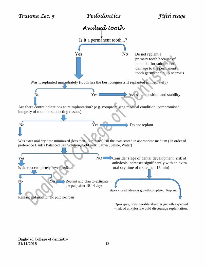
Avulsed tooth
Is it a permanent tooth...?
Yes No Do not replant a
primary tooth because of
potential for subsequent
damage to the permanent
tooth germs and pulp necrosis
Was it replanted immediately (tooth has the best prognosis If replanted immediately)
No Yes Assess the position and stability
Are there contraindications to reimplantation? (e.g. compromising medical condition, compromised
integrity of tooth or supporting tissues)
No Yes Do not replant
Was extra oral dry time minimized (less than 15 minutes)? or the tooth stored in appropriate medium ( In order of
preference Hank's Balanced Salt Solution, Cold milk, Saliva , Saline, Water)
Yes NO Consider stage of dental development (risk of
ankylosis increases significantly with an extra
Is the root completely developed? oral dry time of more than 15 min)
No Yes Replant and plan to extirpate
the pulp after 10-14 days Apex closed, alveolar growth completed: Replant.
Replant and monitor for pulp necrosis
Open apex, considerable alveolar growth expected
– risk of ankylosis would discourage replantation.