preventiveeffectofpinebarkextract(flavangenol)on...
TRANSCRIPT

Hindawi Publishing CorporationEvidence-Based Complementary and Alternative MedicineVolume 2011, Article ID 185913, 9 pagesdoi:10.1093/ecam/nep231
Original Article
Preventive Effect of Pine Bark Extract (Flavangenol) onMetabolic Disease in Western Diet-Loaded Tsumura SuzukiObese Diabetes Mice
Tsutomu Shimada,1 Mitsutaka Kosugi,2 Daisuke Tokuhara,3 Masahito Tsubata,4
Tomoyasu Kamiya,4 Mayu Sameshima,4 Rika Nagamine,4 Kinya Takagaki,4
Ken-ichi Miyamoto,2 and Masaki Aburada1
1 Research Institute of Pharmaceutical Sciences, Musashino University, Shinmachi Nishitokyo-shi, Tokyo 202-8585, Japan2 Department of clinical pharmacy, Graduate School of Natural Science and Technology, Kanazawa University,Kakuma-machi Kanazawa-shi, Ishikawa, Japan
3 Tomei Atsugi Hospital, Atsugi City, Kanagawa, Japan4 Toyo Shinyaku Co. Ltd, Tosu-Shi, Saga, Japan
Correspondence should be addressed to Masaki Aburada, [email protected]
Received 10 August 2009; Accepted 1 December 2009
Copyright © 2011 Tsutomu Shimada et al. This is an open access article distributed under the Creative Commons AttributionLicense, which permits unrestricted use, distribution, and reproduction in any medium, provided the original work is properlycited.
It is known that the metabolic syndrome has a multi-factorial basis involving both genetic and environmental risk factors. In thisstudy, Tsumura Suzuki Obese Diabetes (TSOD) mice, a mouse model of multi-factorial, hereditary, obese type II diabetes, weregiven a Western diet (WTD) as an environmental factor to prepare a disease model (TSOD-WTD) and to investigate the preventiveeffects of Pine bark extract (Flavangenol) against obesity and various features of metabolic disease appearing in this animal model.In contrast to control Tsumura Suzuki Non-obesity (TSNO) mice, TSOD mice were obese and suffered from other metaboliccomplications. WTD-fed TSOD mice developed additional features such as hyperinsulinemia, abnormal glucose/lipid metabolismand fatty liver. The treatment with Flavangenol had a suppressive effect on increase in body weight and accumulation of visceral andsubcutaneous fat, and also showed preventive effects on symptoms related to insulin resistance, abnormal glucose/lipid metabolismand hypertension. Flavangenol also increased the plasma concentration of adiponectin and decreased the plasma concentration ofTNF-α. We next investigated the effect of Flavangenol on absorption of meal-derived lipids. Flavangenol suppressed absorptionof neutral fat in an olive-oil-loading test (in vivo) and showed an inhibitory effect on pancreatic lipase (in vitro). The aboveresults suggest that Flavangenol has a preventive effect on severe metabolic disease due to multiple causes that involve both geneticand environmental risk factors. The mechanism of action might involve a partial suppressive effect of meal-derived lipids onabsorption.
1. Introduction
Obesity, especially accumulation of visceral fat, is associatedwith a potential for harm and provides a pathologicalbasis for various metabolic diseases, the development ofdiabetes/abnormal lipid metabolism/hypertension and othermetabolic complications starting with insulin resistance andprogressing to arteriosclerosis and ischemic cerebral/cardiacdiseases. Prioritized exploratory research has sought materi-als for developing drugs to prevent and treat these disorders.
Pine bark extract (Flavangenol), obtained from thepine trees that grow on the west coast of France (Pinus
maritima), contains oligomeric proanthocyanidin complexes(catechin oligomers) as the major ingredient. Pine barkextract is reported to be an antioxidant [1] and anti-inflammant [2], with the capacity to improve diabeticmicrovascular damage [3], vascular endothelial cell function[4–6] and ischemia/reperfusion-induced renal injury [7].Clinical studies indicate that the extract is effective in thetreatment of chronic venous insufficiency and retinal micro-hemorrhages [8].
The primary causes of obesity and metabolic dis-eases based on obesity include genetic factors [9–11] and

2 Evidence-Based Complementary and Alternative Medicine
environmental factors (overeating, stress, development ofmeans of transportation). We previously developed ananimal model of the disease, Tsumura Suzuki Obese Diabetes(TSOD), which spontaneously develops various featuressimilar to those of human metabolic diseases, and confirmedthat this is a spontaneous obese mouse model of type IIdiabetes [12–14]. In addition, Izumi et al. analyzed TSODmice by the QTL mapping method and reported that this isa multi-factorial hereditary obesity/metabolic disease modelwith mutations in the gene loci controlling body weight,insulin level, fat level and adipocyte size [15, 16].
In this study, TSOD mice with genes for metabolic dis-ease were given a Western diet (WTD) as an environmentalfactor, and the preventive effects of Flavangenol on variousmetabolic disease features were investigated. Furthermore,to clarify the mechanism of action of Flavangenol, weinvestigated the effect of Flavangenol on absorption of meal-derived lipids.
2. Methods
2.1. Experimental Materials. Flavangenol used in this studywas supplied by Toyo Shinyaku Co. Ltd (Saga Prefecture),and contains 72.5% polyphenol (determined by the Folin-Denis method) including 5% proanthocyanidin B1, 2.98%catechin and 0.23% epicatechin.
The WTD, F2WDT (Oriental Yeast Co., Ltd, Tokyo),contains 19.82% casein, 0.3% l-cystine, 3.7458% cornstarch,1.25% α-Cornstarch, 34% sucrose, 1.0% soybean oil, 5.0%cellulose powder, 1.0% AIN-93 vitamin mixture, 3.5 AIN-93G mineral mixture, 0.25% choline bitratrate, 0.0042%t-butylhydroquinone, 20.0% milk fat (butter, etc.), 9.98%maltodextrin and 0.15% cholesterol. The total calories ofnutritional elements were 450.8 kcal/100 g, and the caloriepercentages were 17.8% for protein, 20.0% for lipid and49.0% for carbohydrate. As a control diet, MF (OrientalYeast Co. Ltd) was used as an “ordinary diet” (total calorieof nutritional elements: 360 kcal/100 g, calorie percentages:23.6% for protein, 5.3% for lipid and 6.1% for carbohydrate).
2.2. Experimental Animals. TSOD mice and the corre-sponding control Tsumura Suzuki Non-Obesity (TSNO)mice (derived from the same ancestry but developing nometabolic disease), were purchased from the Institute forAnimal Reproduction (Ibaraki Prefecture) at the age of3 weeks. On the other hand, 5-week-old ddY mice werepurchased from Tokyo Laboratory Animals Science Co. Ltd(Tokyo). The mice were acclimated for one week to thetemperature of 23 ± 2◦C and humidity of 55 ± 10%,and were given an ordinary MF powder diet and purifiedwater ad libitum. After the acclimation, TSOD and TSNOmice were weighed and assigned to five groups (the TSNO-MF group: TSNO mice given MF; the TSOD-MF group:TSOD mice given MF; the TSOD-WTD group: TSOD micegiven WTD; the TSOD-WTD-Flavangenol 3% group: TSODmice given the WTD containing 3% Flavangenol; and theTSOD-WTD-Flavangenol 5% group: TSOD mice given theWTD containing 5% Flavangenol), ensuring that the bodyweight was distributed uniformly. Flavangenol was mixed
well with the WTD so that the content was uniformly 3or 5%. During the 8-week experiment, each group wasgiven water and each defined diet ad libitum. All the animalexperiment procedures were performed pursuant to theAnimal Experiment Ethical Rules of Musashino University.
2.3. Components Examined During the Animal Experiment.All the animals were weighed once a week, and the foodintake was determined every other week [17–20]. For investi-gating the time-course of changes in amounts of visceral andsubcutaneous fat, the amounts of visceral and subcutaneousfat were determined at the start of the experiment and atfollowing 4 and 8 weeks, a total of three times, by X-raycomputed tomography (CT; Latheta, Aloka Co. Ltd. Tokyo)with scanning from the ensiform process to the sacral boneat distance intervals of 1.5 mm under anesthetization withNembutal (50 mg/kg i.p.).
2.4. Glucose-Loading Test and Blood Pressure Determination.The glucose-loading test and blood pressure determinationwere performed 8 weeks after the start of the experiment.In the glucose-loading test, each mouse after fasting forone night was given oral glucose (2 g/kg); blood samplingfrom the orbital venous plexus was performed at the definedtime points under non-anesthetized conditions. The plasmasamples obtained by centrifugation were stored at –80◦Cuntil determination of glucose concentrations.
The blood pressure was determined with a non-invasiveblood pressure meter (Softron Co. Ltd, Tokyo) by fixing eachmouse in a fixing apparatus (Softron Co. Ltd) under non-anesthetized conditions. The cuff was set at the tail root todetermine the systolic/diastolic/mean blood pressures.
2.5. Components Examined at the End of the Experiment.Under non-fasting conditions, each mouse was anesthetizedwith ether and the blood was drawn from the abdominalvena cava. The plasma samples obtained by centrifugationwere stored at –80◦C until blood biochemical tests could beperformed. At the time of autopsy, the liver, mesenteric fat,perinephric fat, epididymal fat and retroperitoneal fat wereisolated and weighed. Using the plasma samples obtained,the glucose, total cholesterol, triacylglycerol and free fattyacid levels were determined with biochemical tests kitsobtained from Wako Pure Chemical Industries Ltd (Tokyo).The insulin level was determined with Rebis Insulin-Mouse-T (Shibayagi Co. Ltd, Gunma Prefecture), the adiponectinlevel with Mouse Adiponectin/Acrp30 Immunoassay (R&DSystems) and the TNF-α level with TNF-α ELISA (Bio CosmoCo. Ltd, Tokyo).
2.6. Olive-Oil-Loading Test and Pancreatic Lipase InhibitionTest. The olive-oil-loading test was performed accordingto the method of Ninomiya et al. [21]. After fasting forone night, 6-week-old ddY mice were orally administeredwith Flavangenol (0.5 or 1 g/kg), and 30 min later, witholive oil (5 mL/kg). Blood sampling from the orbital venousplexus was performed at the defined time points under non-anesthetized conditions. The plasma samples obtained by
Evidence-Based Complementary and Alternative Medicine 3
centrifugation were stored at −80◦C until determination oftriacylglycerol concentrations.
The pancreatic lipase inhibition test was performedaccording to the method of Ninomiya et al. [21], using ageneral kit (Lipase Kit S: Dainippon Sumitomo Pharma Co.Ltd, Osaka) and porcine pancreatic lipase (L3126 Type II,Sigma-Aldrich, St Louis, MO).
2.7. Statistical Analysis. The data were displayed as mean ±standard deviation (SD). In each experiment, the inter-groupdifference was tested for significance by Dunnett’s multiplecomparison procedure with a significance level of 0.05.
3. Results
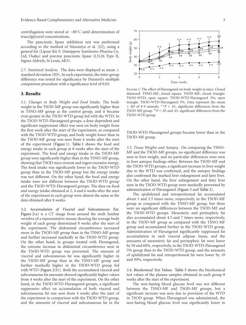
3.1. Changes in Body Weight and Food Intake. The bodyweight in the TSOD-MF group was significantly higher thanin TSNO-MF group as the control group, and it becameeven greater in the TSOD-WTD group fed with the WTD. Inthe TSOD-WTD-Flavangenol groups, a dose-dependent andsignificant suppressant effect was seen on body weight fromthe first week after the start of the experiment, as comparedwith the TSOD-WTD group, and body weight lower than inthe TSOD-MF group was seen from 4 weeks after the startof the experiment (Figure 1). Table 1 shows the food andenergy intake in each group at 8 weeks after the start of theexperiment. The food and energy intake in the TSOD-MFgroup were significantly higher than in the TSNO-MF group,showing that TSOD mice overeat and ingest excessive energy.The food intake was significantly lower in the TSOD-WTDgroup than in the TSOD-MF group but the energy intakewas not different. On the other hand, the food and energyintake were not different between the TSOD-WTD groupand the TSOD-WTD-Flavangenol groups. The data on foodand energy intake obtained at 2, 4 and 6 weeks after the startof the experiment in each group were almost the same as thedata obtained after 8 weeks.
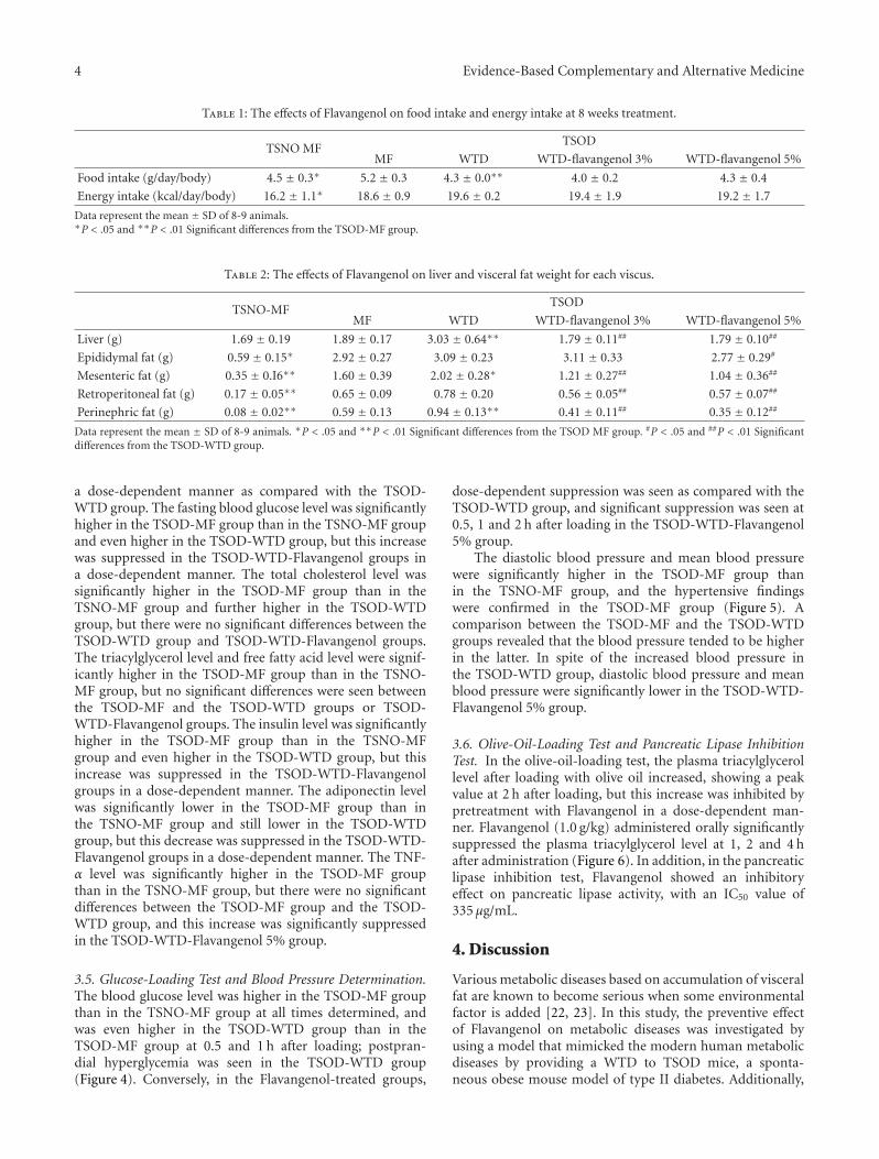
3.2. Accumulation of Visceral and Subcutaneous Fat.Figure 2(a) is a CT image from around the sixth lumbarvertebra of a representative mouse showing the average bodyweight of each group determined 8 weeks after the start ofthe experiment. The abdominal circumference increasedmore in the TSOD-MF group than in the TSNO-MF groupand further increased markedly in the TSOD-WTD group.On the other hand, in groups treated with Flavangenol,the extreme increase in abdominal circumference seen inthe TSOD-WTD group was prevented. The amount ofvisceral and subcutaneous fat was significantly higher inthe TSOD-MF group than in the TSNO-MF group andfurther markedly higher in the TSOD-WTD group fedwith WTD (Figure 2(b)). Both the accumulated visceral andsubcutaneous fat amounts showed significantly higher valuesfrom 4 weeks after the start of the experiment. On the otherhand, in the TSOD-WTD-Flavangenol groups, a significantsuppressive effect on accumulation of both visceral andsubcutaneous fat was seen from 4 weeks after the start ofthe experiment in comparison with the TSOD-WTD group,and the amounts of visceral and subcutaneous fat in the
20
30
40
50
60
70
Bod
yw
eigh
t(g
)
0 2 4 6 8
Time (week)
∗∗
∗∗ ∗∗ ∗∗ ∗∗ ∗∗
###
##
##
##
##
##
##
##
##
##
##
##
##
####
∗∗ ∗∗ ∗∗ ∗∗ ∗∗ ∗∗ ∗∗ ∗∗ ∗∗
Figure 1: The effect of Flavangenol on body weight in mice. Closeddiamond: TSNO-MF; closed square: TSOD-MF; closed triangle:TSOD-WTD; open square: TSOD-WTD-Flavangenol 3%; opentriangle: TSOD-WTD-Flavangenol 5%. Data represent the mean± SD of 8-9 animals. ∗∗P < .01, significant differences from theTSOD-MF group. ##P < .05 and .01, significant differences from theTSOD-WTD group.
TSOD-WTD-Flavangenol groups became lower than in theTSOD-MF group.
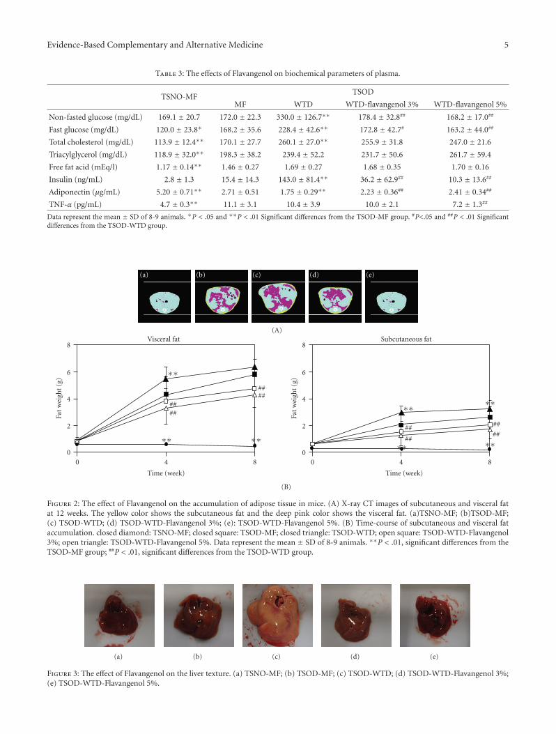
3.3. Tissue Weights and Autopsy. On comparing the TSNO-MF and the TSOD-MF groups, no significant difference wasseen in liver weight, and no particular differences were seenin liver autopsy findings either. Between the TSOD-MF andthe TSOD-WTD groups, a significant increase in liver weightdue to the WTD was confirmed, and the autopsy findingsalso confirmed the marked liver enlargement and fatty liver.On the other hand, the liver enlargement and fatty liverseen in the TSOD-WTD group were markedly prevented byadministration of Flavangenol (Figure 3 and Table 2).
The epididymal and retroperitoneal fat accumulatedabout 5 and 3.5 times more, respectively, in the TSOD-MFgroup as compared with the TSNO-MF group, but therewere no significant differences between the TSOD-MF andthe TSOD-WTD groups. Mesenteric and perinephric fatalso accumulated about 4.5 and 7 times more, respectively,in the TSOD-MF group as compared with the TSNO-MFgroup and accumulated further in the TSOD-WTD group.Administration of Flavangenol significantly suppressed fataccumulation in each visceral adipose tissue, and theamounts of mesenteric fat and perinephric fat were lowerby 50 and 60%, respectively, in the TSOD-WTD-Flavangenol5% group than in the TSOD-WTD group, and the amountsof epididymal fat and retroperitoneal fat were lower by 10and 30%, respectively.
3.4. Biochemical Test Values. Table 3 shows the biochemicaltest values of the plasma samples obtained in each group 8weeks after the start of the experiment.
The non-fasting blood glucose level was not differentbetween the TSNO-MF and TSOD-MF groups, but asignificant increase was seen due to provision of the WTDin TSOD group. When Flavangenol was administered, thenon-fasting blood glucose level was significantly lower in
4 Evidence-Based Complementary and Alternative Medicine
Table 1: The effects of Flavangenol on food intake and energy intake at 8 weeks treatment.
TSNO MFTSOD
MF WTD WTD-flavangenol 3% WTD-flavangenol 5%
Food intake (g/day/body) 4.5 ± 0.3∗ 5.2 ± 0.3 4.3 ± 0.0∗∗ 4.0 ± 0.2 4.3 ± 0.4
Energy intake (kcal/day/body) 16.2 ± 1.1∗ 18.6 ± 0.9 19.6 ± 0.2 19.4 ± 1.9 19.2 ± 1.7
Data represent the mean ± SD of 8-9 animals.∗P < .05 and ∗∗P < .01 Significant differences from the TSOD-MF group.
Table 2: The effects of Flavangenol on liver and visceral fat weight for each viscus.
TSNO-MFTSOD
MF WTD WTD-flavangenol 3% WTD-flavangenol 5%
Liver (g) 1.69 ± 0.19 1.89 ± 0.17 3.03 ± 0.64∗∗ 1.79 ± 0.11## 1.79 ± 0.10##
Epididymal fat (g) 0.59 ± 0.15∗ 2.92 ± 0.27 3.09 ± 0.23 3.11 ± 0.33 2.77 ± 0.29#
Mesenteric fat (g) 0.35 ± 0.I6∗∗ 1.60 ± 0.39 2.02 ± 0.28∗ 1.21 ± 0.27## 1.04 ± 0.36##
Retroperitoneal fat (g) 0.17 ± 0.05∗∗ 0.65 ± 0.09 0.78 ± 0.20 0.56 ± 0.05## 0.57 ± 0.07##
Perinephric fat (g) 0.08 ± 0.02∗∗ 0.59 ± 0.13 0.94 ± 0.13∗∗ 0.41 ± 0.11## 0.35 ± 0.12##
Data represent the mean ± SD of 8-9 animals. ∗P < .05 and ∗∗P < .01 Significant differences from the TSOD MF group. #P < .05 and ##P < .01 Significantdifferences from the TSOD-WTD group.
a dose-dependent manner as compared with the TSOD-WTD group. The fasting blood glucose level was significantlyhigher in the TSOD-MF group than in the TSNO-MF groupand even higher in the TSOD-WTD group, but this increasewas suppressed in the TSOD-WTD-Flavangenol groups ina dose-dependent manner. The total cholesterol level wassignificantly higher in the TSOD-MF group than in theTSNO-MF group and further higher in the TSOD-WTDgroup, but there were no significant differences between theTSOD-WTD group and TSOD-WTD-Flavangenol groups.The triacylglycerol level and free fatty acid level were signif-icantly higher in the TSOD-MF group than in the TSNO-MF group, but no significant differences were seen betweenthe TSOD-MF and the TSOD-WTD groups or TSOD-WTD-Flavangenol groups. The insulin level was significantlyhigher in the TSOD-MF group than in the TSNO-MFgroup and even higher in the TSOD-WTD group, but thisincrease was suppressed in the TSOD-WTD-Flavangenolgroups in a dose-dependent manner. The adiponectin levelwas significantly lower in the TSOD-MF group than inthe TSNO-MF group and still lower in the TSOD-WTDgroup, but this decrease was suppressed in the TSOD-WTD-Flavangenol groups in a dose-dependent manner. The TNF-α level was significantly higher in the TSOD-MF groupthan in the TSNO-MF group, but there were no significantdifferences between the TSOD-MF group and the TSOD-WTD group, and this increase was significantly suppressedin the TSOD-WTD-Flavangenol 5% group.
3.5. Glucose-Loading Test and Blood Pressure Determination.The blood glucose level was higher in the TSOD-MF groupthan in the TSNO-MF group at all times determined, andwas even higher in the TSOD-WTD group than in theTSOD-MF group at 0.5 and 1 h after loading; postpran-dial hyperglycemia was seen in the TSOD-WTD group(Figure 4). Conversely, in the Flavangenol-treated groups,
dose-dependent suppression was seen as compared with theTSOD-WTD group, and significant suppression was seen at0.5, 1 and 2 h after loading in the TSOD-WTD-Flavangenol5% group.
The diastolic blood pressure and mean blood pressurewere significantly higher in the TSOD-MF group thanin the TSNO-MF group, and the hypertensive findingswere confirmed in the TSOD-MF group (Figure 5). Acomparison between the TSOD-MF and the TSOD-WTDgroups revealed that the blood pressure tended to be higherin the latter. In spite of the increased blood pressure inthe TSOD-WTD group, diastolic blood pressure and meanblood pressure were significantly lower in the TSOD-WTD-Flavangenol 5% group.
3.6. Olive-Oil-Loading Test and Pancreatic Lipase InhibitionTest. In the olive-oil-loading test, the plasma triacylglycerollevel after loading with olive oil increased, showing a peakvalue at 2 h after loading, but this increase was inhibited bypretreatment with Flavangenol in a dose-dependent man-ner. Flavangenol (1.0 g/kg) administered orally significantlysuppressed the plasma triacylglycerol level at 1, 2 and 4 hafter administration (Figure 6). In addition, in the pancreaticlipase inhibition test, Flavangenol showed an inhibitoryeffect on pancreatic lipase activity, with an IC50 value of335 μg/mL.
4. Discussion
Various metabolic diseases based on accumulation of visceralfat are known to become serious when some environmentalfactor is added [22, 23]. In this study, the preventive effectof Flavangenol on metabolic diseases was investigated byusing a model that mimicked the modern human metabolicdiseases by providing a WTD to TSOD mice, a sponta-neous obese mouse model of type II diabetes. Additionally,
Evidence-Based Complementary and Alternative Medicine 5
Table 3: The effects of Flavangenol on biochemical parameters of plasma.
TSNO-MFTSOD
MF WTD WTD-flavangenol 3% WTD-flavangenol 5%
Non-fasted glucose (mg/dL) 169.1 ± 20.7 172.0 ± 22.3 330.0 ± 126.7∗∗ 178.4 ± 32.8## 168.2 ± 17.0##
Fast glucose (mg/dL) 120.0 ± 23.8∗ 168.2 ± 35.6 228.4 ± 42.6∗∗ 172.8 ± 42.7# 163.2 ± 44.0##
Total cholesterol (mg/dL) 113.9 ± 12.4∗∗ 170.1 ± 27.7 260.1 ± 27.0∗∗ 255.9 ± 31.8 247.0 ± 21.6
Triacylglycerol (mg/dL) 118.9 ± 32.0∗∗ 198.3 ± 38.2 239.4 ± 52.2 231.7 ± 50.6 261.7 ± 59.4
Free fat acid (mEq/l) 1.17 ± 0.14∗∗ 1.46 ± 0.27 1.69 ± 0.27 1.68 ± 0.35 1.70 ± 0.16
Insulin (ng/mL) 2.8 ± 1.3 15.4 ± 14.3 143.0 ± 81.4∗∗ 36.2 ± 62.9## 10.3 ± 13.6##
Adiponectin (μg/mL) 5.20 ± 0.71∗∗ 2.71 ± 0.51 1.75 ± 0.29∗∗ 2.23 ± 0.36## 2.41 ± 0.34##
TNF-α (pg/mL) 4.7 ± 0.3∗∗ 11.1 ± 3.1 10.4 ± 3.9 10.0 ± 2.1 7.2 ± 1.3##
Data represent the mean ± SD of 8-9 animals. ∗P < .05 and ∗∗P < .01 Significant differences from the TSOD-MF group. #P<.05 and ##P < .01 Significantdifferences from the TSOD-WTD group.
(a) (b) (c) (d) (e)
(A)
0
2
4
6
8
Fat
wei
ght
(g)
0 4 8
Time (week)
Visceral fat
∗∗
####
∗∗
####
∗∗0
2
4
6
8
Fat
wei
ght
(g)
0 4 8
Time (week)
Subcutaneous fat
∗∗
##
##∗∗
∗∗
##
##
∗∗
(B)
Figure 2: The effect of Flavangenol on the accumulation of adipose tissue in mice. (A) X-ray CT images of subcutaneous and visceral fatat 12 weeks. The yellow color shows the subcutaneous fat and the deep pink color shows the visceral fat. (a)TSNO-MF; (b)TSOD-MF;(c) TSOD-WTD; (d) TSOD-WTD-Flavangenol 3%; (e): TSOD-WTD-Flavangenol 5%. (B) Time-course of subcutaneous and visceral fataccumulation. closed diamond: TSNO-MF; closed square: TSOD-MF; closed triangle: TSOD-WTD; open square: TSOD-WTD-Flavangenol3%; open triangle: TSOD-WTD-Flavangenol 5%. Data represent the mean ± SD of 8-9 animals. ∗∗P < .01, significant differences from theTSOD-MF group; ##P < .01, significant differences from the TSOD-WTD group.
(a) (b) (c) (d) (e)
Figure 3: The effect of Flavangenol on the liver texture. (a) TSNO-MF; (b) TSOD-MF; (c) TSOD-WTD; (d) TSOD-WTD-Flavangenol 3%;(e) TSOD-WTD-Flavangenol 5%.

6 Evidence-Based Complementary and Alternative Medicine
0
200
400
600
800
1000
Glu
cose
(mg/
dL)
0 60 120 180
Time (min)
∗∗####
∗∗
##
∗∗
∗∗
##∗∗
#
∗∗ ∗∗
Figure 4: The effect of Flavangenol on the plasma glucose levelin the oral glucose tolerance test. Closed diamond: TSNO-MF;closed square: TSOD-MF; closed triangle: TSOD-WTD; opensquare: TSOD-WTD-Flavangenol 3%; open triangle: TSOD-WTD-Flavangenol 5%. Data represent the mean± SD of 8-9 animals. ∗∗P< .01, significant differences from the TSOD-MF group. ##P < .01,significant differences from the TSOD-WTD group.
0
20
40
60
80
100
120
140
Blo
odpr
essu
re(m
mH
g)
SBP DBP MBP
∗ # ∗ #
TSNO MFTSOD MFTSOD WTDTSOD WTD-Flavangenol 3%TSOD WTD-Flavangenol 5%
Figure 5: The effect of Flavangenol on blood pressure. Data repre-sent the mean ±SD of 8-9 animals. ∗P < .05, significant differencesfrom the TSOD-MF group; #P < .05, significant differences from theTSOD-WTD group.
for the purpose of clarifying the action mechanism ofFlavangenol, we investigated the effect of Flavangenol onabsorption of meal-derived lipids.
As reported previously, the TSOD-MF group showedincreased food intake, obesity with predominant accumu-lation of visceral fat, insulin resistance and other metaboliccomplications, as compared with the TSNO-MF group[17–20]. When TSOD mice were given a WTD as anenvironmental factor, further significant obesity was seenfrom about 2 weeks after the start of the treatment, andsignificant accumulation of visceral fat (especially mesentericfat and perinephric fat) and subcutaneous fat, markedliver enlargement and fatty liver were observed. On bloodbiochemical tests, significant increases of fasting/non-fastingblood glucose levels, total cholesterol and insulin levels anda significant decrease in adiponectin level were confirmed.
0
200
400
600
800
1000
1200
Tria
cylg
lyce
rol(
mgm
L)
0 1 2 3 4 5 6
Time (hr)
∗
∗∗ ∗
Figure 6: The effect of Flavangenol on plasma triacylglycerolelevation in olive-oil-loaded ddY mice Closed diamond: Normal(water); open triangle: Flavangenol 0.5 g/kg p.o.; open square:Flavangenol 1.0 g/kg p.o. Data represent the mean ±SD of sixanimals. ∗P < .05 and ∗∗P < .01, significant differences from thenormal mice.
It was clarified that more severe metabolic diseases appearedin the TSOD-WTD group following administration of aWTD as an environmental factor, as these mice have genesfor metabolic disease. In the present study, the food intakewas significantly lower in the TSOD-WTD group, but theconverted energy intake was not lower.
The effect of Flavangenol added to the diet of the TSOD-WTD group was marked. Flavangenol did not influencethe food and energy intake but, in a dose-dependentmanner, suppressed the body weight increase due to aWTD. In the adipose tissues, which are closely related to anincrease/decrease in body weight, it was clarified by X-ray CTanalysis that Flavangenol showed a significant suppressiveeffect on accumulation of visceral and subcutaneous fat ina time-course manner. Looking at the suppressive effecton fat accumulation by visceral adipose tissue, a strongereffect was observed on mesenteric and perinephric fat,which increased more significantly on the WTD. When wepreviously investigated the effect of Flavangenol in a modeldeveloped by giving a high-fat diet to Sprague Dawley (SD)rats, we observed a significant suppressive effect on increasein body weight, accumulation of visceral fat and an increasein fecal excretion of total lipid in the group given a dietcontaining 2% Flavangenol (unpublished results). In thepresent study, similar results were obtained in the severedisease state that mimicked human disease by combininggenetic and environmental factors. Additionally, it was alsoclarified that Flavangenol showed a preventive effect on liverenlargement and fatty liver. Recently, it has been stronglysuggested that fatty liver is involved in onset and progressionof non-alcoholic steatohepatitis (NASH) [24]. It is thereforeanticipated that Flavangenol may also exert a preventiveeffect against NASH.
It was reported that visceral fat accumulation oradipocyte hypertrophy induces insulin resistance and themechanism of action involves decreased secretion ofadiponectin (which is known to improve arteriosclerosisand various metabolic diseases) and increased secretion
Evidence-Based Complementary and Alternative Medicine 7
Dietary fat
Small intestine
Lumen
Inhibiting pancreatic lipase
Flavangenol
Enterocyte
Decreasingabsorption
Endogenousmechanisms
? Body weight gainvisceral fat accumulation
TNF-α AdiponectinInsulin resistance
Flavangenol
Hyperglycemia Hyperinsulinemia Hypertension Fatty liver
Metabolic syndrome
Flavangenol
Action of flavangenol
Dietary fat
Panceatic lipase
Figure 7: Mechanisms of action of anti-metabolic disease by Flavangenol.
of TNFα (which induces insulin resistance) [25]. In thisstudy, Flavangenol significantly suppressed accumulation ofvisceral fat, significantly prevented the hyperglycemia andhyperinsulinemia observed in the TSOD-WTD group andprevented onset of abnormal glucose tolerance, suggestinga role in improved insulin resistance. In addition, sinceFlavangenol significantly increased the adiponectin level andsignificantly decreased the TNF-α level, it is suggested thatthe abnormal secretion of adipocytokines was normalizedand onset of insulin resistance was prevented as a result,by the suppressive effect of Flavangenol on hypertrophy andaccumulation of adipocytes. In this study, the body weight,amounts of visceral and subcutaneous fat and liver weightwere lower in the TSOD-WTD-Flavangenol groups thanin the TSOD-MF group. Furthermore, the plasma TNFαlevel was significantly lowered by Flavangenol, though notinfluenced by providing a WTD, suggesting that Flavangenolwas effective not only for the obesity induced by a WTD butalso on the original hereditary obesity of TSOD mice.
In this study, Flavangenol did not show a suppressiveeffect on the increases in blood levels of lipids, such as
total cholesterol, triacylglycerol and free fatty acid levels,observed in the TSOD-WTD group. On the other hand, Fla-vangenol significantly suppressed accumulation of visceraland subcutaneous fat and onset of fatty liver. Mochizuki et al.[26] reported that Pine bark extract enhanced lipolysis bystimulation of β-receptors in a study using 3T3 cells. Itis suggested that enhancement of lipolysis by Flavangenolthrough stimulation of β-receptors might be involved inthe preventive effect of Flavangenol on onset of fatty liverand fat accumulation. Conversely, it is also suggested that asuppressive effect of Flavangenol on increased plasma triacyl-glycerol and free fatty acid levels was not seen, due to transferto plasma of free fatty acids formed through lipid decom-position in the liver and adipocytes. It is known that fataccumulation in the liver and skeletal muscles or abnormalsecretion of adipocytokines from hypertrophic adipocytesinduces onset of insulin resistance. It is considered impor-tant that onset of fatty liver and fat accumulation wasprevented by Flavangenol in this study, though Flavangenoldid not show a preventive effect on increases in blood lipidlevels.
8 Evidence-Based Complementary and Alternative Medicine
Hypertension is one of the diagnostic criteria for themetabolic syndrome, and Flavangenol showed a significantantihypertensive effect (on diastolic and mean bloodpressure). Pine bark extract was reported to show vascularprotection due to an antioxidant effect [6], endothelium-dependent vasodilatation [27], improvement of endothelialcell function in hypertension patients [5] and activation ofendothelial nitric oxide synthase (eNOS) in the endothelialcells [6]. Also, it is suggested that the suppressive effect ofFlavangenol on hypertension seen in this study involves adirect effect on the endothelial cells and an indirect effectthrough suppression of the onset of insulin resistance.
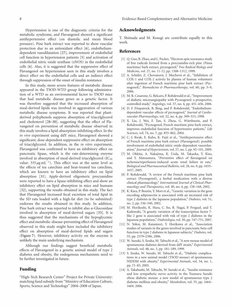
In this study, more severe features of metabolic diseaseappeared in the TSOD-WTD group following administra-tion of a WTD as an environmental factor to TSOD micethat had metabolic disease genes as a genetic factor. Itwas therefore suggested that the increased absorption ofmeal-derived lipids was involved in aggravation of variousmetabolic disease symptoms. It was reported that plant-derived polyphenols suppress absorption of triacylglyceroland cholesterol [28–30], suggesting that the effect of Fla-vangenol on prevention of metabolic disease observed inthis study involves a lipid-absorption-inhibiting effect. In thein vivo experiment using ddY mice, Flavangenol showed asignificant, dose-dependent suppressive effect on absorptionof triacylglycerol. In addition, in the in vitro experiment,Flavangenol was confirmed to have an inhibitory effect onpancreatic lipase, which is the rate-determining enzymeinvolved in absorption of meal-derived triacylglycerol (IC50
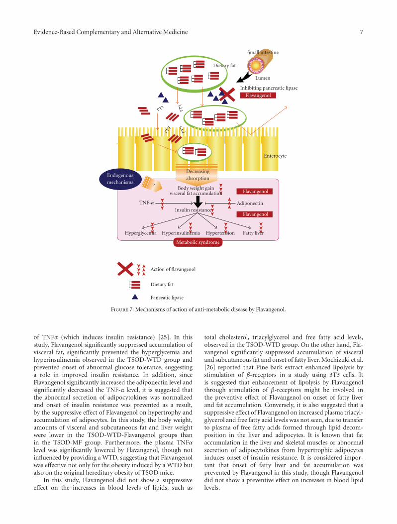
value: 335 μg mL−1). This effect was at the same level asthe effects of tea catechins and heat-treated tea catechins,which are known to have an inhibitory effect on lipidabsorption [31]. Apple-derived oligomeric procyanidinswere reported to have a lipase-inhibiting effect and show aninhibitory effect on lipid absorption in mice and humans[32], supporting the results obtained in this study. The factthat Flavangenol increased fecal excretion of total lipid inthe SD rats loaded with a high-fat diet (to be submitted)endorses the results obtained in this study. In addition,Pine bark extract was reported to inhibit also α-Glucosidaseinvolved in absorption of meal-derived sugars [33]. It isthus suggested that the mechanisms of the hypoglycemiceffect and metabolic-disease-preventing effect of Flavangenolobserved in this study might have included the inhibitoryeffect on absorption of meal-derived lipids and sugars(Figure 7). However, inhibitory activity on the enzyme isunlikely the main underlying mechanism.
Although our findings suggest beneficial metaboliceffects of Flavangenol in a genetic animal model of type 2diabetes and obesity, the endogenous mechanisms need tobe further investigated in future.
Funding
“High-Tech Research Center” Project for Private University:matching fund subsidy from “Ministry of Education Culture,Sports, Science and Technology” 2004–2008 of Japan.
Acknowledgments
T. Shimada and M. Kosugi are contribute equally to thiswork.
References
[1] Q. Guo, B. Zhao, and L. Packer, “Electron spin resonance studyof free radicals formed from a procyanidin-rich pine (Pinusmaritima) bark extract, pycnogenol,” Free Radical Biology andMedicine, vol. 27, no. 11-12, pp. 1308–1312, 1999.
[2] A. Schafer, Z. Chovanova, J. Muchova et al., “Inhibition ofCOX-1 and COX-2 activity by plasma of human volunteersafter ingestion of French maritime pine bark extract (Pyc-nogenol),” Biomedicine & Pharmacotherapy, vol. 60, pp. 5–9,2006.
[3] M. R. Cesarone, G. Belcaro, P. Rohdewald et al., “Improvementof diabetic microangiopathy with Pycnogenol: a prospective,controlled study,” Angiology, vol. 57, no. 4, pp. 431–436, 2006.
[4] D. F. Fitzpatrick, B. Bing, and P. Rohdewald, “Endothelium-dependent vascular effects of pycnogenol,” Journal of Cardio-vascular Pharmacology, vol. 32, no. 4, pp. 509–515, 1998.
[5] X. Liu, J. Wei, F. Tan, S. Zhou, G. Wurthwein, and P.Rohdewald, “Pycnogenol, French maritime pine bark extract,improves endothelial function of hypertensive patients,” LifeSciences, vol. 74, no. 7, pp. 855–862, 2004.
[6] C.-J. Kwak, E. Kubo, K. Fujii et al., “Antihypertensive effectof French maritime pine bark extract (Flavangenol): possibleinvolvement of endothelial nitric oxide-dependent vasorelax-ation,” Journal of Hypertension, vol. 27, no. 1, pp. 92–101, 2009.
[7] M. Ohkita, A. Nakajima, K. Ueda, M. Takaoka, Y. Kiso,and Y. Matsumura, “Preventive effect of flavangenol onischemia/reperfusion-induced acute renal failure in rats,”Biological and Pharmaceutical Bulletin, vol. 28, no. 9, pp. 1655–1657, 2005.
[8] P. Rohdewald, “A review of the French maritime pine barkextract (Pycnogenol), a herbal medication with a diverseclinical pharmacology,” International Journal of Clinical Phar-macology and Therapeutics, vol. 40, no. 4, pp. 158–168, 2002.
[9] K. Kara, P. Boutin, Y. Mori et al., “Genetic variation in the geneencoding adiponectin is associated with an increased risk oftype 2 diabetes in the Japanese population,” Diabetes, vol. 51,no. 2, pp. 536–540, 2002.
[10] M. Horikoshi, K. Hara, C. Ito, R. Nagai, P. Froguel, and T.Kadowaki, “A genetic variation of the transcription factor 7-like 2 gene is associated with risk of type 2 diabetes in theJapanese population,” Diabetologia, vol. 50, pp. 747–751, 2007.
[11] N. Yokoi, M. Kanamori, Y. Horikawa et al., “Associationstudies of variants in the genes involved in pancreatic beta-cellfunction in type 2 diabetes in Japanese subjects,” Diabetes, vol.55, pp. 2379–2386, 2006.
[12] W. Suzuki, S. Iizuka, M. Tabuchi et al., “A new mouse model ofspontaneous diabetes derived from ddY strain,” ExperimentalAnimals, vol. 48, no. 3, pp. 181–189, 1999.
[13] S. Iizuka, W. Suzuki, M. Tabuchi et al., “Diabetic complica-tions in a new animal model (TSOD mouse) of spontaneousNIDDM with obesity,” Experimental Animals, vol. 54, no. 1,pp. 71–83, 2005.
[14] A. Takahashi, M. Tabuchi, W. Suzuki et al., “Insulin resistanceand low sympathetic nerve activity in the Tsumura Suzukiobese diabetic mouse: a new model of spontaneous type 2diabetes mellitus and obesity,” Metabolism, vol. 55, pp. 1664–1669, 2006.

Evidence-Based Complementary and Alternative Medicine 9
[15] I. Hirayama, Z. Yi, S. Izumi et al., “Genetic analysis of obesediabetes in the TSOD mouse,” Diabetes, vol. 48, no. 5, pp.1183–1191, 1999.
[16] S. Mizutani, H. Gomi, I. Hirayama, and T. Izumi, “Chromo-some 2 locus Nidd5 has a potent effect on adiposity in theTSOD mouse,” Mammalian Genome, vol. 17, no. 5, pp. 375–384, 2006.
[17] M. Tsunakawa, T. Shimada, W. Suzuki et al., “Preventive effectsof Daisaikoto on metabolic disorders in spontaneous obesetype II diabetes mice,” Journal of Traditional Medicines, vol. 23,pp. 216–223, 2003.
[18] T. Shimada, T. Kudo, T. Akase, and M. Aburada, “Preventiveeffects of bofutsushosan on obesity and various metabolicdisorders,” Biological and Pharmaceutical Bulletin, vol. 31, no.7, pp. 1362–1367, 2008.
[19] T. Shimada, T. Akase, M. Kosugi, and M. Aburada, “Preventiveeffect of boiogito on metabolic disorders in the TSOD mouse,a model of Spontaneous Obese Type II Diabetes Melli-tus,” Evidence-Based Complementary and Alternative Medicine,2009.
[20] T. Akase, T. Shimada, Y. Harasawa et al., “Preventive effectsof salacia reticulata on obesity and metabolic disorders inTSOD Mice,” Evidence-Based Complementary and AlternativeMedicine, 2009.
[21] K. Ninomiya, H. Matsuda, H. Shimoda et al., “Carnosic acid,a new class of lipid absorption inhibitor from sage,” Bioorganicand Medicinal Chemistry Letters, vol. 14, no. 8, pp. 1943–1946,2004.
[22] S. Akagiri, Y. Naito, H. Ichikawa et al., “A mouse modelof metabolic syndrome; increase in visceral adipose tissueprecedes the development of fatty liver and insulin resistancein HigWTDat diet-fed male KK/Ta mice,” Journal of ClinicalBiochemistry and Nutrition, vol. 42, pp. 150–157, 2008.
[23] S. Tanaka, T. Hayashi, T. Toyoda et al., “HigWTDat dietimpairs the effects of a single bout of endurance exercise onglucose transport and insulin sensitivity in rat skeletal muscle,”Metabolism, vol. 56, pp. 1719–1728, 2007.
[24] J. Jiang and N. Torok, “Nonalcoholic steatohepatitis andthe metabolic syndrome,” Metabolic Syndrome and RelatedDisorders, vol. 6, no. 1, pp. 1–7, 2008.
[25] N. Maeda, M. Takahashi, T. Funahashi et al., “PPARγ ligandsincrease expression and plasma concentrations of adiponectin,an adipose-derived protein,” Diabetes, vol. 50, no. 9, pp. 2094–2099, 2001.
[26] M. Mochizuki and N. Hasegawa, “Pycnogenol stimulateslipolysis in 3T3-L1 cells via stimulation of β-receptor mediatedactivity,” Phytotherapy Research, vol. 18, no. 12, pp. 1029–1030,2004.
[27] K. Nishioka, T. Hidaka, S. Nakamura et al., “Pycnogenol,French maritime pine bark extract, augments endothelium-dependent vasodilation in humans,” Hypertension Research,vol. 30, no. 9, pp. 775–780, 2007.
[28] M. Nakai, Y. Fukui, S. Asami et al., “Inhibitory effects ofoolong tea polyphenols on pancreatic lipase in vitro,” Journalof Agricultural and Food Chemistry, vol. 53, no. 11, pp. 4593–4598, 2005.
[29] M. Yoshikawa, H. Shimoda, N. Nishida, M. Takada, and H.Matsuda, “Salacia reticulata and its polyphenolic constituentswith lipase inhibitory and lipolytic activities have mildantiobesity effects in rats,” Journal of Nutrition, vol. 132, no.7, pp. 1819–1824, 2002.
[30] I. Ikeda, Y. Imasato, E. Sasaki et al., “Tea catechins decreasemicellar solubility and intestinal absorption of cholesterol inrats,” Biochem Biophys Acta, vol. 1127, pp. 141–146, 1992.
[31] I. Ikeda, K. Tsuda, Y. Suzuki et al., “Tea catechins with agalloyl moiety suppress postprandial hypertriacylglycerolemiaby delaying lymphatic transport of dietary fat in rats,” Journalof Nutrition, vol. 135, no. 2, pp. 155–159, 2005.
[32] H. Sugiyama, Y. Akazome, T. Shoji et al., “Oligomericprocyanidins in apple polyphenol are main active componentsfor inhibition of pancreatic lipase and triglyceride absorption,”Journal of Agricultural and Food Chemistry, vol. 55, pp. 4604–4609, 2007.
[33] A. Schafer and P. Hogger, “Oligomeric procyanidins of Frenchmaritime pine bark extract (Pycnogenol) effectively inhibit α-glucosidase,” Diabetes Research and Clinical Practice, vol. 77,no. 1, pp. 41–46, 2007.

Submit your manuscripts athttp://www.hindawi.com
Stem CellsInternational
Hindawi Publishing Corporationhttp://www.hindawi.com Volume 2014
Hindawi Publishing Corporationhttp://www.hindawi.com Volume 2014
MEDIATORSINFLAMMATION
of
Hindawi Publishing Corporationhttp://www.hindawi.com Volume 2014
Behavioural Neurology
EndocrinologyInternational Journal of
Hindawi Publishing Corporationhttp://www.hindawi.com Volume 2014
Hindawi Publishing Corporationhttp://www.hindawi.com Volume 2014
Disease Markers
Hindawi Publishing Corporationhttp://www.hindawi.com Volume 2014
BioMed Research International
OncologyJournal of
Hindawi Publishing Corporationhttp://www.hindawi.com Volume 2014
Hindawi Publishing Corporationhttp://www.hindawi.com Volume 2014
Oxidative Medicine and Cellular Longevity
Hindawi Publishing Corporationhttp://www.hindawi.com Volume 2014
PPAR Research
The Scientific World JournalHindawi Publishing Corporation http://www.hindawi.com Volume 2014
Immunology ResearchHindawi Publishing Corporationhttp://www.hindawi.com Volume 2014
Journal of
ObesityJournal of
Hindawi Publishing Corporationhttp://www.hindawi.com Volume 2014
Hindawi Publishing Corporationhttp://www.hindawi.com Volume 2014
Computational and Mathematical Methods in Medicine
OphthalmologyJournal of
Hindawi Publishing Corporationhttp://www.hindawi.com Volume 2014
Diabetes ResearchJournal of
Hindawi Publishing Corporationhttp://www.hindawi.com Volume 2014
Hindawi Publishing Corporationhttp://www.hindawi.com Volume 2014
Research and TreatmentAIDS
Hindawi Publishing Corporationhttp://www.hindawi.com Volume 2014
Gastroenterology Research and Practice
Hindawi Publishing Corporationhttp://www.hindawi.com Volume 2014
Parkinson’s Disease
Evidence-Based Complementary and Alternative Medicine
Volume 2014Hindawi Publishing Corporationhttp://www.hindawi.com