prevalence of drug-resistant opportunistic microorganisms
TRANSCRIPT

145
Abstract: Drug-resistant opportunistic infections may cause health problems in immunocompromised hosts. Representative microorganisms in opportu-nistic infections of the oral cavity are Staphylococcus aureus, Pseudomonas aeruginosa, and Candida albicans. We investigated the prevalence of drug-resistant opportunistic microorganisms in elderly adults receiving follow-up examinations after primary treatment of oral cancer. Oral microor-ganisms were collected from patients satisfactorily treated for oral cancer (defined as good outcomes to date) and a group of healthy adults (controls). After identification of microorganisms, the prevalence of drug-resistant microorganisms was studied. Pulsed-field gel electrophoresis (PFGE) and staphylococcal cassette chromosome mec (SCCmec) typing were also performed for methicillin-resistant S aureus (MRSA). Statistical analysis revealed no significant differ-ences in the prevalences of the three microorganisms between the groups. Surprisingly, 69.2% of S aureus
isolates showed oxacillin resistance, suggesting that MRSA colonization is increasing among older Japa-nese. These MRSA isolates possessed SCCmec types II and IV but no representative toxin genes. Our results indicate that a basic infection control strategy, including standard precautions against MRSA, is important for elderly adults, particularly after treat-ment for oral cancer. (J Oral Sci 55, 145-155, 2013)
Keywords: drug-resistant microorganism; opportunistic infection; oral cancer.
IntroductionOpportunistic infections are associated with individuals in poor health (referred to as an immunocompromised host or an opportunistic situation) and are caused by several different microorganisms. Among those that originate in the oral cavity, representative microorganisms include Staphylococcus aureus, Pseudomonas aeruginosa, and Candida albicans (1,2). Most of these organisms have become drug-resistant, which has resulted in difficulties in curing the related infectious diseases. In the 1980s and 90s, methicillin-resistant S aureus (MRSA) was preva-
Correspondence to Dr. Masaru Ohara, Department of Advanced General Dentistry, Hiroshima University Hospital, 1-2-3 Kasumi, Minami-ku, Hiroshima 734-8553, JapanFax: +81-82-424-3909 E-mail: [email protected]
Journal of Oral Science, Vol. 55, No. 2, 145-155, 2013
Original
Prevalence of drug-resistant opportunistic microorganismsin oral cavity after treatment for oral cancer
Kaoru Yamashita1), Masaru Ohara2,3), Taro Kojima4), Rumi Nishimura1), Tetsuji Ogawa2), Takamune Hino3), Mitsugi Okada4), Shigeaki Toratani5), Nobuyuki Kamata6),
Motoyuki Sugai7), and Masaru Sugiyama1)
1)Department of Public Oral Health, Institute of Biomedical & Health Sciences, Hiroshima University, Hiroshima, Japan
2)Department of Advanced General Dentistry, Hiroshima University Hospital, Hiroshima, Japan3)Dental Clinic, Hiroshima University Hospital, Higashihiroshima, Japan
4)Special Care Dentistry, Hiroshima University Hospital, Hiroshima, Japan5)Department of Oral and Maxillofacial Surgery, Hiroshima University Hospital, Hiroshima, Japan
6)Department of Oral and Maxillofacial Surgery, Institute of Biomedical & Health Sciences, Hiroshima University, Hiroshima, Japan
7)Department of Bacteriology, Institute of Biomedical & Health Sciences, Hiroshima University, Hiroshima, Japan
(Received December 25, 2012; Accepted April 23, 2013)

146
lent among opportunistic and nosocomial infections and caused pneumonia, sepsis, colitis, and urinary tract infec-tion, among other illnesses (3). Methicillin resistance results from production of a unique low-affinity penicillin binding protein (PBP 2a) encoded by the chromosomal gene mecA, which is acquired by integration of a gene cassette, termed staphylococcal cassette chromosome mec (SCCmec), using recombinase (ccr) carrying genes on their cassettes. An increasing number of recent reports have identified community-acquired or -associated MRSA (C-MRSA) as a factor in various diseases, such as pneumonia, in young patients and others without classic healthcare-associated risk factors (4).
P aeruginosa is another representative bacterium that causes opportunistic infections (5). It is found in soil, water, and skin flora and can survive in poor environ-ments including hypoxic atmosphere. According to the guidelines of the Japanese Infectious Disease Surveillance Center (IDSC), multidrug-resistant P aeruginosa (MDRP) is defined as isolates with the following minimum inhibi-tory concentrations (MICs) of antibiotics: > 16 µg/mL for imipenem, > 32 µg/mL for amikacin, and > 4 µg/mL for ciprofloxacin. The prevalence of MDRP is increasing, and nosocomial infections have become a serious clinical problem in Japan (6).
The Candida species elicit the yeast infectious disease candidiasis, which causes superficial infections such as oral thrush and vaginitis as well as systemic and poten-tially life-threatening diseases, known as candidemia (7). C albicans is a yeast that normally inhabits the human mouth and skin, where it generally uneventfully coexists with a variety of other microorganisms. An infection occurs when the balance of bacteria in the body is disrupted, especially in immunocompromised situations, allowing drug-resistant Candida species to proliferate and overwhelm other healthy microorganisms (8).
Immunocompromised situations are frequently seen in older individuals, infants, people infected with HIV, and individuals with cancer; oral cancer can reduce immunity in the maxillofacial region (1,2). The principal treatments for oral cancer are surgical excision, radiotherapy, and chemotherapy, given alone or in combination (9). Despite successful treatment of cancer, local and systemic immu-nity decreases in affected patients, while surgical excision induces oral dysfunction, due to loss of normal anatomy. For example, swallowing dysfunction after partial tongue excision readily elicits aspiration pneumonia, and radia-tion and chemotherapy reduce myelocyte function, which may result in an immunocompromised host.
In the present study, we investigated drug-resistant opportunistic microorganisms after primary treatment
of oral cancer in elderly adults. A number of reports of opportunistic infections in cancer patients have been published (1,2,5,7). However, the oral conditions of such patients after oral cancer therapy (so-called “follow up-patients”) are not well known. We focused on drug-resistant opportunistic microorganisms in the mouth of patients who had received primary treatment for oral cancer. After hospital discharge, attending physicians check not only for cancer recurrence but also for wound healing, local infection, and oral mucositis. Clinicians are also aware that their patients may become carriers of drug-resistant microorganisms and may spread them to others.
We isolated Staphylococcus, Pseudomonas, and Candida species from the oral cavities of elderly adults after oral cancer treatment, attempted to detect MRSA, MDRP, and drug-resistant Candida species, and compared prevalences between these follow-up patients and a group of healthy adults. In addition, we discuss SCCmec typing and presence of toxin genes in MRSA organisms.
Materials and MethodsSetting and patientsThis study was carried out from June through December 2010 at Hiroshima University Hospital, Department of Oral and Maxillofacial Surgery and Oral Medicine, after receiving approval from the Ethical Committee of Hiroshima University (Clinical Research No. EKI224). In total, 186 elderly patients (age range, 57-98 years, were identified. After excluding people suffering from systemic diseases that affected infection status (i.e., diabetes, autoimmune diseases, and hepatitis), the patients (n = 83) were divided into two groups. The cancer treatment group (n = 46; 20 men, 26 women; average age 67.4 ± 10.3 years) included patients who had undergone treatment of oral cancer at Hiroshima University Hospital and had good outcomes to date (0 months to 11 years after primary treatment). Therapy for oral cancer consisted of radiotherapy, chemotherapy, and surgery, alone or in combination. Tongue cancer was the most prevalent, followed by gingival cancer of the lower jaw. The second group was the control group (n = 37; 11 men, 26 women; average age, 71.3 ± 9.9 years), who had received treatment for cavities or poorly fitting dentures at the same hospital and had no history of any cancer treatment. Activities of daily living (ADL) did not significantly differ between groups.
Collection of microorganisms and culture conditions Microorganism samples were collected by scrubbing the
147
surgical area (in the cancer treatment group), tongue, gingiva, and palate 5 times using a dry sterile cotton swab. Each cotton swab was then immediately inserted into a 1.5-mL sterile tube containing 800 µL of saline for 10 s. After removing the swab, the remaining saline solution was used as the microbial sample. Additionally, 1-mL saliva samples were collected from each subject (saliva sample). In xerostomic participants, the mouth was rinsed with 5 mL of sterile saline for 10 s, and the recovered saline was used as the rinse sample. Each saline sample was centrifuged at 15,000 rpm (20,600 x g) using the angle rotor of a Kubota RA-2724 (Kubota, Tokyo, Japan) with a Kubota 3500 microtube centrifuge at 22°C for 5 min. The resulting precipitate (ppt) was resuspended with 90 µL of saline, after which three 30-µL samples were spread onto three separate agar plates. The microorganisms were cultured on nalidixic acid/cetrimide agar (Eiken, Tokyo, Japan) for the first screening of Pseudomonas species, while Mannit-Salt agar (Eiken) was used for the first screening of Staphy-lococcus species and Candida-GS agar (Eiken) was used for the first screening of Candida species. The nalidixic acid/cetrimide agar and Mannit-Salt agar plates were incubated at 37°C under aerobic conditions for 2 days, while Candida-GS agar plates were incubated at 30°C for 3 days.
To screen for MRSA, colonies grown on the Mannit-Salt agar plates were collected and spread onto MRSA II selective agar (Eiken) plates, then cultured at 37°C for 2 days. When liquid culture was necessary, trypticase soy broth (Becton Dickinson, Tokyo, Japan) was used for Staphylococcus and Pseudomonas species, and Sabouraud liquid broth (Becton Dickinson) was used for Candida species.
Identification of microorganismsThe shapes of the colonies on the plates were checked, and those with different shapes were collected and gram stained for morphological observation under a light microscope. Genomic DNA from each colony was obtained using a Wizard genomic extraction kit (Promega, Madison, WI, USA). For Staphylococcus species, cells were treated with 1 mg/mL of lysostaphin in Tris-EDTA buffer (TE; 10 mM Tris-Cl, 1 mM EDTA, pH 8.0) at 37°C for 1 h before using the kit. Final identification was done by polymerase chain reaction (PCR) amplification with bacterial universal primers for 16S rDNA (for bacteria) or 26S rDNA (for fungus), followed by DNA sequencing (see Supplementary Table).
PCR amplification was carried out using Taq DNA polymerase (Toyobo, Tokyo, Japan) for 25 cycles, with
denaturing at 94°C for 20 s, annealing at 50°C for 30 s, and polymerization at 72°C for 1 min. The PCR products were electrophoresed on 1% agarose gels with 0.5 µg/mL ethidium bromide and visualized using an Atto UV illu-minator (Atto, Tokyo, Japan). DNA fragments of interest were recovered from each gel using a Geneclean kit (MP Biomedicals, Tokyo, Japan). DNA sequencing was performed using an ABI Prism 3100 Genetic Analyzer (Applied Biosystems, Tokyo, Japan) with a Big Dye Cycle Sequencing reaction kit (AB Applied Biosystems). Identification of experimentally determined nucleotide sequences using sequence databases was performed using the Basic Local Alignment Search Tool (BLAST; http://www.ncbi.nlm.nih.gov/blast).
Detection of toxins and related genes in S aureusToxin genes were analyzed in genome DNA isolated from MRSA organisms by PCR. The detected genes were PVL (Panton-Valentine leukocidin), lukE, lukD (leuko-cidin), eta, etb, etd (exfoliative toxin), edinA, edinB, edinC (epidermal cell differentiation inhibitor), and se (staphylococcal enterotoxin) (see Supplementary Table). PCR amplification was performed using the conditions described above. Enterotoxin genes were detected by multiplex PCR, as previously described (10).
Antimicrobial susceptibility testingMICs were determined using the microdilution broth method provided by the Clinical and Laboratory Stan-dards Institute, using a MicroScanWalkAway-96 system (11). The antibiotics tested were oxacillin (MPIPC; sensi-tivity [S] < 0.25 µg/mL, resistance [R] > 2), arbekacin (ABK; S < 4, R > 8), vancomycin (VCM; S < 2, R > 8), teicoplanin (TEIC; S < 8, R > 16), and linezolid (LZD; S < 2, R > 4) for MRSA. For Candida species, we tested fluconazole (FCZ; S < 8, susceptible-dose dependent [S-DD] = 16-32, R > 64), 5-fluorocytosine (5-FC; S < 4, intermediate = 8-16, R > 32), itraconazole (ITZ; S < 0.125, S-DD = 0.25-0.5, R > 1), miconazole (MCZ; S < 0.5, R > 1), amphotericin B (AMPH-B; S < 1, R > 2), voriconazole (VRCZ; S < 1, R > 4), and micafungin (MCFG; S < 1, R > 2). For Pseudomonas species, imipenem/cilastatin (IMP/CS; S < 16, R > 16), amikacin (AMK; S < 32, R > 32), and ciprofloxacin (CPFX; S < 4, R > 4) were tested.
Statistical analysisDifferences between groups were assessed using Pear-son’s χ2 test, followed by Yate's correction.

148
Genotyping of staphylococcal cassette chromosome mecGenotypes of staphylococcal cassette chromosome mec (SCCmec) were defined by two elements: the mec region (mec class A, B, C) and ccr region (ccr types 1-4) (12). SCCmec was classified by PCR amplification, using DNA extracted from MRSA (Supplementary Table), under the conditions described above. PCR products were electrophoresed on 1% agarose gels with 0.5 µg/mL ethidium bromide and visualized using an Atto UV illuminator.
Pulsed-field gel electrophoresisPulsed-field gel electrophoresis (PFGE) was performed using a previously described method (13). Agarose gels embedded with S aureus were treated with 1 mg/mL lyso-staphin in TE at room temperature for 30 min, followed by incubation with 1% sarkosyl in TE at 37°C for 1 h. Sarkosyl-treated samples were incubated in 1 mg/mL protease K in TE at 50°C overnight. After in-gel diges-tion of chromosomal DNA with the restriction enzyme SmaI (New England BioLabs Inc., Ipswich, MA, USA) at 37°C for 18 h, the resulting fragments were separated using a CHEF Mapper PFGE system (Bio-Rad, Tokyo, Japan). Each gel was stained with 0.5 µg/mL ethidium bromide at room temperature for 1 h. After destaining in water for 30 min, the DNA fragment pattern was visual-ized using an Atto Printgraph UV illuminator. Images were captured using an Atto Image Saver AE-6905 and analyzed with the Dice coefficient and unweighted pair group methods using GelCompar II software (Applied Maths BVBA; Sint-Martens-Latem, Belgium).
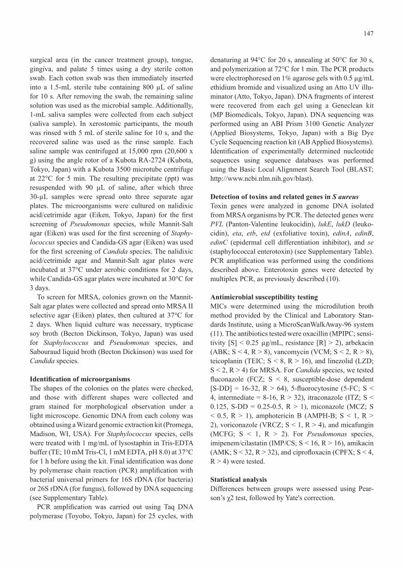
ResultsDetection of opportunistic microorganisms in the oral cavityIn samples from the 83 subjects, 57 Candida species, 71 Staphylococcus species, and 7 Pseudomonas species were isolated (Fig. 1a). In some cases, a single participant had more than two species. For example, S aureus was detected in saliva and S lugdunensis in a tongue swab from the same individual. Thirty-nine participants had Candida species, 28 had Staphylococcus species, and seven had Pseudomonas species (Fig. 1b).
In a comparison between groups of isolated microor-ganisms, Candida, Staphylococcus, and Pseudomonas species were detected in 39 (68.4%), 31 (43.7%), and 4 (57.1%) participants, respectively, in the cancer treatment group, and in 18 (31.6%), 40 (56.3%), and 3 (42.9%) participants, respectively, in the control group (Fig. 1a). Statistical analysis revealed no significant difference
between groups in the number of isolated microorgan-isms or the number of participants with these isolates.
Identification of the isolated organisms by 16S or 26S rDNA sequencing indicated that C albicans (33.3% of Candida species) and C glabrata (31.5%) were dominant among the 57 isolates of Candida species (Fig. 1c). C guilliermondii was isolated from subjects in the control group but not from those in the cancer treatment group. C krusei was found in one participant in the cancer treat-ment group, and C africana was detected in one subject in each group. The numbers of C albicans and C glabrata organisms isolated in the cancer treatment group were greater than those isolated in the control group, although the difference was not significant.
Among Staphylococcus species, S aureus (18.3% of Staphylococcus species) and S epidermidis (19.7%) were dominant, followed by S haemolyticus (15.5%) and S lugdunensis (12.7%) (Fig. 1d). Several minor strains, e.g. S sciuri, S auricularis, and S warner, were only detected in the control group. In a comparison of each Staphylococcus strain between groups, the number of S aureus organisms isolated in the cancer treatment group was greater than that in the control group, although the difference was not significant. All Pseudomonas organ-isms isolated in this study were P aeruginosa.
The presence of two or more microorganisms in patients was also analyzed. No subject had organisms from all three species, although 18 (21.4%) participants had organisms from two species: 11 in the cancer treat-ment group and seven in the control group. Among these patients, 14 (16.7%; 9 in the cancer treatment, 5 in the control group) had a combination of Candida and Staphylococcus species, one from each group had a combination of Candida and Pseudomonas species, and one from each group had a combination of Staphylo-coccus and Pseudomonas species.
Prevalence of antibiotic- and antifungal-resistant opportunistic microorganismsBecause there was no difference between groups in the prevalence of the three opportunistic microorganisms, we next investigated the prevalence of antibiotic-resis-tant microorganisms in the oral cavity. Determination of MICs for Candida species revealed 17 strains with resistance to at least one antifungal agent (Fig. 1c, Table 1). Among drug-resistant Candida species, 14 (82.4%) were C glabrata isolates (11 in the cancer treatment group, 3 in the control group) resistant to itraconazole. C krusei, isolated from subject #60-1, was resistant to three antifungals (5-FC, FCZ, MCZ; Table 1), which is an unsurprising finding because C krusei has been
149
reported to be an intrinsically drug-resistant fungus in which antifungals bind the target molecule cytochrome P-450 with very low affinity (14). The drug resistance of C glabrata is likely partly due to the association with increased expression of genes encoding CDR drug efflux pumps (15,16). However, all the present strains showed susceptibility to MCFG and VRCZ (Table 1). Interest-ingly, one major fungus in the oral cavity, C albicans, did not show resistance to the antifungals used in this
study. Furthermore, only four men carried drug-resistant strains; the other 13 were isolated from women.
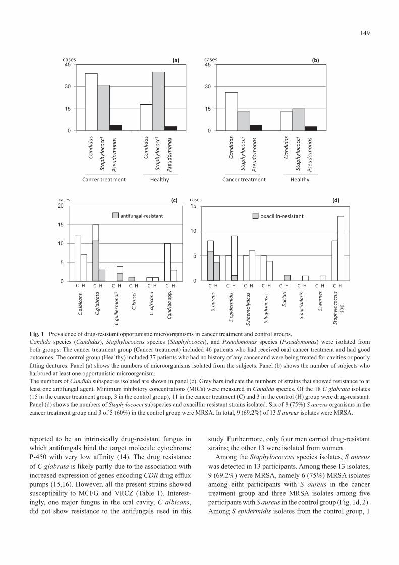
Among the Staphylococcus species isolates, S aureus was detected in 13 participants. Among these 13 isolates, 9 (69.2%) were MRSA, namely 6 (75%) MRSA isolates among eitht participants with S aureus in the cancer treatment group and three MRSA isolates among five participants with S aureus in the control group (Fig. 1d, 2). Among S epidermidis isolates from the control group, 1
Fig. 1 Prevalence of drug-resistant opportunistic microorganisms in cancer treatment and control groups. Candida species (Candidas), Staphylococcus species (Staphylococci), and Pseudomonas species (Pseudomonas) were isolated from both groups. The cancer treatment group (Cancer treatment) included 46 patients who had received oral cancer treatment and had good outcomes. The control group (Healthy) included 37 patients who had no history of any cancer and were being treated for cavities or poorly fitting dentures. Panel (a) shows the numbers of microorganisms isolated from the subjects. Panel (b) shows the number of subjects who harbored at least one opportunistic microorganism. The numbers of Candida subspecies isolated are shown in panel (c). Grey bars indicate the numbers of strains that showed resistance to at least one antifungal agent. Minimum inhibitory concentrations (MICs) were measured in Candida species. Of the 18 C glabrata isolates (15 in the cancer treatment group, 3 in the control group), 11 in the cancer treatment (C) and 3 in the control (H) group were drug-resistant. Panel (d) shows the numbers of Staphylococci subspecies and oxacillin-resistant strains isolated. Six of 8 (75%) S aureus organisms in the cancer treatment group and 3 of 5 (60%) in the control group were MRSA. In total, 9 (69.2%) of 13 S aureus isolates were MRSA.

150
of 9 was oxacillin-resistant (so-called coagulase-negative methicillin-resistant Staphylococcus [MRCNS]), while one MRCNS S sciuri strain was isolated from the control group (Fig. 1d). Susceptibility tests were performed for antibiotics frequently used against MRSA in hospitals (Fig. 2). Isolated MRSA and MRCNS strains were all sensitive to vancomycin, teicoplanin, and linezolid, which are the agents of last resort against MRSA infec-tions. One MRSA strain (subject #22-67) was resistant to arbekacin.
Antibiotic susceptibility of P aeruginosa was exam-ined using the criteria of the Japanese Infectious Disease
Surveillance Center (IDSC). However, no multidrug-resistant P aeruginosa (MDRP) organisms were found.
As noted above, 14 of our 83 subjects had both Candida and Staphylococcus species. Of those, four had both MRSA and drug-resistant Candida species, three of whom were in the cancer treatment group.
History of antibiotic and antifungal treatmentThere was variation in the doses and durations of anti-biotic treatment during oral cancer treatment. Flomoxef, a second-generation oxacephem antibiotic, was the most frequently used antibiotic in this study, followed by
Table 1 Antifungal susceptibility of drug-resistant Candida speciesNo Age, y Sex Sampling site Cancer treatment Identification AMPH 5FC FCZ MCZ ITZ MCFG VRCZ60-1 62 M saliva surgery&RT C.krusei S R R R R S S88-46 79 F surgical scar surgery&chemo&RT C.glabrata S S S-DD I R S S43-3 70 F saliva surgery&chemo&RT C.glabrata S S S-DD I R S S22-67 69 F saliva surgery&chemo&RT C.glabrata S S S-DD I R S S53-8 79 F saliva surgery&chemo C.glabrata S S S-DD I R S S12-3 45 M saliva surgery&chemo C.glabrata S S S-DD I R S S11-8 81 F surgical scar surgery&chemo C.glabrata S S S-DD I R S S94-9 81 F saliva surgery C.glabrata S S S-DD I R S S47-13 87 M saliva surgery C.glabrata S S S-DD I R S S36-12 62 F saliva surgery C.glabrata S S S-DD I R S S25-18 91 F saliva surgery C.glabrata S S S-DD I R S S9-7 74 F saliva surgery&RT C.glabrata S S S I R S S55-9 66 F saliva healthy C.glabrata S S S-DD I R S S66-11 75 F saliva healthy C.glabrata S S S-DD I R S S79-5 80 F saliva healthy C.glabrata S S S S R S S65-3 77 F saliva healthy C.guilliermondii S S S-DD R R S S80-43 74 M saliva healthy C.guilliermondii S S S R R S SAMPH: amphotericin B, 5FC: 5-fluorocytosine, FCZ: fluconazole, MCZ: miconazole, ITZ: itraconazole, MCFG: micafungin, VRCZ: voriconazole, S: sensitive, S-DD: susceptible-dose dependent, I: intermediate, R: resistant, chemo: chemotherapy, RT: radiotherapy
Fig. 2 Phylogenic tree, antibiotic susceptibility, and genotyping of staphylococcal cassette chromosome mec (SCCmec). Pulsed-field gel electrophoresis (PFGE) of the SmaI-digested MRSA genome revealed a phylogenic tree of 9 MRSA organisms. Informa-tion on subjects, including MIC, is shown. Abbreviations – MPIPC: oxacillin, ABK: arbekacin, VCM: vancomycin, TEIC: teicoplanin, LZD: linezolid, S: sensitive, I: intermediate, R: resistant, surg: surgery; RT: radiotherapy.

151
cefazolin, a first-generation cephalosporin antibiotic. In all but two cases, first- and second-generation cephalo-sporin antibiotics were given. Meropenem, which belongs to the carbapenem subgroup, was given to one patient (#22-67, Fig. 2), who carried drug-resistant C glabrata and ABK-resistant MRSA, as described above. In the other case (#15-1, Table 2), clindamycin hydrochloride, a lincosamide antibiotic, was given simultaneously with flomoxef. No drug-resistant organism was detected in this patient. The duration of administration was 3-7 days. There was no association between prevalence of drug-resistant organisms and duration of antibiotic administration.
Prevalence of drug-resistant opportunistic microorganisms among follow up-patients treated for severe oral cancerTen patients in the cancer treatment group had required all three therapeutic modalities (surgery, chemotherapy, and radiotherapy) for treatment of their primary cancer (Table 2). Partial or block resection of primary lesions had been performed. Fluorouracil (5FU), cisplatin (CDDP), docetaxel (DCT), and tegafur (TS1), either singly or combined, were given as chemotherapy. In some cases, intra-arterial injection was selected, to kill cancer cells more effectively. Radiotherapy included
external beam therapy (teletherapy) and internal therapy (brachytherapy) with implanted unsealed radioisotopes. Total dose was 9-70 Gy.
The findings regarding the prevalences of opportunistic microorganisms in these 10 patients were unremarkable. Three patients had both Candida and Staphylococcus species. One patient (#88-46; Fig. 2, Tables 1, 2) carried drug-resistant C glabrata and MRSA. To date, this patient has not developed any subjective or objective health problems.
We also examined the prevalence of opportunistic organisms in the 17 follow-up patients who underwent reconstructive surgery (data not shown). Reconstructive surgery on bone defects included iliac bone graft and titanium plate implantation. Soft tissue reconstructions included pectoralis major myocutaneous (PMMC) flap, temporoparietal fascia flap, rectus abdominis myocu-taneous flap, and skin graft. Our analysis revealed no significant findings.
Phylogenic analysis of isolated MRSAsBecause drug-resistant Candida species seem to be primarily inherited, we next focused on how the mecA gene was acquired among methicillin-resistant Staphy-lococci and the possibility of nosocomial transmission of MRSA. To investigate the origin of MRSA and
Table 2 Prevalence of opportunistic microorganisms in cancer patients who had undergone surgery, chemotherapy, and radiation therapy
No. Age, y Sex Cancer
site Surgery/chemotherapy/radiotherapy Candidas Staphylococci (R or S) Pseudomonas (R or S)
4-1 72 F tongue partial resection + 5FU + external RT 70 Gy C.albicans(S)
8-5 67 M tongue partial resection, RND, skin graft + TS1, 5FU, DCT, CDDP + external RT 60 Gy C.albicans(S)
15-1 67 F tongue partial resection, titanium plate + 5FU + internal RT 60 Gy C.albicans(S)
30-2 66 F oral floor
partial resection + CDDP, 5FU + external RT 66 Gy C.albicans(S) P.aeruginosa (S)
31-3 74 F lower gingiva
block resection, PND, titanium plate + 5FU + external RT 60 Gy
36-12 62 F upper gingiva
partial resection, RND, skin graft + 5FU + external RT 49 Gy C.glabrata(R) S.aureus(S)
43-3 70 F upper gingiva partial resection + 5FU + external RT 60 Gy C.glabrata(R)
60-1 60 M palate partial resection, RND + 5FU + external RT 60 Gy C.krusei(R) S.epidermidis(S)
88-46 79 F lower gingiva partial resection + 5FU + internal RT 60 Gy C.glabrata(R)
S.aureus(R),S.lugdunensis(S),S.haemolyticus(S)
89-3 58 M tongue partial resection + CDDP + internal RT 30 Gy, external RT 32 Gy S.epidermidis(S)
PND, partial neck dissection; RND: radical neck dissection; 5FU: fluorouracil; CDDP: cisplatin; DCT: docetaxel; TS1: tegafur; RT, radiotherapy;R, drug-resistant; S, drug-sensitive

152
nosocomial infection, we performed a PFGE of the SmaI-digested MRSA genome. The results are shown as a phylogenic tree in Fig. 2. A very similar PFGE pattern (an approximately 80% match) was seen in samples #50-1 and #47-13. These subjects had not been admitted to a hospital during the same period and did not live near each other. The other samples showed no such similarity.
The methicillin-resistant phenotype is transferred by acquiring staphylococcal cassette chromosome mec (SCCmec) elements, which carry the penicillin-binding protein 2a (mecA) and cassette chromosome recombinase (ccr) genes. SCCmec of MRSA has been classified into more than eight genotypes (17). Nine isolates of MRSA were classified by PCR, which showed that four MRSA isolates were type II and five were type IV (Fig. 2).
One of the most important toxins produced by MRSA is Panton-Valentine leukocidin (PVL), which induces pore formation in the membranes of host defense cells, espe-cially white blood cells, monocytes, and macrophages. PVL was reported to be present in most community-associated methicillin-resistant S aureus (C-MRSA) infections (18). In the present study, all MRSA strains isolated from oral cavities were PVL-negative (data not shown). Other toxins, including staphylococcal entero-toxins (sea-selr and tst-1), were also negative in all tested strains.
DiscussionWe performed a microbiological study of the major opportunistic microorganisms in the oral cavities of healthy volunteers and adults treated for oral cancer. In the analysis of Candida, Staphylococcus, and Pseudomonas species, we found no significant differences between groups in the numbers of microorganisms isolated. However, 9 (69.2%) of 13 S aureus isolates were MRSA, suggesting that MRSA colonization is increasing among elderly Japanese.
Because we wanted to develop a protocol for assessing risk factors related to oral health after cancer therapy, we chose to study patients who had been treated for oral cancer, had good outcomes, and were receiving periodic examinations at our department. Previous studies investi-gated mucositis in cancer patients by searching published reports; however, some of the findings were controversial (19,20). One report noted that oral mucositis showed no clear pattern in relation to type of cancer treatment or antimicrobial agents (21). Other studies investigated microbiological changes in oral flora among leukemia patients. Systematic reviews of published reports were inconsistent with regard to qualitative or quantitative changes in oral flora during cancer chemotherapy and
showed no clear pattern or association between muco-sitis and oral flora changes (2,22). In contrast, several studies have reported qualitative changes in oral flora during chemotherapy (23-25). Although such changes in oral flora are not well understood, the incidence of Candida species seems to increase after chemotherapy and radiotherapy for cancer (26), a trend also seen in oral flora obtained from immunocompromised hosts (27). In the present study, we found no significant differences in the prevalence of opportunistic microorganisms between the cancer treatment and control groups. It is likely that qualitative changes in oral opportunistic microorgan-isms do not occur in patients who have satisfactorily recovered after oral cancer treatment. We expected that, as compared with the controls, opportunistic microor-ganisms would be much more plentiful in the mouths of the cancer patients, but this was not the case, probably because of immunological recovery and improvement in the oral ecosystem after successful cancer therapy. We do not know how long it takes for the oral ecosystems of patients to recover their oral flora. However, once commensal flora return to the mouths of patients, there may be no difference between the cancer treatment group and control group in the detection rate of opportunistic microorganisms.
Because we found no difference in microorganisms or oral mucosal condition between groups, our focus shifted to the prevalence of drug-resistant opportunistic microorganisms, especially MRSA. MRSA prevalence in the oral cavity is a frequent topic of study. According to a study performed in the United Kingdom, 13 (41.9%) of 31 S aureus isolates from the oral mucosa and pockets of patients with gingivitis/periodontitis were mecA-positive (28). Another study, in the United States, reported that the prevalence of MRSA organisms in the nasal and oral cavities of nursing home residents was 20-35% (29). Furthermore, a Japanese group investigated MRSA colo-nization in neonatal intensive care units and found that 207 (49.9%) of 415 newborns had MRSA organisms (30). As compared with these results, the MRSA prevalence (69.2%) in the present study was quite high, possibly due in part to the tendency to prescribe long-term, high-dose antibiotic treatment in Japan (31).
Genotyping of SCCmec showed that types II and IV were prevalent in our subjects. A previous Japanese study reported that the prevalent SCCmec genotype changed from I or IV (frequently detected in the early 1980s) to type II, which carries a large number of drug-resistant genes on the SCCmec locus (3). Our genotyping results seem to confirm this pattern. SCCmec is also carried by MRCNS organisms, which can be carriers of drug-resis-

153
tant genes. In the present study, 2 MRCNS organisms, S epidermidis and S sciuri, were detected during the experi-ments. In the future, we intend to analyze the molecular characteristics of MRCNS, particularly in isolates from immunocompromised hosts.
Although our analysis of antibiotic use showed no significant relation between prevalence of antibiotic-resistant organisms and antibiotic dose/duration, clinicians should be mindful of antibiotic selection and dose. Continuous monitoring and a basic infection control strategy, including standard precautions, are important for older individuals, especially those receiving follow-up care for oral cancer, as there is always a risk that they may become immunocompromised hosts if cancer recurs or metastasizes. Clinicians must also pay careful atten-tion to systemic illnesses (such as diabetes, hypertension, heart disease) and local conditions (including oral hygiene and smoking status; number of teeth; number of decayed, missing, and filled teeth; saliva volume; and severity of periodontal disease).
AcknowledgmentsWe thank Professor Tetsuji Okamoto for assistance in collecting bacterial samples, Ryoichi Kuwabara for PFGE analysis, and Hokkaido System Sciences and BML for identification of micro-organisms.
References 1. Belazi M, Velegraki A, Koussidou-Eremondi T, Andreadis
D, Hini S, Arsenis G et al. (2004) Oral Candida isolates in patients undergoing radiotherapy for head and neck cancer: prevalence, azole susceptibility profiles and response to anti-fungal treatment. Oral Microbiol Immunol 19, 347-351.
2. Napeñas JJ, Brennan MT, Bahrani-Mougeot FK, Fox PC, Lockhart PB (2007) Relationship between mucositis and changes in oral microflora during cancer chemotherapy. Oral Surg Oral Med Oral Pathol Oral Radiol Endod 103, 48-59.
3. Ma XX, Ito T, Chongtrakool P, Hiramatsu K (2006) Predomi-nance of clones carrying Panton-Valentine leukocidin genes among methicillin-resistant Staphylococcus aureus strains isolated in Japanese hospitals from 1979 to 1985. J Clin Microbiol 44, 4515-4527.
4. Hidron AI, Low CE, Honig EG, Blumberg HM (2009) Emergence of community-acquired meticillin-resistant Staph-ylococcus aureus strain USA300 as a cause of necrotising community-onset pneumonia. Lancet Infect Dis 9, 384-392.
5. Ohara M, Kouda S, Onodera M, Fujiue Y, Sasaki M, Kohara T et al. (2007) Molecular characterization of imipenem-resistant Pseudomonas aeruginosa in Hiroshima, Japan. Microbiol Immunol 51, 271-277.
6. Kouda S, Fujiue Y, Watanabe Y, Ohara M, Kayama S, Kato F et al. (2011) Sporadic isolations of a multi-drug resistant Pseudomonas aeruginosa clone during a 14-month epidemic
in a general hospital in Hiroshima. Infection 39, 247-253. 7. Repetto EC, Giacomazzi CG, Castelli F (2012) Hospital-
related outbreaks due to rare fungal pathogens: a review of the literature from 1990 to June 2011. Eur J Clin Microbiol Infect Dis 31, 2897-2904.
8. Li L, Redding S, Dongari-Bagtzoglou A (2007) Candida glabrata: an emerging oral opportunistic pathogen. J Dent Res 86, 204-215.
9. Myers EN, Suen JY, Myers JN, Hanna EY (2003) Cancer of the head and neck, 4th ed, Saunders, Philadelphia, 675-684.
10. Mizumachi E, Kato F, Hisatsune J, Tsuruda K, Uehara Y, Seo H et al. (2011) Clonal distribution of enterotoxigenic Staphy-lococcus aureus on handles of handheld shopping baskets in supermarkets. J Appl Microbiol 110, 562-567.
11. Clinical and Laboratory Standards Institute (2011) Perfor-mance standards for antimicrobial susceptibility testing. Twenty-first informational supplement. M100-S21. CLSI, Wayne.
12. Kondo Y, Ito T, Ma XX, Watanabe S, Kreiswirth BN, Etienne J et al. (2007) Combination of multiplex PCRs for staphy-lococcal cassette chromosome mec type assignment: rapid identification system for mec, ccr, and major differences in junkyard regions. Antimicrob Agents Chemother 51, 264-274.
13. Yamaguchi T, Yokota Y, Terajima J, Hayashi T, Aepfelbacher M, Ohara M et al. (2002) Clonal association of Staphylococcus aureus causing bullous impetigo and the emergence of new methicillin-resistant clonal groups in Kansai district in Japan. J Infect Dis 185, 1511-1516.
14. Pfaller MA, Diekema DJ, Gibbs DL, Newell VA, Nagy E, Dobiasova S et al. (2008) Candida krusei, a multidrug-resis-tant opportunistic fungal pathogen: geographic and temporal trends from the ARTEMIS DISK Antifungal Surveillance Program, 2001 to 2005. J Clin Microbiol 46, 515-521.
15. Pfaller MA, Diekema DJ (2007) Epidemiology of invasive candidiasis: a persistent public health problem. Clin Microbiol Rev 20, 133-163.
16. Silva S, Negri M, Henriques M, Oliveira R, Williams DW, Azeredo J (2012) Candida glabrata, Candida parapsilosis and Candida tropicalis: biology, epidemiology, pathogenicity and antifungal resistance. FEMS Microbiol Rev 36, 288-305.
17. Li S, Skov RL, Han X, Larsen AR, Larsen J, Sørum M et al. (2011) Novel types of staphylococcal cassette chromosome mec elements identified in clonal complex 398 methicillin-resistant Staphylococcus aureus strains. Antimicrob Agents Chemother 55, 3046-3050.
18. Nastaly P, Grinholc M, Bielawski KP (2010) Molecular characteristics of community-associated methicillin-resistant Staphylococcus aureus strains for clinical medicine. Arch Microbiol 192, 603-617.
19. Bergmann OJ (1991) Alterations in oral microflora and patho-genesis of acute oral infections during remission-induction therapy in patients with acute myeloid leukaemia. Scan J Infect Dis 23, 355-366.
20. O’Sullivan EA, Duggal MS, Bailey CC, Curzon MEJ, Hart P (1993) Changes in the oral microflora during cytotoxic

154
chemotherapy in children being treated for acute leukemia. Oral Surg Oral Med Oral Pathol 76, 161-168.
21. Donnelly JP, Bellm LA, Epstein JB, Sonis ST, Symonds RP (2003) Antimicrobial therapy to prevent or treat oral muco-sitis. Lancet Infect Dis 3, 405-412.
22. Napeñas JJ, Brennan MT, Coleman S, Kent ML, Noll J, Frenette G et al. (2010) Molecular methodology to assess the impact of cancer chemotherapy on the oral bacterial flora: a pilot study. Oral Surg Oral Med Oral Pathol Oral Radiol Endod. 109, 554-560.
23. Fainstein V, Rodriguez V, Turck M, Hermann G, Rosenbaum B, Bodey GP (1981) Patterns of oropharyngeal and fecal flora in patients with acute leukemia. J Infect Dis 144, 10-18.
24. Minah GE, Rednor JL, Peterson DE, Overholser CD, DePaola LG, Suzuki JB (1986) Oral succession of gram-negative bacilli in myelosuppressed cancer patients. J Clin Microbiol 24, 210-213.
25. Sixou JL, De Medeiros-Batista O, Gandemer V, Bonnaure-Mallet M (1998) The effect of chemotherapy on the supragingival plaque of pediatric cancer patients. Oral Oncol 34, 476-483.
26. Almståhl A, Wikström M, Fagerberg-Mohlin B (2008) Micro-
flora in oral ecosystems in subjects with radiation-induced hyposalivation. Oral Dis 14, 541-549.
27. Dahlén G, Blomquist S, Carlén A (2009) A retrospective study on the microbiology in patients with oral complaints and oral mucosal lesions. Oral Dis 15, 265-272.
28. Cuesta AI, Jewtuchowicz VM, Brusca MI, Mujica MT, Rosa AC (2011) Antibiotic susceptibility of Staphylococcus aureus isolates in oral mucosa and pockets of patients with gingivitis-periodontitis. Acta Odontol Latinoam 24, 35-40.
29. Hall DL (2003) Methicillin-resistant Staphylococcus aureus and infection control for restorative dental treatment in nursing homes. Spec Care Dentist 23, 100-107.
30. Uehara Y, Kikuchi K, Nakamura T, Nakama H, Agematsu K, Kawakami Y et al. (2001) Inhibition of methicillin-resistant Staphylococcus aureus colonization of oral cavities in newborns by viridans group streptococci. Clin Infect Dis 32, 1399-1407.
31. Takeda S, Yasunaka K, Kono K, Arakawa K (2000) Meth-icillin-resistant Staphylococcus aureus (MRSA) isolated at Fukuoka University Hospital and hospitals and clinics in the Fukuoka city area. Int J Antimicrob Agents 14, 39-43.

155
Supplementary Table Primers used in the studyGene type Gene name gene primer name sequence 5'-3' sizerDNA bacterial 16s rDNA 16S-Top500(Fw): agagtttgatcctggctcag 16S-Top500(Rv): attaccgcggctgctggc 526bp fungal 26s rDNA NL1(Fw): gcatatcaataagcggaggaaaag NL4(Rv): ggtccgtgtttcaagacgg varies
SCCmec PBP2a mec A TY356 actgctatccaccctcaaac TY357 ctggtgaagttgtaatctgg 163bp mec complex classA mec mI4 caagtgaattgaaaccgcct mI3 caaaaggactggactggagtccaaa ca.180bp classB mec IS5 aacgccactcataacatatggaa mA6 tataccaaacccgacaac ca.2kb classC mec IS2 tgaggttattcagatatttcgatgt mA2 aacgttgtaaccaccccaaga ca.3kb ccr complex type 1 ccrA beta2 attgccttgataatagccitct alpha1 aacctatatcatcaatcagtacgt 695bp type 2 ccrA alpha2 (+beta2) taaaggcatcaatgcacaaacact 937kb type 3 ccrA alpha3 (+beta2) agctcaaaagcaagcaatagaa 1791kb ccrC gammaF cgtctattacaagatgttaaggataat cctttatagactggattattcaaaatat 518bp ccrB4A4 alpha4 gtatcaatgcaccagaactt beta4 ttgcgactctcttggcgttt 1287bp
toxins exofoliative toxin eta TY1 ctatttactgtaggagctag TY2 atttatttgatgctctctat 741bp etb TY3 atacacacattacggataat TY4 caaagtgtctccaaaagtat 629bp etd TY122 aactatcatgtatcaagg TY123 cagaatttcccgactcag 376bp Panton-Valentine Leukocidin PVL PVL1 atcattggtaaaatgtctggacatgatcca PVL2 gcatcaastgtattggatagcaaagc 433bp epidermal differentiation inhibitor edinA TY45 taaatgggggaataaactta TY46 cgatacttgtcaaataatct 248bp edinB TY370 ttagtttgatagatacatctt TY371 atttagttttgacacaagcg 422bp edinC TY372 agactcataagcaaatcaagt TY373 gaagaaaaacctttaacactgg 926bp Leukocidin lukE TY834 ggatatgaattaactaaacg TY835 tggaccatttggactttgtacg 368bp lukD TY836 acatcaaatgcctttattggcgc TY837 ttagtttctttagaatccgtacc 263bp
enterotoxin A sea SEA-3 cctttggaaacggttaaaacg SEA-4 tctgaaccttcccatcaaaaac 127bp enterotoxin B seb SEB-1 tcgcatcaaactgacaaacg SEB-4 gcaggtactctataagtgcctgc 477bp enterotoxin C sec SEC-3 ctcaagaactagacataaaagctagg SEC-4 tcaaaatcggattaacattatcc 271bp enterotoxin D sed SED-3 ctagtttggtaatatctcctttaaacg SED-4 ttaatgctatatcttatagggtaaacatc 319bp enterotoxin E see SEE-3 cagtacctatagataaagttaaaacaagc SEE-2 taacttaccgtggacccttc 178bp
enterotoxin G seg SEG-1 aagtagacatttttggcgttcc SEG-2 agaaccatcaaactcgtatagc 287bp enterotoxin H sehe SEH-1 gtcataatggaggtacaacact SEH-2 gacctttacttatttcgctgtc 213bp enterotoxin I sei SEI-1 ggtgatattggtgtaggtaac SEI-2 atccatattctttgcctttaccag 454bp enterotoxin J selj SEJ-1 atagcatcagaactgttgttccg SEJ-2 ctttctgaattttaccaccaaagg 152bp enterotoxin P selp SEP-3 tgatttattagtagaccttgg SEP-4 ataaccaaccgaatcaccag 396bp
enterotosin K selk SEK-1 taggtgtctctaataatgcca SEK-2 tagatattcgttagtagctg 293bp enterotoxin M selm SEM-1 ggataattcgacagtaacag SEM-2 tcctgcattaaatccagaac 379bp enterotoxin O selo SEO-1 tgtgtaagaagtcaagtgtag SEO-2 tctttagaaatcgctgatga 214bp toxic shock syndrome toxin 1 tst1 TST-3 aagccctttgttgcttgcg TST-6 atcgaactttggcccatacttt 447bp
enterotoxin L sell SEL-1 taacggcgatgtaggtccagg SEL-2 catctatttcttgtgcggtaac 383bp enterotoxin N seln SEN-1 tatgttaatgctgaagtagac SEN-2 atttccaaaatacagtccata 282bp enterotoxin Q selq SEQ-1 aatctctgggtcaatggtaagc SEQ-2 ttgtattcgttttgtaggtattttcg 122bp enterotoxin R selr SER-1 ggataaagcggtaatagcag SER-4 gtattccaaacacatctaac 166bp