pressure ulcers in neonatal patients
TRANSCRIPT

Pressure Ulcers in Neonatal Patients
Rene Amaya, MDPediatric Specialists of Houston –Infectious Disease/Wound Care

Objectives Review skin anatomy and understand why neonatal
skin is at increased risk for injury
Define pressure ulcers and review the stages of pressure ulcers in neonates
Explain the serious legal nature of pressure ulcers and how they are a recognized “Never Event”
Examine current staging tools used to screen patients for pressure ulcer development.
Explain which factors among neonates place them at risk for pressure ulcer development
Address the potential for development of a neonatal skin care teams
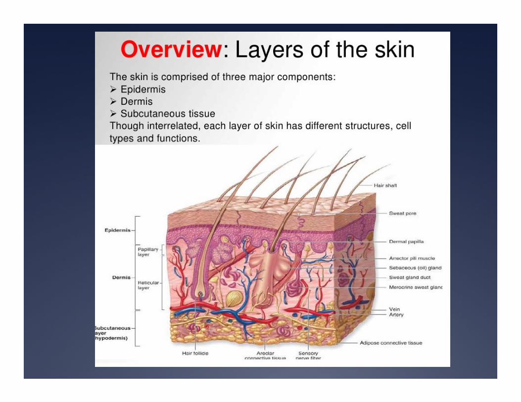

Functions of Skin
Provides physical barrier to protect underlying tissue and organs.
Provides a key role in immune system
Involved in temperature regulation
Key role in sensory perception
Neonatal Skin vs Mature skinSome critical differences
Structural differences
increase risk of trauma and infection

Neonatal Skin Differences
Dermis
Epidermis
Fibrils• Fibrils connect the epidermis and dermis
• More widely spaced and fewer in neonates than in mature skin
• Diminished cohesion leaves the neonate more susceptible to injury from shear and pressure forces

Neonatal Skin Differences
Stratum Corneum is thinner in neonates especially premature infants
Increases susceptibility to infections and topical agents
Also predisposes to excessive evaporative heat and fluid loss
Stratum Corneum

Neonatal Skin Differences
Dermis of newborn is 60% thinner than than that of mature skin
Deficient in collagen
Increases risk for injury to underlying tissues
Dermis
So what is a pressure ulcer?
I thought these only arose in the
elderly?
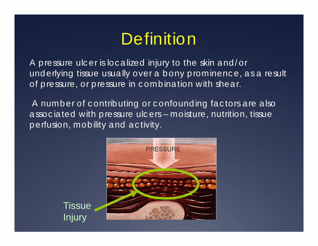
DefinitionA pressure ulcer is localized injury to the skin and/or underlying tissue usually over a bony prominence, as a result of pressure, or pressure in combination with shear.
A number of contributing or confounding factors are also associated with pressure ulcers – moisture, nutrition, tissue perfusion, mobility and activity.
Tissue Injury
Incidence and Prevalence The literature on the incidence and prevalence of
neonatal pressure ulcers remains limited.
From pediatric data that does exist, most of the studies have focused on populations considered high-risk for pressure ulcers: NICU, PICU and Pedi cardiac.
Most available studies cite an incidence rate ranging from 5% to 23% in neonatal patients.
Regardless of the incidence, the goal should be zero
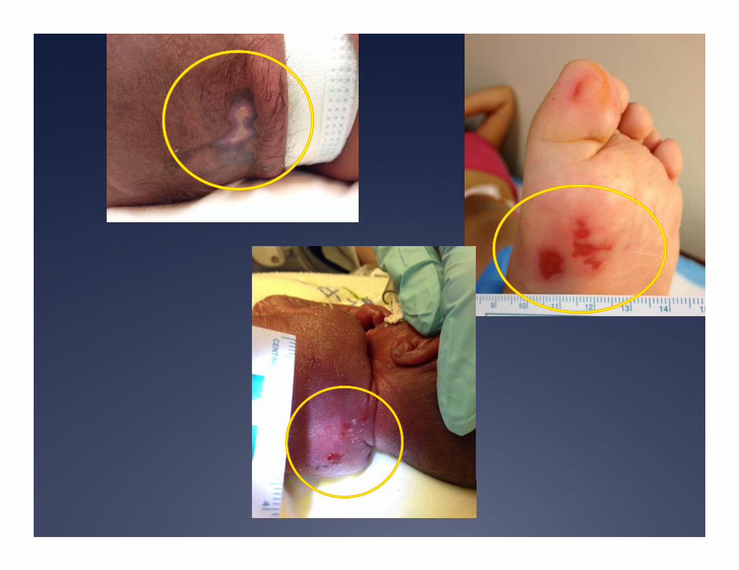
Staging of Pressure Ulcers
Stage I
Stage II
Stage III
Stage IV
Suspected Deep Tissue Injury
Unstageable
Staging of Pressure Ulcers
The staging of pressure ulcers as defined by national guidelines (NPUAP, CMS) allows for uniform documentation and classification of pressure ulcers by healthcare professionals
The staging of pressure ulcers reflects the amount of tissue damage


Stage I: Non-blanchable erythema Intact skin with non-blanchable
redness of a localized area usually over a bony prominence.
Darkly pigmented skin may not have visible blanching; its color may differ from the surrounding area.
The area may be painful, firm, soft, warmer or cooler as compared to adjacent tissue.
Category I may be difficult to detect in individuals with dark skin tones.
May indicate “at risk” persons.


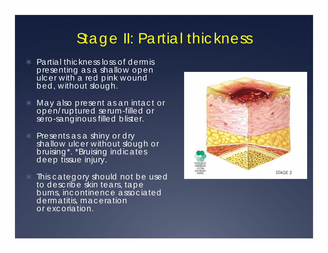
Stage II: Partial thickness Partial thickness loss of dermis
presenting as a shallow open ulcer with a red pink wound bed, without slough.
May also present as an intact or open/ruptured serum-filled or sero-sanginous filled blister.
Presents as a shiny or dry shallow ulcer without slough or bruising*. *Bruising indicates deep tissue injury.
This category should not be used to describe skin tears, tape burns, incontinence associated dermatitis, maceration or excoriation.

Stage III: Full thickness skin loss
Full thickness tissue loss.
Subcutaneous fat may be visible but bone, tendon or muscle are not exposed.
Slough may be present but does not obscure the depth of tissue loss.
May include undermining and tunneling.
Bone/tendon is not visible or directly palpable.


Stage IV: Full thickness tissue loss
Full thickness tissue loss with exposed bone, tendon or muscle.
Slough or eschar may be present.
Often includes undermining and tunneling.
Category/Stage IV ulcers can extend into muscle and/or supporting structures (e.g., fascia, tendon or joint capsule) making osteomyelitis likely to occur.

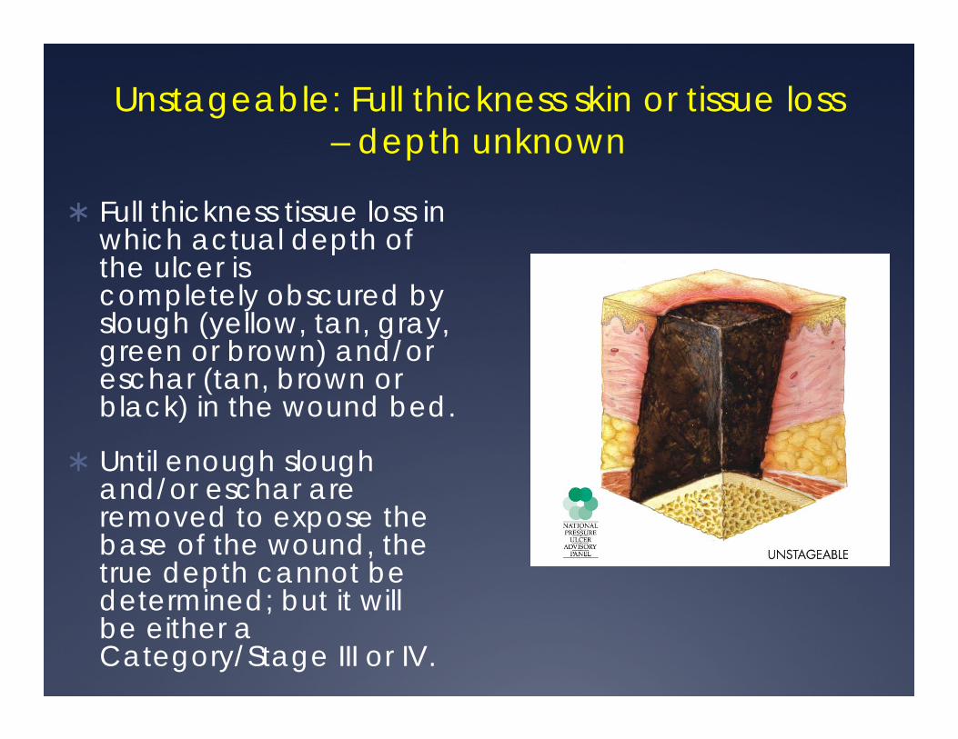
Unstageable: Full thickness skin or tissue loss – depth unknown
Full thickness tissue loss in which actual depth of the ulcer is completely obscured by slough (yellow, tan, gray, green or brown) and/or eschar (tan, brown or black) in the wound bed.
Until enough slough and/or eschar are removed to expose the base of the wound, the true depth cannot be determined; but it will be either a Category/Stage III or IV.


Suspected Deep Tissue Injury –depth unknown
Purple or maroon localized area of discolored intact skin or blood-filled blister due to damage of underlying soft tissue from pressure and/or shear.
The area may be preceded by tissue that is painful, firm, mushy, boggy, warmer or cooler as compared to adjacent tissue.
Deep tissue injury may be difficult to detect in individuals with dark skin tones.
Evolution may include a thin blister over a dark wound bed. The wound may further evolve and become covered by thin eschar.
Evolution may be rapid exposing additional layers of tissue even with optimal treatment.

Pressure Ulcers and CMS/Medicare Pressure ulcers have been classified as a NEVER-EVENT by
CMS
Never-Events are defined as hospital associated problems that can be prevented.
Other examples of Never-Events include surgery on wrong patient, surgery on wrong limb, foreign object left in pt after surgery, infant discharged to the wrong person, ….
Never-Events will NOT be reimbursed by insurance
Never-Events must be reported and can lead to mistrust by public.
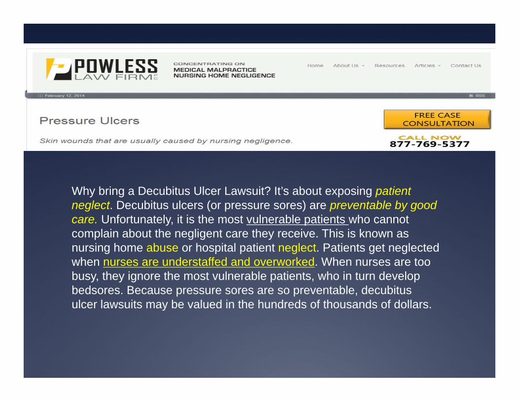
Why bring a Decubitus Ulcer Lawsuit? It’s about exposing patient neglect. Decubitus ulcers (or pressure sores) are preventable by good care. Unfortunately, it is the most vulnerable patients who cannot complain about the negligent care they receive. This is known as nursing home abuse or hospital patient neglect. Patients get neglected when nurses are understaffed and overworked. When nurses are too busy, they ignore the most vulnerable patients, who in turn develop bedsores. Because pressure sores are so preventable, decubitus ulcer lawsuits may be valued in the hundreds of thousands of dollars.

Skin Assessment Scales
The key to keeping neonatal pressure ulcer rates low lies in reliable skin assessment scales
and identification of risk factors associated with ulcer
development.

Skin Assessment Scales Skin assessment scales are tools that can be used to
identify patients at risk for pressure ulcers.
Unfortunately, many of the recognized risk factors are not captured by skin assessment scales.
Nonetheless, NICU personnel should adopt and utilize these tools to identify babies at risk early and this prevent an ulcer from developing.
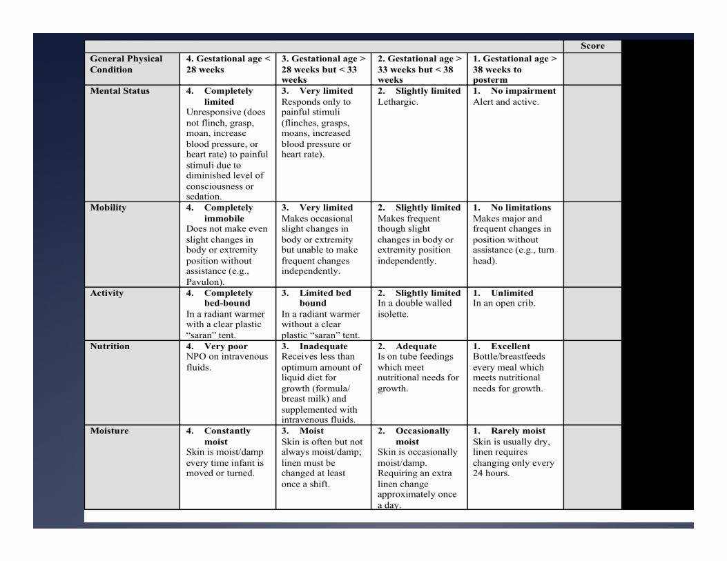
Three pediatric skin assessment tools that have been validated include the Braden Q Scale, Glamorgan Q scale and the Neonatal Skin Risk Assessment Scale (NSRAS).

Neonatal Skin Risk Assessment Scale
(NSRAS)

NSRAS NSRAS modeled after the Braden Scale, measures 6
subscales pertinent to neonates
Reliability and validity testing of the NSRAS was performed with 32 NICU patients (26-40 weeks of gestation)
Using a cutoff score of 5, the sensitivity and specificity of NSRAS was 83% and 81% respectively.
Limitations of the NSRAS scale includes a small sample size, the need for further clarification in subscales’ operational definitions, and improved reliability.

NSRAS
Gestational Age
Mental Status
Mobility
Activity
Nutrition
Moisture

NSRAS Gestational Age
< 28 weeks ………. > 38 weeks posterm
Mental Status Unresponsive even to pain……… Alert and Active
Mobility Completely immobile ….. Major changes in
position w/o assistance
NSRAS
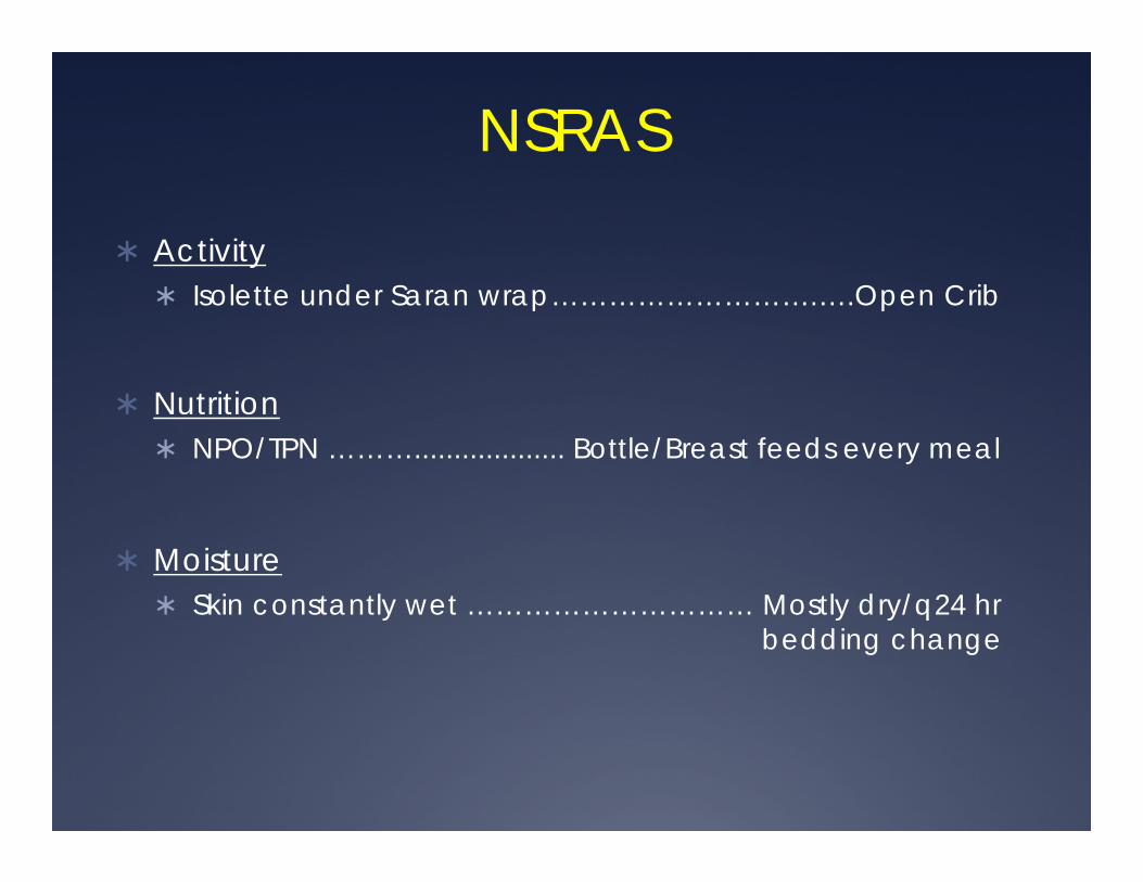
Activity Isolette under Saran wrap……………………….….Open Crib
Nutrition NPO/TPN ………................... Bottle/Breast feeds every meal
Moisture Skin constantly wet ………………………… Mostly dry/q24 hr
bedding change
NSRASA score of >13 should prompt the unit to monitor carefully for signs of a pressure ulcer.
Initiatives to reduce the risk for acquiring a pressure ulcer should to be started.

Risk factors for Neonatal Pressure Ulcers
Identifying babies at risk for pressure ulcers is the key to their prevention
Among neonates and children, 50% of pressure ulcers are equipment and device related (nasal prongs, CPAP masks, tubing, lines, tracheostomy devices, O2 monitors and bedding)
Acutely ill and immobilized neonates are at high risk for pressure injuries. Such patients are often nutritionally challenged which directly affects skin integrity.
Extremely premature infants less than 32 weeks

Risk Factors - Bedding
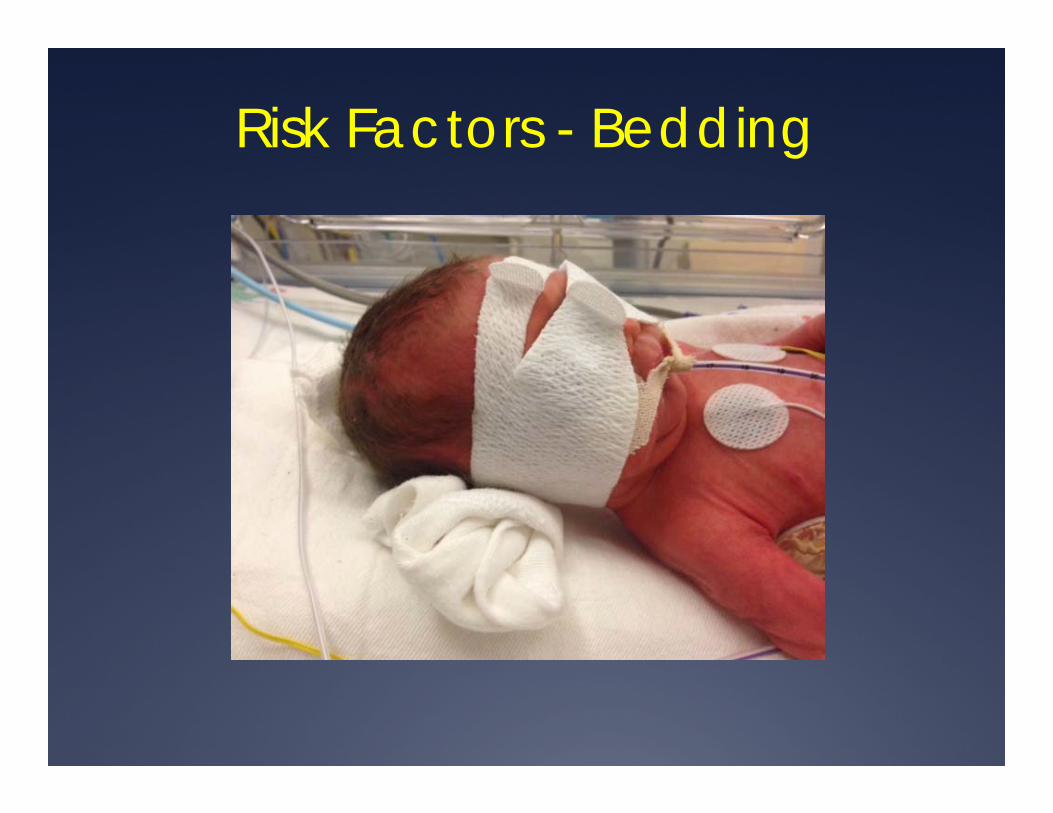
Risk Factors - Bedding

Risk Factors - Devices

Risk Factors - Devices

Risk Factors - Devices

Risk Factors - Devices

Risk Factors - Edema
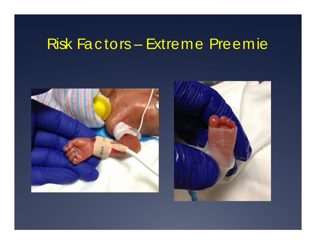
Risk Factors – Extreme Preemie
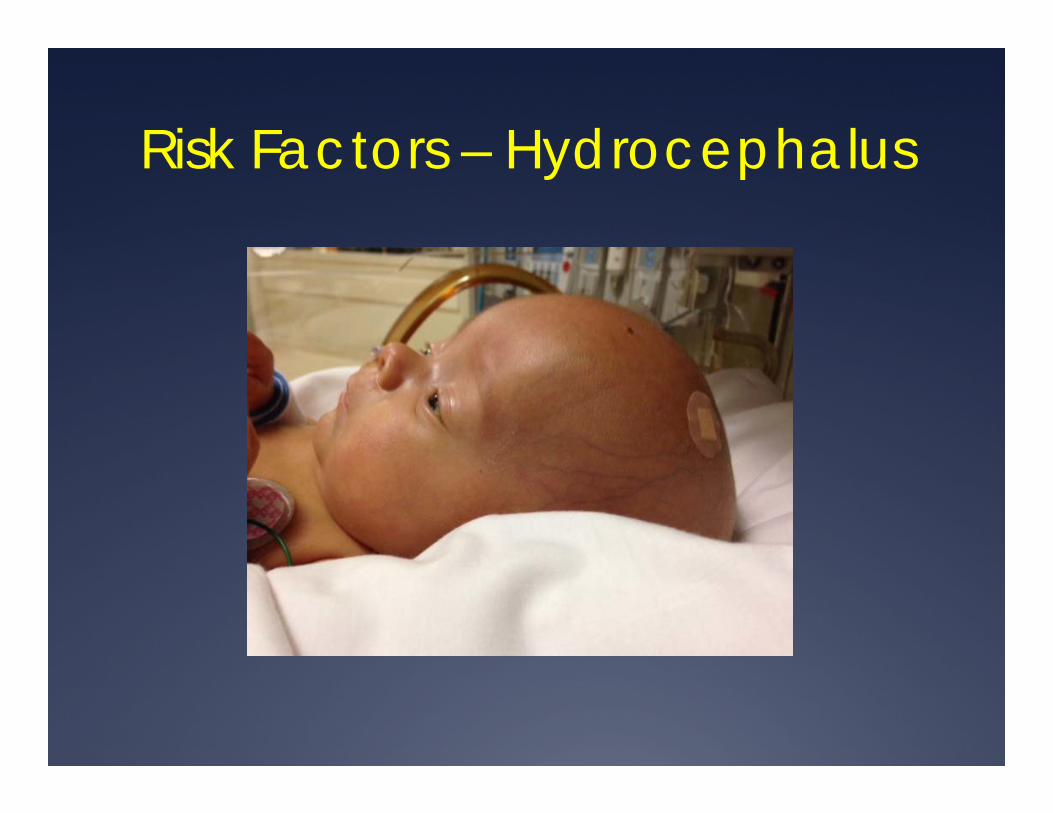
Risk Factors – Hydrocephalus

Risk Factors - Hydrocephalous


NICU Pressure Ulcer PreventionTeam
The goal of each institution is to reduce the incidence of stage 2-4 pressure ulcers in neonates to 0%
One intervention which has resulted in good success is the creation of a Pressure Ulcer Prevention Team “PUP”
Multidisciplinary teams composed of nursing staff, respiratory care, nutrition specialists, NNP/Physicians whose role includes frequent assessment with rounding and data collection.
Function to provide education, identify babies at risk and initiate interventions to prevent PU from developing.

NICU Pressure Ulcer PreventionTeam
Would provide proper reporting, staging and documentation of pressure injuries
Analyze trends to determine if a change in equipment, bedding or procedures is necessary to prevent additional PU from developing.
Ensure that proper wound care intervention is initiated if WC team is not available in the facility.
Summary
Neonatal skin has unique properties which increase the risk for trauma and injury
Pressure ulcers arise on susceptible areas of the body due to combination of pressure, moisture, immobility, shear forces as well as direct injury from medical devices.
Pressure ulcers are classified as Grades1-4 and also include Unstagable and Suspected Deep Tissue Injury
Pressure ulcers may have significant legal implications that directly affect nurses and are considered a “Never Event” by CMS/Medicare.

Summary
Identifying babies at risk using various skin assessment tools such as NSRAS can determine which babies require close observation and monitoring.
Creating a multidisciplinary team to prevent pressure ulcers from developing is one way neonatal ICU’s lower rates to ZERO!

Thank You!
Dr. Rene Amaya
713-464-9776