presentasi prof djamhoer ed
TRANSCRIPT
7/27/2019 Presentasi Prof Djamhoer Ed
http://slidepdf.com/reader/full/presentasi-prof-djamhoer-ed 1/22
INVASIVE MOLE, IN
BANDUNG TROPHOBLASTIC
CENTERAli Budi Harsono
Djamhoer Martaadisoebrata
Division of Gynecology Oncology
Department of Obstetrics and Gynecology
School of Medicine, University of Padjadjaran
Bandung, Indonesia
7/27/2019 Presentasi Prof Djamhoer Ed
http://slidepdf.com/reader/full/presentasi-prof-djamhoer-ed 2/22
2
Introduction
Hasan Sadikin Hospital
(Trophoblastic Center for the
West Java ) the same problems
Invasive mole is rather unique.
Our prior studied indicated
that latent period from mole
to IM was shorter than CC
Trophoblastic Disease
(GTD) is still important
for Indonesia
{incident >,
spread >,
the risk factors > ,
prognosis <}

7/27/2019 Presentasi Prof Djamhoer Ed
http://slidepdf.com/reader/full/presentasi-prof-djamhoer-ed 3/22
3
Malignant transformation after mole, without
histological findings (PTG,GTN )
Is it necessary to differentiate IM and CC before
treatment?
Sasaki S4 had
tried to make a
diagnosis of
Clinical IM and
Clinical CC
This paper is to share our
experience of IM cases,
demographic and also the
diagnostic and treatment
procedures
7/27/2019 Presentasi Prof Djamhoer Ed
http://slidepdf.com/reader/full/presentasi-prof-djamhoer-ed 4/22
4
Materials and methods
Cross sectional retrospective study (1995 -
2004), conducted in the Department of
Obstetrics and Gynecology, Hasan Sadikin
Hospital, Bandung
During that period 27 cases of I
M,
conformed by histological.
7/27/2019 Presentasi Prof Djamhoer Ed
http://slidepdf.com/reader/full/presentasi-prof-djamhoer-ed 5/22
5
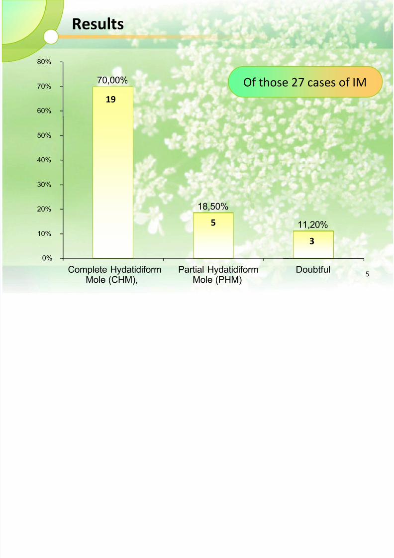
Results
70,00%
18,50%
11,20%
0%
10%
20%
30%
40%
50%
60%
70%
80%
Complete Hydatidiform Mole (CHM),
Partial Hydatidiform Mole (PHM)
Doubtful
19
5
3
Of those 27 cases of IM
7/27/2019 Presentasi Prof Djamhoer Ed
http://slidepdf.com/reader/full/presentasi-prof-djamhoer-ed 6/22
6
The average values of age were 34,4 years,
parity 4.4 pregnancy , transformation(latent) period 2.6 months and uterine
size 14.3 weeks

7/27/2019 Presentasi Prof Djamhoer Ed
http://slidepdf.com/reader/full/presentasi-prof-djamhoer-ed 7/22
7
45%
40%
10%
5,00%
0%
5%
10%
15%
20%
25%
30%
35%
40%
45%
50%
IM ChCa CMH doubtful
Based on USG examination (20 cases)
9
8
2
1
7/27/2019 Presentasi Prof Djamhoer Ed
http://slidepdf.com/reader/full/presentasi-prof-djamhoer-ed 8/22
8
Two cases caused by perforation to
parametrium, and one to abdominalcavity
Perforation occurred in 18 cases (66.7%),
61.1% to abdominal cavity, 5.5% to uterinecavity, 5.5% to uterine and parametrium, 11.1
% to abdominal abdominal and uterine cavity
and 16.7% to abdominal cavity and
parametrium.

7/27/2019 Presentasi Prof Djamhoer Ed
http://slidepdf.com/reader/full/presentasi-prof-djamhoer-ed 9/22
9
There were 2 cases with metastases
(7.4%).
One to the vagina and one to the lungs.
Both of them survived
7/27/2019 Presentasi Prof Djamhoer Ed
http://slidepdf.com/reader/full/presentasi-prof-djamhoer-ed 10/22
10
There were three dead cases (11.1 %), all ofthem due
to perforation
In 25 (92.6%) cases
ATH were performed, 1 (3.7% explorative laparotomy
and one 3.7%) chemotherapy.

7/27/2019 Presentasi Prof Djamhoer Ed
http://slidepdf.com/reader/full/presentasi-prof-djamhoer-ed 11/22
11
Discussion
The transformation or latent period ranges
from zero to 5.5 months, with the average
value : 2.6 months.
When one says that the latent period is zero, it
means that both CHM and IM occur in the same
time. How do we explain it ? ( 2 Cases)

7/27/2019 Presentasi Prof Djamhoer Ed
http://slidepdf.com/reader/full/presentasi-prof-djamhoer-ed 12/22
12
The average age and parity
value : 34.4 years (17 -48 years)
and 4.4 pregnancy (112).
Five occurred in young nullipara, and 2 withonly one living child.
All of them deprived of their future fertility, a
rather costly sacrifice to survive
7/27/2019 Presentasi Prof Djamhoer Ed
http://slidepdf.com/reader/full/presentasi-prof-djamhoer-ed 13/22
13
Detection of IM is much earlier than ChCa, based on
short latent period and small uterine size
It ranges from 6 to 24 weeks, with an average value of
14.3 weeks
The size is smaller because it perforates earlier

7/27/2019 Presentasi Prof Djamhoer Ed
http://slidepdf.com/reader/full/presentasi-prof-djamhoer-ed 14/22
14
The most common
route is to abdominal
cavity, causing
abdominal
hemorrhage
The other two routes are
to parametrium or back to
the uterine cavity.
These two types of
perforation do not cause
acute clinical signs
7/27/2019 Presentasi Prof Djamhoer Ed
http://slidepdf.com/reader/full/presentasi-prof-djamhoer-ed 15/22
15
The transformation process (Pathogenesis)
from HM to IM, ??!!
1. When the patients harbor HM, some of its chorionic
villous have already invaded the myometrium.
2. When we evacuate the mole tissues, the chorionic
villous remain in situ
3. Generally, the chorionic villous will be absorbed by the
body, and the patient recover completely.

7/27/2019 Presentasi Prof Djamhoer Ed
http://slidepdf.com/reader/full/presentasi-prof-djamhoer-ed 16/22
16
4. In a small portion of cases, by some unknown mechanism,
the chorionic villous will grow into grapelike vesicles.
5. Since there is only a limited space in the myometrium, the
growing vesicles must look for more spacious place
Perforation, is not the only complication
in IM (Hyperthyroid, severe anemia,
shock)

7/27/2019 Presentasi Prof Djamhoer Ed
http://slidepdf.com/reader/full/presentasi-prof-djamhoer-ed 17/22
17
Could IM be diagnosed without
histological findings ?
From Our data
Clinical, laboratory as well as
imaging,
Show that there are similarity, in
most of IM cases
Mose JC
claimed that there is a different ultrasound
appearance between IM and ChCa
7/27/2019 Presentasi Prof Djamhoer Ed
http://slidepdf.com/reader/full/presentasi-prof-djamhoer-ed 18/22
18
Sasaki Classified Persistent Trophoblastic Disease :
1) post molar persistent hCG
2) invasive or metastatic mole and
3) choriocarcinoma.
{Diagnostic Score }
7/27/2019 Presentasi Prof Djamhoer Ed
http://slidepdf.com/reader/full/presentasi-prof-djamhoer-ed 19/22
19
Surgical intervention had been performed in
26 cases
But not all of
them
hysterectomy
Based on the fact that IM is similar toHM, it is our policy not give
chemotherapy in IM cases, as long as
there is no distortion in BhCG curve, and
there is not signs of metastasis.

7/27/2019 Presentasi Prof Djamhoer Ed
http://slidepdf.com/reader/full/presentasi-prof-djamhoer-ed 20/22
20
Conclusions and Suggestions
IM should be suspected in middle aged women, high parity,
with hystory HM, bleeding, sub involution of the uterus,
short transformation period and increase level of BhCG
Although it has a low grade of malignancy but it can be fatal

7/27/2019 Presentasi Prof Djamhoer Ed
http://slidepdf.com/reader/full/presentasi-prof-djamhoer-ed 21/22
21
The role of USG as a diagnostic procedure is promising,
but it still further prospective study. It will be a great advantage to the management and
prognosis" if IM can be diagnosed in a non invasive
manner

7/27/2019 Presentasi Prof Djamhoer Ed
http://slidepdf.com/reader/full/presentasi-prof-djamhoer-ed 22/22
SEE YOU IN NEXT
18TH WORLD CONGRESS OF ISSTD
BANDUNG
2015