present state of the systematics of planktonic coccoid...
TRANSCRIPT
PHYTOPLANKTON Review Paper
Present state of the systematics of planktonic coccoid greenalgae of inland waters
Lothar Krienitz • Christina Bock
Received: 23 September 2011 / Accepted: 7 March 2012 / Published online: 30 March 2012
� Springer Science+Business Media B.V. 2012
Abstract This review discusses the main develop-
ments in the systematics of coccoid green algae over
the last three decades. The relationships of key groups
of planktonic coccoid green algae are shown in the
phylogenetic trees of Chlorophyceae and Trebouxio-
phyceae. The trees clearly show that the morphology
of these algae do not adequately reflect their phylo-
genetic position. Different phylogenetic species can be
hidden under one and the same morphotype. As most
of the genera have a polyphyletic origin, they are in
need of a systematic re-evaluation. Species classifica-
tion using the phylogenetic species concept resulted in
the establishment of new genera with smaller numbers
of species and the description of new species that are
not distinguishable by light microscopy. An overview
of genera is given in tables and the revised designa-
tions of species as contained in the harmonized taxon
list of the European Water Framework Directive lists
is provided. In this transitional phase from an artificial
to a more natural systematics of algae, field biologists
and ecologists as well as molecular biologists must
strengthen their interdisciplinary cooperation. The
alignment of eco-functional groups of algae with true
species identities using the barcoding conception will
provide a better understanding of the interaction
between organisms and their environment.
Keywords Coccoid green algae � Chlorophyceae �Trebouxiophyceae � Systematics � Genus and species
concept � Barcodes � Functional groups
Introduction
The classification of algae is presently going through
an extremely interesting stage. The old, artificial
classification system is being replaced by a new, more
natural, phylogenetic system. This is especially the
case for the coccoid green algae, mainly because
numerous organisms in this morphological group do
not propagate sexually. Hence, phenotypic criteria
have to a large extent been used in the establishment of
taxonomic clades. Initially, the introduction of molec-
ular phylogenetic methods into the systematics of
green algae led to a fundamental revision of the
concepts of higher taxonomic lineages such as divi-
sions, classes and orders (Melkonian & Surek, 1995;
Friedl, 1997; Chapman et al., 1998; Leliaert et al.,
Guest editors: N. Salmaso, L. Naselli-Flores, L. Cerasino,
G. Flaim, M. Tolotti & J. Padisak / Phytoplankton responses to
human impacts at different scales: 16th workshop of the
International Association of Phytoplankton Taxonomy and
Ecology (IAP)
L. Krienitz (&)
Leibniz-Institute of Freshwater Ecology and Inland
Fisheries, Alte Fischerhutte 2, 16775 Stechlin, Germany
e-mail: [email protected]
C. Bock
Department of General Botany, Faculty of Biology,
University of Duisburg-Essen, Universitatsstraße 5,
45141 Essen, Germany
123
Hydrobiologia (2012) 698:295–326
DOI 10.1007/s10750-012-1079-z

2012). Following these new phylogenetic conceptions
the orders that contain coccoid green algae have been
considerably changed. The systematics of lower tax-
onomic levels such as families and genera remain
provisional because detailed taxon sampling for
molecular sequence analyses have not been carried
out for the taxa. Nevertheless, key groups of planktonic
coccoid greens have in the recent past been studied by
molecular methods, and as such a picture of their
phylogenetic designation has started to take shape.
The central unit for biological classification is the
species. The most established species concept is the
biological species (Mayr, 1942), which refers to
groups of interbreeding natural populations that are
reproductively isolated. Because of its restriction to
sexually reproducing organisms, this concept cannot
be applied to most of the coccoid green algae. In an
effort to deal with this limitation, limnologists adopted
the morphological species concept using morphology-
based diacritical characteristics. The climax of the
morphological species concept for the coccoid green
algae was reached following the publication of the
famous handbook on ‘‘Chlorococcales’’ by Komarek
& Fott (1983). Approximately 1,200 taxa from water,
soil, and other habitats were compiled in this com-
prehensive work. The works of Hindak entitled
‘‘Studies on the Chlorococcal Algae (Chlorophyceae)
I–V’’ (1977, 1980, 1984, 1988, 1990), which were
published partly before and after the handbook of
Komarek & Fott, have inspired many phytoplankton
researchers. Combining a sharp eye for detail and
experience, Hindak identified several new morpho-
species. His observations triggered lively discussions
on diacritic morphological characteristics such as
presence or absence of pyrenoids, cell wall incrusta-
tions and mucilaginous envelopes.
Running parallel to these traditional approaches, an
ultrastructural concept to classify green algae based on
anatomy of flagellated cells and cytokinesis during
mitosis was initiated by Melkonian (1982, 1984) and
Mattox & Stewart (1984). Based on the orientation of
the basal bodies of the flagellar apparatus, three main
types have been suggested: counter clockwise orien-
tation (CCW, 11–5 o’clock), clockwise orientation
(CW, 1–7 o’clock), and directly opposite orientation
(DO, 12–6 o’clock). Later studies on molecular
characteristics by Lewis et al. (1992) revealed that
the relationships established on the basis of ultrastruc-
tural data were closely supported by molecular data.
As discussions on the relations between the various
concepts of classifying green algae continued, the
phylogenetic concept, which utilizes molecular mark-
ers to delineate taxa, steadily made its way into the
daily routine of phycologists. The evidence that has so
far accumulated clearly suggests that biological and
morphological conceptions cannot solve the main
questions on natural grouping of coccoid green algae.
A polyphasic approach combining morphological,
ecophysiological and molecular phylogenetic methods
should contribute to modern systematics (Proschold &
Leliaert, 2007). In this review, we focus on the main
developments in the systematics of coccoid green
algae following the landmarks set by the works of
Komarek & Fott, Hindak, and Mattox & Stewart in the
middle of the 1980s.
Higher taxonomic lineages which contain coccoid
green algae
The green algae evolved about 1,500 million years ago
(Yoon et al., 2004) into two large lineages (divisions),
the Chlorophyta and the Charophyta (Lewis &
McCourt, 2004). In this review, we follow the work
of Lewis & McCourt (2004) who suggested a ‘‘work-
ing classification of green algae and land plants’’
(Table 1).
The Charophyta has often been labeled the Strep-
tophyta by Bremer et al. (1987). This division contains
seven classes that include the Charophyceae (stone-
worts), the Embryophyceae (higher land plants), the
Zygnemophyceae (conjugates), and the Chlorokybo-
phyceae (has only one species Chlorokybus atmo-
phyticus, an aerophytic coccoid green alga). The
Zygnemophyceae has within the order Desmidiales, a
special type of coccoid green algae with a striking
morphology characterized by two symmetrical halves
(semicells). The majority of coccoid green algae
considered here occur in several orders of Chlorophy-
ceae, Trebouxiophyceae, and Prasinophyceae within
the division Chlorophyta (Melkonian, 1990a; Fawley
et al., 2000; Krienitz et al., 2003). Conventionally,
these coccoid taxa were lumped in the order Chloro-
coccales sensu lato (s.l.) by Komarek & Fott (1983)
and represented one of the most diverse groups of
photoautotrophic cryptogams. However, this classical
approach based on Pascher’s (1918) idea of establish-
ing orders according to life forms has not been
296 Hydrobiologia (2012) 698:295–326
123

supported by phylogenetic methods and is therefore
not applicable in modern systematic considerations of
green algae. Consequently, most of the taxa that do not
belong to the Chlorococcales s.l. have been transferred
to other orders.
In the division Chlorophyta, coccoid taxa occur in
three different classes; the Chlorophyceae, Trebouxi-
ophyceae, and Prasinophyceae (Table 1). In the
Chlorophyceae, the coccoid taxa occur in two orders;
the Chlorococcales sensu stricto (s.str.) and Sphaero-
pleales. The only remaining taxa in the order Chloro-
coccales s.str. are those related to the polyphyletic
genus Chlorococcum and some other genera that all
are still under revision. It has also become evident that
some taxa of Chlamydomonadales, Dunaliellales, and
Volvocales occur in the order Chlorococcales s.str.
(Nakayama et al., 1996; Booton et al., 1998; Chapman
et al., 1998; Proschold et al., 2001). The ultrastructures
of all these taxa are characterized by a CW orientation
of the basal apparatus of the flagella. Apart from the
Chlorococcales s.str. and Sphaeropleales, several
other lineages (incertae sedis) which do not corre-
spond to the named clades have coccoid taxa. Future
revisions of the CW-group of Chlorophyceae may
probably lead to the establishment of several new
orders. Furthermore, the content of ambiguous orders
such as Actinochloridales, Chlorosarcinales, and Tet-
rasporales must be emendated.
A majority of chlorophycean members of Chloro-
phyta are placed in the order Sphaeropleales (Fig. 1).
Some members of these taxa such as Sphaeropleaceae,
Hydrodictyaceae, and Bracteacoccus produce zoo-
spores with DO-orientation of the flagellar apparatus
(Lewis, 1997; Buchheim et al., 2001; Wolf et al.,
2002a; Shoup & Lewis, 2003). However, many non
motile unicellular or colonial taxa such as Selenastr-
aceae and Mychonastes are also included in this order
(Krienitz et al., 2001, 2003, 2011a, b, 2012). The
Scenedesmaceae also belong to this order. Members of
this family are commonly non motile. However,
presence of zoospores in cultures of Acutodesmus
has been reported (Trainor, 1963). Keller et al. (2008)
confirmed the monophyly of Sphaeropleales; how-
ever, from several other studies the relations between
the clade containing the type genus Sphaero-
plea (Sphaeropleales s.str.) and the other clades
Table 1 Overview on main lineages of green algae
Kingdom Chlorobionta
Division Chlorophyta
Class Chlorophyceae
Order Chlamydomonadales
Order Chlorococcales* (Chlorococcum)
Order Sphaeropleales* (Ankyra, Pediastrum,
Scenedesmus, Selenastrum)
Order Oedogoniales
Order Chaetopeltidales
Order Chaetophorales
Incertae sedis* (Actinochloris, Nautococcus)
Class Ulvophyceae
Order Ulotrichales
Class Trebouxiophyceae
Order Trebouxiales* (Trebouxia, Chloroidium,
Myrmecia)
Order Microthamniales
Order Prasiolales
Order Chlorellales* (Chlorella, Dictyosphaerium,
Micractinium)
Incertae sedis* (Choricystis, Coccomyxa,
Botryococcus)
Class Prasinophyceae
Order Pyramimonadales* (Halosphaera,
Pachysphaera)
Order Mamiellales* (Bathycoccus, Ostreococcus)
Order Pseudoscourfieldiales* (Pycnococcus)
Order Prasinococcales* (Prasinococcus)
Order Chlorodendrales
Incertae sedis* (Picocystis)
Division Charophyta
Class Mesostigmatophyceae
Class Chlorokybophyceae
Order Chlorokybales* (Chlorokybus)
Class Klebsormidiophyceae
Class Zygnemophyceae
Order Zygnematales
Order Desmidiales* (Cosmarium)
Class Coleochaetophyceae
Subdivision Streptophytina
Class Charophyceae
Class Embryophyceae
The orders containing coccoid phenotypes are indicated by an
asterisk, and some examples of genera belonging into these
orders are given
Hydrobiologia (2012) 698:295–326 297
123

298 Hydrobiologia (2012) 698:295–326
123

(e.g., Hydrodictyon-clade, Scenedesmus-clade, Ankis-
trodesmus-clade, Bracteacoccus-clade) of the Sph-
aeropleales s.l. remain ambiguous (Fawley et al.,
2005a; Proschold & Leliaert, 2007; Krienitz et al.,
2011a). Better-supported conclusions will require
more data, based on sequences of a larger number of
molecular markers.
Two major orders of coccoid taxa can be identified
in the class Trebouxiophyceae; the Trebouxiales and
Chlorellales (Fig. 2). Several other clades such as the
Choricystis-clade and Oocystis-clade are distinctive
and may probably be established as an independent
order in future. Other clades (incertae sedis) hosting
Coccomyxa, Botryococcus, Coenocystis and Pseudo-
chlorella await delineation into higher taxonomic
ranks. The Trebouxiales mainly comprises taxa from
edaphic and aerophytic habitats as well as endos-
ymbionts of lichens (Ettl & Gartner, 1995; Friedl,
1995). Within Chlorellales (Fig. 3) are numerous new
genera and species that were recently described from
freshwaters (Fawley et al., 2005b; Bock et al., 2010,
2011a, b, c; Proschold et al., 2010).
The class Prasinophyceae contains small flagellated
and scaled green algae. A few of them have lost one or
both the flagella and scales and now have a coccoid
phenotype. Coccoid prasinophytes occur in different
orders of the class Prasinophyceae (Table 1). It has
been shown that this class is an artificial taxonomic
lineage because these organisms have a paraphyletic
origin; hence should better be named as prasinophytes
(Steinkotter et al., 1994). The prasinophytes evolved in
several independent lineages which probably represent
independent classes. A report by Guillou et al. (2004)
identifies seven different clades of the prasinophytes.
Initial taxonomic revision of some of these clades has
resulted in the description of the classes Nephrosel-
midophyceae (Cavalier-Smith, 1993), Chlorodendro-
phyceae (Masjuk, 2006), and Mamiellophyceae
(Marin & Melkonian, 2010). The coccoid prasino-
phytes are restricted to marine habitats or saline inland
waters (Guillou et al., 2004; Krienitz et al., 2012).
Recent systematics of key groups of coccoid green
algae of inland waters
The ranking of the groups discussed in this section
follow the topology in the phylogenetic trees (Figs. 1,
2, 3). These trees are based on a selection of sequences
obtained from Genbank [National Center for Biotech-
nology Information (NCBI) http://www.ncbi.nlm.
nih.gov/]. The accession numbers of these sequences
are indicated in Figs. 1, 2, and 3).
Chlorophyceae
Selenastraceae
Morphologically, this group comprises of ‘‘needles
and capricorns.’’ Members of this family are very
common in freshwaters and exhibit a high morpho-
logical diversity. They exclusively propagate by
autospores. Discussions on the diacritical features of
this group have been very intense. Marvan et al. (1984)
investigated 18 genera of the Selenastraceae by means
of numerical evaluation of morphological and onto-
genetic characteristics. The shape of the cells or
colonies, the arrangement of autospores inside the
mother cell, the development of mucilage and incrus-
tations on the cell wall, and the presence, number, and
type of pyrenoids were used to differentiate the
genera. According to this conception, several genera
differ from each other only by one of the above
characters. For example, Selenastrum differs from
Ankistrodesmus by the curvature of the cells (Komarek
& Comas, 1982), Raphidocelis differs from Kirch-
neriella by cell wall incrustations (Hindak, 1977),
and Chlorolobion differs from Monoraphidium by
the presence of a pyrenoid with starch envelope
(Komarek, 1979; Heynig & Krienitz, 1982). Fawley
et al. (2005a) correlated the morphological features of
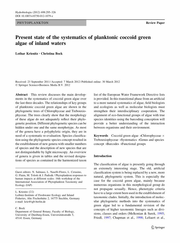
Fig. 1 Molecular phylogeny of the Chlorophyceae based on
SSU rRNA gene sequence comparisons. The phylogenetic tree
shown was inferred using the maximum likelihood (ML)
method (with substitution model: J1 [Optimum, Empirical]:G
[Optimum]:5), based on 1558 aligned positions of 87 taxa using
Treefinder (Jobb, 2008). Bayesian values ([0.95) (MB) were
calculated by MrBayes 3.1 using GTR settings (Ronquist &
Huelsenbeck, 2003; Posada & Buckley, 2004). The stationary
distribution was assumed after 4 million generations when the
average standard deviations of split frequencies between two
runs was lower than 0.01. To test the tree confidence, bootstrap
values ([50%) ML (1,000 replicates), neighbor-joining (NJ)
(1,000 replicates; calculated using Paup 4.0), and maximum
parsimony (MP) [1,000 replicates; calculated using Paup 4.0
(Swofford, 2002)] were determined. Support values are shown
at the branches in the order: MB, ML, MP, NJ. Scale barindicates substitutions per site. The sequences were obtained
from Genbank [National Center for Biotechnology Information
(NCBI)]. For each taxon, the NCBI accession number is given in
brackets
b
Hydrobiologia (2012) 698:295–326 299
123

300 Hydrobiologia (2012) 698:295–326
123

several morphospecies of Selenastraceae with their
molecular characteristics and found that one morpho-
type can cover different phylotypes. The data sug-
gested that a broad morphospecies concept would
result in a substantial underestimation of species
diversity. Subsequent molecular studies have con-
firmed the existence of ‘‘small’’ genera in Selenastr-
aceae containing only a few species (Krienitz et al.,
2001, 2011b). From the molecular studies, 10 different
genera have been confirmed in this family (Table 2).
Several other genera are yet to be subjected to detailed
molecular investigation the outcome of which may be
the description of new genera. This is especially the
case for the relatives of the needle-shaped Ankis-
trodesmus s.l. and Monoraphidium s.l. which are
found in nine different clades and probably represent
different genera (Krienitz et al., 2011b). However,
further taxon sampling is essential before conclusions
on the taxonomic identities of these clades can be
drawn.
The adjustment of the diacritical value of the main
morphological characteristics in line with the molec-
ular findings has revealed the following: the solitary
versus colonial life form, the general shape of cells and
colonies are relatively good criteria. Although changes
in environmental conditions can result in some
colonies disintegrating, the arrangement of autospores
in the mother cells can be used for the differentiation
of genera. The establishment of mucilage and incrus-
tations are of limited taxonomic value because of the
wide variability. Similarly, views on the value of
starch formation and the presence of pyrenoids have
changed over time. Traditionally, most of the genera
were thought to be devoid of pyrenoids. However, this
position was later, revised following the observation
of pyrenoids in some genera. The first report on the
presence of pyrenoids was made by Eloranta (1979),
who used the TEM to observe pyrenoids in Mono-
raphidium griffithii. This finding was later confirmed
for other species (Krienitz et al., 1985, 2001; Krienitz
& Scheffler, 1994). In subsequent studies, pyrenoids
were detected in all taxa studied; some exhibited a
naked matrix, while others were equipped with starch
grains covering the pyrenoid matrix (Krienitz et al.,
2011b). In aerated cultures, it was observed that the
concentration of CO2 influenced pyrenoid formation
(Miyachi et al., 1986). In Monoraphidium terreste,
pyrenoids were well developed under normal air
conditions. However, they were found to disappear
under CO2 enrichment (Krienitz & Klein, 1988).
Hence the presence or absence of pyrenoids cannot be
used as a basis for the differentiation of members of
the Selenastraceae family.
Based on molecular phylogeny, several taxa tradi-
tionally included in the Selenastraceae were removed
from this family. Choricystis minor is member of a
picoplanktonic lineage in the Trebouxiophyceae
(Krienitz et al., 1996a; Darienko et al., 2010).
Keratococcus bicaudatus and Pseudococcomyxa sim-
plex are closely related to Choricystis (Friedl, 1996).
The authentic strain of P. simplex was recovered in the
Elliptochloris-clade and found to be closely related to
other strains of Coccomyxa (Proschold et al., 2011).
Consequently, the taxon Coccomyxa simplex has been
re-established. Hence, other members of Pseud-
ococcomyxa need a taxonomic revision. Dicloster
acuatus is a needle-shaped coenobial member of
Chlorellaceae (Hegewald & Hanagata, 2000). Clos-
teriopsis acicularis is a solitary, needle-shaped mem-
ber of the Chlorellaceae and is closely related to
Dicloster (Ustinova et al., 2001). The ultrastructure of
its pyrenoid is comparable to that of Chlorella and
other Chlorellaceae (Hegewald & Schnepf, 1986). The
apochloric Hyaloraphidium curvatum is a member of
the lower fungi (Ustinova et al., 2000).
Scenedesmaceae
This is the largest group of coccoid green algae in
freshwater ecosystems. Among the genera, the genus
Scenedesmus s.l. with its extremely wide morpholog-
ical variability is a nightmare for field ecologists who
Fig. 2 Molecular phylogeny of the Trebouxiophyceae based on
SSU rRNA gene sequence comparisons. The phylogenetic tree
shown was inferred using the maximum likelihood (ML)
method (with substitution model: J1 [Optimum, Empirical]:G
[Optimum]:5), based on 1,429 aligned positions of 58 taxa using
Treefinder (Jobb, 2008). Bayesian values ([0.95) (MB) were
calculated by MrBayes 3.1 using GTR settings (Ronquist &
Huelsenbeck, 2003; Posada & Buckley, 2004). The stationary
distribution was assumed after 4 million generations when the
average standard deviations of split frequencies between two
runs was lower than 0.01. To test the tree confidence, bootstrap
values ([50%) for ML (1,000 replicates), NJ (1,000 replicates;
calculated using Paup 4.0), and MP [1,000 replicates; calculated
using Paup 4.0 (Swofford, 2002)] were calculated. Support
values are shown at the branches in the order: MB, ML, MP, and
NJ. Scale bar indicates substitutions per site. The sequences
were obtained from Genbank [National Center for Biotechnol-
ogy Information (NCBI)]. For each taxon, the NCBI accession
number is given in brackets
b
Hydrobiologia (2012) 698:295–326 301
123
302 Hydrobiologia (2012) 698:295–326
123

wish to determine a taxon in a fixed sample under the
inverted microscope. The ‘‘Annotated catalogue of
Scenedesmus and nomenclaturally related genera,’’
published by Hegewald & Silva (1988) is a milestone
in the history of conventional systematics of green
algae. More than 800 taxa were included in details that
provide a clear testimony of how the game of nature
creates a multitude of morphospecies. Although this
catalogue is a good guide for microscopists, at the end
of the day it is difficult to decide on the species
delineation in Scenedesmaceae. Several publications,
each justifying the different views of the authors, have
been written.
According to Hegewald (1997), the great morpho-
logical variability of Scenedesmaceae can be attrib-
uted to the strictly non-sexual propagation. This
argument may also be valid for many other coccoid
green algae such as the Selenastraceae. The simple
reproduction by means of autospores results in a
situation whereby all mutations occurring, which do
not significantly influence growth and ability of the
mutants to compete, remain and are not lost by genetic
processes. This discussion leads to the old question on
the possible occurrence of flagellated stages in
Scenedesmus, first reported by a Ukrainian phycolo-
gist J. J. Valz more than 130 years ago (according to
Hegewald, 1997). Trainor (1963) recorded the pres-
ence of flagellates in cultures of Scenedesmus, while
Lukavsky (1991) and Cepak (1993) found them in
outdoor mass cultures. Trainor (1996) has described
the production and germination of zygospores. How-
ever, the majority of these algae propagate asexually
by autospores, and the reasons given by Hegewald to
explain the wide morphological variability remain
valid.
Revisions of the class followed the introduction of
molecular methods. First, it became evident that the
morphologically differentiated subgenera should be
upgraded to generic status. Consequently, the genus
Desmodesmus was separated from Scenedesmus (An
et al., 1999). Whereas Desmodesmus comprises spe-
cies characterized by many substructures on the cell
wall and are equipped with teeth, rosettes, warts and
spines, the species of Scenedesmus have a smooth,
non-ornamented cell wall (Hegewald, 2000). The
genus Acutodesmus, which comprises of more or less
ellipsoidal, spindle-shaped taxa that show longitudinal
ridges under the TEM, was established by Tsarenko &
Petlevanny (2001). Later, Pectinodesmus was erected
for taxa with similar morphology as Acutodesmus but
differing in molecular phylogeny (Hegewald et al.,
2010). Several genera which are presently monotypic
such as Hylodesmus and Comasiella have recently
been established (Elias et al., 2010; Hegewald et al.,
2010).
A major surprise resulting from the molecular
studies is the clustering of Coelastrum-morphotypes
with Scenedesmus suggesting that the flat coenobia
with cells arranged in one or two rows of Scenedes-
mus-relatives are phylogenetically closely related to
the spherical coenobia of Coelastrum and allied
species (Fig. 1) (Hegewald et al., 2010). Following
confirmation through molecular studies of the separate
position of Coelastrum reticulatum, the old name
Hariotina was reintroduced (Hegewald et al., 2002).
At present, 13 genera of Scenedesmaceae are
phylogenetically and morphologically well defined
(Table 3). Several other genera of this family, such as
all the crucigenoid groups, are yet to be revised.
Hydrodictyaceae
The Hydrodictyaceae comprises microscopic colonies
of Pediastrum, Euastropsis, and Sorastrum as well as
macroscopic colonies of Hydrodictyon. The most
Fig. 3 Molecular phylogeny of the Chlorellaceae based on a
partitioned dataset of SSU, ITS1, 5.8S, and ITS2 gene
sequences. The phylogenetic tree shown is based on 2,536
manually aligned base positions of 50 taxa, calculated by
Treefinder (Jobb, 2008) using the maximum likelihood (ML)
method under different substitutional models for each partition.
The substitution models were as follows: 18S (1,686 bases) J2
[Optimum, Empirical]:GI [Optimum]:5; ITS1 (388 bases) J1
[Optimum, Empirical]:G [Optimum]:5; 5.8S (137 bases) HKY
[{3,1,1,1,1,3}, Empirical]:G [Optimum]:5; and ITS2 (325
bases) GTR [Optimum, Empirical]:G [Optimum]:5. The Bayes-
ian values ([0.95) (MB) were calculated by MrBayes 3.1. A
general time reversible model with gamma shape parameter and
proportion of invariable sites (GTR?I?G) was applied to each
partition. The parameters were unlinked and allowed to vary
across the partitions (Ronquist & Huelsenbeck, 2003; Posada &
Buckley, 2004). The stationary distribution was assumed after 4
million generations when the average standard deviations of
split frequencies between two runs was lower than 0.01. To test
the tree confidence, bootstrap values ([50%) for ML (1,000
replicates), NJ (1,000 replicates; using Paup 4.0), and MP (1,000
replicates; using Paup 4.0 (Swofford, 2002) were calculated.
Support values are shown at the branches in the order: MB, ML,
MP, and NJ. Scale bar indicates substitutions per site. The
sequences were obtained from Genbank [National Center for
Biotechnology Information (NCBI)]. For each taxon, the NCBI
accession number is given in brackets
b
Hydrobiologia (2012) 698:295–326 303
123

Table 2 Genera of Selenastraceae confirmed by 18S rRNA gene phylogeny and their main diacritic morphological characteristics
Genus Drawing Main diacritic morphology
Ankistrodesmus Needle-shaped cells, in colonies, parallel arrangement of
autospores
Kirchneriella Semilunate- to crescent-shaped cells, in colonies, serial
arrangement of autospores
Monoraphidium Needle- to rod-shaped cells, solitary, serial arrangement of
autospores
Nephrochlamys Semilunate-shaped cells, in colonies, serial arrangement of
autospores, widening mother cell wall
Podohedriella Needle-shaped cells, solitary, heteropolar, serial
arrangement of autospores
Quadrigula Cylindrical cells with rounded ends, in quadricellular
colonies, parallel arrangement of autospores
Raphidocelis Capricorn-shaped cells, arcuated, solitary or in irregular
colonies, serial arrangement of autospores
Rhombocystis Cells rhomboidal with slightly thickened poles, solitary or
colonial, parallel arrangement of autospores
Selenastrum Semilunate-shaped cells in regular colonies, parallel
arrangement of autospores
Tetranephris Bean-shaped cells in quadricellular colonies, touched at the
poles, serial arrangement of autospores
Drawings after Komarek & Fott (1983)
304 Hydrobiologia (2012) 698:295–326
123

recent review reduced the number of Pediastrum-
species to 24 (Komarek & Jankovska, 2001). Hydro-
dictyaceae exhibit distinct reproduction strategies:
they produce asexually inside the parental cells
through biflagellated zoospores that aggregate after
swarming to daughter colonies which are released
from the mother-sporangiae in a gelatinous bubble.
Sexually, Hydrodictyaceae reproduce by isogametes.
The ultrastructure of the flagellar apparatus is charac-
terized by a directly opposite (DO) configuration in
Hydrodictyon and Pediastrum (Wilcox & Floyd,
1988).
Phylogenetic analysis of Hydrodictyon, Pedia-
strum, and Sorastrum has revealed a pattern of
colony-form evolution within the family from two-
dimensionality to three-dimensionality (McManus &
Lewis, 2005). Another molecular phylogenetic study
of 28 hydrodictyacean strains revealed polyphyly in
Pediastrum and resulted in taxonomic conclusions
(Buchheim et al., 2005). Beside Pediastrum, the
genera Monactinus, Parapediastrum, Pseudopedia-
strum, and Stauridium were delineated. It is interesting
to note that Pediastrum duplex with a complex
morphology of colonies evolved polyphyletically
(McManus & Lewis, 2011). Consequently, a new
genus Lacunastrum was erected (McManus et al.,
2011). It has also been shown that members of the
genus Tetraedron evolved as a sister clade to the
Hydrodictyaceae. However, the evolutionary link
between the tetraedric unicells of autosporic Tetrae-
dron, zoosporic Chlorotetraedron, and the zoosporic
colonial Hydrodictyaceae remains obscure (McEntee
et al., 1977; Komarek & Kovacik, 1985; Hegewald
et al., 2001; Buchheim et al., 2005). Presently, the
Hydrodictyaceae has 10 genera as confirmed by
morphological and molecular analyses (Table 4).
Sphaeropleaceae
This group provides a good example for demonstrating
that life form sensu Pascher (1918) is not suitable for
the natural grouping of algae. Two different life forms
in this family, the filamentous (Sphaeroplea) and the
coccoid (Ankyra) green algae cluster together (Fig. 1).
Sphaeroplea is a filamentous green algal genus with
multinucleate (coenocytic) cells (Buchheim et al.,
2001). Its propagation is by asexual division of the cells
in the unbranched filaments and oogamous sexual
reproduction. Actractomorpha produces extremely
long needle-shaped solitary cells that are coenocytic
in character and can propagate asexually by zoospores
and sexually by anisogamy or seldom by oogamy
(Hoffman, 1983). Normally, this alga occurs mostly in
soil. However, it has also been observed in freshwaters
(Schmidt & Feher, 1999–2000). Freshwater forms are
not always correctly designated (e.g., as Closteriopsis
longissima f. gigantea Heynig, 1980). Such planktonic
cells can grow to lengths of 1,000–1,900 lm. The most
frequently observed members of Sphaeropleaceae in
the plankton are species of Ankyra, which produce
spindle-shaped heteropolar cells with an anchor (Rey-
mond & Hegewald, 1988). Species of Ankyra dominate
in the clear water stages of stagnant waters (Barone &
Naselli Flores, 1994). Although Ankyra mainly prop-
agate asexually by zoospores, propagation in the genus
needs more detailed investigation. Several types of
unidentified aplanospores or resting stages have been
observed in Ankyra cultures and field samples (Fott,
1971; Krienitz & Heynig, 1982).
Other clades
The Mychonastes-clade contains tiny, mostly spheri-
cal or oval cells of small size that occur as solitary cells
or in colonies previously considered as two separate
genera, Mychonastes and Pseudodictyosphaerium.
Members of these genera belong to the most common
pico- or small nanoplankton green algae in fresh or
brackish waters. Molecular phylogenetic analyses
have shown that both genera are mixed in the same
clade (Krienitz et al., 1999, 2011a). Most species were
previously described under the generic name of
Pseudodictyosphaerium (Hindak, 1978a, b, 1988).
However, since the genus Mychonastes was described
four month earlier in 1978 (Simpson & Van Valken-
burg, 1978), it therefore has nomenclatural priority.
Consequently, the species of Pseudodictyosphaerium
have been transferred to Mychonastes.
The Bracteacoccus-clade comprises mainly of soil
algae. However, Planktosphaeria gelatinosa, which is
a freshwater plankton commonly present, has been
found to be a close relative of Bracteacoccus and
Radiococcus (Wolf et al., 2003b). Unfortunately, this
morphotype has been under-represented in recent
taxon samplings for molecular considerations and
needs further investigation. The edaphic ‘‘Mychonas-
tes’’ zofingiensis does not belong to the true Mychon-
astes genus, and it is very likely that a new generic
Hydrobiologia (2012) 698:295–326 305
123
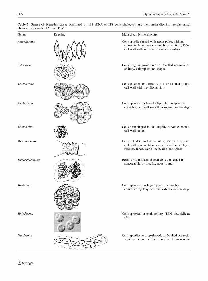
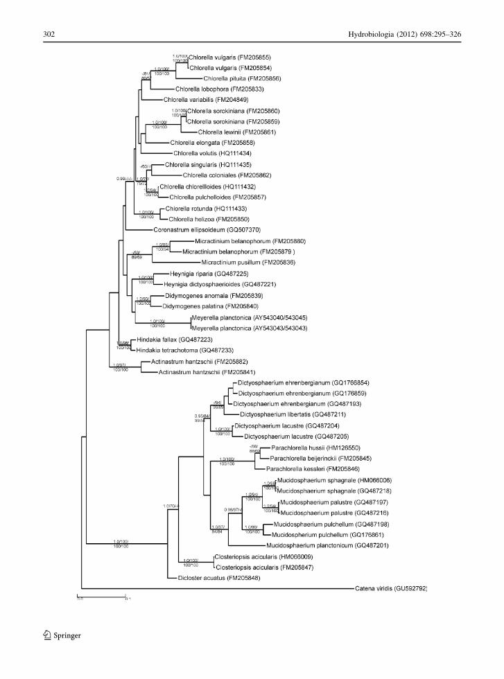
Table 3 Genera of Scenedesmaceae confirmed by 18S rRNA or ITS gene phylogeny and their main diacritic morphological
characteristics under LM and TEM
Genus Drawing Main diacritic morphology
Acutodesmus Cells spindle-shaped with acute poles, without
spines, in flat or curved coenobia or solitary, TEM:
cell wall without or with few weak ridges
Asterarcys Cells irregular ovoid, in 4- or 8-celled coenobia or
solitary, chloroplast net-shaped
Coelastrella Cells spherical or ellipsoid, in 2- or 4-celled groups,
cell wall with meridional ribs
Coelastrum Cells spherical or broad ellipsoidal, in spherical
coenobia, cell wall smooth or rugose, no mucilage
Comasiella Cells bean-shaped in flat, slightly curved coenobia,
cell wall smooth
Desmodesmus Cells cylindric, in flat coenobia, often with special
cell wall ornamentations on an fourth outer layer,
rosettes, tubes, warts, teeth, ribs, and spines
Dimorphococcus Bean- or semilunate-shaped cells connected in
syncoenobia by mucilaginous strands
Hariotina Cells spherical, in large spherical coenobia
connected by long cell wall extensions, mucilage
Hylodesmus Cells spherical or oval, solitary, TEM: few delicate
ribs
Neodesmus Cells spindle- to drop-shaped, in 2-celled coenobia,
which are connected in string-like of syncoenobia
306 Hydrobiologia (2012) 698:295–326
123

designation will be necessary for this taxon (Krienitz
et al., 2011a).
The Golenkinia-clade is a member of the Chloro-
coccales s.str. (Fig. 1). The morphological and onto-
genetic peculiarities, especially the CW basal body
orientation shown by Hegewald & Schnepf (1984),
have been supported by molecular analyses (Wolf
et al., 2003a). Members of the polyphyletic genus
Chlorococcum mostly occur in soils. However, after a
heavy downpour, they can be washed out and trans-
ported into water bodies where they are able to
propagate as observed in the case of Chlorococcum
robustum (Krienitz et al., 1997).
Trebouxiophyceae
Chlorellaceae
This clade contains the classical ‘‘green balls.’’ Follow-
ing the description of the archetypical form of coccoid
green algae, Chlorella vulgaris by Beijerinck (1890),
more than 100 species of this genus have been described
from freshwater, marine, and soil habitats. However,
most of them need to be revised and transferred to other
genera and other families. It has been shown that
Chlorella-like green spheres evolved independently in
different evolutionary lineages of Chlorophyceae, Tre-
bouxiophyceae, and prasinophytes (Friedl, 1997;
Chapman et al., 1998; Proschold & Leliaert, 2007;
Darienko et al., 2010). Based on the examination of
biochemical and molecular data, Huss et al. (1999) have
reduced the Chlorella genus to four species.
In the year 2000, a new development followed the
work of Hegewald & Hanagata (2000) who found out
that the coenobial ellipsoid D. acuatus, formerly
classified in Scenedesmaceae, was closely related to
Chlorella kessleri. The needle-shaped C. acicularis,
traditionally considered as member of Selenastraceae,
was also found to cluster in this lineage (Ustinova et al.,
2001). Actinastrum hantzschii, a coenobial taxon
formerly in the Coelastraceae, has also been trans-
ferred to Chlorellaceae (Wolf et al., 2002b). Krienitz
et al. (2004) have identified two separate lineages
within Chlorellaceae, designated as a Chlorella-clade
and a Parachlorella-clade. Presently, these two clades
have both old and new genera (Table 5; Figs. 2, 3).
The Chlorella-clade has eight different lineages
that form clusters designated as genera. The scope of
this genus has been extended to include 14 species.
Among these species are solitary cells with or without
mucilage and colonial forms that exhibit a morphol-
ogy resembling Dictyosphaerium (Bock et al., 2011a).
Chlorella-species occur in freshwater, soil and as
endosymbionts (Proschold et al., 2011). Fawley et al.
(2005b) found Meyerella, a tiny sphere without
pyrenoid, within the Chlorella-clade. According to
Table 3 continued
Genus Drawing Main diacritic morphology
Pectinodesmus Cells spindle-shaped, in flat or curved coenobia,
TEM: cell wall with strong longitudinal ridges
Scenedesmus Cells oval or cylindrical with obtuse or truncate
poles, without spines, in flat or slightly curved
coenobia, cell wall smooth
Westella Cells spherical to ovoid, in 4-celled square-shaped
coenobia, which are connected to syncoenobia
Drawings after Komarek & Fott (1983) and Krienitz (1990). Micrograph of Hylodesmus after Elias et al. (2010)
Hydrobiologia (2012) 698:295–326 307
123
Table 4 Genera of Hydrodictyaceae confirmed by 18S or 26S rRNA gene phylogeny and their main diacritic morphological
characteristics
Genus Drawing Main diacritic morphology
Chlorotetraedron Cells tetrahedral or polyhedral, with elongated cell wall
protuberances at the corners, solitary
Hydrodictyon Cells cylindrical in macroscopic net-like, one-layered
coenobia
Lacunastrum Flat coenobia with large intercellular spaces, marginal
cells with two lobes
Monactinus Flat coenobia with large intercellular spaces, marginal
cells with one tapering lobe
Parapediastrum Flat coenobia with intercellular spaces, marginal cells
with 2 lobes each divided into 2 projections
Pediastrum Flat coenobia with large intercellular spaces, marginal
cells with 2 projections
Pseudopediastrum Flat coenobia without intercellular spaces, marginal
cells with two tapering lobes in one plane
Sorastrum Three-dimensional coenobia, cells with 2 or 4
projections
Stauridium Flat coenobia without intercellular spaces, marginal
cells incised trapezoid or with projections in 2 planes
308 Hydrobiologia (2012) 698:295–326
123

Luo et al. (2010), the genera Actinastrum, Didymog-
enes, Hegewaldia and Micractinium also belong to
this clade. Hegewaldia comprises taxa with facultative
bristle production and oogamy (Proschold et al.,
2010). Micractinium usually occur in colonies and
produce bristles. However, the colonies can disinte-
grate to form single cells without bristles. The
formation of bristles can be triggered by substances
produced by grazers such as the rotifer Brachionus
(Luo et al., 2006). In addition to the several species of
Chlorella, the Chlorella-clade has two other lineages
with a colonial morphology similar to that of Dict-
yosphaerium. The two lineages belong to the genera
Heynigia and Hindakia (Bock et al., 2010).
Six genera occur in the Parachlorella-clade. The
genus Parachlorella has three species that are solitary
or colonial, and covered by a mucilaginous envelope
(Krienitz et al., 2004; Bock et al., 2011b). According to
Krienitz et al. (2010), the Dictyosphaerium-morpho-
type evolved independently in different lineages of
Chlorellaceae. The scope of the genus Dictyosphaeri-
um has been reduced to three species, the type species
D. ehrenbergianum (Bock et al., 2011c) and two new
taxa. The genus Mucidosphaerium was established
based on differences in its molecular phylogeny from
that of Dictyosphaerium. Mucidosphaerium contains
two former Dictyosphaerium-species, D. pulchellum,
and D. sphagnale, as well as two new species. The taxa
of the genera Dictyosphaerium and Mucidosphaerium
are of great importance as they play an important role
of establishing plankton communities. The spindle- to-
needle-shaped genera Closteriopsis and Dicloster, as
well as the marine, spherical Marinichlorella also
belong to the Parachlorella-clade (Aslam et al., 2007).
Oocystaceae
The family of Oocystaceae is a natural lineage in
Trebouxiophyceae confirmed by ultrastructural and
molecular criteria. Many members of this group are
very common in the plankton of stagnant and flowing
waters. The extended cell wall is multilayered and
constructed from crystalline cellulose fibers. Molec-
ular phylogenetic data have revealed the monophyly
of Oocystaceae. However, the species concept in this
group has not been confirmed (Hepperle et al., 2000;
Pazoutova et al., 2010; Krienitz & Bock, 2011).
Because of the reduced taxon availability in strain
collections and remarkable uncertainties on the con-
cept of the type genus Oocystis, the circumscription of
the genera of Oocystaceae has remained obscure.
Hindak (1988) re-applied Lemmermanns (1903) cri-
teria for distinguishing between Oocystis (without
pyrenoids) and Oocystella (with pyrenoids). Hence,
the taxonomic relevance of pyrenoids in Oocystaceae
must be resolved. Several genera are characterized by
incrustations (Amphikrikos, Granulocystis, Granulo-
cystopsis, and Siderocelis); however, their origins and
taxonomical values are still the subject of ongoing
discussions.
Other clades
Choricystis and Botryococcus. Choricystis-species
exhibit tiny bean-shaped, solitary cells and were
traditionally assigned to the Chlorophyceae family.
However molecular data have revealed that they have
a close affiliation to the Trebouxiophyceae (Krienitz
et al., 1996a, 1999). Surprisingly, the colonial and oil-
producing alga Botryococcus braunii clusters close to
Choricystis (Senousy et al., 2004). Komarek &
Marvan (1992) established a multitude of Botryococ-
cus species based on morphology. However, accord-
ing to Plain et al. (1993) the morphological features of
B. braunii vary depending on growth conditions hence
it is not reliable to define more species. The figures
provided by Plain et al. (1993) were exclusively those
of B. braunii. This study did not include any of the new
Table 4 continued
Genus Drawing Main diacritic morphology
Tetraedron Cells flat or twisted, 3-, 4- or 5-sided, with rounded or
elongated or spined corners, solitary
Drawings after Komarek & Fott (1983) and McManus et al. (2011)
Hydrobiologia (2012) 698:295–326 309
123

Table 5 Genera of Chlorellaceae confirmed by combined 18S rRNA and ITS gene phylogeny and their main diacritic morphological
characteristics
Genus Drawing Main diacritic morphology
Actinastrum Cells rod-shaped, elongated,
radially arranged in coenobia
Chlorella Spherical or broad oval cells, with one pyrenoid,
solitary or in mucilage covered colonies
Closteriopsis Needle-shaped cells with several pyrenoids
in the lateral chloroplast, solitary
Dictyosphaerium Broad oval cells interconnected by strands attaching
the elongated cell side, in mucilaginous colonies
Dicloster Cells ellipsoidal arcuated with convex sides attached
in 2- or 4-celled coenobia, 1 or 2 pyrenoids
Didymogenes Cells cylindrical curved with convex side attached
in 2-, 4- or 16-celled coenobia, one pyrenoid, spines
Hegewaldia Spherical cells with or without bristles, solitary
or in colonies, 1 pyrenoid, facultative oogamy
Hindakia Broad oval cells interconnected by strands attaching
the apical pole, in mucilaginous colonies, 1 pyrenoid
Heynigia Spherical cells interconnected by mucilaginous strands
in mucilaginous colonies, 1 pyrenoid
310 Hydrobiologia (2012) 698:295–326
123

species described by Komarek & Marvan (1992) such
as B. terribilis that has been shown to be very common
in freshwater phytoplankton of diverse climatic
regions (Krienitz et al., 1996b; Ballot et al., 2009;
Fanes Trevino et al., 2009). The conversion of the
braunii-morphotype into the terribilis-morphotype
and vice versa was never demonstrated. It is therefore,
important to as a matter of urgency include more
Botryococcus-taxa into molecular studies. From an
ecological point of view, the alliance of the pico-
planktonic, invasive Choricystis, and the microplank-
tonic, attuning Botryococcus shown in Fig. 2, is not
explainable. Padisak et al. (2009, 2010) grouped
Choricystis into the functional group X1 and Botryo-
coccus in group F. Later considerations will probably
reveal the existence of some undiscovered lineages
that have evolved between these two clades. Henley
et al. (2004) documented a close relationship between
C. minor and Paradoxia multiseta based on 18S rRNA
phylogeny. However, according to the micrograph
given (showing globular cells) in the catalogue of the
UTEX strain collection, the identity of the Paradoxia
strain investigated is doubtful.
Between the Choricystis-clade and the Chlorella-
ceae, several clades of green coccoid soil and
aerophytic algae, such as Chloroidium, Watanabea,
Dictyochloris, and the lichen symbionts of Trebouxi-
ales have evolved (Friedl, 1995; Darienko et al., 2010;
Rindi et al., 2010). These algae occasionally occur in
the plankton of freshwaters. However, if a sufficient
starting inoculum of these air- and soil-born algae is
available and if the algae are able to propagate under
the new conditions, then colonization of standing
water bodies can occur rapidly (Happy-Wood, 1988).
Evolution of morphological peculiarities of coccoid
green algae and their possible ecological functions
One of the most striking peculiarities of numerous
coccoid green algae is the development of a uniform
morphology hidden in the picoplankton size group
which is nearly unidentifiable by LM. Convergent
evolution resulted in a tiny, more or less spherical
morphotype which covers an extremely high diversity
with regard to phylogeny and physiology (Potter et al.,
1997; Hepperle & Krienitz, 2001; Krienitz et al.
2011a). Based on its fast growth and high rates of
reproduction and primary production, picophyto-
plankton can play a key role in food webs of
Table 5 continued
Genus Drawing Main diacritic morphology
Micractinium Spherical cells with long bristles,
in colonies, 1 pyrenoid
Meyerella Cells short cylindrical, small, without mucilage,
solitary, pyrenoid missing
Mucidosphaerium Spherical cells interconnected by mucilaginous
strands in mucilaginous colonies, 1 pyrenoid
Parachlorella Spherical cells covered by mucilage,
solitary or in groups, 1 pyrenoid
Drawings after Komarek & Fott (1983), Krienitz, et al. (2004), and Bock et al. (2010). Micrograph of Meyerella after Fawley et al.
(2005b)
Hydrobiologia (2012) 698:295–326 311
123

freshwater, marine, and saline habitats (Stockner &
Antia, 1986; Raven, 1999). Picoplankton may estab-
lish large populations under nearly all levels of trophy
(Stockner, 1991; Weisse, 1993; Padisak et al., 1997;
Hehmann et al., 2001; Hepperle & Krienitz, 2001).
Even under very saline conditions they can dominate
all the succession stages of eukaryotic primary
producers (Henley et al., 2004; Somogyi et al., 2011;
Krienitz et al., 2012).
Picoplanktons evolved as adaptive strategies in all
the classes considered here. In the Chlorophyceae, the
Mychonastes-clade contains several species of pico-
plankton (Krienitz et al., 2011a). In the Trebouxio-
phyceae, a number of lineages, which include
Picochloron and Chloroparva from salt pans (Henley
et al., 2004; Somogyi et al., 2011), Choricystis, and
Nannochloris from freshwaters (Krienitz et al., 1996a;
Yamamoto et al., 2003), have picoplankton species.
Marvania is a picoplankton species from different
inland waters characterized by vegetative propagation
through budding (Hindak, 1976; Reymond et al.,
1986). Among the prasinophytes, there are many
picoplankton lineages which are usually abundant in
the oceans (Guillou et al., 2004; Leliaert et al. 2012).
Extreme saline inland waters have one lineage with
Picocystis (Lewin et al., 2000; Hollibaugh et al., 2001;
Roesler et al., 2002). Picocystis salinarum from saline
inland waters represents a link between picoplankton
from marine and freshwater habitats and is therefore of
great ecological and phylogenetic interest (Krienitz &
Kotut, 2010; Krienitz et al., 2012).
Comparison of the fatty acid’s contents of green
(Choricystis, Mychonastes) and eustigmatophycean
(Nannochloropsis) freshwater picoplankton revealed
surprising differences in the composition of polyunsat-
urated fatty acids. The sums of n-6 and n-3 fatty acids are
ten times higher in Nannochloropsis limnetica than in
the green picoplankton (Krienitz et al., 2000; Krienitz &
Wirth, 2006). Hence, the eustigmatophycean picoplank-
ton has a higher nutritional value for grazers than green
algal picoplankton. This raises the question on how the
grazers can differentiate between the nutritious eustig-
matophycean and the less nutritious green algae. As a
follow-up to Hartmann & Kunkel’s (1991) statement
‘‘The paradigm of invariate, nonselective feeding by
zooplankton is rejected,’’ limnologists have come up
with possible mechanism of food selection that include:
chemosensory, electrical charge, surface hydrophobic-
ity, and chemical cues (Weisse, 2004). All these
interactions between green algae and possible grazers
need to be further elucidated.
Incrustations on the surface of coccoid green algae
are common across the whole range of systematic
groups of green algae. For example, they have been
observed in Selenastraceae (Raphidocelis), Scene-
desmaceae (Scenedesmus), Oocystaceae (Amphikri-
kos, Siderocelis), and Radiococcaceae (Coenochloris)
(Hindak, 1977; Crawford, 1978; Krienitz, 1986;
Vanormelingen et al., 2007). These incrustations are
precipitates of ferric and manganic hydroxides and are
of crystalline or amorphous nature (Crawford & Heap,
1978). The genesis and structure of this cell wall
deposits varies with taxon suggesting a genetic
influence and give the impression that this process is
under the control of the cell (Crawford & Heap, 1978).
So far, no vesicle driven expression through pores
from inner to outer cell wall has been observed. The
ecological function of these incrustations remains a
subject of discussion. From our field observation, it
was apparent that numerous algae tend to develop
incrustations under highly disturbed conditions in
rivers (Krienitz, 1990, 1998). It can therefore be
hypothesized that the incrustations increase the weight
of the cells, and this allows the cells to sink to the
lower less disturbed habitats, which are conducive for
propagation.
Spines and bristles of the plankton algae are
adaptive features that promote buoyancy and reduce
grazing pressure (Van den Hoek et al., 1995). A large
number of publications have documented the interac-
tion of kairomon-producing grazers and spine- and
bristle-formations by coccoid green algae (summa-
rized by Van Donk, 2005). Spines and bristles have
different origins and compositions. Scenedesmaceae
produce rigid, tube-like spines as elaborated parts of
the outer sporopollenin-like cell wall layer (Schnepf
et al., 1980). In contrast to spines, bristles develop
after cell wall formation and lack cellulosic fibers and
algaenan substances (Schnepf et al., 1980; Hegewald
& Schnepf, 1984). Didymogenes and Micractinium
produce bristles of the same type (Schnepf & Hege-
wald, 1993). The spines of Golenkinia contain cellu-
losic fibers and are produced after cell wall formation
(Hegewald & Schnepf, 1984). The morphological and
biochemical differentiation of spines and bristles in
the various groups of coccoid green algae closely
agree with their differences based on molecular data.
The spine producing Scenedesmaceae belong to the
312 Hydrobiologia (2012) 698:295–326
123
Chlorophyceae (Hegewald, 1997). The close relation-
ship between Didymogenes and Micractinium as
members of Trebouxiophyceae was demonstrated by
Luo et al. (2006). The polyphyletic origin of bristles
within the Trebouxiophyceae was confirmed by
Proschold et al. (2010). The separate position of
Golenkinia as a member of Chlorococcales s.l. was
revealed by Wolf et al. (2003a).
The production of mucilage by the cells is one of
the most readily observed phenomena in coccoid
green algae. It is commonly observed in all groups
under discussion and has a polyphyletic origin. The
production of mucilage largely depends on environ-
mental influences and interaction with other species
such as grazing pressure and resource competition
(Reynolds, 2007). It may protect the algae from
ingestion or, even if ingested, against digestion while
passing through the intestinal tract of zooplankton
(Porter, 1973). On the other side, the mucilaginous
envelope can act as microhabitat for bacterial flora that
produces substances with a nutritional value or have
stimulatory effects (Cole, 1982). The mucilage can
also act as a depository of nutrients (Decho, 1990).
Furthermore, mucilage affects the buoyancy of the
phytoplankton (Boney, 1981). The multitude of eco-
logical functions makes it understandable that mor-
photypes characterized by mucilage evolved in
different lineages and at different times as a response
to the diverse interactions. This observation has
clearly been demonstrated in the Dictyosphaerium-
morphotype (Bock et al., 2010, 2011c; Krienitz et al.,
2010). Even in one and the same genus such as
Chlorella or Mychonastes mucilage possession has
appeared and disappeared several times (Bock et al.,
2011a; Krienitz et al., 2011a). Hence, the question on
which of the contrasting features, ‘‘with mucilage’’ or
‘‘without mucilage,’’ is more ancestral remains open.
A very complicated case is the elucidation of the
origin of the radiococcacean morphotype. Kostikov
et al. (2002) revised this ‘‘family’’ based on a detailed
examination of morphology and ontogeny. However, a
few molecular phylogenetic studies have indicated a
polyphyletic evolution of members of this family
(Wolf et al., 2003b; Bock et al., 2011b). The study of
this group has been hampered by scarcity of cultures,
because of their poor growth performance in culture.
Future studies subjecting single colonies to PCR can
help to shed some light on the phylogeny of this
‘‘slimy colonial green spheres.’’
Coccoid green algae represent the most diverse
group among plankton algae. It is one of the groups
well suitable for cultivation and therefore provides a
wide range of experimental opportunities to study the
genesis and ecological advantages of these organisms.
Based on autecological features and functional char-
acteristics, Reynolds et al. (2002) and Padisak et al.
(2009) have placed most of the coccoid green algae in
codon X1 (shallow mixed layers in enriched condi-
tions), and some of them (Botryococcus, Dictyosp-
haerium, and Oocystis) in codon F (clear epilimnia),
and others (Coelastrum, Pediastrum, and Scenedes-
mus) in codon J (shallow enriched lakes, ponds, and
rivers). Recent observations on species characterisics
that include drastic changes in size through colony
disintegration, and the periodic appearance and dis-
appearance of features, such as mucilage, incrusta-
tions, and spines depending on interactions with
abiotic and biotic environments suggest that a species
can possibly deviate from this ecofunctional
classification.
The phylogenetic species concept
The debate on the ‘‘right’’ species concept is certainly
old and ongoing. In the post-Darwinian time, numer-
ous concepts were proposed with different criteria for
species delineation (e.g., Mayr, 1942; Henning, 1966;
de Queiroz, 1998). As the biological (or reproductive)
species concept of reproductive isolation (Mayr, 1942)
is not applicable to asexually reproducing taxa (which
is the case for many protists lineages, and especially the
coccoid green algae), and the morphological species
concept is very subjective, the phylogenetic species
concept (or diagnostic concept sensu Mallet (2006))
has gained a lot of ground (Cracraft, 1989). This
concept is based on genetic markers and recognizes the
smallest monophyletic clusters of taxa worthy of
taxonomic recognition as individual species. The
populations/strains must share the same ancestor and
all its descendants to be considered as individual
species (Johansen & Casamatta, 2005; Mallet, 2006).
This concept allows the delimitation of species by
specific data like base changes in gene sequences.
Hence, the number of recognized species largely
depends on the chosen marker; the more conserved
the analyzed region, the fewer the number of species
recognized and vice versa (Hoef-Emden, 2007; Rindi
Hydrobiologia (2012) 698:295–326 313
123

et al., 2009). The same applies to the chosen threshold
of genetic divergence between sequences. A higher
threshold for the congruence of sequences (e.g., 98%
congruence) results in more species recognized than a
lower threshold (e.g., 94%). The small subunit (SSU)
of the ribosomal rRNA (18S) gene sequence is
conventionally used as marker in phylogenetic studies
(Chapman et al., 1998; Huss et al., 1999). The 18S
rRNA is a universal gene and plays a major role in
protein translation. It contains highly conserved
regions which favors the development of universal
primers and simplifies sequence alignment of distant
taxa (Long & David, 1980; Sogin et al., 1986). As it is
present in numerous copies within the genome, it is
easy to amplify during a PCR. These criteria makes the
18S rDNA a useful tool for the phylogenetic resolution
of higher algal ranks such as classes and orders (Friedl,
1995; Krienitz et al., 2003, 2011a). Exhaustive studies
combining molecular and morphological data have
shown that the 18S is too conserved to separate closely
related species in coccoid green algal lineages that are
clearly distinguishable by morphological and/or eco-
logical factors (e.g., Krienitz et al., 2004; Bock et al.,
2010; Darienko et al., 2010).
New insights and methodological opportunities in the
molecular techniques have shifted the focus to the
internal transcribed spacer 2 (ITS2). The ITS2 is a fast
evolving marker region, situated between the 5.8S and
the 28S rRNA on the ribosomal gene. To obtain a mature
rRNA, the ITS2 needs to be excised during the maturing
stage so that it folds up into a characteristic secondary
structure. The primary sequence of the ITS2 is highly
variable, even between closely related taxa. However,
the ITS2 secondary structure motive seems to be
extremely conserved in all eukaryotes (Mai & Coleman,
1997; Coleman, 2003, 2007; Schultz et al., 2005). Indeed
the ITS2 folds normally in four helices, with helix III
being the longest and the most conserved (Coleman,
2007). Due to this specific folding pattern, it is possible to
align closely related sequences unambiguously.
The Compensatory Base pair Changes (CBC)
concept refers to base changes within the secondary
structure where a matching pair of bases in a double
stranded section of the structure in one taxon is
exchanged by a different matching pair in a second
taxon (Gutell, 1994). Mating experiments in sexual
protist lineages in combination with CBCs within the
ITS2 have pushed the discussion toward concepts
merging Mayr’s biological species concept with
molecular data (Coleman, 2000; Denboh et al., 2003;
Behnke et al., 2004; Hoef-Emden, 2007). In protist
lineages with at least occasional sexual reproduction,
the occurrence of CBCs in conserved regions of the
ITS coincides with the sexual incompatibility between
species (Mai & Coleman, 1997; Muller et al., 2007;
Coleman, 2009). As a consequence, the presence of
CBCs or hemi-CBCs (only one-sided base changes) is
often used for species delineation in morphological
difficult groups or when only asexual reproduction is
known (Krienitz et al., 2004; Hoef-Emden, 2007).
Additionally, the CBC concept includes the step of
calculating the secondary structure. Software for
secondary structure prediction can help to facilitate
the procedure but are not reliable if the entire ITS2
sequence is directly submitted to a RNA folding
server. The sequences have to be submitted in small
pieces which correspond to the different expected
helices to get consistent results (Zuker, 2003; Hoef-
Emden, 2007; Schultz & Wolf, 2009).
Apart from the nuclear regions (18S, ITS, 28S), a
variety of markers have been used for species delin-
eation. Among these markers is the chloroplast
encoded large subunit of the ribulose-bisphosphate
carboxylase (rbcL) gene. This protein coding region
results in a straightforward alignment and tends to be
more variable than the 18S but not as variable as the
ITS regions. Another advantage of this marker over
the nuclear markers is that since it is a plastid gene, the
risk of amplifying contaminants like fungi is reduced.
As a protein-coding gene, the rbcL sequence can be
partitioned into codons with first, the second, and the
third base positions. The third base (wobble) position
has a higher evolution rate than the first or the second
base positions because of its substitutional saturation
(Rindi et al., 2009). However, the usually applied rbcL
sequence has been criticized because it is not variable
enough for the different substitution rates for the
wobble position may cause problems within the
phylogeny. Several other markers have been tested
on closely related algae in an effort to separate species
but none has proved to be exhaustive for a large
number of groups. Examples are the trnGucc intron
sequences for Desmidiales (Neustupa et al., 2010;
Nemjova et al., 2011), actin I locus sequences for
Asterochloris (Skaloud & Peksa, 2010); the psbA/rbcL
spacer in combination with the rbcL gene for the
Tribonemataceae (Rybalka et al., 2009) and many
more.
314 Hydrobiologia (2012) 698:295–326
123

The recent discussion proposing the use of a
‘‘barcode’’ to facilitate the identification of taxa has
resulted in the recognition of the need to identify a
specific DNA region for species delineation again
(Hebert et al., 2003). The ideal barcode should be a
short sequence, easy to amplify by universal primers
and with the power to resolve organisms at species
level (e.g., Hebert et al., 2003; Zimmermann et al.,
2011). Under this context, various barcodes have been
proposed for different organisms. For animals, part of
the mitochondrial cytochrome oxidase c subunit I gene
(COI, cox1) has been proposed and is now widely used
(Hajibabaei et al., 2007). Evans et al. (2007) have
proposed the cox1 as barcode for diatoms. The V4
subregion on the 18S rRNA gene was proposed by
Zimmermann et al. (2011) for diatoms. Moniz &
Kaczmarska (2010) have suggested use of the 5.8S and
part of the ITS2 for diatoms, and the same was also
successfully tested on Chlorella-related strains (Bock
et al., 2011a). The ITS2 is favored by many scientists
because of its variability combined with the conserved
folding pattern (Buchheim et al., 2011). As the
ribosomal operon may contain several versions of
ITS2, indels of numerous nucleotides may be present
in the less-conserved parts of the different ITS2
versions, which prevents direct sequencing and
requires cloning or the use of specific primers
(Proschold et al., 2005). Hence, the establishment of
a barcode conception for algae is of great practical
importance. Unambiguous designation of species is
essential for water quality studies. Barcodes can be
used for the identification of standard organisms
because morphological concepts often fail to provide
accurate identification of algae (Zimmermann et al.,
2011). It is especially important to identify barcodes
for indicator species similar to the functional group
conception established by Reynolds et al. (2002).
There are many other indices in different parts of the
world. However, it is important that whichever index
is used, it should give a correct species identification.
The role of the field workers and experts
of phenotypic and ecological characterization
of algae
In the assessment of the quality of inland waters,
phytoplankton community structure provides a useful
indicator tool (Salmaso et al., 2006; Pearl et al., 2007).
This is due to the following important attributes:
– being the main pelagic primary producers the
phytoplankton play a key role in the functioning of
standing water bodies,
– as a consequence of its high reproduction rates,
phytoplankton gives a rapid response to changes in
environmental conditions,
– phytoplankton communities are generally more
diverse than other eukaryotic populations within
aquatic food webs,
– species composition of the phytoplankton com-
munity, i.e., its biodiversity, has a critical influence
on many kinds of water-utilization by man.
Hence phytoplanktologists worldwide contribute to
the establishment of water quality assessment meth-
ods. In Europe, the Water Framework Directive is the
key approach to the evaluation and protection of the
inland surface waters (Padisak et al., 2006; Anneville
et al., 2008). To support the practical field work,
harmonized taxon lists have been established. For the
phytoplankton, such a list was developed by Mischke
(2006) and Mischke & Nixdorf (2008). About 300 taxa
of coccoid green algae are included in this list.
However, the list is under continuous improvement,
and there are initiatives to assist the field-workers with
information systems on taxonomy of algae such as
AlgaTerra (Jahn & Kusber, 2006), and AlgaeBase
(Guiry & Guiry, 2011). The value of ecological data is
to a large extent dependent on the correct identifica-
tion of organisms as any incorrectly identified samples
cannot be improved by any statistical treatment or
other sophisticated methods (Kotut & Krienitz, 2011).
In this paper (Table 6), we have included 84 coccoid
green algae from the harmonized taxon list, which
have already been subjected to molecular phyloge-
netic examinations, and provided their old and new
taxonomic designations. It has become evident that
many taxa are still missing, and joint research
activities are necessary to facilitate the acquisition of
more cultures from field samples that can be included
in the morphological, ecophysiological, and molecular
analyses.
There is no doubt that there are difficulties in the
microscopic identification of many species of coccoid
green algae. Different phylogenetic species can be
hidden under one and the same morphotype (Potter
et al., 1997; Slapeta et al., 2005). On the other hand,
Hydrobiologia (2012) 698:295–326 315
123

Table 6 Coccoid green algae from the harmonized taxon list of the European Water Framework Directive, subjected to molecular
phylogenetic examination, and their old, revised, or confirmed designations
Old designation Revised or confirmed designation Reference
Actinastrum hantzschii* Actinastrum hantzschii* Lagerheim Wolf et al. (2002b)
Amphikrikos sp. Amphikrikos sp. Hepperle et al. (2000)
Ankistrodesmus bibraianus Selenastrum bibraianum* Reinsch Krienitz et al. (2011b)
Ankistrodesmus fusiformis Ankistrodesmus fusiformis Corda Krienitz et al. (2001)
Ankistrodesmus gracilis To be included in a new genus not yet designated Krienitz et al. (2011b)
Ankistrodesmus nannoselene To be included in a new genus not yet designated Krienitz et al. (2011b)
Ankistrodesmus stipitatus Ankistrodesmus stipitatus (Chodat) Komarkova-Legnerova Krienitz et al. (2001)
Ankyra judayi Ankyra judayi (G.M. Smith) Fott Wolf et al. (2002a)
Ankyra lanceolata Ankyra lanceolata (Korshikov) Fott Wolf et al. (2002a)
Botryococcus braunii* Botryococcus braunii* Kutzing Senousy et al. (2004)
Chlorella ellipsoidea Chloroidium ellipsoideum (Gerneck) Darienko et al. Darienko et al. (2010)
Chlorella minutissima Mychonastes homosphaera (Skuja) Kalina & Puncocharova Krienitz et al. (2011a)
Chlorella pyrenoidosa Pseudochlorella pyrenoidosa (Zeitler) Lund Darienko et al. (2010)
Chlorella vulgaris* Chlorella vulgaris* Beijerinck Huss et al. (1999)
Chlorotetraedron incus Chlorotetraedron incus (Teiling) Komarek & Kovacik Hegewald et al. (2001)
Choricystis minor* Choricystis minor* (Skuja) Fott Krienitz et al. (1996a, b)
Closteriopsis acicularis Closteriopsis acicularis (G.M. Smith) Belcher & Swale Ustinova et al. (2001)
Coelastrum astroideum Coelastrum astroideum De Notaris Hegewald et al. (2010)
Coelastrum microporum Coelastrum microporum Nageli Hegewald et al. (2010)
Coelastrum morum Coelastrum morum W. et G.S. West Hegewald et al. (2010)
Coelastrum pseudomicroporum Coelastrum pseudomicroporum Korshikov Hegewald et al. (2010)
Coelastrum reticulatum Hariotina reticulata* Dangeard Hegewald et al. (2010)
Coelastrum sphaericum* Coelastrum sphaericum* Nageli Hegewald et al. (2010)
Coenochloris hindakii Parachlorella hussii C. Bock, Pazoutova & Krienitz Bock et al. (2011c)
Coenochloris polycocca* Radiococcus polycoccus (Korshikov) Kostikov et al. Wolf et al. (2003b)
Coronastrum ellipsoideum To be included in a new genus not yet designated Bock et al. (2011c)
Crucigeniella rectangularis To be included in a new genus not yet designated Krienitz et al. (2003)
Dictyosphaerium chlorelloides Chlorella chlorelloides (Naumann) C. Bock, Krienitz & Proschold Bock et al. (2011a)
Dictyosphaeriumehrenbergianum
Dictyosphaerium ehrenbergianum Nageli Krienitz et al. (2010)
Dictyosphaerium pulchellum Mucidosphaerium pulchellum (Wood) C. Bock, Proschold &
Krienitz
Bock et al. (2011b)
Dictyosphaeriumtetrachotomum
Hindakia tetrachotoma (Printz) C. Bock, Proschold & Krienitz Bock et al. (2010)
Didymocystis inermis Didymocystis inermis (Fott) Fott An et al. (1999)
Didymogenes palatina* Didymogenes palatina* Schmidle Luo et al. (2010)
Golenkinia radiata* Golenkinia radiata* Chodat Wolf et al. (2003a)
Kirchneriella aperta Kirchneriella aperta Teiling Krienitz et al. (2001)
Kirchneriella obesa* Kirchneriella obesa* (W. West) Schmidle Krienitz et al. (2011b)
Kirchneriella dianae Kirchneriella dianae (Bohlin) Comas Krienitz et al. (2011b)
Kirchneriella subcapitata Raphidocelis subcapitata (Korshikov) Nygaard et al. Krienitz et al. (2011b)
Lagerheimia genevensis* Lagerheimia genevensis* (Chodat) Chodat Krienitz et al. (2003)
Micractinium pusillum* Micractinium pusillum* Fresenius Luo et al. (2006)
Monoraphidium contortum Monoraphidium contortum (Thuret) Komarkova-Legnerova Krienitz et al. (2011b)
316 Hydrobiologia (2012) 698:295–326
123

Table 6 continued
Old designation Revised or confirmed designation Reference
Monoraphidium convolutum To be included in a new genus not yet designated Krienitz et al. (2011b)
Monoraphidium dybowskii To be included in a new genus not yet designated Krienitz et al. (2011b)
Monoraphidium griffithii* Monoraphidium griffithii* (Berkeley) Komarkova-Legnerova Krienitz et al. (2001)
Monoraphidium minutum Nephrochlamys subsolitaria (G.S. West) Korshikov Krienitz et al. (2011b)
Monoraphidium pusillum To be included in a new genus not yet designated Krienitz et al. (2011b)
Neodesmus danubialis Neodesmus danubialis Hindak Hegewald & Hanagata
(2000)
Nephrochlamys subsolitaria Nephrochlamys subsolitaria G.S. West Krienitz et al. (2011b)
Oocystis marssonii Oocystis marssonii Lemmermann Hepperle et al. (2000)
Oocystis solitaria To be included in a new genus not yet designated Hepperle et al. (2000)
Paradoxia multiseta* Paradoxia multiseta* Svirenko Henley et al. (2004)
Pediastrum biradiatum Parapediastrum biradiatum (Meyen) E. Hegewald Buchheim et al. (2005)
Pediastrum boryanum Pseudopediastrum boryanum (Turpin) E. Hegewald Buchheim et al. (2005)
Pediastrum duplex* Pediastrum duplex* Meyen Buchheim et al. (2005)
Pediastrum kawraiskyi Pseudopediastrum kawraiskyi (Schmidle) E. Hegewald Buchheim et al. (2005)
Pediastrum simplex Monactinus simplex (Meyen) Corda Buchheim et al. (2005)
Pediastrum tetras Stauridium tetras (Ehrenberg) E. Hegewald Buchheim et al. (2005)
Planktosphaeria gelatinosa* Planktosphaeria gelatinosa* G.M. Smith Wolf et al. (2003b)
Polyedriopsis spinulosa* Polyedriopsis spinulosa* (Schmidle) Schmidle Hegewald et al. (2001)
Pseudococcomyxa simplex Coccomyxa sp. Proschold et al. (2011)
Pseudodictyosphaerium jurisii Mychonastes jurisii (Hindak) Krienitz et al. Krienitz et al. (2011a)
Quadricoccus ellipticus Quadricoccus ellipticus Hortobagyi Krienitz & Bock (2011)
Quadrigula closterioides* Quadrigula closterioides* (Bohlin) Printz Krienitz et al. (2001)
Scenedesmus arcuatus Comasiella arcuata* (Lemmermann) E. Hegewald et al. Hegewald et al. (2010)
Scenedesmus acuminatus Acutodesmus acuminatus (Lagerheim) Tsarenko Hegewald & Wolf (2003)
Scenedesmus armatus Desmodesmus armatus (Chodat) E. Hegewald Hegewald et al. (2010)
Scenedesmusarthrodesmiformis
Desmodesmus arthrodesmiformis (Schroder) An, Friedl & E.
Hegewald
An et al. (1999)
Scenedesmus costato-
granulatusDesmodesmus costato-granulatus (Skuja) E. Hegewald Vanormelingen et al. (2007)
Scenedesmus denticulatus Desmodesmus denticulatus (Lagerheim) An, Friedl & E. Hegewald An et al. (1999)
Scenedesmus falcatus Pectinodesmus pectinatus* (Meyen) E. Hegewald et al. Hegewald et al. (2010)
Scenedesmus obliquus Acutodesmus obliquus (Turpin) Tsarenko Hegewald & Wolf (2003)
Scenedesmus obtusus Scenedesmus obtusus Meyen Hegewald & Wolf (2003)
Scenedesmus opoliensis Desmodesmus opoliensis (P. Richter) E. Hegewald Hegewald et al. (2010)
Scenedesmus ovalternus Scenedesmus ovalternus Chodat Kessler et al. (1997)
Scenedesmus quadricauda Desmodesmus communis (E. Hegewald) E. Hegewald Kessler et al. (1997)
Scenedesmus serratus Desmodesmus serratus (Corda) An, Friedl & E. Hegewald An et al. (1999)
Scenedesmus subspicatus Desmodesmus subspicatus (Chodat) E. Hegewald & Ant. Schmidt Hegewald et al. (2005)
Schroederia setigera* Schroederia setigera* (Schroder) Lemmermann Buchheim et al. (2001)
Tetrachlorella alternans* Tetrachlorella alternans* (G.M. Smith) Korshikov Hepperle et al. (2000)
Tetraedron caudatum Tetraedron caudatum (Corda) Hansgirg Buchheim et al. (2005)
Tetraedron minimum Tetraedron minimum (A. Braun) Hansgirg Buchheim et al. (2005)
Hydrobiologia (2012) 698:295–326 317
123
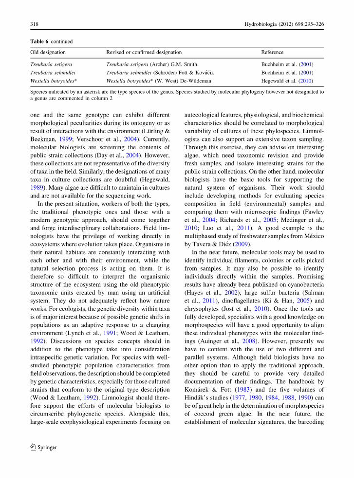
one and the same genotype can exhibit different
morphological peculiarities during its ontogeny or as
result of interactions with the environment (Lurling &
Beekman, 1999; Verschoor et al., 2004). Currently,
molecular biologists are screening the contents of
public strain collections (Day et al., 2004). However,
these collections are not representative of the diversity
of taxa in the field. Similarly, the designations of many
taxa in culture collections are doubtful (Hegewald,
1989). Many algae are difficult to maintain in cultures
and are not available for the sequencing work.
In the present situation, workers of both the types,
the traditional phenotypic ones and those with a
modern genotypic approach, should come together
and forge interdisciplinary collaborations. Field lim-
nologists have the privilege of working directly in
ecosystems where evolution takes place. Organisms in
their natural habitats are constantly interacting with
each other and with their environment, while the
natural selection process is acting on them. It is
therefore so difficult to interpret the organismic
structure of the ecosystem using the old phenotypic
taxonomic units created by man using an artificial
system. They do not adequately reflect how nature
works. For ecologists, the genetic diversity within taxa
is of major interest because of possible genetic shifts in
populations as an adaptive response to a changing
environment (Lynch et al., 1991; Wood & Leatham,
1992). Discussions on species concepts should in
addition to the phenotype take into consideration
intraspecific genetic variation. For species with well-
studied phenotypic population characteristics from
field observations, the description should be completed
by genetic characteristics, especially for those cultured
strains that conform to the original type description
(Wood & Leatham, 1992). Limnologist should there-
fore support the efforts of molecular biologists to
circumscribe phylogenetic species. Alongside this,
large-scale ecophysiological experiments focusing on
autecological features, physiological, and biochemical
characteristics should be correlated to morphological
variability of cultures of these phylospecies. Limnol-
ogists can also support an extensive taxon sampling.
Through this exercise, they can advise on interesting
algae, which need taxonomic revision and provide
fresh samples, and isolate interesting strains for the
public strain collections. On the other hand, molecular
biologists have the basic tools for supporting the
natural system of organisms. Their work should
include developing methods for evaluating species
composition in field (environmental) samples and
comparing them with microscopic findings (Fawley
et al., 2004; Richards et al., 2005; Medinger et al.,
2010; Luo et al., 2011). A good example is the
multiphased study of freshwater samples from Mexico
by Tavera & Diez (2009).
In the near future, molecular tools may be used to
identify individual filaments, colonies or cells picked
from samples. It may also be possible to identify
individuals directly within the samples. Promising
results have already been published on cyanobacteria
(Hayes et al., 2002), large sulfur bacteria (Salman
et al., 2011), dinoflagellates (Ki & Han, 2005) and
chrysophytes (Jost et al., 2010). Once the tools are
fully developed, specialists with a good knowledge on
morphospecies will have a good opportunity to align
these individual phenotypes with the molecular find-
ings (Auinger et al., 2008). However, presently we
have to content with the use of two different and
parallel systems. Although field biologists have no
other option than to apply the traditional approach,
they should be careful to provide very detailed
documentation of their findings. The handbook by
Komarek & Fott (1983) and the five volumes of
Hindak’s studies (1977, 1980, 1984, 1988, 1990) can
be of great help in the determination of morphospecies
of coccoid green algae. In the near future, the
establishment of molecular signatures, the barcoding
Table 6 continued
Old designation Revised or confirmed designation Reference
Treubaria setigera Treubaria setigera (Archer) G.M. Smith Buchheim et al. (2001)
Treubaria schmidlei Treubaria schmidlei (Schroder) Fott & Kovacik Buchheim et al. (2001)
Westella botryoides* Westella botryoides* (W. West) De-Wildeman Hegewald et al. (2010)
Species indicated by an asterisk are the type species of the genus. Species studied by molecular phylogeny however not designated to
a genus are commented in column 2
318 Hydrobiologia (2012) 698:295–326
123
conception, will allow an unambiguous designation of
organisms. The alignment of eco-functional groups of
algae with true species identities using the barcoding
conception will provide a better understanding of the
interaction between organisms and their environment.
The reasons why and how an individual species adapts
to different environmental conditions by producing
spines, mucilage or incrustations, or losing them,
establishing colonies or disintegrating them, exhibit-
ing striking colonial appearances or returning to the
simple green ball habit as a survival tactic will become
evident.
Acknowledgments We thank Eva Willen for her helpful
discussions and improvement of the manuscript. Kiplagat Kotut
revised the text with the main focus of improving on the
language. We thank Ute Mischke for providing information on
the harmonized taxon list of the European Water Framework
Directive. The E. Schweizerbart’sche Verlagsbuchhandlung
(Nagele & Obermiller), Stuttgart (www.schweizerbart.de) pro-
vided permission to copy figures of algae in Tables 2, 3, 4, and 5.
References
An, S. S., T. Friedl & E. Hegewald, 1999. Phylogenetic rela-
tionships of Scenedesmus and Scenedesmus-like coccoid
green algae as inferred from ITS-2 rDNA sequence com-
parisons. Plant Biology 1: 418–428.
Anneville, O., C. Kaiblinger, R. D. Tadonleke, J.-C. Druart &
M. T. Dokulil, 2008. Contribution of long-term monitoring
to the European Water Framework Directive implemen-
tation. In Sengupta, M. & R. Dalwani (eds), Proceedings of
Taal2007: The 12th World Lake Conference, Jaipur:
1122–1131.
Aslam, Z., W. Shin, M. K. Kim, W.-T. Im & S.-T. Lee, 2007.
Marinichlorella kaistiae gen. et sp. nov. (Trebouxiophy-
ceae, Chlorophyta) based on polyphasic taxonomy. Journal
of Phycology 43: 576–584.
Auinger, B. M., K. Pfandl & J. Boenigk, 2008. An improved
methodology for the identification of protists and micro-
algae from plankton samples preserved in Lugol’s iodine
solution: combining microscopic analysis with single cell
PCR. Applied Environmental Microbiology 74: 2505–
2510.
Ballot, A., K. Kotut, E. Novelo & L. Krienitz, 2009. Changes of
phytoplankton communities in Lakes Naivasha and Oloi-
dien, examples of degradation and salinization of lakes in
the Kenyan Rift Valley. Hydrobiologia 632: 359–363.
Barone, R. & L. Naselli Flores, 1994. Phytoplankton dynamics
in a shallow, hypertrophic reservoir (Lake Arancio, Sicily).
Hydrobiologia 289: 199–214.
Behnke, A., T. Friedl, V. A. Chepurnov & D. G. Mann, 2004.
Reproductive compatibility and rDNA sequence analyses
in the Sellaphora pupula species complex (Bacilla-
riophyta). Journal of Phycology 40: 193–208.
Beijerinck, M. W., 1890. Culturversuche mit Zoochlorellen,
Lichenengoniden und anderen niederen Algen I-III.
Botanische Zeitung 48: 726–740.
Bock, C., T. Proschold & L. Krienitz, 2010. Two new Dict-yosphaerium-morphotype lineages of the Chlorellaceae
(Trebouxiophyceae): Heynigia gen. nov. and Hindakiagen. nov. European Journal of Phycology 45: 267–277.
Bock, C., L. Krienitz & T. Proschold, 2011a. Taxonomic reas-
sessment of the genus Chlorella (Trebouxiophyceae) using
molecular signatures (barcodes), including description of
seven new species. Fottea 11: 293–312.
Bock, C., M. Pazoutova & L. Krienitz, 2011b. Phylogenetic
relationship of Coronastrum ellipsoideum and description
of Parachlorella hussii sp. nov. Biologia 66: 585–594.
Bock, C., T. Proschold & L. Krienitz, 2011c. Updating the genus
Dictyosphaerium and description of Mucidosphaera gen.
nov. (Trebouxiophyceae) based on morphological and
molecular data. Journal of Phycology 47: 638–652.
Boney, A. D., 1981. Mucilage: the ubiquitous algal attribute.
British Phycological Journal 16: 115–132.
Booton, G. C., G. L. Floyd & P. A. Fuerst, 1998. Polyphyly of
tetrasporalean green algae inferred from nuclear small-
subunit ribosomal DNA. Journal of Phycology 34:
306–311.
Bremer, K., C. J. Humphries, B. D. Mishler & S. P. Churchill,
1987. On cladistic relationships in green plants. Taxon 36:
339–349.
Buchheim, M. A., E. A. Michalopulos & J. A. Buchheim, 2001.
Phylogeny of the Chlorophyceae with special reference to
the Sphaeropleales: a study of 18S and 26S rDNA data.
Journal of Phycology 37: 819–835.
Buchheim, M. A., J. Buchheim, T. Carlson, A. Braband, D.
Hepperle, L. Krienitz, M. Wolf & E. Hegewald, 2005.
Phylogeny of the Hydrodictyaceae (Chlorophyceae):
inferences from rDNA data. Journal of Phycology 41:
1039–1054.
Buchheim, M. A., A. Keller, C. Koetschan, F. Forster, B. Merget
& M. Wolf, 2011. Internal Transcribed Spacer 2 (nu ITS2
rRNA) sequence-structure phylogenetics: towards an
automated reconstruction of the green algal tree of life.
PLoS ONE 6: e16931.
Cavalier-Smith, T., 1993. The origin, losses and gains of chlo-
roplasts. In Lewin, R. A. (ed.), Origin of Plastids: Symbi-
ogenesis, Prochlorophytes and the Origins of Chloroplasts.
Chapman and Hall, New York: 291–348.
Cepak, V., 1993. Scenedesmus obliquus zoospores in outdoor
mass culture – reality or artefact? Algological Studies 71:
37–42.
Chapman, R. L., M. A. Buchheim, C. F. Delwiche, T. Friedl, V.
A. R. Huss, K. G. Karol, L. A. Lewis, J. Manhart, R.
M. McCourt, J. L. Olsen & D. A. Waters, 1998. Molecular
systematics of the green algae. In Soltis, D. E., P. S. Soltis
& J. J. Doyle (eds), Molecular Systematics of Plants II.
DNA Sequencing. Kluwer Academic Publishers, Boston,
Dordrecht, London: 508–540.
Cole, J. J., 1982. Interactions between bacteria and algae in
aquatic ecosystems. Annual Review of Ecology Evolution
and Systematics 13: 291–314.
Coleman, A. W., 2000. The significance of a coincidence
between evolutionary landmarks found in mating affinity
and a DNA sequence. Protist 151: 1–9.
Hydrobiologia (2012) 698:295–326 319
123

Coleman, A. W., 2003. ITS2 is a double-edged tool for
eukaryote evolutionary comparisons. Trends in Genetics
19: 370–375.
Coleman, A. W., 2007. Pan-eukaryote ITS2 homologies
revealed by RNA secondary structure. Nucleic Acids
Research 35(10): 3322–3329.
Coleman, A. W., 2009. Is there a molecular key to the level of
‘‘biological species’’ in eukaryotes? A DNA guide.
Molecular Phylogenetics and Evolution 50: 197–203.
Cracraft, J., 1989. Speciation and its ontology: the empirical
consequences of alternative species concepts for under-
standing patterns and processes of differentiation. In Otte,
D. & J. Endler (eds), Speciation and Its Consequences.
Sinauer Associates, Sunderland, MA: 28–59.
Crawford, R. M., 1978. A new forma and a new species of the
genera Scenedesmus and Siderocelis (Chlorophyceae)
from the nannoplankton of a brackish-water coastal pool.
Protoplasma 96: 351–360.
Crawford, R. M. & P. F. Heap, 1978. Transmission elec-
tron microscopy X-ray microanalysis of two algae of the
genera Scenedesmus and Siderocelis. Protoplasma 96:
361–367.
Darienko, T., L. Gustavs, O. Mudimu, C. Rad Menendez, R.
Schumann, U. Karsten, T. Friedl & T. Proschold, 2010.
Chloroidium, a common terrestrial coccoid green alga
previously assigned to Chlorella (Trebouxiophyceae,
Chlorophyta). European Journal of Phycology 45: 79–95.
Day, J. G., J. Lukavsky, M. Lorenz, T. Friedl, J. Elster, C.
N. Cambell & J. J. Brand, 2004. Pringsheim’s living leg-
acy: CAUP, CCALA, CCAP, SAG and UTEX culture
collections of algae. Nova Hedwigia 79: 27–37.
de Queiroz, K., 1998. The general lineage concept of species,
species criteria, and the process of speciation: a conceptual
unification and terminological recommendations. In
Howard, D. J. & S. H. Berlocher (eds), Endless Forms:
Species and Speciation. Oxford University Press, Oxford,
England: 57–75.
Decho, A. W., 1990. Microbial exopolymer secretions in ocean
environments – their role(s) in food webs and marine
processes. Oceanography and Marine Biology 28: 73–153.
Denboh, T., T. Ichimura, D. Hendrayanti & A. W. Coleman,
2003. Closterium moniliferum-ehrenbergii (Charophy-
ceae; Chorophyta) Specie complex viewed from 1506
group I intron and nuclear rDNA. Journal of Phycology 39:
960–977.
Elias, M., Y. Nemkova, P. Skaloud, J. Neustupa, V. Kaufnerova
& L. Sejnohova, 2010. Hylodesmus singaporensis gen. et
sp. nov., a new autosporic subaerial green alga (Scene-
desmaceae, Chlorophyta) from Singapore. International
Journal of Systematic and Evolutionary Microbiology 60:
1224–1235.
Eloranta, V., 1979. The compound internal pyrenoid in cultured
cells of the green alga Monoraphidium griffithii (Berkel.)
Komar.-Legner. Protoplasma 99: 229–235.
Ettl, H. & G. Gartner, 1995. Syllabus der Boden-, Luft- und
Flechtenalgen. Gustav Fischer, Stuttgart.
Evans, K. M., A. H. Wortley & D. G. Mann, 2007. An assess-
ment of potential diatom ‘‘barcode’’ genes (cox1, rbcL, 18S
and ITS rDNA) and their effectiveness in determining
relationships in Sellaphora (Bacillariophyta). Protist 158:
349–364.
Fanes Trevino, I., P. Sanchez-Castillo & A. Comas Gonzalez,
2009. Contribution to the taxonomic study of the family
Botryococcaceae (Trebouxiophyceae, Chlorophyta) in
southern Spain. Cryptogamie, Algologie 30: 17–30.
Fawley, M. W., Y. Yun & M. Qin, 2000. Phylogenetic analyses
of 18S rDNA sequences reveal a new coccoid lineage of the
Prasinophyceae (Chlorophyta). Journal of Phycology 36:
387–393.
Fawley, M. W., K. P. Fawley & M. A. Buchheim, 2004.
Molecular diversity among communities of freshwater
microchlorophytes. Microbial Ecology 48: 489–499.
Fawley, M. W., M. L. Dean, S. K. Dimmer & K. P. Fawley,
2005a. Evaluating the morphospecies concept in the Sel-
enastraceae (Chlorophyceae, Chlorophyta). Journal of
Phycology 42: 142–154.
Fawley, M. W., K. P. Fawley & H. A. Owen, 2005b. Diversity
and ecology of small coccoid green algae from Lake Itasca,
Minnesota, USA, including Meyerella planktonica, gen. et
sp. nov. Phycologia 44: 35–48.
Fott, B., 1971. Algenkunde. VEB Gustav Fischer Verlag, Jena:
581 pp.
Friedl, T., 1995. Inferring taxonomic positions and testing genus
level assignments in coccoid green lichen algae: a phylo-
genetic analysis of 18S ribosomal RNA sequences from
Dictyochloropsis reticulata and from members of the
genus Myrmecia (Chlorophyta, Trebouxiophyceae cl.
nov.). Journal of Phycology 31: 630–637.
Friedl, T., 1996. Systematik und Phylogenie terrestrischer Grunal-
gen unter besonderer Berucksichtigung von Flechtenalgen:
molekulare und morphologische Merkmalsanalysen. Univer-
sity of Bayreuth, Habilitation: 122 pp.
Friedl, T., 1997. The evolution of the green algae. Plant Sys-
tematics and Evolution 11: 87–101.
Guillou, L., W. Eikrem, M.-J. Chretiennot-Dinet, F. Le Gall, R.
Massana, K. Romari, C. Pedros-Alio & D. Vaulot, 2004.
Diversity of picoplanktonic prasinophytes assessed by
direct nuclear SSU rDNA sequencing of environmental
samples and novel isolates retrieved from oceanic and
coastal marine ecosystems. Protist 155: 193–214.
Guiry, M. D. & G. M. Guiry, 2011. AlgaeBase. World-wide
electronic publication, National University of Ireland,
Galway [available on internet at http://www.algaebase.org
]; searched on 28 June 2011.
Gutell, R. R., 1994. Collection of small-subunit (16S- and 16S-
like) ribosomal-RNA structures. Nucleic Acids Research
22: 3502–3507.
Hajibabaei, M., G. A. C. Singer, P. D. N. Hebert & D. A. Hickey,
2007. DNA barcoding: how it complements taxonomy,
molecular phylogenetics and population genetics. Trends
in Genetics 23: 167–172.
Happy-Wood, C. M., 1988. Ecology of freshwater planktonic
green algae. In Sandgren, C. D. (ed.), Growth and Repro-
ductive Strategies of Freshwater Phytoplankton. Cam-
bridge University Press, Cambridge: 175–226.
Hartmann, H. J. & D. D. Kunkel, 1991. Mechanisms of food
selection in Daphnia. Hydrobiologia 225: 129–154.
Hayes, P. K., G. L. A. Barker, J. Batley, S. J. Beard, B.
A. Handley, P. Vacharapiyasophon & A. E. Walsby, 2002.
Genetic diversity within populations of cyanobacteria
assessed by analysis of single filaments. Antonie van
Leeuwenhoek 81: 197–202.
320 Hydrobiologia (2012) 698:295–326
123

Hebert, P. D. N., A. Cywinska, S. L. Ball & J. R. DeWaard,
2003. Biological identifications through DNA barcodes.
Proceedings of the Royal Society of London. Series B:
Biological Sciences 270: 313–321.
Hegewald, E., 1989. The Scenedesmus strains of the culture
collection at Austin, Texas (UTEX). Archiv fur Hydrobi-
ologie, Supplement 82, Algological Studies 55: 153–189.
Hegewald, E., 1997. Taxonomy and phylogeny of Scenedes-
mus. Algae (The Korean Journal of Phycology) 12:
235–246.
Hegewald, E., 2000. New combinations in the genus Des-modesmus (Chlorophyceae, Scenedesmaceae). Algological
Studies 96: 1–18.
Hegewald, E. & N. Hanagata, 2000. Phylogenetic studies on
Scenedesmaceae (Chlorophyta). Archiv fur Hydrobiolo-
gie, Supplement 136, Algological Studies 100: 29–49.
Hegewald, E. & E. Schnepf, 1984. Zur Struktur und Taxonomie
bestachelter Chlorococcales (Micractiniaceae, Golenkini-
aceae, Siderocystopsis). Nova Hedwigia 39: 297–383.
Hegewald, E. & E. Schnepf, 1986. Zur Struktur und Taxonomie
spindelformiger Chlorellales (Chlorophyta): Schroederia,
Pseudoschroederia gen. nov., Closteriopsis. Archiv fur
Hydrobiologie, Supplement 73, 1, Algological Studies 42:
21–48.
Hegewald, E. & P. C. Silva, 1988. Annotated Catalogue of
Scenedesmus and nomenclaturally related genera, includ-
ing original descriptions and figures. Bibliotheca Phyco-
logica 80: 1–587.
Hegewald, E. & M. Wolf, 2003. Phylogenetic relationships of
Scenedesmus and Acutodesmus (Chlorophyta, Chlorophy-
ceae) as inferred from 18S rDNA and ITS-2 sequence
comparisons. Plant Systematics and Evolution 241:
185–191.
Hegewald, E., D. Hepperle, M. Wolf & L. Krienitz, 2001.
Phylogenetic placement of Chlorotetraedron incus, C.polymorphum and Polyedriopsis spinulosa (Neochlorida-
ceae, Chlorophyceae). Phycologia 40: 399–402.
Hegewald, E., P. F. M. Coesel & P. Hegewald, 2002. A phy-
toplankton collection from Bali, with the description of a
new Desmodesmus species (Chlorophyta, Scenedesma-
ceae). Algological Studies 105: 51–78.
Hegewald, E., A. Schmidt, A. Braband & P. Tsarenko, 2005.
Revision of the Desmodesmus (Sphaeropleales, Scene-
desmaceae) species with lateral spines. 2. The multi-spined
to spineless taxa. Archiv fur Hydrobiologie, Supplement
157, Algological Studies 116: 1–38.
Hegewald, E., M. Wolf, A. Keller, T. Friedl & L. Krienitz, 2010.
ITS2 sequence-structure phylogeny in the Scenedesmaceae
with special reference to Coelastrum (Chlorophyta, Chlo-
rophyceae), including the new genera Comasiella and
Pectinodesmus. Phycologia 49: 325–335.
Hehmann, A., L. Krienitz & R. Koschel, 2001. Long-term
phytoplankton changes in an artificially divided top-down
manipulated humic lake. Hydrobiologia 448: 83–96.
Henley, W. J., J. L. Hironaka, L. Guillou, M. A. Buchheim, J.
A. Buchheim, M. W. Fawley & K. P. Fawley, 2004. Phy-
logenetic analysis of the ‘Nannochloris-like’ algae
and diagnoses of Picochlorum oklahomensis gen. et sp.
nov. (Trebouxiophyceae, Chlorophyta). Phycologia 43:
641–652.
Henning, W., 1966. Phylogenetic Systematics. University of
Illinois Press, Urbana.
Hepperle, D. & L. Krienitz, 2001. Systematics and ecology of
chlorophyte picoplankton in German inland waters along a
nutrient gradient. International Revue of Hydrobiology 86:
269–284.
Hepperle, D., E. Hegewald & L. Krienitz, 2000. Phylogenetic
position of the Oocystaceae (Chlorophyta). Journal of
Phycology 36: 590–595.
Heynig, H., 1980. Interessante Phytoplankter aus Gewassern des
Bezirks Halle (DDR). III. Archiv fur Protistenkunde 123:
349–357.
Heynig, H. & L. Krienitz, 1982. Monoraphidium neglectumn.sp. sowie einige Bemerkungen zu den Gattungen
Monoraphidium, Chlorolobion und Keratoccus (Chloro-
cocclaes). Archiv fur Protistenkunde 125: 335–344.
Hindak, F., 1976. Marvania geminata gen. nov. et sp. nov., a
new green alga. Algological Studies 16: 261–270.
Hindak, F., 1977. Studies on the chlorococcal algae (Chloro-
phyceae). I. Biologicke Prace 28(4): 1–263.
Hindak, F., 1978a. New taxa and reclassifications in the Chlo-
rococcales (Chlorophyceae). Preslia 50: 97–109.
Hindak, F., 1978b. The genus Lagerheimia Chod. and Lager-heimia-like unicells in the genus Scenedesmus Meyen
(Chlorophyceae). Biologia 33: 795–808.
Hindak, F., 1980. Studies on the chlorococcal algae (Chloro-
phyceae). II. Biologicke Prace 26(6): 1–195.
Hindak, F., 1984. Studies on the chlorococcal algae (Chloro-
phyceae). III. Biologicke Prace 30(1): 1–308.
Hindak, F., 1988. Studies on the chlorococcal algae (Chloro-
phyceae). IV. Biologicke Prace 34: 1–263.
Hindak, F., 1990. Studies on the chlorococcal algae (Chloro-
phyceae). V. Biologicke Prace 36: 1–225.
Hoef-Emden, K., 2007. Revision of the genus Cryptomonas(Cryptophyceae) II: incongruences between the classical
morphospecies concept and molecular phylogeny in
smaller pyrenoid-less cells. Phycologia 46: 402–428.
Hoffman, L. R., 1983. Atractomorpha echinata gen. et sp. nov.,
a new anisogamous member of the Sphaeropleaceae
(Chlorophyceae). Journal of Phycology 19: 76–86.
Hollibaugh, J. T., P. S. Wong, N. Bano, S. K. Pak, E. M. Prager
& C. Orrego, 2001. Stratification of microbial assemblages
in Mono Lake, California, and response to a mixing event.
Hydrobiologia 466: 45–60.
Huss, V. A. R., C. Frank, E. C. Hartmann, M. Hirmer, A. Klo-
boucek, B. M. Seidel, P. Wenzeler & E. Kessler, 1999.
Biochemical taxonomy and molecular phylogeny of the
genus Chlorella sensu lato (Chlorophyta). Journal of
Phycology 35: 587–598.
Jahn, R. & W. H. Kusber (eds), 2006. AlgaTerra Information
System (online). Botanic Garden and Botanical Museum
Berlin-Dahlem, FU-Berlin [available on internet at
http://www.algaterra.orgS].
Jobb, G., 2008. Treefinder, version of October 2008. Munich,
Germany. Distributed by the author at www.treefinder.de.
Johansen, J. R. & D. A. Casamatta, 2005. Recognizing cyano-
bacterial diversity through adoption of a new species par-
adigm. Algological Studies 116: 71–93.
Jost, S., R. Medinger & J. Boenigk, 2010. Cultivation-inde-
pendent species identification of Dinobryon species
Hydrobiologia (2012) 698:295–326 321
123

(Chrysophyceae) by means of multiplex single-cell PCR.
Journal of Phycology 46: 901–906.
Keller, A., T. Schleicher, F. Forster, B. Ruderisch, T. Dandekar,
T. Muller & M. Wolf, 2008. ITS2 data corroborate a
monophyletic chlorophycean DO-group (Sphaeropleales).
BMC Evolutionary Biology 8: 218.
Kessler, E., M. Schafer, C. Hummer, A. Kloboucek & V.A.R.
Huss, 1997. Physiological, biochemical, and molecular
characters for the taxonomy of the subgenera of Scene-desmus (Chlorococcales, Chlorophyta). Botanica Acta
110: 244–250
Ki, J.-S. & M.-S. Han, 2005. Sequence-based diagnostics and
phylogenetic approach of uncultured freshwater dinofla-
gellate Peridinium (Dinophyceae) species, based on single-
cell sequencing of rDNA. Journal of Applied Phycology
17: 147–153.
Komarek, J., 1979. Anderungen in der Taxonomie der chloro-
coccalen Algen. Algological Studies 24: 239–263.
Komarek, J. & B. Fott, 1983. Chlorophyceae (Grunalgen)
Ordnung: Chlorococcales. In: Huber-Pestalozzi, G. (ed.),
Das Phytoplankton des Sußwassers 7. Teil, 1. Halfte.
Schweizerbart’sche Verlagsbuchhandlung (Nagele u.
Obermiller), Stuttgart.
Komarek, J. & A. G. Comas, 1982. Taxonomical definition of
the genera and several species of Ankistrodesmus and Se-lenastrum (Chlorococcales). Archiv fur Hydrobiologie,
Supplement 63.3, Algological Studies 32: 259–277.
Komarek, J. & V. Jankovska, 2001. Review of the green algal
genus Pediastrum; implication for pollen-analytical
research. Bibliotheca Phycologica 108: 1–127.
Komarek, J. & L. Kovacik, 1985. The genus ChlorotetraedronMcEntee et al. (Protosiphonales, Chlorophyceae). Preslia
57: 289–297.
Komarek, J. & P. Marvan, 1992. Morphological differences in
natural populations of the genus Botryococcus (Chloro-
phyceae). Archiv fur Protistenkunde 141: 65–100.
Kostikov, I., T. Darienko, A. Lukesova & L. Hoffmann, 2002.
Revision of the classification system of Radiococca-
ceae Fott ex Komarek (except the subfamily Dictyochlo-
relloideae) (Chlorophyta). Algological Studies 104:
23–58.
Kotut, K. & L. Krienitz, 2011. Does the potentially toxic cya-
nobacterium Microcystis exist in the soda lakes of East
Africa? Hydrobiologia 664: 219–225.
Krienitz, L., 1986. Drei neue Arten coccaler Grunalgen (Chlo-
rellales) aus dem Plankton der Elbe. Archiv fur Protis-
tenkunde 132: 299–311.
Krienitz, L., 1990. Coccale Grunalgen der mittleren Elbe.
Limnologica 21: 165–231.
Krienitz, L., 1998. Amphikrikos variabilis sp. nov. (Chloro-
phyta), a common species of inland waters of Namibia.
Archiv fur Hydrobiologie, Supplement 126, Algological
Studies 91: 1–10.
Krienitz, L. & C. Bock, 2011. Elongatocystis ecballocystiformisgen. et comb. nov., and some reflections on systematics of
Oocystaceae (Trebouxiophyceae, Chlorophyta). Fottea 11:
271–278.
Krienitz, L. & H. Heynig, 1982. Beobachtungen an Ankyralanceolata (Kors. 1953) Fott 1957 und Ankyra spatulifera(Kors. 1953) Fott 1957 (Chlorococcales) im Freiland. Ar-
chiv fur Protistenkunde 126: 265–271.
Krienitz, L. & G. Klein, 1988. Morphologie und Ultrastruktur
einiger Arten der Gattung Monoraphidium (Chlorellales)
III. Monoraphidium terrestre (Bristol) nov. comb. Algo-
logical Studies 49: 447–463.
Krienitz, L. & K. Kotut, 2010. Fluctuating algal food popula-
tions and the occurrence of Lesser Flamingos (Phoenic-onaias minor) in three Kenyan Rift Valley lakes. Journal of
Phycology 46: 1088–1096.
Krienitz, L. & W. Scheffler, 1994. The Selenastraceae of the
oligotrophic Lake Stechlin (Brandenburg, Germany). Bi-
ologia 49: 463–471.
Krienitz, L. & M. Wirth, 2006. The high content of polyunsat-
urated fatty acids in Nannochloropsis limnetica (Eustig-
matophyceae) and its implication for food web
interactions, freshwater aquaculture and biotechnology.
Limnologica 36: 204–210.
Krienitz, L., G. Klein & H. Bohm, 1985. Zur Morphologie und
Ultrastruktur von Selenastrum gracile Reinsch 1867
(Chlorellales). Archiv fur Protistenkunde 130: 79–92.
Krienitz, L., V. A. R. Huss & C. Hummer, 1996a. Picoplank-
tonic Choricystis species (Chlorococcales, Chlorophyta)
and problems surrounding the morphologically similar
‘Nannochloris-like algae’. Phycologia 35: 332–341.
Krienitz, L., P. Kasprzak & R. Koschel, 1996b. Long term study
on the influence of eutrophication, restoration and bioma-
nipulation on the structure and development of phyto-
plankton communities in Feldberger Haussee (Baltic Lake
District, Germany). Hydrobiologia 330: 89–110.
Krienitz, L., A. Hehmann & S. J. Casper, 1997. The unique
phytoplankton community of a highly acidic bog lake in
Germany. Nova Hedwigia 65: 411–430.
Krienitz, L., H. Takeda & D. Hepperle, 1999. Ultrastructure, cell
wall composition, and phylogenetic position of Pseud-odictyosphaerium jurisii (Chlorococcales, Chlorophyta)
including a comparison with other picoplanktonic green
algae. Phycologia 38: 100–107.
Krienitz, L., D. Hepperle, H.-B. Stich & W. Weiler, 2000.
Nannochloropsis limnetica (Eustigmatophyceae), a new
species of picoplankton from freshwater. Phycologia 39:
219–227.
Krienitz, L., I. Ustinova, T. Friedl & V. A. R. Huss, 2001.
Traditional generic concepts versus 18S rRNA gene phy-
logeny in the green algal family Selenastraceae (Chloro-
phyceae, Chlorophyta). Journal of Phycology 37: 852–865.
Krienitz, L., E. Hegewald, D. Hepperle & M. Wolf, 2003. The
systematics of coccoid green algae: 18S rRNA gene
sequence data versus morphology. Biologia 58: 437–446.
Krienitz, L., E. Hegewald, D. Hepperle, V. A. R. Huss, T. Rohr
& M. Wolf, 2004. Phylogenetic relationship of Chlorellaand Parachlorella gen. nov. (Chlorophyta, Trebouxio-
phyceae). Phycologia 43: 529–542.
Krienitz, L., C. Bock, W. Luo & T. Proschold, 2010. Polyphy-
letic origin of the Dictyosphaerium-morphotype within
Chlorellaceae (Trebouxiophyceae). Journal of Phycology
46: 559–563.
Krienitz, L., C. Bock, P. K. Dadheech & T. Proschold, 2011a.
Taxonomic reassessment of the genus Mychonastes(Chlorophyceae, Chlorophyta) including the description of
eight new species. Phycologia 50: 89–106.
Krienitz, L., C. Bock, H. Nozaki & M. Wolf, 2011b. SSU rRNA
gene phylogeny of morphospecies affiliated to the bioassay
322 Hydrobiologia (2012) 698:295–326
123

alga ‘‘Selenastrum capricornutum’’ recovered the poly-
phyletic origin of crescent-shaped chlorophyta. Journal of
Phycology 47: 880–893.
Krienitz, L., C. Bock, K. Kotut & W. Luo, 2012. Picocystissalinarum (Chlorophyta) in saline lakes and hot springs of
East Africa. Phycologia 51: 22–32.
Leliaert, F., D. R. Smith, H. Moreau, M. D. Herron, H. Verb-
ruggen, C. F. Delwiche & O. De Clerck, 2012. Phylogeny
and molecular evolution of the green algae. Critical
Reviews in Plant Sciences 31: 1–46.
Lemmermann, E., 1903. Brandenburgische Algen. Zeitschrift
fur Fischerei 11: 73–123.
Lewin, R. A., L. Krienitz, R. Goericke, H. Takeda & D. Hep-
perle, 2000. Picocystis salinarum gen. et sp. nov. (Chlo-
rophyta) – a new picoplanktonic green alga. Phycologia 39:
560–565.
Lewis, L. A., 1997. Diversity and phylogenetic placement of
Bracteacoccus Tereg (Chlorophyceae, Chlorophyta) based
on 18S ribosomal RNA gene sequence data. Journal of
Phycology 33: 279–285.
Lewis, L. A. & R. M. McCourt, 2004. Green algae and the origin
of land plants. American Journal of Botany 91: 1535–1556.
Lewis, L. A., L. W. Wilcox, P. A. Fuerst & G. L. Floyd, 1992.
Concordance of molecular and ultrastructural data in the
study of zoosporic chlorococcalean green algae. Journal of
Phycology 28: 375–380.
Long, E. O. & I. B. David, 1980. Repeated genes in eukaryotes.
Annual Review Biochemistry 49: 727–764.
Lukavsky, J., 1991. Motile cells in Scenedesmus obliquus in
outdoor mass culture. Archiv fur Protistenkunde 140:
345–348.
Luo, W., S. Pflugmacher, T. Proschold, N. Walz & L. Krienitz,
2006. Genotype versus phenotype variability in Chlorellaand Micractinium (Chlorophyta, Trebouxiophyceae). Pro-
tist 157: 315–333.
Luo, W., T. Proschold, C. Bock & L. Krienitz, 2010. Generic
concept in Chlorella-related coccoid green algae (Chlo-
rophyta, Trebouxiophyceae). Plant Biology 12: 545–553.
Luo, W., C. Bock, H. R. Li, J. Padisak & L. Krienitz, 2011.
Molecular and microscopic diversity of planktonic
eukaryotes in the oligotrophic Lake Stechlin (Germany).
Hydrobiologia 661: 133–143.
Lurling, M. & W. Beekman, 1999. Grazer induced defenses in
Scenedesmus (Chlorococclaes; Chlorophyceae): coenobi-
um and spine formation. Phycologia 38: 368–376.
Lynch, M., W. Gabriel & A. M. Wood, 1991. Adaptive and
demographic responses of plankton populations to envi-
ronmental change. Limnology and Oceanography 36:
1301–1312.
Mai, J. C. & A. W. Coleman, 1997. The internal transcribed
spacer 2 exhibits a common secondary structure in green
algae and flowering plants. Journal of Molecular Evolution
44: 258–271.
Mallet, J., 2006. Species concepts. In Fox, C. W. J. (ed.), Evo-
lutionary Genetics: Concepts and Case Studies. Oxford
University Press, Oxford: 367–373.
Marin, B. & M. Melkonian, 2010. Molecular phylogeny and
classification of the Mamiellophyceae class. nov. (Chloro-
phyta) based on sequence comparisons of the nuclear- and
plastid-encoded rRNA operons. Protist 161: 304–336.
Marvan, P., J. Komarek & A. Comas, 1984. Weighting and
scaling of features in numerical evaluation of coccal
green algae (genera of the Selenastraceae). Archiv fur
Hydrobiologie, Supplement 67, Algological Studies 37:
363–399.
Masjuk, N. P., 2006. Chlorodendrophyceae class nov. (Chlo-
rophyta, Viridiplantae) in the Ukrainian flora: I. Phyloge-
netic relations and taxonomical status. Ukrainian Botanical
Journal 63: 601–614. (in Ukrainian).
Mattox, K. R. & K. D. Stewart, 1984. Classification of the green
algae: a concept based on comparative cytology. In Irvine,
D. E. G. & D. M. John (eds), Systematics of the green
algae. The Systematics Association, Special Volumes, No.
27. Academic Press, London, Orlando: 29–72.
Mayr, E., 1942. Systematics and the Origin of Species from the
Viewpoint of a Zoologist. Harvard University Press,
Cambridge, MA: 1–372.
McEntee, F. J., H. C. Bold & P. A. Archibald, 1977. Notes on
some edaphic algae of the South Pacific and Malaysian
areas, with special reference to Pseudotetraedron poly-morphum gen. et spec. nov. Soil Science 124: 161–166.
McManus, H. A. & L. A. Lewis, 2005. Molecular phylogenetics,
morphological variation and colony-form evolution in the
family Hydrodictyaceae (Sphaeropleales, Chlorophyta).
Phycologia 44: 582–595.
McManus, H. A. & L. A. Lewis, 2011. Molecular phylogenetic
relationships in the freshwater family Hydrodictyaceae
(Sphaeropleales, Chlorophyceae), with an emphasis on
Pediastrum duplex. Journal of Phycology 47: 152–163.
McManus, H. A., L. A. Lewis & E. T. Schultz, 2011. Distin-
guishing multiple lineages of Pediastrum duplex with
morphometrics and a proposal for Lacunastrum gen. nov.
Journal of Phycolgy 47: 123–130.
Medinger, R., V. Nolte, R. V. Pandey, S. Jost, B. Ottenwaelder,
C. Schlotterer & J. Boenigk, 2010. Diversity in a hidden
world: potential and limitation of next generation
sequencing for surveys of molecular diversity of eukaryotic
microorganisms. Molecular Ecology 19: 32–40.
Melkonian, M., 1982. Structural and evolutionary aspects of the
flagellar apparatus in green algae and land plants. Taxon
31: 255–265.
Melkonian, M., 1984. Flagellar apparatus ultrastructure in
relation to green algal classification. In Irvine, D. E. G. &
D. M. John (eds), Systematics of the green algae. The
Systematics Association, Special Volumes, No. 27. Aca-
demic Press, London, Orlando: 73–120.
Melkonian, M., 1990a. Phylum Chlorophyta. Class Prasini-
phyceae. In Margulis, L., et al. (eds), Handbook of Pro-
toctista. Jones and Bartlett, Boston, MA: 600–607.
Melkonian, M., 1990b. Phylum Chlorophyta. Class Chloro-
phyceae. In Margulis, L., et al. (eds), Handbook of Pro-
toctista. Jones and Bartlett, Boston, MA: 608–616.
Melkonian, M. & B. Surek, 1995. Phylogeny of the Chlorophyta
– congruence between ultrastructural and molecular evi-
dence. Bulletin de la Societe Zoologique de France 120:
191–208.
Mischke, U., 2006. Downloads zum Bewertungsverfahren
Phytoplankton nach WRRL. Leibniz-Institut fur Gewas-
serokologie und Binnenfischerei [available on internet at
http://www.igb-berlin.de/abt2/mitarbeiter/mischke/].
Hydrobiologia (2012) 698:295–326 323
123

Mischke, U. & B. Nixdorf (eds), 2008. Bewertung von Seen
mittels Phytoplankton zur Umsetzung der EU-Wasserrah-
menrichtlinie, BTUC-AR 2/2008: 266 pp. ISBN 978-3-
940471-06-2 ISSN 1434-6834.
Miyachi, S., M. Tsuzuki, I. Mruyama, M. Gantar, S. Miyachi &
H. Matsushima, 1986. Effects of CO2 concentration during
growth on the intracellular structure of Chlorella and
Scenedesmus (Chlorophyta). Journal of Phycology 22:
313–319.
Moniz, M. B. J. & I. Kaczmarska, 2010. Barcoding of diatoms:
nuclear encoded ITS revisited. Protist 161: 7–34.
Muller, T., N. Philippi, T. Dandekar, J. Schultz & M. Wolf,
2007. Distinguishing species. RNA 13: 1469–1472.
Nakayama, T., S. Watanabe, K. Mitsui, H. Uchida & I. Inouye,
1996. The phylogenetic relationship between Chlamydo-
monadales and Chlorococcales inferred from 18S rDNA
sequence data. Phycological Research 44: 47–55.
Nemjova, K., J. Neustupa, J. St’astny, P. Skaloud & J. Vesela,
2011. Species concept and morphological differentiation of
strains traditionally assigned to Micrasterias truncata.
Phycological Research 59: 208–220.
Neustupa, J., P. Skaloud & J. St’astny, 2010. The molecular
phylogenetic and geometric morphometric evaluation of
Micrasterias Crux-Melitensis/M-Radians species com-
plex1. Journal of Phycology 46: 703–714.
Padisak, J., L. Krienitz, R. Koschel & J. Nedoma, 1997. Deep
layer autotrophic picophytoplankton maximum in the oli-
gotrophic Stechlinsee, Germany: origin, activity, devel-
opment and erosion. European Journal of Phycology 32:
403–416.
Padisak, J., G. Borics, I. Grigorszky & E. Soroczki-Pinter, 2006.
Use of phytoplankton assemblages for monitoring eco-
logical status of lakes within the Water Framework
Directive: the assemblage index. Hydrobiologia 553: 1–14.
Padisak, J., L. O. Crossetti & L. Naselli-Flores, 2009. Use and
misuse in the application of the phytoplankton functional
classification: a critical review with updates. Hydrobiolo-
gia 621: 1–19.
Padisak, J., E. Hajnal, L. Krienitz, J. Lakner & V. Uveges, 2010.
Rarity, ecological memory, rate of floral change in phyto-
plankton – and the mystery of the Red Cock. Hydrobiologia
653: 45–64.
Pascher, A., 1918. Von einer allen Algenreihen gemeinsamen
Entwicklungsregel. Berichte der Deutschen Botanischen
Gesellschaft 36: 390–409.
Pazoutova, M., P. Skaloud & K. Nemjova, 2010. Phylogenetic
position of Ooplanctella planoconvexa, gen. et comb. nova
and Echinocoleum elegans (Oocystaceae, Trebouxiophy-
ceae, Chlorophyta). Fottea 10: 75–83.
Pearl, H. W., L. M. Valdes, A. R. Joyner & V. Winkelmann,
2007. Phytoplankton indicators of ecological change in the
eutrophying Pamlico Sound system, North Carolina. Eco-
logical Applications 17: S88–S101.
Plain, N., C. Largeau, S. Derenne & A. Coute, 1993. Variabilite
morphologique de Botryococcus braunii (Chlorococcales,
Chlorophyta): correlations avec les conditions de crois-
sance et la teneur en lipides. Phycologia 32: 259–265.
Porter, K. G., 1973. Selective grazing and differential digestion
of algae by zooplankton. Nature 244: 179–180.
Posada, D. & T. R. Buckley, 2004. Model selection and
model averaging in phylogenetics: advantages of Akaike
information criterion and Bayesian approaches over like-
lihood ratio tests. System Biology 53: 793–808.
Potter, D., T. C. Lajeunesse, G. W. Saunders & R. A. Anderson,
1997. Convergent evolution masks extensive biodiversity
among marine coccoid picoplankton. Biodiversity and
Conservation 6: 99–107.
Proschold, T. & F. Leliaert, 2007. Systematics of the green
algae: conflict of classic and modern approaches. In Bro-
die, J. & J. Lewis (eds), Unravelling the Algae: The Past,
Present, and Future of the Algae. Taylor and Francis,
London: 123–153.
Proschold, T., B. Marin, U. G. Schlosser & M. Melkonian, 2001.
Molecular phylogeny and taxonomic revision of Chla-mydomonas (Chlorophyta). I. Emendation of Chlamydo-monas Ehrenberg and Chloromonas Gobi, and description
of Oogamochlamys gen. nov. and Lobochlamys gen. nov.
Protist 152: 265–300.
Proschold, T., E. H. Harris & A. W. Coleman, 2005. Portrait of a
species: Chlamydomonas reinhardtii. Genetics 170:
1601–1610.
Proschold, T., C. Bock, W. Luo & L. Krienitz, 2010. Polyphy-
letic distribution of bristle formation in Chlorellaceae:
Micractinium, Diacanthos, Didymogenes and Hegewaldiagen. nov. (Trebouxiophyceae, Chlorophyta). Phycological
Research 58: 1–8.
Proschold, T., T. Darienko, P. C. Silva, W. Reisser & L. Krie-
nitz, 2011. The systematics of ‘‘Zoochlorella’’ revisited
employing an integrative approach. Environmental
Microbiology 13: 350–364.
Raven, J. A., 1999. Picophytoplankton. In Round, F. E. & D.
J. Chapman (eds), Progress in phycological research, Vol.
13. Biopress Ltd, Bristol: 33–105.
Reymond, O. L. & E. Hegewald, 1988. A morphological and
cytological assessment of Paradoxia and Ankyra (Chlo-
rococcales, Chlorophyceae). Archiv fur Protistenkunde
135: 167–172.
Reymond, O. L., F. Hindak & H. J. Sluiman, 1986. Morphologie
cellulaire et cycle de developpement chez l’algue verte
Marvania geminata Hindak. Archives des Sciences, Gen-
eve 39: 243–255.
Reynolds, C. S., 2007. Variability in the provision and function
of mucilage in phytoplankton: facultative responses to the
environment. Hydrobiologia 578: 37–45.
Reynolds, C. S., V. Huszar, C. Kruk, L. Naselli-Flores & S.
Melo, 2002. Towards a functional classification of the
freshwater phytoplankton. Journal of Plankton Research
24: 417–428.
Richards, T. A., A. A. Vepritskiy, D. E. Gouliamova & S.
A. Nierzwicki-Bauer, 2005. The molecular diversity of
freshwater picoeukaryotes from an oligotrophic lake
reveals diverse, distinctive and globally dispersed lineages.
Environmental Microbiology 7: 1413–1425.
Rindi, F., D. W. Lam & J. M. Lopez-Bautista, 2009. Phyloge-
netic relationships and species circumscription in Tren-tepohlia and Printzina (Trentepohliales, Chlorophyta).
Molecular Phylogenetics and Evolution 52: 329–339.
Rindi, F., H. A. Allali, D. W. Lam & J. M. Lopez-Bautista, 2010.
An overview of the biodiversity and biogeography of ter-
restrial green algae. In Rescigno, V. & S. Maletta (eds),
Biodiversity Hotspots. Nova Science Publishers, New
York: 105–122.
324 Hydrobiologia (2012) 698:295–326
123

Roesler, C. S., C. W. Culbertson, S. M. Etheridge, R. Goericke,
R. P. Kiene, L. G. Miller & R. S. Oremland, 2002. Distri-
bution, production, and ecophysiology of Picocystis strain
ML in Mono Lake, California. Limnology and Oceanog-
raphy 47: 440–452.
Ronquist, F. & J. P. Huelsenbeck, 2003. MrBayes 3: Bayesian
phylogenetic inference under mixed models. Bioinfor-
matics 19: 1572–1574.
Rybalka, N., R. A. Andersen, I. Kostikov, K. I. Mohr, A. Mas-
salski, M. Olech & T. Friedl, 2009. Testing for endemism,
genotypic diversity and species concepts in Antarctic ter-
restrial microalgae of the Tribonemataceae (Strameno-
piles, Xanthophyceae). Environmental Microbiology 11:
554–565.
Salman, V., R. Amann, A.-C. Girnth, L. Polerecky, J. V. Bailey,
S. Hegslund, G. Jessen, S. Par & H. N. Schulz-Vogt, 2011.
A single-cell sequencing approach to the classification of
large, vacuolated sulfur bacteria. Systematic and Applied
Microbiology 34: 243–259.
Salmaso, N., G. Morabito, F. Buzzi, L. Garibaldi, M. Simona &
R. Mosello, 2006. Phytoplankton as an indicator of the
water quality of the deep lakes south of the Alps. Hydro-
biologia 563: 167–187.
Schmidt, A. & G. Feher, 1999–2000. Adatol del-Magyarorszagi
Vizek Algainal ismeretehez IV. Botanikai Kozlemenyek
86–87: 95–105.
Schnepf, E. & E. Hegewald, 1993. Didymogenes palatinaSchmidle and Didymogenes anomala (G.M. Smith) Hind.
(Chlorococcales): Taxonomy, ultrastructure, autosporo-
genesis and autospore wall assembly. Archiv fur Protis-
tenkunde 143: 41–53.
Schnepf, E., G. Deichgraber, M. Glaab & E. Hegewald, 1980.
Bristles and spikes in Chlorococcales: ultrastructural
studies in Acanthosphaera, Micractinium, Pediastrum,
Polyedriopsis, Scenedesmus, and Siderocystopsis. Journal
of Ultrastructure Research 72: 367–379.
Schultz, J. & M. Wolf, 2009. ITS2 sequence-structure analysis
in phylogenetics: a how-to manual for molecular system-
atics. Molecular Phylogenetics and Evolution 52: 250–252.
Schultz, J., S. Maisel, D. Gerlach, T. Muller & M. Wolf, 2005. A
common core of secondary structure of the internal tran-
scribed spacer 2 (ITS2) throughout the Eukaryota. RNA
11: 361–364.
Senousy, H. H., G. W. Beakes & E. Hack, 2004. Phylogenetic
placement of Botryococcus braunii (Trebouxiophyceae)
and Botryococcus sudeticus isolate UTEX 1629 (Chloro-
phyceae). Journal of Phycology 40: 412–423.
Shoup, S. & L. A. Lewis, 2003. Polyphyletic origin of parallel
basal bodies in swimming cells of chlorophycean green
algae (Chlorophyta). Journal of Phycology 39: 789–796.
Simpson, P. D. & S. D. Van Valkenburg, 1978. The ultra-
structure of Mychonastes ruminatus gen. et sp. nov., a new
member of the Chlorophyceae isolated from brackish
water. British Phycological Journal 13: 117–130.
Skaloud, P. & O. Peksa, 2010. Evolutionary inferences based on
ITS rDNA and actin sequences reveal extensive diversity
of the common lichen alga Asterochloris (Trebouxiophy-
ceae, Chlorophyta). Molecular Phylogenetics and Evolu-
tion 54: 36–46.
Slapeta, J., D. Moreira & P. Lopez-Garcia, 2005. The extent of
protist diversity: insights from molecular ecology of
freshwater eukaryotes. Proceedings of the Royal Society of
London. Series B: Biological Sciences 272: 2073–2081.
Sogin, M. L., M. T. Swanton, J. H. Gunderson & H. J. Elwood,
1986. Sequence of the small subunit ribosomal RNA gene
from the hypotrichous ciliate Euplotes aediculatus. Journal
of Protozoology 33: 26–29.
Somogyi, B., T. Felfoldi, K. Solymosi, J. Makk, Z. G. Homo-
nnay, G. Horvath, E. Turcsi, B. Boddi, K. Marialigeti & L.
Voros, 2011. Chloroparva pannonica gen. et sp. nov.
(Trebouxiophyceae, Chlorophyta) – a new picoplanktonic
green alga from a turbid, shallow soda pan. Phycologia 50:
1–10.
Steinkotter, J., D. Bhattacharya, I. Semmelroth, C. Bibeau & M.
Melkonian, 1994. Prasinophytes form independent lin-
eages within the Chlorophyta: evidence from ribosomal
RNA sequence comparisons. Journal of Phycology 30:
340–345.
Stockner, J. G., 1991. Autotrophic picoplankton in freshwater
ecosystems: the view from the summit. Internationale
Revue der Gesamten Hydrobiologie 76: 483–492.
Stockner, J. G. & N. J. Antia, 1986. Algal picoplankton from
marine and freshwater ecosystems: a multidisciplinary
perspective. Canadian Journal of Fisheries and Aquatic
Sciences 43: 2472–2503.
Swofford, D. L., 2002. Phylogenetic Analysis Using Parsimony
(* and Other Methods), Version 4.0b 10. Sinauer Associ-
ates, Sunderland, MA.
Tavera, R. & B. Diez, 2009. Multifaceted approach for the
analysis of the phototrophic microbial community in a
freshwater recreational area of Xochimilco, Mexico.
Hydrobiologia 636: 353–368.
Trainor, F. R., 1963. Zoospores in Scenedesmus obliquus. Sci-
ence 142: 1673–1674.
Trainor, F. R., 1996. Reproduction in Scenedesmus. Algae (The
Korean Journal of Phycology) 11: 183–201.
Tsarenko, P. M. & O. A. Petlevanny, 2001. Addition to the
diversity of algae of Ukraine. Algologia, Supplement: 1–130.
Ustinova, I., L. Krienitz & V. A. R. Huss, 2000. Hyaloraphidiumcurvatum is not a green alga, but a lower fungus; Amoe-bidium parasiticum is not a fungus, but a member of the
DRIPs. Protist 151: 253–262.
Ustinova, I., L. Krienitz & V. A. R. Huss, 2001. Closteriopsisacicularis (G.M. Smith) Belcher et Swale is a fusiform
member of the genus Chlorella, closely related to C.kessleri (Chlorophyta, Trebouxiophyceae). European
Journal of Phycology 36: 341–351.
Van den Hoek, C., D. G. Mann & H. G. Jahns, 1995. Algae: An
Introduction to Phycology. Cambridge University Press,
Cambridge: 627 pp.
Van Donk, E., 2005. Planktonic interactions: developments and
perspectives. Verhandlungen der Internationalen Vereini-
gung fur theoretische und angewandte Limnologie 29:
61–72.
Vanormelingen, P., E. Hegewald, A. Braband, M. Kitschke, T.
Friedl, K. Sabbe & W. Vyverman, 2007. The systematics of a
small spineless Desmodesmus taxon, D. costato-granulatus(Sphaeropleales, Chlorophyceae), based on ITS2rDNA
sequence analyses and cell wall morphology. Journal of
Phycology 43: 378–396.
Verschoor, A. M., I. Van Der Stap, N. R. Helmsing, M. Lurling
& E. Van Donk, 2004. Inducible colony formation within
Hydrobiologia (2012) 698:295–326 325
123
the Scenedesmaceae: adaptive response to infochemicals
from two different herbivore taxa. Journal of Phycology
40: 808–814.
Weisse, T., 1993. Dynamics of autotrophic picoplankton in
marine and freshwater ecosystems. In Jones, G. (ed.),
Advances in Microbial Ecology, Vol. 13. Plenum Press,
New York: 327–370.
Weisse, T., 2004. Pelagic microbes – protozoa and the microbial
food web. In O‘Sullivan, P. E. & C. S. Reynolds (eds), The
Lakes Handbook, Vol. 1 Limnology and Limnetic Ecol-
ogy. Blackwell Publishing, Malden, Oxford, Carlton:
417–460.
Wilcox, L. W. & G. L. Floyd, 1988. Ultrastructure of the gamete
of Pediastrum duplex (Chlorophyceae). Journal of Phy-
cology 24: 140–146.
Wolf, M., M. Buchheim, E. Hegewald, L. Krienitz & D. Hep-
perle, 2002a. Phylogenetic position of the Sphaeropleaceae
(Chlorophyta). Plant Systematic and Evolution 230:
161–171.
Wolf, M., L. Krienitz, E. Hegewald & D. Hepperle, 2002b.
Phylogenetic position of Actinastrum hantzschii (Chloro-
phyta, Trebouxiophyceae). Algological Studies 104:
59–67.
Wolf, M., E. Hegewald, D. Hepperle & L. Krienitz, 2003a.
Phylogenetic position of the Golenkiniaceae (Chlorophyta)
as inferred from 18S rDNA sequence data. Biologia 58:
433–436.
Wolf, M., D. Hepperle & L. Krienitz, 2003b. On the phylogeny
of Radiococcus, Planktosphaeria and Schizochlamydella(Radiococcaceae, Chlorophyta). Biologia 58: 759–765.
Wood, M. A. & T. Leatham, 1992. The species concept in
phytoplankton ecology. Journal of Phycology 28: 723–729.
Yamamoto, M., H. Nozaki, Y. Miyazawa, T. Koide & S.
Kawano, 2003. Relationships between presence of a
mother cell wall and speciation in the unicellular micro
alga Nannochloris (Chlorophyta). Journal of Phycology
39: 172–284.
Yoon, H. S., J. Hackett, C. Ciniglia, G. Pinto & D. Bhattacharya,
2004. A molecular timeline for the origin of phytosynthetic
eukaryotes. Molecular Biology and Evolution 21:
809–818.
Zimmermann, J., R. Jahn & B. Gemeinholzer, 2011. Barcoding
diatoms: evaluation of the V4 subregion on the 18S rRNA
gene, including new primers and protocols. Organisms,
Diversity and Evolution 11: 173–192.
Zuker, M., 2003. Mfold web server for nucleic acid folding and
hybridization prediction. Nucleic Acids Research 31:
3406–3415.
326 Hydrobiologia (2012) 698:295–326
123