presence of rituximab ajay k. gopal, stanley r. riddell,...
TRANSCRIPT

1
Preserved Activity of CD20-Specific Chimeric Antigen Receptor-Expressing T cells in the Presence of Rituximab Gregory A. Rufener,1 Oliver W. Press,1,4 Philip Olsen,1 Sang Yun Lee,1 Michael C. Jensen,1,2 Ajay K. Gopal,1,4 Barbara Pender,1 Lihua E. Budde,3 Jeffrey K. Rossow,1 Damian J. Green,1,4 David G. Maloney,1,4 Stanley R. Riddell,1,4 and Brian G. Till1,4 1 Clinical Research Division, Fred Hutchinson Cancer Research Center, Seattle, WA 2 Seattle Children’s Research Institute, Seattle, WA 3 City of Hope Medical Center, Duarte, CA 4 Division of Medical Oncology, University of Washington School of Medicine, Seattle, WA Running title: Effect of Rituximab on CD20 CAR T Cell Function Keywords: Chimeric antigen receptors, Adoptive T cell therapy, Rituximab, Immunotherapy, Lymphoma Financial support: Damon Runyon Cancer Research Foundation (grant 49-C10) (PI: Till), Giuliani Family Foundation (PI: Press), NIH/NCI K23CA154874 (PI: Till), NIH/NCI Cancer Center Support Grant P30CA015704 both for Shared Resources (P.I.: Gary Gilliland) and also as a Recruitment Award (Sub-project PI: Till), NIDDK DK56465 (PI: Shelly Heimfeld, core resources) NIH/NCI P01CA044991, NIH/NCI K24CA184039 (PI: Gopal), Lymphoma Research Foundation MCL-1003440 (PI: Gopal), NIH/NCI K08CA151682 (PI: Green) Corresponding author: Brian G. Till Fred Hutchinson Cancer Research Center 1100 Fairview Ave N., D3-100 Seattle, WA 98109 [email protected] Phone: 206-667-7269 Fax: 206-667-1874 Potential conflicts of interest: BGT and OWP have received research funding from Roche/Genentech. MCJ and SRR have received research funding, patents and royalties, and equity ownership of Juno Therapeutics. Word count: 5008 Figures: 6 Tables: 0
on June 7, 2018. © 2016 American Association for Cancer Research. cancerimmunolres.aacrjournals.org Downloaded from
Author manuscripts have been peer reviewed and accepted for publication but have not yet been edited. Author Manuscript Published OnlineFirst on April 21, 2016; DOI: 10.1158/2326-6066.CIR-15-0276

2
Abstract CD20 is an attractive immunotherapy target for B-cell non-Hodgkin lymphomas, and adoptive
transfer of T cells genetically modified to express a chimeric antigen receptor (CAR) targeting
CD20 is a promising strategy. A theoretical limitation is that residual serum rituximab might
block CAR binding to CD20 and thereby impede T cell–mediated anti-lymphoma responses. The
activity of CD20 CAR-modified T cells in the presence of various concentrations of rituximab
was tested in vitro and in vivo. CAR binding sites on CD20+ tumor cells were blocked by
rituximab in a dose-dependent fashion, although at 37˚C blockade was incomplete at
concentrations up to 200 μg/ml. T cells with CD20 CARs also exhibited modest dose-dependent
reductions in cytokine secretion and cytotoxicity, but not proliferation, against lymphoma cell
lines. At rituximab concentrations of 100 μg/ml, CAR T cells retained ≥ 50% of baseline activity
against targets with high CD20 expression, but were more strongly inhibited when target cells
expressed low CD20. In a murine xenograft model using a rituximab-refractory lymphoma cell
line, rituximab did not impair CAR T-cell activity, and tumors were eradicated in > 85% of mice.
Clinical residual rituximab serum concentrations were measured in 103 lymphoma patients after
rituximab therapy, with the median level found to be only 38 μg/ml (interquartile range 19-72
μg/ml). Thus, despite modest functional impairment in vitro, the in vivo activity of CD20-
targeted CAR T cells remains intact at clinically relevant levels of rituximab, making use of
these T cells clinically feasible.
on June 7, 2018. © 2016 American Association for Cancer Research. cancerimmunolres.aacrjournals.org Downloaded from
Author manuscripts have been peer reviewed and accepted for publication but have not yet been edited. Author Manuscript Published OnlineFirst on April 21, 2016; DOI: 10.1158/2326-6066.CIR-15-0276

3
Introduction
Adoptive transfer of genetically modified T cells has emerged as a potent therapy for lymphoid
malignancies. The most widely employed strategy has been infusion of patient-derived T cells
expressing chimeric antigen receptors (CARs) that target tumor-associated antigens. This
approach has numerous theoretical advantages, including the ability to target T cells to any cell
surface antigen, circumvent loss of major histocompatibility complex as a tumor escape
mechanism, and employ a single vector construct to treat any patient, regardless of human
leukocyte antigen haplotype. CAR clinical trials for B-cell non-Hodgkin lymphoma (NHL) have,
to date, targeted CD19, CD20, or CD22—antigens that are expressed both on malignant
lymphoid cells and on normal B cells (1-10). Most investigators target CD19 because it is
expressed from earlier stages of B-cell differentiation than CD20 or CD22. CAR T cells
targeting CD19 can therefore be used to treat a slightly wider range of B-cell malignancies,
including acute lymphoblastic leukemia, which arises at the pro or pre-B cell stage of
differentiation.
CD20 remains an appealing antigen, however, having an extensive clinical record as a
successful immunotherapy target for monoclonal antibodies (mAbs) like rituximab (11-14). In
contrast to CD19, which is readily internalized upon Ab binding (15), CD20 is much more
slowly endocytosed. (16,17). This stability could theoretically have a positive impact on the
quality of the immunological synapse, resulting in more robust CAR triggering and T cell
activation. Loss of CD19 expression on tumor cells is an escape mechanism in patients treated
with CD19-targeted T cells (18). Although CD20 loss has also been described following CD20
mAb therapy, CD20-specific CAR T cells provide an alternative target that would allow
on June 7, 2018. © 2016 American Association for Cancer Research. cancerimmunolres.aacrjournals.org Downloaded from
Author manuscripts have been peer reviewed and accepted for publication but have not yet been edited. Author Manuscript Published OnlineFirst on April 21, 2016; DOI: 10.1158/2326-6066.CIR-15-0276

4
sequential therapy, or could be used in concert with CD19 CAR T cells to target multiple
antigens simultaneously, reducing the risk of immune escape by antigen loss.
One potential limitation of CD20 as a target antigen for CARs is that patients with
relapsed or refractory lymphoma who are likely to be candidates for CAR T cell therapy trials
will often have been treated recently with rituximab-containing regimens. Since Ab can persist in
the serum for months, residual rituximab could theoretically block the binding of CARs to CD20
and prevent or weaken T-cell activation, potentially rendering therapy ineffective. In our
previous CD20 CAR T-cell trials (9,10), eligibility criteria excluded patients recently treated
with rituximab. However, this approach significantly impacts accrual and would ultimately limit
the availability of this therapy for patients most in need of novel treatment options.
A few previous observations led us to question the assumption that residual CD20 mAb
would represent a major constraint for CD20-targeted CAR T cells. The activity of bispecific
mAbs that bind both CD3 and CD20 and mediate cellular cytotoxicity by conjugating T cells to
CD20+ tumor cells, is not blocked by rituximab concentrations up to 100 μg/ml (19), suggesting
that only a few binding sites for CD20 need be available for sufficient T-cell activation.
Additionally, experiments using first-generation CD20 CARs demonstrated partial inhibition of
cytokine secretion by rituximab, but T cells retained 40-60% of baseline function at
concentrations of 20-50 μg/ml (20,21). In a broader context, cytokine secretion and cytotoxicity
of CAR T cells targeting carcinoembryonic antigen, Lewis-Y antigen, or CD30 are largely
unimpaired in the presence of soluble cognate antigen concentrations of up to 10 μg/ml (22-25),
although levels higher than this are potentially inhibitory (22).
In this study we sought to test the effect of various rituximab concentrations on the
activity of T cells expressing anti-CD20 CARs both in vitro and in vivo, and found that CD20
on June 7, 2018. © 2016 American Association for Cancer Research. cancerimmunolres.aacrjournals.org Downloaded from
Author manuscripts have been peer reviewed and accepted for publication but have not yet been edited. Author Manuscript Published OnlineFirst on April 21, 2016; DOI: 10.1158/2326-6066.CIR-15-0276

5
CAR T cell function was largely preserved in the presence of clinically relevant rituximab
concentrations.
Materials and Methods
Cell lines
Raji, Daudi, and Ramos (Burkitt lymphoma), Rec-1 (mantle cell lymphoma), and K562 (CD20-
negative erythroid leukemia) tumor cell lines were obtained from ATCC. Granta-519 (mantle
cell lymphoma) was obtained from DSMZ, and FL-18 (transformed follicular lymphoma) was
obtained from Dr. David Maloney (Fred Hutchinson Cancer Research Center [FHCRC]). Cells
were originally obtained between 2004 and 2010 and passaged for ≤ 1 month before
experiments, and CD20 expression was authenticated by flow cytometry on all cell lines prior to
experiments. Cell lines were cultured in RPMI 1640 with 25 mM HEPES, 10% FBS, 1%
penicillin/streptomycin, and 1% L-glutamine and incubated at 37˚C in 5% CO2. K562 cells were
transduced with a lentiviral vector to express human CD80 and again with a retroviral vector to
express CD20. Low, medium, and high CD20-expressing K562-CD80 cell lines were obtained
by selection after limiting dilution cloning. Raji-ffLuc cells were produced by transduction of
Raji cells with retrovirus encoding firefly luciferase-Thy1.1-Neo and selected with G418 as
previously described (26). Rituximab-refractory Raji-ffLuc cells were generated with repeated,
intermittent cycles of escalating rituximab concentrations as previously described (27).
Flow cytometry to assess CD20 blocking by rituximab
Ramos cell lines were incubated with rituximab concentrations ranging from 0 to 200 μg/ml at
room temperature for 30 minutes. After CD20 blocking, CD20-PE mAb (clone L27 [Leu16], BD
Biosciences) was added, and cells were incubated at either 4˚C or 37˚C for 30 minutes. Cells
on June 7, 2018. © 2016 American Association for Cancer Research. cancerimmunolres.aacrjournals.org Downloaded from
Author manuscripts have been peer reviewed and accepted for publication but have not yet been edited. Author Manuscript Published OnlineFirst on April 21, 2016; DOI: 10.1158/2326-6066.CIR-15-0276

6
were washed with cold FACS buffer (0.5% fetal bovine serum and 2.5 mM EDTA in PBS) and
analyzed on a BD Canto 2 flow cytometer. Data were analyzed using FlowJo version 7.6.1
(TreeStar). In a separate experiment, FL-18 cells were blocked with varying concentrations of
rituximab, washed once with FACS buffer, and then anti-CD20-FITC Ab (clone 1F5, produced
in-house from a hybridoma) (28) was added and incubated with blocked cells for 15 minutes at
4˚C. Cells were then washed and analyzed as described above. Similar experiments were also
conducted in which rituximab was replaced by ofatumumab, a fully human mAb to CD20 that
binds to a different epitope.
Vector constructs
The CD20-specific Leu16-28-BB-z-tEGFR vector was constructed by amplifying the Leu16
single-chain variable-region fragment (scFv) (29,30) by PCR and cloning into NheI and RsrII
sites of an epHIV7 lentiviral vector encoding an IgG4-Fc, CD28 and 41BB domains, and CD3ζ
domain (31) (GenBank accession # KX055828). The Leu16-28-z vector was generated by splice
overlap PCR of the Leu16-28-BB-z-tEGFR vector to remove the 41BB domain and truncated
EGFR (GenBank accession # KX055829). The lentiviral vector encoding the CD20-specific
1F5-28-BB-z CAR has been previously described (32), but was transferred to the HIV-1-based
RRL.sin.cPPT.PGK.GFP.wpre self-inactivating third-generation lentiviral vector backbone (33)
(from Dr. Hans-Peter Kiem, FHCRC). The Fc spacer region of this construct was modified to
abrogate binding to Fcγ receptors by substituting the IgG1 hinge linker with the IgG2 hinge
linker and adding an N297Q mutation as previously described (34,35), to create the 1F5-NQ-28-
BB-z vector (GenBank accession # KX055830). To generate the 1.5.3-NQ-28-BB-z CAR
construct, a novel scFv sequence was produced by synthesizing the VL and VH sequences from
the 1.5.3 fully human anti-CD20 Ab(36) (patent WO 2006/130458) using a codon optimization
on June 7, 2018. © 2016 American Association for Cancer Research. cancerimmunolres.aacrjournals.org Downloaded from
Author manuscripts have been peer reviewed and accepted for publication but have not yet been edited. Author Manuscript Published OnlineFirst on April 21, 2016; DOI: 10.1158/2326-6066.CIR-15-0276

7
algorithm (GenScript), separated by a 15-amino-acid glycine-serine linker, preceded by the GM-
CSF signal peptide. An overlapping fragment produced by splice overlap PCR was used to
replace the scFv domain of the 1F5-NQ-28-BB-z vector, cloning it into AgeI/SacII restriction
sites (GenBank accession # KX055831). The inducible caspase 9 suicide gene and downstream
2A sequence were removed from this construct by splice overlap PCR. The 1.5.3-NQ-28-z
construct was generated by removing the 41BB domain from 1.5.3-NQ-28-BB-z by splice
overlap PCR (GenBank accession # KX055832). All constructs were confirmed by Sanger
sequencing. Lentivirus was produced using 293T cells transiently transfected with the described
backbone vectors as well as the packaging vectors pCGHP-2, pCMV-Rev2, and pCMV-G, and
supernatants containing packaged lentivirus were concentrated 100-fold by centrifugation.
T cell isolation and transduction
Peripheral blood mononuclear cells (PBMC) were obtained either by apheresis from healthy
donors consented under Institutional Review Board (IRB)-approved research protocols at the
FHCRC or from used Pall leukocyte filters purchased from the Puget Sound Blood Center.
PBMC isolated by centrifugation with Ficoll-Paque density gradient medium underwent red
blood cell lysis with ammonium-chloride-potassium (ACK) buffer and were cryopreserved in
10% DMSO and 90% FBS. For in vitro experiments, T cells were negatively selected from
thawed PMBC by MACS using a Pan T cell Isolation Kit II (Miltenyi Biotec). For cytotoxicity
experiments, CD8+ T cells were positively selected from healthy donor apheresis PBMC by
MACS using CD8 mAb-coated beads (Miltenyi Biotec) prior to cryopreservation. For some
experiments, central memory T cells (TCM) were isolated from healthy donor apheresis PBMC
prior to cryopreservation by negative selection using an AutoMACS device after incubation with
CliniMACS anti-CD14 and anti-CD45RA beads (Miltenyi Biotec), followed by positive
on June 7, 2018. © 2016 American Association for Cancer Research. cancerimmunolres.aacrjournals.org Downloaded from
Author manuscripts have been peer reviewed and accepted for publication but have not yet been edited. Author Manuscript Published OnlineFirst on April 21, 2016; DOI: 10.1158/2326-6066.CIR-15-0276

8
selection with CliniMACS anti-CD62L beads. T cells were stimulated with anti-CD3/anti-CD28
mAb-coated Human T-Expander Beads (Invitrogen) at a 3:1 bead:T-cell ratio. Activated T cells
were spin-transduced (2100 rpm for 60 minutes at 32˚C) the next day with lentiviral vector
encoding one of the CD20 CAR constructs (multiplicity of infection of 2-6) plus polybrene (4-8
μg/ml). Transduced T cells were cultured in media containing recombinant human interleukin 2
(rhIL2, 50 IU/ml) and rhIL15 (10 ng/ml, Miltenyi Biotec), incubated 5 days after stimulation
before magnetic removal of anti-CD3/anti-CD28 beads, and analyzed by flow cytometry to
confirm CAR expression. CAR+ T cells were then used in functional assays.
For in vivo mouse experiments, TCM were thawed, activated, and transduced the next day
with concentrated 1.5.3-NQ-28-BB-z lentiviral supernatant. CD3/CD28 beads were removed on
day 5, cells were expanded in rhIL2 (50 IU/mL), restimulated on day 10 with irradiated CD20+
lymphoblastoid cells (LCL) at a 1:1 responder:stimulator ratio, and injected into mice 11 days
after restimulation with LCL.
Proliferation and cytokine secretion assays
Target cells were irradiated with 8000-10000 cGy and incubated for 30 minutes at room
temperature with various rituximab (or ofatumumab, as indicated) concentrations. T cells (2 x
105 total cells) stained with 5 μM carboxyfluorescein succinimidyl ester (CFSE) were then
cocultured at 1:1 ratios with rituximab-blocked tumor target lines. Supernatant was collected 24
hours after plating and stored at -20˚C until subsequent cytokine analysis by Luminex assay as
previously described (10) to quantify interferon-gamma (IFNγ), interleukin-2 (IL2), and tumor
necrosis factor-alpha (TNFα). After 4-5 days, cells were stained with anti-CD3-APC
(BioLegend), and CFSE dilution of CD3-gated lymphocytes as a measure of proliferation was
determined by flow cytometry. Cell size as another measure of activation was determined by
on June 7, 2018. © 2016 American Association for Cancer Research. cancerimmunolres.aacrjournals.org Downloaded from
Author manuscripts have been peer reviewed and accepted for publication but have not yet been edited. Author Manuscript Published OnlineFirst on April 21, 2016; DOI: 10.1158/2326-6066.CIR-15-0276

9
flow cytometry using the geometric mean of the forward scatter (FSC-A) parameter using
FlowJo (v7.6.1) software, and subtracting the cell size of resting T cells.
Cytotoxicity assays
51Cr-labeled target cell lines were incubated at various rituximab (or ofatumumab, as indicated)
concentrations (ranging from 0 to 200 μg/mL) for 30 minutes (at double the final concentration
during the initial incubation to yield final concentrations of 10, 25, 50, 100, and 200 μg/ml)
before addition of CAR+CD8+ T cells at various effector to target (E:T) ratios. Cells were
cultured in duplicate at 37°C for 5 hours in medium containing heat-inactivated FBS, with 51Cr-
labeled rituximab-blocked target cells in U-bottom 96-well plates. Control wells contained target
cells incubated in rituximab-containing medium without T cells (denoted in figures as “0:1” E:T
ratio) to exclude the possibility of rituximab/ofatumumab-induced complement-dependent
cytotoxicity. Maximal 51Cr release was determined by directly measuring the 51Cr content of
supernatants of labeled cells lysed with 5% IGEPAL CA-630. Supernatants were harvested into
96-well Lumaplates, air-dried overnight, and counts were assayed with a TopCount
(PerkinElmer). Percent cytotoxicity was calculated by the equation: [Sample - Minavg] / [Maxavg -
Minavg]*100.
In vivo assessment of rituximab blocking on CAR T cell efficacy
Groups of 8-10 NOD.Cg-PrkdcscidIl2rgtm1Wjl/SzJ (NOD/SCID/γ-/- [NSG]) mice 6-10 weeks of age
(Jackson Laboratory) were inoculated with 5 x 10⁵ rituximab-resistant Raji-ffLuc lymphoma
cells 5 days prior to intraperitoneal (i.p.) administration of 25 or 200 μg of rituximab. The
following day (6 days after tumor inoculation), 107 CAR+ Tcm-derived cells were injected by tail
vein. Mouse serum was obtained by centrifugation of clotted blood specimens from the
retroorbital plexus on days 6 and 13 after tumor inoculation, and serum rituximab levels were
on June 7, 2018. © 2016 American Association for Cancer Research. cancerimmunolres.aacrjournals.org Downloaded from
Author manuscripts have been peer reviewed and accepted for publication but have not yet been edited. Author Manuscript Published OnlineFirst on April 21, 2016; DOI: 10.1158/2326-6066.CIR-15-0276

10
measured using an ELISA assay to determine rituximab concentrations as previously described
(37,38). Bioluminescence imaging to determine tumor growth was performed as previously
described (26). Binning and exposure were adjusted to achieve maximum sensitivity without
leading to image saturation. Survival curves were generated using the Kaplan-Meier method with
GraphPad Prism 6 software.
To test for persistence of adoptively transferred T cells, whole blood collected on day 28
by retro-orbital bleeding was lysed by ACK lysing buffer (Quality Biological). Fc receptors of
isolated cells were blocked with intravenous immunoglobulin (IVIG), and cells were stained
with mAbs to mCD45 (30-F11, Biolegend), hCD3 (HTT3a, Biolegend), and hCD19 (HIB19, BD
Bioscience). Data were collected with a BD Canto 2 and analyzed on FlowJo Software
(Treestar). Mouse studies were approved by the FHCRC Institutional Animal Care and Use
Committee.
Patient serum samples
Human serum samples were provided by B-cell lymphoma patients following IRB approval and
informed consent obtained in accordance with the Declaration of Helsinki. Serum samples were
collected within 4 months after rituximab-containing salvage chemoimmunotherapy, and serum
rituximab concentrations were determined (38).
Results
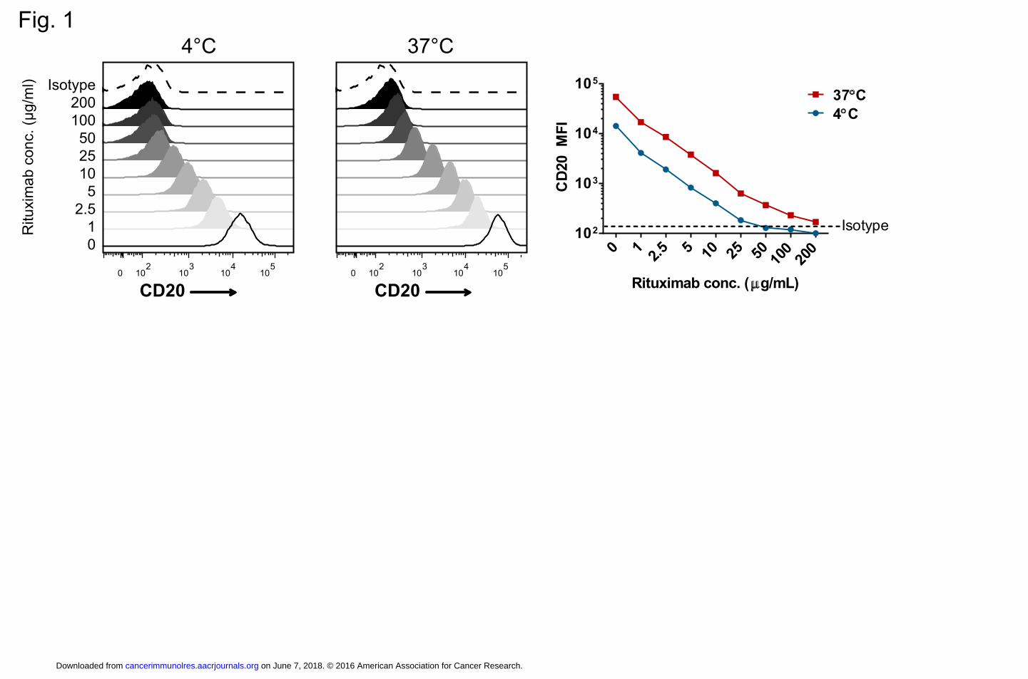
Rituximab blocks CD20 binding of antibodies used to derive CAR scFvs.
We previously reported results testing CD20-directed CARs using scFvs derived from two
different murine mAbs, Leu16 (L27) or 1F5 (9, 10, 29, 32), each of which binds to epitopes on
the large extracellular loop of the CD20 molecule (39). These CD20 epitopes overlap with the
on June 7, 2018. © 2016 American Association for Cancer Research. cancerimmunolres.aacrjournals.org Downloaded from
Author manuscripts have been peer reviewed and accepted for publication but have not yet been edited. Author Manuscript Published OnlineFirst on April 21, 2016; DOI: 10.1158/2326-6066.CIR-15-0276

11
rituximab epitope (39), and thus rituximab would be expected to block the binding of these
CARs. Using flow cytometry, we tested the ability of various concentrations of rituximab to
block binding of the Leu16 mAb coupled to phycoerythrin (Leu16-PE) to CD20 expressed on
Ramos lymphoma cells by pre-incubating these cells with rituximab before incubation with the
Leu16-PE. As expected, we found a dose-dependent blockade of CD20, with near complete
blockade with rituximab at 50 μg/ml at 4˚C. However, when Leu16-PE was incubated at the
physiologically relevant 37˚C, it bound CD20 to a small extent even at 200 μg/ml of rituximab
(Fig. 1). Similar findings were observed in experiments using the 1F5 mAb on FL-18 cells (data
not shown). Thus, rituximab binds to overlapping epitopes with our CD20 CARs and has the
potential to interfere with CAR T-cell activity against CD20+ target cells.
Impact of rituximab on in vitro function of CAR T cells
We assessed the impact of CD20 blocking by rituximab on the function of CD20 CAR T cells by
measuring proliferation, cytokine secretion, and cytotoxicity using five different CD20 CAR
lentiviral constructs after incubation with a variety of CD20+ B cell NHL cell lines. The CAR
constructs (Supplemental Fig. S1A) were the third-generation Leu16-28-BB-z-tEGFR and 1F5-
28-BBz constructs (32), the second-generation Leu16-28-z construct, and two CD20 CARs
(1.5.3-NQ-28-BB-z and 1.5.3-NQ-28-z) derived from the fully human 1.5.3 mAb to CD20,
which also binds to an overlapping epitope with rituximab (36). CAR expression was typically
achieved in 40-80% of the T cells (Supplemental Fig. S1B).
Proliferation of CFSE-labeled CAR T cells was largely unimpaired when cultured with
various NHL target cell lines (Raji, Daudi, Rec-1, and FL-18) in the presence of rituximab. CAR
T cells stimulated with target cells in the presence of rituximab at concentrations up to 200 μg/ml
exhibited > 96% of the proliferation observed after stimulation in the absence of rituximab (Fig.
on June 7, 2018. © 2016 American Association for Cancer Research. cancerimmunolres.aacrjournals.org Downloaded from
Author manuscripts have been peer reviewed and accepted for publication but have not yet been edited. Author Manuscript Published OnlineFirst on April 21, 2016; DOI: 10.1158/2326-6066.CIR-15-0276

12
2A; Supplemental Fig. S2). Cell size is another measure of T-cell activation (40). We analyzed
CAR+ T cells by flow cytometry for forward scatter as an estimate of cell size and found that
after stimulation with Raji, Daudi, or Rec-1 cells pre-incubated with rituximab, CAR T cells
exhibited a median size > 85% of the size of control cells not exposed to rituximab (Fig. 2A). T
cells incubated with FL-18 cells exhibited a slightly more pronounced but still modest reduction
in cell size after incubation with rituximab (73% of control cell size at 200 μg/ml).
In contrast to proliferation, we found that cytokine secretion by CAR T cells decreased in
the presence of increasing rituximab (Fig. 2B). However, even at 100 μg/ml of rituximab, IFNγ,
IL2, and TNFα were produced at 34-51%, 70-92%, and 79-108% of baseline, respectively.
Similar findings were observed using as targets K562 cells genetically modified to express CD80
and CD20, with CD20– K562-CD80 cells as a control to demonstrate antigen specificity of CD20
CAR T-cell activity (Supplemental Fig. S3).
We also examined the impact of rituximab on the cytolytic activity of CAR+ T cells
against various CD20+ NHL target cell lines. Using standard 51Cr-release assays with
CAR+CD8+ T cells as effectors and Raji, FL-18, Granta, or Rec-1 as targets, we found that
cytotoxicity was minimally impaired in rituximab concentrations of up to 50 μg/ml (Fig. 3), and
> 65% of baseline cytolytic activity was retained in rituximab concentrations of 100 μg/ml
against all target cell lines tested.
We tested the in vitro functionality of the fully human 1.5.3-NQ-28-z and 1.5.3-NQ-28-
BB-z CAR T cells in the presence of rituximab, and found that, as with the Leu16 and 1F5
CARs, cytokine secretion and cytotoxicity, but not proliferation, decreased modestly in a dose-
dependent manner against rituximab pre-treated target cells (Supplemental Fig. S4).
Effect of CD20 antigen expression level
on June 7, 2018. © 2016 American Association for Cancer Research. cancerimmunolres.aacrjournals.org Downloaded from
Author manuscripts have been peer reviewed and accepted for publication but have not yet been edited. Author Manuscript Published OnlineFirst on April 21, 2016; DOI: 10.1158/2326-6066.CIR-15-0276

13
We hypothesized that the amount of CD20 expression on tumor cells might impact sensitivity to
rituximab blockade and tested this by selecting K562-CD80 cell lines with low, medium, and
high CD20 expression after limiting dilution cloning (Supplemental Fig. S5). We again assessed
in vitro CAR T cell function in the presence of varying concentrations of rituximab. As with the
NHL cell lines, proliferation of CAR T cells was completely intact regardless of target cell CD20
expression (Fig. 4A). Cell size was undiminished when CD20high cells were used as targets,
although a modest reduction in cell size occurred with cells expressing lower levels of CD20. In
contrast to proliferation and cell size, cytokine secretion was significantly impaired upon
stimulation with CD20low target cells, with IFNγ, IL2, and TNFα levels as low as 5%, 17%, and
22% of baseline values, respectively, at 100-200 μg/ml of rituximab (Fig. 4B; Supplemental Fig.
S6), whereas T cells stimulated with CD20high targets retained > 75% of baseline activity at
rituximab concentrations of 100 μg/ml.
CD20 antigen density impacted the rituximab-mediated inhibition of CAR T-cell
cytolytic activity (Fig. 4C). T cell killing of target cells expressing high CD20 was minimally
impacted by rituximab, even at low E:T ratios. However, T cell cytotoxicity decreased in a dose-
dependent manner against CD20low and CD20medium K562-CD80 targets, which was most
pronounced at lower E:T ratios. Cytolytic activity against CD20low targets was retained at 47% of
baseline at a 50:1 E:T ratio at 200 μg/ml rituximab, but was only 16% of baseline at a 2:1 E:T
ratio.
In vivo antitumor activity of CD20 CAR T cells in the presence of residual rituximab
The in vitro experiments above suggested that CD20 CAR T cells retain significant functionality
against CD20+ tumors despite the presence of moderate levels of rituximab. To evaluate how
on June 7, 2018. © 2016 American Association for Cancer Research. cancerimmunolres.aacrjournals.org Downloaded from
Author manuscripts have been peer reviewed and accepted for publication but have not yet been edited. Author Manuscript Published OnlineFirst on April 21, 2016; DOI: 10.1158/2326-6066.CIR-15-0276

14
these observations would translate to the in vivo setting, we tested the impact of residual
rituximab on CAR T cell activity in a mouse lymphoma model.
Rituximab as a single agent has significant antitumor activity against Raji cells in
immunocompromised mouse xenograft models (41). To overcome a potential confounding
therapeutic effect from rituximab in these combination therapy experiments, we generated a
rituximab-refractory Raji cell line (RR-Raji) using previously described methods (27), and found
that CD20 expression was retained in this cell line (Supplemental Fig. S7).
We inoculated NSG mice i.v. with RR-Raji cells and treated some groups with high- or
low-dose rituximab 5 days after inoculation, once tumors were established. CD20 CAR+ T cells
were administered i.v. the next day (Fig. 5A). Mice that received rituximab alone demonstrated a
modest, transient antitumor effect, but all died of tumor progression by day 24, whereas mice
treated with CAR T cells alone had significant tumor regression, with tumor eradication in 40%
of mice and a doubling of median survival (52 days). Mice that received rituximab the day
before T-cell infusion did not have impaired in vivo CAR T-cell activity compared with mice
receiving CAR T cells alone; tumors were eradicated in all mice in the rituximab 200 μg/ml
group and in all but one mouse in the 25 μg/ml group (Fig. 5B and C; Supplemental Fig. S8).
To confirm that these tumor remissions occurred in the presence of physiologically
relevant serum concentrations of rituximab, we collected serum from rituximab-treated mice on
the day of T-cell infusion and one week later and measured serum rituximab. Mice receiving
rituximab at 200 μg/ml had an initial median serum rituximab concentration of 138.5 μg/ml
(range 54.5-173.6) and 39.7 μg/ml (range 1.6-51.9) a week later, and mice receiving 25 μg/ml
had a median concentration of 11.7 μg/ml (range 2.8-17.8) at baseline and 0 μg/ml at 1 week
after T-cell infusions (Fig. 5D).
on June 7, 2018. © 2016 American Association for Cancer Research. cancerimmunolres.aacrjournals.org Downloaded from
Author manuscripts have been peer reviewed and accepted for publication but have not yet been edited. Author Manuscript Published OnlineFirst on April 21, 2016; DOI: 10.1158/2326-6066.CIR-15-0276

15
We quantified relative circulating CAR T cell numbers by flow cytometry 28 days after
tumor injection. Mice receiving CAR T cells alone or rituximab plus CAR T cells had similar
numbers, suggesting that the presence of rituximab did not impair in vivo persistence of CAR T
cells (Supplemental Fig. S9).
Serum rituximab concentrations of patients treated with salvage rituximab-containing regimens
Because the ultimate goal of these experiments is to inform future CD20 CAR clinical trials, we
defined a clinically relevant range of residual serum rituximab concentrations in the intended
patient population by querying a database of patients with B-cell NHL who underwent
autologous stem cell transplantation on investigational protocols at our center and had a pre-
transplant serum rituximab measurement available (37). We identified 103 patients who received
a rituximab-containing chemotherapy regimen within 4 months of the serum blood draw (range
0.5–3.8 months, median 1.8), and the median rituximab concentration in these patients was 38.3
μg/ml, with an interquartile range of 19.1–71.7 μg/ml (Fig. 5E). The rituximab concentration was
100 μg/ml or lower in 86% of patients.
Effect of ofatumumab on CD20 CAR T-cell function
To determine the importance of epitope location on the effect of CD20 mAbs on CAR function,
we repeated the in vitro assays with ofatumumab, a fully human mAb to CD20 that binds to a
distinct epitope involving the smaller extracellular loop of CD20 as well as a different area of the
large loop (42,43).
We first evaluated the ability of ofatumumab to block binding of the Leu16 mAb by flow
cytometry and found that despite the different epitope, binding of the second antibody was
profoundly blocked by ofatumumab, at even lower concentrations than rituximab (Supplemental
on June 7, 2018. © 2016 American Association for Cancer Research. cancerimmunolres.aacrjournals.org Downloaded from
Author manuscripts have been peer reviewed and accepted for publication but have not yet been edited. Author Manuscript Published OnlineFirst on April 21, 2016; DOI: 10.1158/2326-6066.CIR-15-0276

16
Fig. S10). We then performed in vitro functional assays on Rec-1 and Raji-ffLuc lymphoma cells
that had been pre-incubated with varying concentrations of ofatumumab (Fig. 6). The results
were similar to those with rituximab, in that proliferation and cell size were minimally affected,
but cytokine production was more impacted, in a dose-dependent manner. Compared with
rituximab, cytotoxicity was more profoundly impaired in the presence of ofatumumab. These
findings suggested that the inhibitory effect of CD20 mAb is due not to direct blocking of the
CAR binding epitope, but rather from steric inhibition, and that the stronger inhibitory effect of
ofatumumab resulted from a slower off-rate compared with rituximab. This was supported by
competitive cell-binding flow cytometry studies at 4˚C or 37˚C (Supplemental Fig. S10) that
confirmed a much lower dissociation of ofatumumab, consistent with previously reported data
(44).
Discussion
In this study we addressed the theoretical concern that CD20-targeted CAR T cells may be
rendered ineffective by residual levels of rituximab. This question is relevant to future clinical
trials enrolling patients with relapsed or refractory B cell lymphomas treated recently with
rituximab-containing salvage chemotherapy, who may have significant residual serum rituximab
concentrations at the time of CAR T cell infusion.
We defined the range of serum levels of rituximab that would likely be encountered in a
CAR T cell trial by examining a large cohort of patients treated at our center who had received
rituximab-containing therapy within the previous 4 months. We found that the vast majority of
patients had rituximab of 100 μg/ml or less, with a median value of less than 40 μg/ml. Within
this range of rituximab concentrations, CD20-specific CAR T cells maintained significant
on June 7, 2018. © 2016 American Association for Cancer Research. cancerimmunolres.aacrjournals.org Downloaded from
Author manuscripts have been peer reviewed and accepted for publication but have not yet been edited. Author Manuscript Published OnlineFirst on April 21, 2016; DOI: 10.1158/2326-6066.CIR-15-0276

17
activity both in vitro and in vivo despite partial blockade of CAR binding sites, presumably due
to a significant dissociation (off-rate) of rituximab at 37˚C that permits CAR triggering to occur.
Cytokine secretion and cytotoxicity were impaired in vitro in a dose-dependent manner;
however, even at rituximab concentrations of 100 μg/ml, CAR T cells generally retained at least
half, and usually 60-70% or more of their baseline activity, while proliferation was not impacted
at up to 200 μg/ml of rituximab. Most importantly, CAR T-cell function was not impacted in
vivo in a mouse model in the presence of rituximab concentrations that were significantly higher
than the anticipated values of most human patients in future CAR T-cell trials.
The results of the mouse experiment demonstrating superior outcomes in the groups
receiving combination therapy compared with mice treated with CAR T cells alone are most
likely explained by the transient therapeutic response to rituximab that occurred, despite the
relative rituximab resistance of the Raji cell line. This resulted in a smaller tumor burden at the
time of CAR T cell infusion and likely conferred an advantage over mice receiving CAR T cells
alone. However, the possibility that a synergistic response occurred in the mice that received
both rituximab and CAR T cells cannot be excluded. Regardless, the central observation is that
CAR T cells remained active and were not impaired by the presence of rituximab. Furthermore,
these experiments also illustrate the clinically relevant point that CAR T cells are potentially
effective in treating rituximab-resistant tumors, as long as the mechanism of escape from
antibody-based therapy is not antigen loss.
The experiments with ofatumumab provide insight into the mechanism of interference of
CD20 mAb on CAR T cell function. Ofatumumab inhibited CAR T-cell function despite the fact
that it binds to a nonoverlapping epitope on CD20 compared with our CARs, suggesting that the
primary inhibitory mechanism is steric interference between CD20 and the CAR (or T cell),
on June 7, 2018. © 2016 American Association for Cancer Research. cancerimmunolres.aacrjournals.org Downloaded from
Author manuscripts have been peer reviewed and accepted for publication but have not yet been edited. Author Manuscript Published OnlineFirst on April 21, 2016; DOI: 10.1158/2326-6066.CIR-15-0276

18
rather than direct competitive blockade of the binding epitope. The functions most impacted by
the presence of ofatumumab were, in descending order, cytotoxicity (4 hour assay), cytokine
secretion (24 hour assay), and proliferation (96 hour assay). This highlights the importance of
off-rate: the slower dissociation of ofatumumab compared with rituximab manifested as
profound impairment of CAR T-cell function over a short time period, but CAR T cells in
continuous close contact with target cells over a day or more could become activated.
Our results also provide insights into CAR T cell biology, with respect to the effect of
high or low target antigen expression on CAR T-cell activation, since varying degrees of
blockade of CD20 binding sites at different rituximab concentrations may be thought of as a
surrogate for antigen density. The observation that CAR T cells effectively kill target cells in the
presence of high concentrations of rituximab, even on target cells with low CD20 expression,
suggests that very little antigen is required for CAR T-cell lytic activity, consistent with recently
reported data (45). In fact, high antigen density may be suboptimal for CAR T cell-activation, at
least with respect to cytokine secretion, because low-to-intermediate concentrations of rituximab
on target cells with high antigen expression actually increased cytokine production. This concept
that an optimal CAR-to-antigen-density ratio exists is consistent with previous reports (46).
The results presented here provide reassurance that adoptively transferred CD20-specific
CAR T cells are likely to retain major activity even in patients recently treated with rituximab,
but a further consideration is that even at high mAb concentrations sufficient to block CAR T
cell activity in vitro, complete saturation of intratumoral CD20 sites may not occur. In a clinical
trial, four patients with refractory malignant B-cell lymphomas treated with continuous
intravenous infusions of the murine 1F5 mAb to CD20 had minimal 1F5 binding to malignant
lymph node sites at low-to-intermediate serum concentrations (28). Only at high concentrations
on June 7, 2018. © 2016 American Association for Cancer Research. cancerimmunolres.aacrjournals.org Downloaded from
Author manuscripts have been peer reviewed and accepted for publication but have not yet been edited. Author Manuscript Published OnlineFirst on April 21, 2016; DOI: 10.1158/2326-6066.CIR-15-0276

19
(>150 μg/ml) was significant Ab binding observed, suggesting poor penetration of the mAb into
tumors. It is possible that T cells, by virtue of their ability to extravasate and migrate actively
through lymphoid tissue, could access tumor cells in areas not penetrated by Ab, thus further
circumventing the potential negative impact of competitively binding Ab therapy.
Although these experiments were limited to the evaluation of the CD20 antigen, our
results have potential implications for patients receiving CAR T-cell therapy targeted against any
antigen previously targeted with a therapeutic Ab. We provide proof-of-principle that Ab therapy
targeting the same epitope as a CAR does not necessarily preclude using CAR T cells afterwards.
This concept is likely to become increasingly relevant as more mAb and mAb-drug conjugates
are used to treat a variety of malignancies for which CAR T cells may also be employed.
In current clinical practice rituximab is the most relevant mAb for CD20 CAR T cells,
but other CD20 mAbs are becoming more routinely used. The experiments reported here may
need to be repeated in the future using these alternative mAb, particularly those with slower off-
rates than rituximab, since our experiments with ofatumumab suggest that they may have a more
inhibitory effect than rituximab on CD20 CAR T-cell function.
In summary, our results suggest that residual rituximab, within the range of
concentrations likely to be encountered in B-cell lymphoma patients entering CAR T cell clinical
trials, leads to a modest reduction of CAR T-cell function in vitro, but in vivo antitumor activity
remains intact. Thus, prior therapy with rituximab is unlikely to be a major barrier to treatment
with CD20 CAR T cells in most patients with lymphoma.
Acknowledgments
on June 7, 2018. © 2016 American Association for Cancer Research. cancerimmunolres.aacrjournals.org Downloaded from
Author manuscripts have been peer reviewed and accepted for publication but have not yet been edited. Author Manuscript Published OnlineFirst on April 21, 2016; DOI: 10.1158/2326-6066.CIR-15-0276

20
BGT is a Damon Runyon-Pfizer Clinical Investigator (Grant 49-C10). This work was also
supported by the Giuliani Family Foundation, NIH/NCI K23CA154874, NIH/NCI Cancer Center
Support Grant P30CA015704, NIDDK DK56465, NIH/NCI P01CA044991, NIH/NCI
K24CA184039, NIH/NCI K08CA151682, and Lymphoma Research Foundation MCL-1003440.
References 1. Brentjens RJ, Davila ML, Riviere I, Park J, Wang X, Cowell LG, et al. CD19-Targeted T
Cells Rapidly Induce Molecular Remissions in Adults with Chemotherapy-Refractory Acute Lymphoblastic Leukemia. Sci Transl Med 2013;5(177):177ra38.
2. Haso W, Lee DW, Shah NN, Stetler-Stevenson M, Yuan CM, Pastan IH, et al. Anti-CD22-chimeric antigen receptors targeting B-cell precursor acute lymphoblastic leukemia. Blood 2013;121(7):1165-74.
3. James SE, Greenberg PD, Jensen MC, Lin Y, Wang J, Till BG, et al. Antigen sensitivity of CD22-specific chimeric TCR is modulated by target epitope distance from the cell membrane. J Immunol 2008;180(10):7028-38.
4. Kalos M, Levine BL, Porter DL, Katz S, Grupp SA, Bagg A, et al. T cells with chimeric antigen receptors have potent antitumor effects and can establish memory in patients with advanced leukemia. Sci Transl Med 2011;3(95):95ra73.
5. Kochenderfer JN, Dudley ME, Kassim SH, Somerville RP, Carpenter RO, Stetler-Stevenson M, et al. Chemotherapy-refractory diffuse large B-cell lymphoma and indolent B-cell malignancies can be effectively treated with autologous T cells expressing an anti-CD19 chimeric antigen receptor. J Clin Oncol 2015;33(6):540-9.
6. Lee DW, Kochenderfer JN, Stetler-Stevenson M, Cui YK, Delbrook C, Feldman SA, et al. T cells expressing CD19 chimeric antigen receptors for acute lymphoblastic leukaemia in children and young adults: a phase 1 dose-escalation trial. Lancet 2015;385(9967):517-28.
7. Porter DL, Hwang WT, Frey NV, Lacey SF, Shaw PA, Loren AW, et al. Chimeric antigen receptor T cells persist and induce sustained remissions in relapsed refractory chronic lymphocytic leukemia. Sci Transl Med 2015;7(303):303ra139.
8. Savoldo B, Ramos CA, Liu E, Mims MP, Keating MJ, Carrum G, et al. CD28 costimulation improves expansion and persistence of chimeric antigen receptor-modified T cells in lymphoma patients. J Clin Invest 2011;121(5):1822-6.
9. Till BG, Jensen MC, Wang J, Chen EY, Wood BL, Greisman HA, et al. Adoptive immunotherapy for indolent non-Hodgkin lymphoma and mantle cell lymphoma using genetically modified autologous CD20-specific T cells. Blood 2008;112(6):2261-71.
10. Till BG, Jensen MC, Wang J, Qian X, Gopal AK, Maloney DG, et al. CD20-specific adoptive immunotherapy for lymphoma using a chimeric antigen receptor with both CD28 and 4-1BB domains: pilot clinical trial results. Blood 2012;119(17):3940-50.
on June 7, 2018. © 2016 American Association for Cancer Research. cancerimmunolres.aacrjournals.org Downloaded from
Author manuscripts have been peer reviewed and accepted for publication but have not yet been edited. Author Manuscript Published OnlineFirst on April 21, 2016; DOI: 10.1158/2326-6066.CIR-15-0276

21
11. Coiffier B, Lepage E, Briere J, Herbrecht R, Tilly H, Bouabdallah R, et al. CHOP chemotherapy plus rituximab compared with CHOP alone in elderly patients with diffuse large-B-cell lymphoma. N Engl J Med 2002;346(4):235-42.
12. Lenz G, Dreyling M, Hoster E, Wormann B, Duhrsen U, Metzner B, et al. Immunochemotherapy with rituximab and cyclophosphamide, doxorubicin, vincristine, and prednisone significantly improves response and time to treatment failure, but not long-term outcome in patients with previously untreated mantle cell lymphoma: results of a prospective randomized trial of the German Low Grade Lymphoma Study Group (GLSG). J Clin Oncol 2005;23(9):1984-92.
13. Marcus R, Imrie K, Solal-Celigny P, Catalano JV, Dmoszynska A, Raposo JC, et al. Phase III study of R-CVP compared with cyclophosphamide, vincristine, and prednisone alone in patients with previously untreated advanced follicular lymphoma. J Clin Oncol 2008;26(28):4579-86.
14. Pfreundschuh M, Kuhnt E, Trumper L, Osterborg A, Trneny M, Shepherd L, et al. CHOP-like chemotherapy with or without rituximab in young patients with good-prognosis diffuse large-B-cell lymphoma: 6-year results of an open-label randomised study of the MabThera International Trial (MInT) Group. Lancet Oncol 2011;12(11):1013-22.
15. Pulczynski S, Boesen AM, Jensen OM. Antibody-induced modulation and intracellular transport of CD10 and CD19 antigens in human B-cell lines: an immunofluorescence and immunoelectron microscopy study. Blood 1993;81(6):1549-57.
16. Press OW, Howell-Clark J, Anderson S, Bernstein I. Retention of B-cell-specific monoclonal antibodies by human lymphoma cells. Blood 1994;83(5):1390-7.
17. Pulczynski S, Boesen AM, Jensen OM. Modulation and intracellular transport of CD20 and CD21 antigens induced by B1 and B2 monoclonal antibodies in RAJI and JOK-1 cells--an immunofluorescence and immunoelectron microscopy study. Leuk Res 1994;18(7):541-52.
18. Grupp SA, Kalos M, Barrett D, Aplenc R, Porter DL, Rheingold SR, et al. Chimeric antigen receptor-modified T cells for acute lymphoid leukemia. N Engl J Med 2013;368(16):1509-18.
19. Gall JM, Davol PA, Grabert RC, Deaver M, Lum LG. T cells armed with anti-CD3 x anti-CD20 bispecific antibody enhance killing of CD20+ malignant B cells and bypass complement-mediated rituximab resistance in vitro. Exp Hematol 2005;33(4):452-9.
20. Jensen M, Tan G, Forman S, Wu AM, Raubitschek A. CD20 is a molecular target for scFvFc:zeta receptor redirected T cells: implications for cellular immunotherapy of CD20+ malignancy. Biology of blood and marrow transplantation : journal of the American Society for Blood and Marrow Transplantation 1998;4(2):75-83.
21. Jensen MC, Cooper LJ, Wu AM, Forman SJ, Raubitschek A. Engineered CD20-specific primary human cytotoxic T lymphocytes for targeting B-cell malignancy. Cytotherapy 2003;5(2):131-8.
22. Hombach A, Heuser C, Gerken M, Fischer B, Lewalter K, Diehl V, et al. T cell activation by recombinant FcepsilonRI gamma-chain immune receptors: an extracellular spacer domain impairs antigen-dependent T cell activation but not antigen recognition. Gene Ther 2000;7(12):1067-75.
on June 7, 2018. © 2016 American Association for Cancer Research. cancerimmunolres.aacrjournals.org Downloaded from
Author manuscripts have been peer reviewed and accepted for publication but have not yet been edited. Author Manuscript Published OnlineFirst on April 21, 2016; DOI: 10.1158/2326-6066.CIR-15-0276

22
23. Hombach A, Koch D, Sircar R, Heuser C, Diehl V, Kruis W, et al. A chimeric receptor that selectively targets membrane-bound carcinoembryonic antigen (mCEA) in the presence of soluble CEA. Gene Ther 1999;6(2):300-4.
24. Nolan KF, Yun CO, Akamatsu Y, Murphy JC, Leung SO, Beecham EJ, et al. Bypassing immunization: optimized design of "designer T cells" against carcinoembryonic antigen (CEA)-expressing tumors, and lack of suppression by soluble CEA. Clin Cancer Res 1999;5(12):3928-41.
25. Westwood JA, Murray WK, Trivett M, Haynes NM, Solomon B, Mileshkin L, et al. The Lewis-Y carbohydrate antigen is expressed by many human tumors and can serve as a target for genetically redirected T cells despite the presence of soluble antigen in serum. J Immunother 2009;32(3):292-301.
26. James SE, Orgun NN, Tedder TF, Shlomchik MJ, Jensen MC, Lin Y, et al. Antibody-mediated B-cell depletion before adoptive immunotherapy with T cells expressing CD20-specific chimeric T-cell receptors facilitates eradication of leukemia in immunocompetent mice. Blood 2009;114(27):5454-63.
27. Czuczman MS, Olejniczak S, Gowda A, Kotowski A, Binder A, Kaur H, et al. Acquirement of rituximab resistance in lymphoma cell lines is associated with both global CD20 gene and protein down-regulation regulated at the pretranscriptional and posttranscriptional levels. Clin Cancer Res 2008;14(5):1561-70.
28. Press OW, Appelbaum F, Ledbetter JA, Martin PJ, Zarling J, Kidd P, et al. Monoclonal antibody 1F5 (anti-CD20) serotherapy of human B cell lymphomas. Blood 1987;69(2):584-91.
29. Wang J, Jensen M, Lin Y, Sui X, Chen E, Lindgren CG, et al. Optimizing Adoptive Polyclonal T Cell Immunotherapy of Lymphomas, Using a Chimeric T Cell Receptor Possessing CD28 and CD137 Costimulatory Domains. Hum Gene Ther 2007;18(8):712-25.
30. Wang J, Press OW, Lindgren CG, Greenberg P, Riddell S, Qian X, et al. Cellular immunotherapy for follicular lymphoma using genetically modified CD20-specific CD8+ cytotoxic T lymphocytes. Mol Ther 2004;9(4):577-86.
31. Hudecek M, Lupo-Stanghellini MT, Kosasih PL, Sommermeyer D, Jensen MC, Rader C, et al. Receptor Affinity and Extracellular Domain Modifications Affect Tumor Recognition by ROR1-Specific Chimeric Antigen Receptor T Cells. Clin Cancer Res 2013.
32. Budde LE, Berger C, Lin Y, Wang J, Lin X, Frayo SE, et al. Combining a CD20 Chimeric Antigen Receptor and an Inducible Caspase 9 Suicide Switch to Improve the Efficacy and Safety of T Cell Adoptive Immunotherapy for Lymphoma. PLoS One 2013;8(12):e82742.
33. Becker PS, Taylor JA, Trobridge GD, Zhao X, Beard BC, Chien S, et al. Preclinical correction of human Fanconi anemia complementation group A bone marrow cells using a safety-modified lentiviral vector. Gene Ther 2010;17(10):1244-52.
34. Hudecek M, Sommermeyer D, Kosasih PL, Silva-Benedict A, Liu L, Rader C, et al. The Nonsignaling Extracellular Spacer Domain of Chimeric Antigen Receptors Is Decisive for In Vivo Antitumor Activity. Cancer immunology research 2014.
35. Hombach A, Hombach AA, Abken H. Adoptive immunotherapy with genetically engineered T cells: modification of the IgG1 Fc 'spacer' domain in the extracellular
on June 7, 2018. © 2016 American Association for Cancer Research. cancerimmunolres.aacrjournals.org Downloaded from
Author manuscripts have been peer reviewed and accepted for publication but have not yet been edited. Author Manuscript Published OnlineFirst on April 21, 2016; DOI: 10.1158/2326-6066.CIR-15-0276

23
moiety of chimeric antigen receptors avoids 'off-target' activation and unintended initiation of an innate immune response. Gene Ther 2010;17(10):1206-13.
36. Bornstein GG, Queva C, Tabrizi M, van Abbema A, Chavez C, Wang P, et al. Development of a new fully human anti-CD20 monoclonal antibody for the treatment of B-cell malignancies. Invest New Drugs 2010;28(5):561-74.
37. Gopal AK, Press OW, Wilbur SM, Maloney DG, Pagel JM. Rituximab blocks binding of radiolabeled anti-CD20 antibodies (Ab) but not radiolabeled anti-CD45 Ab. Blood 2008;112(3):830-5.
38. Maloney DG, Grillo-Lopez AJ, White CA, Bodkin D, Schilder RJ, Neidhart JA, et al. IDEC-C2B8 (Rituximab) anti-CD20 monoclonal antibody therapy in patients with relapsed low-grade non-Hodgkin's lymphoma. Blood 1997;90(6):2188-95.
39. Polyak MJ, Deans JP. Alanine-170 and proline-172 are critical determinants for extracellular CD20 epitopes; heterogeneity in the fine specificity of CD20 monoclonal antibodies is defined by additional requirements imposed by both amino acid sequence and quaternary structure. Blood 2002;99(9):3256-62.
40. Grumont R, Lock P, Mollinari M, Shannon FM, Moore A, Gerondakis S. The mitogen-induced increase in T cell size involves PKC and NFAT activation of Rel/NF-kappaB-dependent c-myc expression. Immunity 2004;21(1):19-30.
41. Hernandez-Ilizaliturri FJ, Jupudy V, Ostberg J, Oflazoglu E, Huberman A, Repasky E, et al. Neutrophils contribute to the biological antitumor activity of rituximab in a non-Hodgkin's lymphoma severe combined immunodeficiency mouse model. Clin Cancer Res 2003;9(16 Pt 1):5866-73.
42. Du J, Yang H, Guo Y, Ding J. Structure of the Fab fragment of therapeutic antibody Ofatumumab provides insights into the recognition mechanism with CD20. Mol Immunol 2009;46(11-12):2419-23.
43. Teeling JL, Mackus WJ, Wiegman LJ, van den Brakel JH, Beers SA, French RR, et al. The biological activity of human CD20 monoclonal antibodies is linked to unique epitopes on CD20. J Immunol 2006;177(1):362-71.
44. Teeling JL, French RR, Cragg MS, van den Brakel J, Pluyter M, Huang H, et al. Characterization of new human CD20 monoclonal antibodies with potent cytolytic activity against non-Hodgkin lymphomas. Blood 2004;104(6):1793-800.
45. Watanabe K, Terakura S, Martens AC, van Meerten T, Uchiyama S, Imai M, et al. Target Antigen Density Governs the Efficacy of Anti-CD20-CD28-CD3 zeta Chimeric Antigen Receptor-Modified Effector CD8+ T Cells. J Immunol 2014.
46. Alvarez-Vallina L, Russell SJ. Efficient discrimination between different densities of target antigen by tetracycline-regulatable T bodies. Hum Gene Ther 1999;10(4):559-63.
on June 7, 2018. © 2016 American Association for Cancer Research. cancerimmunolres.aacrjournals.org Downloaded from
Author manuscripts have been peer reviewed and accepted for publication but have not yet been edited. Author Manuscript Published OnlineFirst on April 21, 2016; DOI: 10.1158/2326-6066.CIR-15-0276

24
Figure Legends
Figure 1. Rituximab blocks antigen binding of Ab used to derive CAR scFv. Ramos cells
(CD20+) were incubated with the indicated rituximab concentrations for 30 minutes, followed by
incubation with PE-labeled anti-CD20 Ab (clone Leu16) or isotype control at either 4˚C or 37˚C
for 30 minutes. Cells were washed and analyzed by flow cytometry to determine available CD20
binding sites as measured by PE fluorescence intensity. The right panel summarizes the
geometric mean fluorescence intensity (MFI) at either 4˚C or 37˚C as a function of rituximab
concentration. The data are representative of 3 independent experiments.
Figure 2. Effect of rituximab on CAR T cell function in vitro. The indicated B-cell NHL cell
lines were irradiated and incubated for 30 minutes at room temperature with varying rituximab
concentrations (at 2x the concentrations during incubation to yield the indicated final
concentrations after addition of T cells). CFSE-stained T cells expressing the Leu16-28-BB-z-
tEGFR CD20-specific CAR were added to the target cells at a 1:1 volume and ratio. Proliferation
of the T cells was analyzed 4 days later by flow cytometry for CFSE dilution (A). The percent
divided CD3+ T cells relative to unstimulated T cells are shown on the left axis (filled bars).
Histograms of CFSE intensity are shown in Supplemental Figure 2. Cell size of CD3+ T cells as
determined by geometric mean of forward scatter (subtracting size of cells in media only) is
shown on the right axis (open bars). (B) Cytokine secretion of these T cells was measured by
Luminex assay using supernatants from 24 hours after restimulation. IL2 concentrations are
shown on the left y-axis and IFN-γ and TNF-α on the right y-axis. The data shown are
representative of 3 independent experiments.
on June 7, 2018. © 2016 American Association for Cancer Research. cancerimmunolres.aacrjournals.org Downloaded from
Author manuscripts have been peer reviewed and accepted for publication but have not yet been edited. Author Manuscript Published OnlineFirst on April 21, 2016; DOI: 10.1158/2326-6066.CIR-15-0276

25
Figure 3. Effect of rituximab on CAR T cell-mediated cytotoxicity. The indicated 51Cr-labeled
target cells were pre-incubated for 30 minutes with rituximab (at 2x the concentrations during
incubation to yield the indicated final concentrations after addition of T cells), and then CD8+ T
cells expressing the Leu16-28-z CAR were added at the E:T ratios shown in a standard 5-hour
51chromium-release assay. Mock-transduced T cells, and samples with rituximab and target cells
only (“0:1”) were used as negative controls. The average value of duplicate wells is shown, with
error bars representing standard deviation. The data are representative of results from 4
independent experiments.
Figure 4. Sensitivity to rituximab blockade is dependent on CD20 antigen density on target cells.
K562 cells transduced with CD80 and CD20 (“K80-20”) were cloned by limiting dilution,
selected for high, medium, or low levels of CD20 expression (Supplemental Figure 5), and used
as target cells in assays for (A) proliferation and cell size (geometric mean forward scatter of
gated CD3+ cells minus the size of cells in media only) using CFSE-labeled Leu16-28-z CAR-
transduced T cells as described in Figure 2; (B) cytokine secretion of the Leu16-28-z CAR-
transduced T cells at 24 hours from (A) above, measured by Luminex assay; and (C) cytotoxicity
using Leu16-28-z CAR-transduced CD8+ T cells by 51Cr-release assay as described in Figure 3.
Data are representative of 3 independent experiments. Absolute values for cytokine secretion are
shown in Supplemental Figure 6.
Figure 5. In vivo effect of rituximab on CD20 CAR T cell function. NSG mice were injected i.v.
with 5 x 10⁵ rituximab-refractory Raji-ffLuc lymphoma cells, followed by one of the following
treatments: no treatment, rituximab only (25 μg or 200) μg i.p 5 days later, 107 1.5.3-NQ-28-BBz
on June 7, 2018. © 2016 American Association for Cancer Research. cancerimmunolres.aacrjournals.org Downloaded from
Author manuscripts have been peer reviewed and accepted for publication but have not yet been edited. Author Manuscript Published OnlineFirst on April 21, 2016; DOI: 10.1158/2326-6066.CIR-15-0276

26
CAR T cells only 6 days after tumor, or rituximab 25 or 200 i.p. at 5 days followed by 107 CAR
T cells at 6 days after tumor. Mice were imaged twice weekly for bioluminescence. A) Schema
of mouse experiment. B) Average tumor burden per group over time as measured by total body
bioluminescence. The geometric mean luminescence values with 95% confidence intervals are
shown, and to prevent misleading fluctuations in tumor volume graphs, the last bioluminescence
level of each mouse was carried forward after it was killed until no mice in that group remained.
Individual bioluminescence traces are shown in Supplemental Figure 8. C) Kaplan-Meier plot
showing overall survival of each treatment group. D) Serum rituximab levels on the day of T cell
infusion (day 6) and 1 week post T cell infusion (day 13). The red lines denote the median values
for each group of mice. E) Serum rituximab concentrations from lymphoma patients who
underwent rituximab-containing salvage chemotherapy within the 4 preceding months. The red
line indicates the median, and black lines indicate the interquartile range (25-75%).
Figure 6. Effect of ofatumumab on CD20 CAR T cell function in vitro. Irradiated Rec-1 or Raji-
ffLuc cells (A and B) or nonirradiated 51Cr-labeled Rec-1 cells (C) were pre-incubated for 30
minutes with 2x the indicated concentrations of ofatumumab, followed by experiments to
determine function of T cells expressing the 1.5.3-NQ-28-BB-z CAR, using the
methodologies described in the legend of Figs. 2 and 3. (A) The percent divided CD3+ T cells
relative to unstimulated T cells are shown on the left axis (filled bars). Cell size of CD3+ T cells
as determined by geometric mean of forward scatter (subtracting size of cells in media only) is
shown on the right axis (open bars). (B) Cytokine secretion of these T cells was measured by
Luminex assay using supernatants from 24 hours after restimulation. IL2 concentrations are
shown on the left y-axis and IFNγ on the right y-axis. (C) Cytotoxicity of 1.5.3-NQ-28-BB-z
on June 7, 2018. © 2016 American Association for Cancer Research. cancerimmunolres.aacrjournals.org Downloaded from
Author manuscripts have been peer reviewed and accepted for publication but have not yet been edited. Author Manuscript Published OnlineFirst on April 21, 2016; DOI: 10.1158/2326-6066.CIR-15-0276

27
CAR T cells was determined using a standard 4-hour 51Cr-release assay with Rec-1 target cells.
The average value of duplicate wells is shown, with error bars representing standard deviation.
The data shown each represent a single experiment, with assessment of Rec-1 and Raji targets in
independent experiments.
on June 7, 2018. © 2016 American Association for Cancer Research. cancerimmunolres.aacrjournals.org Downloaded from
Author manuscripts have been peer reviewed and accepted for publication but have not yet been edited. Author Manuscript Published OnlineFirst on April 21, 2016; DOI: 10.1158/2326-6066.CIR-15-0276
Isotype
100
2550
105
2.5
0
200
1
4°C
CD20
37°C
CD20
Ritu
xim
ab c
onc.
(μg/
ml)
0 1 2.5 5 10 25 50 100
200
102
103
104
105
Rituximab conc. (µg/mL)
CD2
0 M
FI
4°C37°C
Isotype
Fig. 1
on June 7, 2018. © 2016 American Association for Cancer Research. cancerimmunolres.aacrjournals.org Downloaded from
Author manuscripts have been peer reviewed and accepted for publication but have not yet been edited. Author Manuscript Published OnlineFirst on April 21, 2016; DOI: 10.1158/2326-6066.CIR-15-0276

Mock 0 10 25 5010
020
00
20
40
60
80
100
0.0
0.5
1.0
1.5
2.0
2.5
Raji
% d
ivid
ed
FSC-A (x 10,000)
Rituximab (µg/ml)Mock 0 10 25 50
100
200
0
20
40
60
80
100
0.0
0.5
1.0
1.5
2.0
2.5
Daudi
% d
ivid
ed
FSC-A (x 10,000)
Rituximab (µg/ml)Mock 0 10 25 50
100
200
0
20
40
60
80
100
0.0
0.5
1.0
1.5
2.0Rec-1
% d
ivid
ed
FSC-A (x 10,000)
Rituximab (µg/ml)Mock 0 10 25 50
100
200
0
20
40
60
80
100
0.0
0.5
1.0
1.5
2.0
FL-18
% d
ivid
ed
FSC-A (x 10,000)
Rituximab (µg/ml)
A
B
Media
Mock 0 10 25 50100 200
0
200
400
600
800
1000
0
100
200
300
400Raji
pg/m
l pg/ml
IFN-γIL-2TNF-α
Rituximab (µg/ml)Med
iaMock 0 10 25 50 10
020
00
200
400
600
800
0
100
200
300Daudi
pg/m
l pg/ml
IFN-γIL-2TNF-α
Rituximab (µg/ml)Med
iaMock 0 10 25 50 10
020
00
500
1000
1500
2000
0
100
200
300
400
500Rec-1
pg/m
l pg/ml
IFN-γIL-2TNF-α
Rituximab (µg/ml) Media
Mock 0 10 25 50 100
200
0
200
400
600
0
100
200
300FL-18
pg/m
l pg/ml
IFN-γIL-2TNF-α
Rituximab (µg/ml)
Fig. 2
on June 7, 2018. © 2016 American Association for Cancer Research. cancerimmunolres.aacrjournals.org Downloaded from
Author manuscripts have been peer reviewed and accepted for publication but have not yet been edited. Author Manuscript Published OnlineFirst on April 21, 2016; DOI: 10.1158/2326-6066.CIR-15-0276
Mock 0 10 25 50 100 20
00
20
40
60
80
Raji
% s
pecif
ic c
yto
toxic
ity 50:1
10:1
2:1
0:1
Rituximab (µg/ml)Mock 0 10 25 50 100 20
00
20
40
60
80
FL-18
% s
pecif
ic c
yto
toxic
ity
Rituximab (µg/ml)
Mock 0 10 25 50 100 20
00
20
40
60
80
Granta
% s
pecif
ic c
yto
toxic
ity
Rituximab (µg/ml)Mock 0 10 25 50 100 20
00
20
40
60
80
Rec-1
% s
pecif
ic c
yto
toxic
ity
Rituximab (µg/ml)
Fig. 3
on June 7, 2018. © 2016 American Association for Cancer Research. cancerimmunolres.aacrjournals.org Downloaded from
Author manuscripts have been peer reviewed and accepted for publication but have not yet been edited. Author Manuscript Published OnlineFirst on April 21, 2016; DOI: 10.1158/2326-6066.CIR-15-0276

A
B
C
Mock 0 10 25 50 100
200
0
50
100
IFN-γ
% o
f no
ritux
imab
MediaK80K80-20low
CAR T cells + target+ indicated rituximab conc. ( µg/ml)
K80-20med
K80-20high
Mock 0 10 25 5010
020
0 0
50
100
IL-2
% o
f no
ritux
imab
CAR T cells + target+ indicated rituximab conc. ( µg/ml)
Mock 0 10 25 5010
020
0 0
50
100TNF-α
% o
f no
ritux
imab
CAR T cells + target+ indicated rituximab conc. ( µg/ml)
mock 0 10 25 5010
020
00
1
2
3Cell size
FSC-
A (x
10,
000)
K80K80-20low
K80-20med
K80-20high
CAR T cells + target+ indicated rituximab conc. ( µg/ml)
mock 0 10 25 5010
020
00
50
100Proliferation
% d
ivid
ed
MediaK80K80-20low
K80-20med
K80-20high
CAR T cells + target+ indicated rituximab conc. ( µg/ml)
Mock
CAR + K80 0 10 25 50 100
200
0
50
100
15050:1
% o
f no
ritux
imab
K80-20low
K80-20med
K80-20high
CAR+ T cells + K80-20+ indicated rituximab conc. ( µg/ml)
Mock
CAR + K80 0 10 25 50 100
200
0
50
100
15010:1
% o
f no
ritux
imab
CAR+ T cells + K80-20+ indicated rituximab conc. ( µg/ml)
Mock
CAR + K80 0 10 25 50 100
200
0
50
100
1502:1
% o
f no
ritux
imab
CAR+ T cells + K80-20+ indicated rituximab conc. ( µg/ml)
Fig. 4
on June 7, 2018. © 2016 American Association for Cancer Research. cancerimmunolres.aacrjournals.org Downloaded from
Author manuscripts have been peer reviewed and accepted for publication but have not yet been edited. Author Manuscript Published OnlineFirst on April 21, 2016; DOI: 10.1158/2326-6066.CIR-15-0276

A
B C
D E
Day 0 5 6
RR Raji-!Luc
i.v.
Ritux-
imab
i.p.
CAR
T cells
i.v.
Bioluminescent imaging
0
50
100
150
200
250
Rit
ux
ima
b (µ
g/m
l)
25 µg 200 µg 25 µg 200 µg 0
50
100
150
200
Rit
uxim
ab
(µ
g/m
l) Day 6Day 13
Rituximabonly
Rituximab +CAR T cells
0 10 20 30 40 50 60 70 800
25
50
75
100
Days After Tumor
Perc
en
t s
urv
iva
l
0 10 20 30 40 50 60 70 80106
107
108
109
1010
Days After Tumor
To
tal b
od
y F
lux (
p/s
)
No treatment
Rituximab 25 µg
Rituximab 200 µg
CAR T cells only
Ritux 25 µg + CAR T cells
Ritux 200 µg + CAR T cells
No Tumor
Fig. 5
on June 7, 2018. © 2016 American Association for Cancer Research. cancerimmunolres.aacrjournals.org Downloaded from
Author manuscripts have been peer reviewed and accepted for publication but have not yet been edited. Author Manuscript Published OnlineFirst on April 21, 2016; DOI: 10.1158/2326-6066.CIR-15-0276

Published OnlineFirst April 21, 2016.Cancer Immunol Res Gregory A. Rufener, Oliver W Press, Philip Olsen, et al. Receptor-Expressing T cells in the Presence of RituximabPreserved Activity of CD20-Specific Chimeric Antigen
Updated version
10.1158/2326-6066.CIR-15-0276doi:
Access the most recent version of this article at:
Material
Supplementary
http://cancerimmunolres.aacrjournals.org/content/suppl/2016/04/20/2326-6066.CIR-15-0276.DC1
Access the most recent supplemental material at:
Manuscript
Authoredited. Author manuscripts have been peer reviewed and accepted for publication but have not yet been
E-mail alerts related to this article or journal.Sign up to receive free email-alerts
Subscriptions
Reprints and
To order reprints of this article or to subscribe to the journal, contact the AACR Publications
Permissions
Rightslink site. Click on "Request Permissions" which will take you to the Copyright Clearance Center's (CCC)
.http://cancerimmunolres.aacrjournals.org/content/early/2016/04/21/2326-6066.CIR-15-0276To request permission to re-use all or part of this article, use this link
on June 7, 2018. © 2016 American Association for Cancer Research. cancerimmunolres.aacrjournals.org Downloaded from
Author manuscripts have been peer reviewed and accepted for publication but have not yet been edited. Author Manuscript Published OnlineFirst on April 21, 2016; DOI: 10.1158/2326-6066.CIR-15-0276