in vitro characterization of the anti-pd-1 antibody in...
TRANSCRIPT

Research Article
In Vitro Characterization of the Anti-PD-1 AntibodyNivolumab, BMS-936558, and In Vivo Toxicology inNon-Human Primates
Changyu Wang1, Kent B. Thudium1, Minhua Han1, Xi-Tao Wang1, Haichun Huang1, Diane Feingersh1,Candy Garcia1, Yi Wu1, Michelle Kuhne1, Mohan Srinivasan1, Sujata Singh1, Susan Wong1, Neysa Garner1,Heidi Leblanc1, R. Todd Bunch2, Diann Blanset3, Mark J. Selby1, and Alan J. Korman1
AbstractThe programmed death-1 (PD-1) receptor serves as an immunologic checkpoint, limiting bystander tissue
damage and preventing the development of autoimmunity during inflammatory responses. PD-1 is expressed byactivated T cells and downmodulates T-cell effector functions upon binding to its ligands, PD-L1 and PD-L2, onantigen-presenting cells. In patientswith cancer, the expression of PD-1 on tumor-infiltrating lymphocytes and itsinteraction with the ligands on tumor and immune cells in the tumor microenvironment undermine antitumorimmunity and support its rationale for PD-1 blockade in cancer immunotherapy. This report details thedevelopment and characterization of nivolumab, a fully human IgG4 (S228P) anti-PD-1 receptor-blockingmonoclonal antibody. Nivolumab binds to PD-1 with high affinity and specificity, and effectively inhibits theinteraction between PD-1 and its ligands. In vitro assays demonstrated the ability of nivolumab to potentlyenhance T-cell responses and cytokine production in the mixed lymphocyte reaction and superantigen orcytomegalovirus stimulation assays. No in vitro antibody-dependent cell-mediated or complement-dependentcytotoxicity was observedwith the use of nivolumab and activated T cells as targets. Nivolumab treatment did notinduce adverse immune-related eventswhen given to cynomolgusmacaques at high concentrations, independentof circulating anti-nivolumab antibodies where observed. These data provide a comprehensive preclinicalcharacterization of nivolumab, for which antitumor activity and safety have been demonstrated in humanclinical trials in various solid tumors. Cancer Immunol Res; 2(9); 846–56. �2014 AACR.
IntroductionCancer can be considered as an inability of the host to
eliminate transformed cells. Although the immune system isthe principal mechanism of cancer prevention, transformedcells counteract immunosurveillance. Natural controlmechan-isms that limit T-cell activation, thereby preventing collateraldamage from unrestrained T-cell activity, may be exploited bytumors to evade immune responses (1). Restoring the capacityof immune effector cells—especially T cells—to recognize andeliminate cancer is the goal of immunotherapy. The concept ofinhibitory receptor blockade, also known as checkpoint block-ade, has been validated in humanswith the approval of the anti-CTLA-4 antibody ipilimumab for metastatic melanoma (2, 3).
Programmed death-1 (PD-1) is an additional inhibitoryreceptor expressed by T cells. Engagement of PD-1 by itsligands, PD-L1 and PD-L2, induces an inhibitory signal result-ing in reduced T-cell proliferation, cytokine production, andcytotoxic activity (4, 5). PD-1 deletion in mice can lead toautoimmunity (6, 7), most notably when bred onto back-grounds of autoimmune-susceptible mouse strains (8). Elevat-ed PD-1 expression on T cells, observed during chronic viralinfections in humans and mice, is associated with reduced T-cell functionality or "exhaustion." T cells become progressivelymore nonresponsive as they express additional inhibitoryreceptors (9). Tumor-infiltrating T cells may also be function-ally inert, due in part to the expression of PD-1 alongwith otherinhibitory receptors (10, 11). In multiple syngeneic mousetumor models, blockade of PD-1 or its ligands promotesantitumor activity (12–14); anti-PD-1 activity in vivo can beenhanced by combination with antibodies to other T-cellnegative regulators, such as CTLA-4 and LAG-3 (15–17).
PD-L1 is expressed by many human tumors, includingmelanoma, lung, and kidney (10, 18, 19). PD-L1 engagementof PD-1 may be one mechanism whereby tumors evade immu-nosurveillance by directly limiting effector T-cell activity. Sev-eral studies support the notion that PD-L1 expression and,in some cases, PD-L2 expression is associated with tumoraggressiveness and adverse patient outcome (14, 20–22).
1BiologicsDiscoveryCalifornia, Bristol-Myers SquibbCompany, RedwoodCity, California; 2Bristol-Myers Squibb Company, Mount Vernon, Indiana;and 3Medarex, Princeton, New Jersey
Note: Supplementary data for this article are available at Cancer Immu-nology Research Online (http://cancerimmunolres.aacrjournals.org/).
Corresponding Author: Alan J. Korman, Bristol-Myers Squibb, 700 BayRoad, Redwood City, CA 94063. Phone: 650-260-9586; Fax: 650-260-9898; E-mail: [email protected]
doi: 10.1158/2326-6066.CIR-14-0040
�2014 American Association for Cancer Research.
CancerImmunology
Research
Cancer Immunol Res; 2(9) September 2014846
on June 4, 2018. © 2014 American Association for Cancer Research. cancerimmunolres.aacrjournals.org Downloaded from
Published OnlineFirst May 28, 2014; DOI: 10.1158/2326-6066.CIR-14-0040

Alternatively, PD-L1 expression in metastatic melanoma, upre-gulated by the expression of IFNg through locally activated Tcells, may indicate preexisting antitumor activity. Accordingly,patients with PD-L1þ tumors had improved survival relativeto those with PD-L1� tumors (23). Although greater responsesto PD-1 blockade in humans are associated with PD-L1 expres-sion on 5% or more of melanoma tumor cells, responses havealso been seen in PD-L1� patients (24).Here, we describe the selection and characterization of the
anti-PD-1 antibody nivolumab (BMS-936558, MDX-1106, andONO-4538). Nivolumab was generated in transgenic mice,which contain a human immunoglobulin minilocus for theheavy chain together with the human immunoglobulin lightchain kappa locus along with mutations that prevent theproduction of murine antibodies. Antibodies arising fromimmunization of these mice are fully human and have lowimmunogenicity in human patients. Nivolumab has shownpromising early results in patients with advanced malignan-cies, including melanoma, lung, and renal cancer, with gener-ally manageable side effects (25–27).
Materials and MethodsAntibody generationTransgenic mice comprising germline configuration human
immunoglobulin miniloci in an endogenous IgH and Igkknockout background (28, 29) were used to generate humananti-PD-1 monoclonal antibodies (mAb). The transgenic micewere immunized with recombinant human PD-1-Fc proteinconsisting of the extracellular domain of PD-1 (amino acids 1–167) and the Fc portion of human IgG1, and Chinese hamsterovary (CHO) cells expressing human PD-1 (CHO-PD-1 cells).Spleen cells from immunized mice were fused with SP2/0myeloma cells and screened for hybridomas producing humanmAbs reactive to PD-1-Fc by enzyme-linked immunosorbentassay (ELISA). The CHO cell line was provided by Dr. LawrenceChasin (Columbia University, New York, NY). The SP2/0 andSK-MEL-3 cell lines were purchased from the ATCC. All celllines were confirmed to be Mycoplasma-free by RT-PCR anal-ysis. No other authentication assays were performed.
Nivolumab binding to human and cynomolgus PD-1CD4þ T cells purified from human peripheral blood mono-
nuclear cells (PBMC) using a CD4þ T-cell positive selection kit(Dynal) were activated with plate-coated anti-CD3 antibody(clone UCHT-1; BD Biosciences) for 4 days and tested fornivolumab binding in a fluorescence-activated cell sorting(FACS)–based assay using a fluorescein isothiocyanate(FITC)–conjugated anti-human kappa antibody (JacksonImmunoResearch). Binding kinetics of nivolumab toPD-1weredetermined using recombinant human PD-1-Fc (R&D Sys-tems) or FLAG-tagged cynomolgus PD-1 protein (containingamino acids 1–169 of the cynomolgus PD-1 extracellulardomain) coated on a CM5 (Biacore) sensor chip or capturedon a CM5 sensor chip precoated with anti-FLAG mAb M2(Sigma-Aldrich) with low antigen density, respectively. Nivo-lumab was flowed over the antigen-coated chip, and aviditywas determined using surface plasmon resonance (Biacore).Alternatively, affinity was determined by capture of nivolumab
on an anti-CH1 antibody precoated CM5 chip over whichhuman PD-1-Fc protein was applied.
ImmunohistochemistryThenivolumab tissue-binding profilewas assessed in a small
panel of normal human tissues, including tonsil (hyperplasia,three samples), spleen, cerebellum, heart, kidney, liver, lung,and pituitary (five samples). Snap-frozen, optimal cuttingtemperature compound–embedded, unfixed tissues were pur-chased from Analytical Biological Services Inc., Asterand Inc.,Cooperative Human Tissue Network, and National DiseaseResearch Interchange (Philadelphia, PA). FITC-conjugatednivolumab (0.2–10 mg/mL) was applied to acetone-fixed sec-tions, followed by anti-FITC as a bridging antibody, andvisualized using the EnVisionþ System (Dako).
In vitro functional assaysMixed lymphocyte reaction. Dendritic cells (DC) were
generated by culturing monocytes isolated from PBMCs usinga monocyte purification kit (Miltenyi Biotec) in vitro for 7 dayswith 500 U/mL interleukin-4 (IL-4) and 250 U/mL GM-CSF(R&D Systems). CD4þ T cells (1� 105) and allogeneic DCs (1�104) were cocultured with or without dose titrations of nivo-lumab added at the initiation of the assay. After 5 days, IFNgsecretion in culture supernatants was analyzed by ELISA (BDBiosciences), and cells were labeled with 3H-thymidine for anadditional 18 hours to measure T-cell proliferation.
Staphylococcal enterotoxin B stimulation of PBMCs.PBMCs from healthy human donors (N¼ 18) were cultured for3 days with nivolumab or an isotype control antibody (20 mg/mL) at the initiation of the assay together with serial dilutionsof staphylococcal enterotoxin B (SEB; Toxin Technology). IL-2levels in culture supernatants were measured by ELISA anal-ysis (BD Biosciences).
Antigen-specific recall response in vitro. In a cytomeg-alovirus (CMV)-restimulation assay, 2 � 105 PBMCs from aCMV-positive donor (Astarte) were stimulated using lysate ofCMV-infected cells (Astarte), with serial dilutions of nivolumabadded at the initiation of the assay. After 4 days, supernatantswere assayed for IFNg .
Suppression assay with regulatory T cells. CD4þCD25þ
regulatory T cells (Tregs) and CD4þCD25� responder T cellswere purified from PBMCs (CD4þCD25þ Treg isolation kit;Miltenyi Biotec). In an allogeneic mixed lymphocyte reaction(MLR) assay, Tregs (5 � 104) were cocultured with 1 � 105
responder T cells and 2 � 104 monocyte-derived DCs, with 20mg/mL nivolumab. After 5 days, IFNg production was assessedin supernatants, and cells were labeled with 3H-thymidine foran additional 18 hours for proliferation analysis.
Antibody-dependent cell-mediated cytotoxicityAntibody-dependent cell-mediated cytotoxicity (ADCC)was
assayed using the DELFIA Cell Cytotoxicity Kit (PerkinElmer).PBMCs were incubated overnight with 50 U/mL IL-2 (R&DSystems) and used as effector cells. Activated CD4þ T cellslabeled with BATDA reagent were used as target cells at aneffector-to-target cell ratio of 50:1. Serial dilutions of nivolu-mab or positive control [anti–major histocompatibility
Characterization of the Anti-PD-1 Antibody Nivolumab
www.aacrjournals.org Cancer Immunol Res; 2(9) September 2014 847
on June 4, 2018. © 2014 American Association for Cancer Research. cancerimmunolres.aacrjournals.org Downloaded from
Published OnlineFirst May 28, 2014; DOI: 10.1158/2326-6066.CIR-14-0040

complex (MHC) class I antibody; Bristol-Myers Squibb] wereadded; the cells were incubated for 3 hours at 37�C. Tomeasurecytotoxicity, supernatant was mixed with Europium solutionand read using a RUBYstar Model 460microplate reader (BMGLABTECH).
Pharmacokinetics, toxicity, and immunogenicity ofnivolumab in cynomolgus macaques
In a single-dose pharmacokinetic (PK) study, cynomolgusmonkeys (Macaca fascicularis) received i.v. nivolumab,1 mg/kg (3 males and 3 females) or 10 mg/kg (3 males).The optical densities (OD) of a set of nivolumab concen-tration standards were determined and used to plot an ODversus concentration standard curve that was analyzed byfour-parameter curve fit. Nivolumab serum concentrationswere determined from the standard curve using SOFTmaxPro version 4.3 software. Anti-nivolumab antibodies weremeasured using a bridge ELISA and were detected withbiotinylated nivolumab. Post-dose samples with mean OD> 1.5 � predose mean OD were reported as positive for anti-nivolumab antibody response. Each positive sample at day28 was further characterized by dilutional titration andrecovery of spiked nivolumab.
In a 3-month toxicity study, cynomolgus monkeys (6 malesand 6 females per dose group)were injected i.v. with 0 (vehicle),10, or 50 mg/kg nivolumab twice weekly for a total of 27 doses.Dosing levels were based on results from a 1-month toxicitystudy, inwhich doses up to 50mg/kgweeklywerewell tolerated(results not shown). Twenty-four monkeys (4/gender/group)were euthanatized 1 day following the last dose for primarynecropsy. The remaining 12 monkeys (2/gender/group) wereeuthanatized 28 days after the last dose for recovery necropsy.Analyses included body weight, cardiovascular, neurologic,and respiratory assessments; urinalysis; clinical pathology[hematologic assessments and analysis of plasma hormones:triiodothyronine (T3), thyroxine (T4), thyroid-stimulating hor-mone (TSH), growth hormone, ACTH, and a-MSH]; organweights; and macroscopic and microscopic pathology.
Immunization of SK-MEL-3 melanoma cells andhepatitis B virus surface antigen in cynomolgusmacaques
Vaccination studies were undertaken to examine the effectsof nivolumab on activation of immune responses. Groupsof 6 cynomolgus monkeys were dosed monthly with 10 mg/kg i.v. nivolumab, ipilimumab, or saline control (three dosestotal). In addition, all groups simultaneously received mito-mycin C–inactivated SK-MEL-3 melanoma cells (5� 106 cells)and hepatitis B virus surface antigen (Engerix-B; GlaxoSmithK-line) injected subcutaneously at three independent sites.Peripheral blood samples from all animals were drawn imme-diately before and 2 weeks following each immunization.Antibodies to HBsAg were quantified using a commerciallyavailable kit (DiaSorin).
Antibody responses to the SK-MEL-3 cellular vaccine weremeasured with SK-MEL-3 cells incubated withmonkey plasmasamples at 4�C for 30minutes. Afterwashing, bound antibodieswere detected with a phycoerythrin (PE)-conjugated F(ab0)2
goat anti-human IgG, Fcg-specific antibody (Jackson Immu-noResearch), and analyzed by FACS. Anti-human leukocyteantigen (HLA)-A2404 titer was measured in a 96-well platecoated with A2404 monomer (Baylor College of Medicine,Houston, TX) at 2 mg/mL.
ResultsBinding analysis of nivolumab and inhibition of ligandbinding
One clone (PD1.5) was selected from a panel of humanantibodies generated by immunization of human immuno-globulin transgenic mice based on its ability to bind PD-1 withhigh affinity and specificity, to inhibit PD-1 ligand binding toPD-1, and to promote T-cell function. The variable regions ofthis antibody were sequenced and grafted onto human kappaand IgG4 constant region sequences containing an S228Pmutation, and the resulting antibody (nivolumab) was ex-pressed and purified from a transfected CHO cell line. Thecomplete characterization of nivolumab is described below;preliminary data have been reported previously (25, 30).
Nivolumabbound toCHOcells expressing PD-1with anEC50
of 1.66 nmol/L, but did not bind to the parental CHO cell line(data not shown). To confirm that nivolumab recognizednative PD-1, binding of nivolumab to activated humanCD4þ T cells was assessed (Supplementary Fig. S1A). Nivolu-mab bound to PD-1 on activated T cells with an EC50 of 0.64nmol/L. Additional flow cytometric analysis of human T-cellsubsets revealed that nivolumab stained memory and effector,but not na€�ve CD4þ or CD8þ T cells from human peripheralblood (Supplementary Fig. S1B). CD4þCD25hi Tregs were alsobound by nivolumab (Supplementary Fig. S1C). By Scatchardanalysis, nivolumab bound to PD-1 on activated human CD4þ
T cells, which expressed approximately 10,000 PD-1 receptorsper activated T cell, with an affinity of 2.6 nmol/L (data notshown). Nivolumab demonstrated a similar affinity for cyno-molgus PD-1 (1.7 nmol/L) by assessing binding to activated Tcells by Scatchard analysis (data not shown). CynomolgusPD-1 has a 96% identity with human PD-1 in the extracel-lular domain (Genbank NP_001271065.1). Using surfaceplasmon resonance, the affinity of nivolumab for recombi-nant human PD-1 protein was 3.06 nmol/L when the chipwas coated with low antigen density, and 2.64 nmol/L whenantibody was captured on the chip using anti-IgG, in goodagreement with the Scatchard analysis. The affinity forcynomolgus PD-1 was 3.92 nmol/L. Using Bio-layer Inter-ferometry (ForteBio), the affinity of nivolumab for PD-1-Fcprotein was substantially higher, at 2.7 pmol/L (data notshown). The reason for this difference is unclear.
The molecular epitope of nivolumab on human PD-1 wasdetermined using mass spectrometry. Two peptides from pro-tease-treated human PD-1, 29SFVLNWYR-MSPSNQTDKLAAF-PEDR53 (putative glycosylation site underlined) and 85SGTYLC-GAISLAPKAQIKE103, bound to nivolumab (Supplementary Fig.S2A). Previous studies identified several human PD-1 residuesas critical for PD-L1 and PD-L2 binding (31–34), and theseresidues are contained within the two sequences (Supplemen-tary Fig. S2B). The amino acid sequence of the nivolumabepitope is identical between cynomolgus and human species.
Wang et al.
Cancer Immunol Res; 2(9) September 2014 Cancer Immunology Research848
on June 4, 2018. © 2014 American Association for Cancer Research. cancerimmunolres.aacrjournals.org Downloaded from
Published OnlineFirst May 28, 2014; DOI: 10.1158/2326-6066.CIR-14-0040

Nivolumab inhibits the interaction between PD-1 and itsligands, PD-L1 and PD-L2, with IC50 values of 2.52 and 2.59nmol/L, respectively, as shown by surface plasmon reso-nance (Supplementary Fig. S3). In a previous study usingFACS to evaluate ligand binding to PD-1 expressed on CHOcells, the IC50 values for nivolumab-mediated inhibition ofPD-1 binding to PD-L1 or PD-L2 were similar (1.04 and 0.97nmol/L, respectively; ref. 25).
Binding specificity and immunohistochemistry ofnivolumab in normal human tissuesNivolumab binds specifically to PD-1 and not to other
immunoglobulin superfamily proteins, such as CD28, CTLA-4,ICOS, and BTLA (25). The specificity and tissue-binding prop-erties of nivolumab were assessed by immunohistochemistryusing a panel of normal human tissues. In tonsil, there wasstrong, specific staining by nivolumab in a subset of small- tomedium-sized lymphocytes (Fig. 1A and B). These PD-1-pos-itive cells were primarily in the periphery of reactive germinalcenters (centrocytes), with a few scattered PD-1-positive cellsin the mantle zone and the interfollicular region. When folliclezonationwas observed, positive cells were primarily in the lightzone. These results are consistent with PD-1þ expression onTFH cells (35, 36).
In four of five pituitary samples, immunoreactivity wasdetectable in a very small number of scattered endocrine cells(Fig. 1G–I); stainingwas primarily cytoplasmic and observed athigher antibody concentrations (5 or 10 mg/mL). In twosamples, staining was localized mainly in large cytoplasmicspherical organelles, likely enigmatic bodies (large lysosomes),which are characteristic structures of corticotroph cells. Pitu-itary immunoreactivity required a 20-fold increase in antibodyconcentration, suggesting that PD-1 expression in the pituitaryis very low or that nivolumab binds a cross-reactive pituitarytissue antigen with low affinity. However, PD-1 expression hasnot been reported in this cell type, and RT-PCR analysis forPD-1 mRNA in pituitary cells was also negative (data notshown). There was no specific staining in the other tissuesexamined (Fig. 1C–F), including cerebellum, heart, liver, lung,kidney, and spleen.
In vitro activity of nivolumabThe ability of nivolumab to promote T-cell responses was
evaluated in vitro using human T cells; these assays include anallogeneic MLR, stimulation of human PBMCs by the super-antigen SEB, and antigen-specific stimulation of T cells fromCMV-responsive donors. In an allogeneic MLR, PD-1 blockadewith nivolumab systematically resulted in a titratable
GC
MZ
MZ
A
GC
MZ
GC
MZ
B
D
CGL
ML
E
Br
F
Gl
IG H
20.0 µm
20.0 µm
50.0 µm 50.0 µm20.0 µm50.0 µm
100 µm 100 µm
Figure 1. Limited PD-1 expression in normal human tissues. Immunohistochemistry in positive control tissue, hyperplastic tonsil (A), using FITC-conjugatednivolumab. Strong immunoreactivity was distributed in subsets of lymphocytes primarily in germinal center of the tonsil; FITC-conjugated human IgG4was used as an isotype control in tonsil (B). No specific stainingwas observed in cerebellum (C), heart (D), lung (E), or kidney (F). Positive stainingwas revealedin a very small number of scattered endocrine cells (G–I) in four of five pituitary samples. G and H represent two positive samples, and I representsnegative pituitary tissue. Insets in G andH are high-power views showing strong immunoreactivity in large cytoplasmic spherical organelles, enigmatic body-like structures, with weak cytoplasmic staining. Br, bronchiole of the lung; GC, germinal center of the tonsil; Gl, glomerulus of the kidney; GL, granular layerof the cerebellum cortex; ML, molecular layer of the cerebellum cortex; MZ, mantle zone of the tonsil.
Characterization of the Anti-PD-1 Antibody Nivolumab
www.aacrjournals.org Cancer Immunol Res; 2(9) September 2014 849
on June 4, 2018. © 2014 American Association for Cancer Research. cancerimmunolres.aacrjournals.org Downloaded from
Published OnlineFirst May 28, 2014; DOI: 10.1158/2326-6066.CIR-14-0040

enhancement of IFNg release, and in some donor T-cell/DCpairs, enhanced T-cell proliferation was observed (Fig. 2A).Nivolumab also enhanced IL-2 secretion over isotype control inresponse to SEB using PBMCs (Fig. 2B). Addition of nivolumabincreased IL-2 secretion by amean of 97% to 139% over control(Supplementary Table S1). Using a CMV-restimulation assay,nivolumab, compared with an isotype control, resulted in aconcentration-dependent augmentation of IFNg secretion
from CMV-responsive donors (Fig. 2C). Although PD-1expression can be observed in T cells before stimulation byallogeneic DCs, SEB, or CMV antigen, PD-1 expression isupregulated after T-cell activation in all of these assays(Supplementary Fig. S4A and S4B and data not shown). Inaddition, PD-L1 expression and upregulation can beobserved in multiple cell subsets in these assays (Supple-mentary Fig. S4B and data not shown).
0.1 1 10 100 1,000 10,0000
2,500
5,000
7,500
10,000
12,500
15,000
17,500
hIgG4 control
Nivolumab
0
SEB (ng/mL)
IL-2
(pg
/mL)
B
0.0001 0.001 0.01 0.1 1 10 1000
5
10
15
hIgG4 control
Nivolumab
Antibody concentration (μg/mL)
IFN
γ(n
g/m
L)
C
Nivolumab
hIgG4 control
CD4 + T + DC
CD4 + T only
A
0.0001 0.01 1 100 10,000
0
2,000
4,000
6,000
Antibody concentration (nmol/L)
IFN
γ (p
g/m
L)
T/DC T/DC/Treg Treg/DC0
20,000
40,000
60,000No Ab
Control Ab
Nivolumab
CP
M
T/DC T/DC/Treg Treg/DC0
500
1,000
1,500
2,000
2,500No Ab
Control Ab
Nivolumab
IFN
γ (p
g/m
L)
D
0.0001 0.01 1 100 10,000
0
50,000
100,000
150,000
200,000
Antibody concentration (nmol/L)
CP
M
Figure 2. PD-1 blockade enhances T-cell function. A, 105 purified CD4þ T cells were cocultured with 104 allogeneic monocyte-derived DCs in the presence of atitration of nivolumab or isotype control antibody in triplicates for 6 days. Supernatants were collected at day 5 andmeasured for IFNg production by ELISA. Thecultured cells were labeled with 1 mCi 3H-thymidine for another 18 hours before being analyzed for proliferation. Representative data frommultiple donor DC/T-cell pairs are shown. CPM, counts per minute. B, 105 PBMCs were stimulated with serial dilutions of SEB in the presence of a fixed amount of nivolumabor isotype control antibody in solution (20 mg/mL). Supernatants were collected after 3 days for measurement of IL-2 by ELISA. Representative data frommultiple healthy donors (n¼ 18) are shown.C, 2� 105PBMCs fromaCMV-positivedonorwere stimulatedwith lysate fromCMV-infected cells in the presence ofnivolumab or isotype control. Supernatants were collected after 4 days and assayed for IFNg secretion by ELISA. D, 5 � 104 CD4þCD25þ Tregs werecocultured with 105 CD4þCD25� responder T cells and 2 � 104 DCs in the presence of 20 mg/mL of nivolumab or isotype control antibody in an allogeneicMLR for 6 days. IFNg was analyzed from the supernatants collected at day 5 and proliferation was measured at day 6 after 18 hours of 3H-thymidine labeling.
Wang et al.
Cancer Immunol Res; 2(9) September 2014 Cancer Immunology Research850
on June 4, 2018. © 2014 American Association for Cancer Research. cancerimmunolres.aacrjournals.org Downloaded from
Published OnlineFirst May 28, 2014; DOI: 10.1158/2326-6066.CIR-14-0040

As Tregs also express PD-1, nivolumab was assessed in anallogeneic MLR in which Tregs suppressed the proliferationand cytokine secretion of purified CD4þCD25� responder Tcells, which were stimulated by allogeneic DCs. In this assay,nivolumab completely restored proliferation and partiallyrestored IFNg release by the alloreactive T cells (Fig. 2D).Taken together, these data show that nivolumab can, at very
low concentrations (�1.5 ng/mL), enhance T-cell reactivityin the presence of a T-cell receptor stimulus. However, nivo-lumab had no stimulatory effect in the absence of antigen or T-cell receptor stimulus. Specifically, there was no significantrelease of inflammatory cytokines, including IFNg , TNFa, IL-2,IL-4, IL-6, or IL-10, from unstimulated whole blood aftercoincubation with nivolumab. In contrast, positive controlanti-CD3 antibody potently increased cytokine release (Sup-plementary Table S2). These results demonstrate that nivolu-mab does not cause nonspecific lymphocyte activation.Finally, the ability of nivolumab (tested from 0.003 to 50 mg/
mL) to mediate ADCC activity in vitro was tested. Using IL-2-activated PBMCs as a source of natural killer (NK) cells andactivated human CD4þ T cells expressing high levels of cell-surface PD-1 as target cells, nivolumab [IgG4 (S228P)] did notmediate ADCC (Fig. 3). Limited ADCC activity was observedat high concentrations with the parental form of nivolumab,an IgG1 antibody purified from hybridoma supernatant,whereas positive control anti-MHC class I antibody was ableto mediate ADCC of T cells at low antibody concentrations. Inaddition, nivolumab did not mediate complement-mediatedcytotoxicity (CDC) of activated human CD4þ T cells in thepresence of human complement (data not shown).
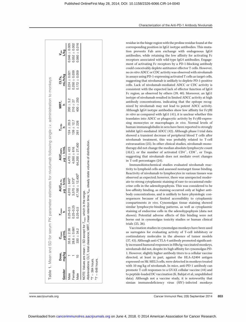
Pharmacokinetics, immunogenicity, and toxicity ofnivolumab in cynomolgus monkeysSingle, i.v. administration of nivolumab to cynomolgus
monkeys at 1 and 10 mg/kg was well tolerated with no effectson body weight or clinical observations. Mean concentration–time profiles for serum nivolumab were qualitatively similarfor males and females at 1 mg/kg and for males at 10 mg/kg.Mean concentrations declined in a multi-phasic manner fromCmax, observed within 0.5 hour at both doses. Serum PKparameter estimates are shown in Table 1. Mean apparentterminal elimination half-life estimates for males and femalesat 1 mg/kg were similar (124 and 139 hours, respectively), andthe mean half-life estimate for males at 10 mg/kg was 261hours. Anti-nivolumab antibodies were detected on day 28, butseemed to have no substantial impact on PK assessment [i.e.,mean retention time (MRT), total clearance (CLT), and steady-state volumeof distribution (Vss)]. In general, serumnivolumabhad a relatively slow clearance with limited extra vasculardistribution, as demonstrated by a Vss value consistent withplasma volume. Five of the 6 animals in group 1 (1 mg/kgnivolumab), and 2 of 3 in group 2 (10 mg/kg nivolumab) werepositive for an anti-nivolumab antibody response (includingneutralizing antibodies) at day 28 (data not shown), but withno observable adverse effects.In a 3-month toxicity study in cynomolgus monkeys, twice
weekly i.v. administration of nivolumab at doses of 10 mg/kgand 50 mg/kg was also well tolerated, with no effect on body
weight, and no other clinical findings. Serum chemistrychanges were limited to a reversible 28% decrease in T3 atweek 13 in females treated with 50 mg/kg. T4 and TSH levelswere unchanged. Inmales treatedwith 50mg/kg, therewere nochanges inT3, T4, or TSH levels. Nivolumab exposure increasedin an approximately dose-proportional manner between 10and 50 mg/kg, with no substantial sex differences noted (datanot shown). Anti-nivolumab antibodies were detected in only 1of 24 animals, although high nivolumab concentrations couldhave interfered with the assay. The highest well-tolerated dosein this study, 50 mg/kg twice weekly, is at least 20 times greaterthan doses reported to demonstrate antitumor activity inhumans (�10 mg/kg, every other week; ref. 26).
In phenotypic analyses of PBMCs 1 day after the last dose, nosignificant difference in total T- and B-cell numbers wasobserved between groups (data not shown). There were sig-nificantly more CD8þ effector memory T cells in the 50 mg/kggroup than in the 10 mg/kg and untreated groups (Supple-mentary Fig. S5A), and a nonsignificant trend toward moreCD8þ central memory T cells in the nivolumab group, espe-cially in the 50 mg/kg group. Na€�ve T-cell populations weredecreased in the 50 mg/kg group, suggesting that PD-1 block-ademay facilitate activation anddifferentiation of na€�veT cells.Finally, there were more CD11cþ DCs in the 50 mg/kg groupthan in the untreated group (P ¼ 0.014; Supplementary Fig.S5B), suggesting a possible role for PD-1 blockade in promotingDC differentiation.
Immune responses in cynomolgus monkeys to cellularand particulate virus-like particle vaccinescoadministered with nivolumab or ipilimumab
Enhancement of vaccine responses can demonstrate theactivity or potency of immunomodulatory antibodies in non-human primates. Previous studies demonstrated that CTLA-4blockade potentiated immune responses to an HBsAg vaccineor SK-MEL-3melanoma cell vaccine (37). A similar experimentwas conducted to examine the ability of PD-1 blockade topotentiate vaccine responses. As previously observed, ipilimu-mab strongly enhanced humoral immune responses to HBsAgas compared with control, whereas nivolumab showed noeffect on HBsAg titers over control treatment (Fig. 4A). Allgroups had normal anamnestic responses with measurableantibody titers following the second vaccine dose, whichpeaked after the third dose and declined thereafter.
Responses to the SK-MEL-3 vaccine were elevated in bothnivolumab- and ipilimumab-treated groups comparedwith con-trol (Fig. 4B). There was a modest increase in the humoralvaccine response in thenivolumabgroup,with a greater increasein the ipilimumab group. Titers increased markedly followingthe second vaccine dose, but did not change substantially afterthe third dose, followedby a rapid decline in all groups. Antibodytiters to HLA-A2404 (an allele of HLA expressed by SK-MEL-3cells) were also increased in animals treated with ipilimumab(4.3-fold) or nivolumab (2.4-fold) on day 71 (Fig. 4C).
DiscussionNivolumab is a fully human IgG4 PD-1 antibody that binds to
human and cynomolgus PD-1 with high affinity and blocks the
Characterization of the Anti-PD-1 Antibody Nivolumab
www.aacrjournals.org Cancer Immunol Res; 2(9) September 2014 851
on June 4, 2018. © 2014 American Association for Cancer Research. cancerimmunolres.aacrjournals.org Downloaded from
Published OnlineFirst May 28, 2014; DOI: 10.1158/2326-6066.CIR-14-0040

interaction of PD-1 with both PD-L1 and PD-L2 ligands. Infunctional in vitro assays, nivolumab enhanced cytokine pro-duction in human T-cell/DC MLR, SEB, and CMV recallresponse assays. In addition, antigen-specific CD8þ T-cellresponses from patients with melanoma increased afterincubation with nivolumab and peptide antigen, and notby stimulation with an irrelevant peptide (30). Importantly,while anti-PD-1 antibody enhanced antigen-specific T-cellresponses, it did not stimulate nonspecific responses byhuman blood cells, as determined by cytokine release upon
incubation with antibody alone. In a Treg suppression assay,nivolumab completely restored CD4þ T-responder cell prolif-eration and partially restored IFNg production. Although it isunclear whether nivolumab acts directly on CD4þ T-responderor Tregs, previous data have demonstrated that nivolumabcould overcomeTreg suppression of CD8þT cells by increasingresistance to Treg suppression, and also by directly limitingTreg-suppressive capacity (38).
The heavy chain constant region of nivolumab is a humanIgG4 isotype with an S228P mutation, which replaces a serine
0.001 0.01 0.1 1 10 100−10
0
10
20
30
40PD-1 IgG1
PD-1 IgG4
Anti-MHC-I
Control IgG1
Control IgG4
Antibody concentration (µg/mL)
0.001 0.01 0.1 1 10 100−10
0
10
20
30PD-1 IgG1
PD-1 IgG4
Anti-MHC-I
Control IgG1
Control IgG4
Antibody concentration (µg/mL)
0 102 103 104 1050
20
40
60
80
100
% o
f max
0 102 103 104 1050
20
40
60
80
100
% o
f max
0 102 103 104 1050
20
40
60
80
100
% o
f max
0.001 0.01 0.1 1 10 100−10
0
10
20
30
40
50
60
70PD-1 IgG1
PD-1 IgG4
Anti-MHC-I
Control IgG1
Control IgG4
Antibody concentration (µg/mL)
A
B
C
% S
peci
fic ly
sis
% S
peci
fic ly
sis
% S
peci
fic ly
sis
Figure 3. The absence of ADCC by nivolumab in vitro. IL-2–activated human PBMCs (effector cells) were incubated with activated human CD4þ T cells (targetcells) in an effector-to-target cell ratio of 50:1 in the presence of serial dilutions of nivolumab or a positive control anti-MHC class I antibody for 3 hour at 37�C.A–C, data from three individual ADCC assays using cells from different donors are shown. Purified CD4þ T cells were activated by coated anti-CD3antibody (4 mg/mL) plus soluble anti-CD28 antibody (1 mg/mL) and IL-2 (100U/mL) for 3 days. PD-1 expression on activated CD4þ T cells in each of the ADCCassays is shown in the right (solid line for PD-1, gray line for isotype control).
Wang et al.
Cancer Immunol Res; 2(9) September 2014 Cancer Immunology Research852
on June 4, 2018. © 2014 American Association for Cancer Research. cancerimmunolres.aacrjournals.org Downloaded from
Published OnlineFirst May 28, 2014; DOI: 10.1158/2326-6066.CIR-14-0040
residue in the hinge regionwith the proline residue found at thecorresponding position in IgG1 isotype antibodies. This muta-tion prevents Fab arm exchange with endogenous IgG4antibodies, while retaining the low affinity for activating Fcreceptors associated with wild-type IgG4 antibodies. Engage-ment of activating Fc receptors by a PD-1-blocking antibodycould conceivably deplete antitumor effector T cells. However,no in vitroADCC or CDC activity was observed with nivolumabin assays using PD-1-expressing activated T cells as target cells,suggesting that nivolumab is unlikely to deplete PD-1-positivecells. Lack of nivolumab-mediated ADCC or CDC activity isconsistent with the expected lack of effector function of IgG4Fc region, as observed by others (39, 40). Moreover, an IgG1isotype of nivolumab resulted in limited ADCC activity at highantibody concentrations, indicating that the epitope recog-nized by nivolumab may not lead to potent ADCC activity.Although IgG4 isotype antibodies show low affinity for FcgRIin vitro as compared with IgG1 (41), it is unclear whether thistranslates into ADCC or phagocytic activity by FcgRI-expres-sing monocytes or macrophages in vivo. Normal levels ofhuman immunoglobulin in sera have been reported to stronglyinhibit IgG1-mediated ADCC (42). Although phase I trial datashowed a transient decrease of peripheral blood T cells afternivolumab treatment, this was probably related to T-cellextravasation (25). In other clinical studies, nivolumab mono-therapy did not change themedian absolute lymphocyte count(ALC), or the number of activated CD4þ, CD8þ, or Tregs,suggesting that nivolumab does not mediate overt changesin T-cell percentages (24).
Immunohistochemical studies evaluated nivolumab reac-tivity to lymphoid cells and assessed nontarget tissue binding.Reactivity of nivolumab to lymphocytes in various tissues wasobserved as expected; however, there was unexpected moder-ate-to-strong cytoplasmic staining of rare-to-occasional endo-crine cells in the adenohypophysis. This was considered to below-affinity binding, as staining occurred only at higher anti-body concentrations, and is unlikely to have physiologic con-sequences because of limited accessibility to cytoplasmiccompartments in vivo. Cynomolgus tissue staining showedsimilar lymphocyte-binding patterns, as well as cytoplasmicstaining of endocrine cells in the adenohypophysis (data notshown). Potential adverse effects of this binding were notborne out in cynomolgus toxicity studies or human clinicaltrials (25, 26).
Vaccination studies in cynomolgusmonkeys have been usedas surrogates for evaluating activity of T-cell inhibitory orcostimulatory molecules in the absence of tumor models(37, 43). Although anti-CTLA-4 antibody promoted significant-ly increased humoral responses inHBsAg-vaccinatedmonkeys,nivolumab did not, despite its high affinity for cynomolgus PD-1. However, slightly higher antibody titers to a cellular vaccinedirected, at least in part, against the HLA-A2404 antigenexpressed on SK-MEL3 cells, were detected inmonkeys treatedwith 10 mg/kg of nivolumab. In mice, anti-PD-1 antibody canpromote T-cell responses to a GVAX cellular vaccine (44) andto peptide-loadedDC vaccination (K. Bahjat et al.; unpublisheddata). Although not a vaccine study, it is noteworthy thatsimian immunodeficiency virus (SIV)–infected monkeys
Tab
le1.
Mea
nan
dSDforse
rum
PKparam
eter
estim
ates
fornivo
lumab
follo
wingsing
lei.v.a
dministrationto
mon
keys
Gen
der
Dose
,mg/kg
Cmax,
mg/m
LTmax,
hAUC(0-T
),mg�
h/mL
AUC(INF),
mg�
h/mL
T(1/2),
hMRT,
hCLT
s,mL/h/kg
Vss,
L/kg
Male
133
.8�
2.36
0.25
–0.5
4,01
0�
645a
4,47
0�
423
124�
20.3
200�
5.2
0.22
4�
0.02
10.04
6�
0.00
5Fe
male
128
.6�
0.68
10.25
–0.25
3,57
0�
573a
4,05
0�
616
139�
12.7
210�
270.25
0�
0.03
90.05
2�
0.00
2Male
1033
0�
34.2
0.25
–0.5
47,100
�12
,400
b64
,200
�27
,400
261�
226
400�
250
0.17
2�
0.05
90.06
0�
0.01
6
NOTE
:Value
saremea
n�
SDex
ceptforT m
ax,w
hich
istherang
e.Abbreviations
:CLT
,total
clea
ranc
e;MRT,
mea
nretentiontim
e;Vss,s
tead
ystatevo
lumeof
distribution.
aT¼
384ho
urs.
bT¼
648ho
urs.
Characterization of the Anti-PD-1 Antibody Nivolumab
www.aacrjournals.org Cancer Immunol Res; 2(9) September 2014 853
on June 4, 2018. © 2014 American Association for Cancer Research. cancerimmunolres.aacrjournals.org Downloaded from
Published OnlineFirst May 28, 2014; DOI: 10.1158/2326-6066.CIR-14-0040

treated with an anti-PD-1 antibody showed increased humoralresponses to SIV antigens (45).
Nivolumab was well tolerated when administered to cyno-molgus monkeys as twice-weekly i.v. injections for 3 months atdoses up to 50 mg/kg, with no adverse effects on any para-meters. Although there was a low incidence of anti-nivolumabantibodies, they were not associated with any adverse effects
(i.e., hypersensitivity reactions), and had no substantial impacton PK parameters. Furthermore, anti-drug antibody responsesin animals are not considered predictive of responses inhumans (46). Thus, the results of the nonclinical studies inmonkeys suggested a favorable risk:benefit ratio to supportinitial clinical trials with nivolumab. Although nivolumabseems to lack toxicity in monkeys, toxicities have beenobserved in human clinical trials. In a phase I trial, nivolumabhad a favorable safety profile (26). Adverse events were gen-erally similar to those observed with ipilimumab, althoughwith lower incidence and of less severity, and comprisedgastrointestinal, endocrine, and skin toxicities, and pulmonaryinflammation. Interestingly, pneumonitis has been observed inPD-1-deficient mice bred onto the MRL genetic background(8), but not in PD-1-deficient mice with other genetic back-grounds (6, 7). In cynomolgus toxicity studieswith anti-CTLA-4(ipilimumab), no or only rare toxicities were observed,although they were evident in human studies. These observa-tions highlight the difficulty of predicting toxicities in humanswith antibodies mediating checkpoint blockade, such as anti-PD-1 or anti-CTLA-4 antibodies, from results in mice and non-human primates.
Increased numbers of CD8þ T-effector memory cells weredetected in cynomolgus monkey peripheral blood afterrepeated treatment at the highest dose of nivolumab for 3months. Although recent data (24) suggest that activated Tcells are not potential pharmacodynamic markers of nivo-lumab treatment, it remains to be determined whethernivolumab treatment increases CD8þ effector memory cellsin humans with cancer. A marked accumulation of CD8þ
effector memory cells in lymphoid organs and tissues ofPD-1-deficient mice has been described previously (47).Increases in CD11cþ DCs were also observed in the cyno-molgus monkey safety study, although the underlying mech-anism is unclear.
In early clinical trials, nivolumab produced durableresponses and stable disease, and an encouraging survivalprofile, in patients with advanced melanoma, lung, and renalcancers. In some patients, tumor regression persisted afterdiscontinuation of nivolumab (26, 27, 48). Nivolumab wasgenerally well tolerated, even with prolonged dosing(26, 27, 48). Another PD-1-blocking antibody, pembrolizu-mab, has shown similar activity and safety in metastaticmelanoma (49). These studies further validate the concept ofmodulating immune responses with checkpoint blockade forcancer immunotherapy, as first demonstrated in clinicaltrials with ipilimumab (1, 2).
Exploration of nivolumabcombinationswith other immuno-oncology approaches, as well as standard of care therapies,is warranted. PD-1 pathway blockade combined with anti-CTLA-4 or anti-LAG-3 antibody showed synergistic antitumoractivity superior to the single agents in murine tumor models(15–17). Preliminary clinical data in patients with melanomareceiving nivolumab plus ipilimumab showed rapid and dura-ble responses: 31% of responders had tumor regression of 80%or more by week 12, a superior profile to monotherapies (50).The combination is currently being clinically evaluated inmultiple tumor types.
0
10,000
20,000
30,000
40,000
SalineNivolumab
Study day
Ipilimumab
0 14 30 43 58 71 85 100
Test article dosing
Ant
i-HB
sAg
titer
(m
IU/m
L)
0 14 30 43 58 71 85 1000
50
100
150
200
SalineNivolumabIpilimumab
Test article dosing
Study day
MF
I on
SK
-ME
L-3
cells
0.0
0.5
1.0
1.5
2.0
2.5
Saline
IpilimumabNivolumab
0 14 30 43 58 71 85 100
Test article dosing
Study day
Mea
n O
D to
HLA
-A24
04A
B
C
Figure 4. Effect of nivolumab or ipilimumab on immune responses tovaccination in cynomolgusmonkeys. A, humoral immune responses to aparticulate HBsAg vaccine by ELISA. Plasma samples obtained at theindicated times were analyzed for anti-HBsAg Abs. B, antibodyresponses to an SK-MEL-3 vaccine as assessed by flow cytometry.Vaccine-specific antibody responses were measured by incubation ofSK-MEL-3 cells with plasma collected at 2-week intervals. Data pointsrepresent the mean � SEM of the mean fluorescence intensity (MFI)values in each treatment group at each collection time point. C, antibodyresponses to HLA-A2404 were determined from plasma by ELISA. Datapoints represent the mean � SEM of the mean OD values in eachtreatment group. All sampleswere analyzed at least two timeswith similarresults.
Wang et al.
Cancer Immunol Res; 2(9) September 2014 Cancer Immunology Research854
on June 4, 2018. © 2014 American Association for Cancer Research. cancerimmunolres.aacrjournals.org Downloaded from
Published OnlineFirst May 28, 2014; DOI: 10.1158/2326-6066.CIR-14-0040

Disclosure of Potential Conflicts of InterestC. Wang, M.J. Selby, X.-T. Wang, and A.J. Korman have ownership
interest (including patents) in Bristol-Myers Squibb stock. No potential conflictsof interest were disclosed by the other authors.
DisclaimerAll pivotal toxicology studies were conducted in compliance with the Good
Laboratory Practice Regulations for nonclinical Laboratory Studies of the U.S.Food and Drug Administration (21 CFR Part 58) and were approved by thelaboratory's Institutional Animal Care and Use Committee.
Authors' ContributionsConception and design: C. Wang, X.-T. Wang, H. Huang, D. Blanset, M.J. Selby,A.J. KormanDevelopment of methodology: C. Wang, K.B. Thudium, X.-T. Wang, H. Huang,D. Feingersh, M. Srinivasan, S. Wong, N. Garner, D. Blanset, M.J. SelbyAcquisition of data (provided animals, acquired and managed patients,provided facilities, etc.): C. Wang, K.B. Thudium, X.-T. Wang, H. Huang,D. Feingersh, C. Garcia, Y. Wu, M. Kuhne, M. Srinivasan, S. Singh, S. Wong, N.Garner, M.J. SelbyAnalysis and interpretation of data (e.g., statistical analysis, biostatistics,computational analysis): C. Wang, K.B. Thudium, M. Han, X.-T. Wang,H. Huang, D. Feingersh, C. Garcia, Y. Wu, M. Kuhne, M. Srinivasan, S. Singh,S. Wong, N. Garner, T. Bunch, D. Blanset, M.J. Selby, A.J. Korman
Writing, review, and/or revision of themanuscript:C.Wang, K.B. Thudium,M. Han, X.-T. Wang, H. Huang, D. Feingersh, M. Kuhne, M. Srinivasan, S. Wong,N. Garner, H. Leblanc, T. Bunch, D. Blanset, M.J. Selby, A.J. KormanAdministrative, technical, or material support (i.e., reporting or orga-nizing data, constructing databases): C. Wang, K.B. Thudium, H. Huang,M. Kuhne, S. Wong, M.J. SelbyStudy supervision: C. Wang, H. Huang, H. Leblanc, D. Blanset, M.J. Selby,A.J. Korman
AcknowledgmentsThe authors thank Rangan Vangipuram, Alison Witte, Huiming Li, Peter
Brams, Shrikant Deshpande, and Pina Cardarelli for their contributions to thePD-1 project. Professional medical writing and editorial assistance was providedby Cailin Moira Wilke and Emily de Looze at StemScientific.
Grant SupportThis study was funded by Bristol-Myers Squibb.The costs of publication of this article were defrayed in part by the payment of
page charges. This article must therefore be hereby marked advertisement inaccordance with 18 U.S.C. Section 1734 solely to indicate this fact.
Received March 12, 2014; revised May 23, 2014; accepted May 23, 2014;published OnlineFirst May 29, 2014.
References1. Korman AJ, Peggs KS, Allison JP. Checkpoint blockade in cancer
immunotherapy. Adv Immunol 2006;90:297–339.2. Hodi FS, O'Day SJ, McDermott DF, Weber RW, Sosman JA, Haanen
JB, et al. Improved survival with ipilimumab in patients with metastaticmelanoma. N Engl J Med 2010;363:711–23.
3. Robert C, Thomas L, Bondarenko I, O'Day S, Weber J, Garbe C, et al.Ipilimumab plus dacarbazine for previously untreated metastatic mel-anoma. N Engl J Med 2011;364:2517–26.
4. Freeman GJ, Long AJ, Iwai Y, Bourque K, Chernova T, Nishimura H,et al. Engagement of thePD-1 immunoinhibitory receptor by anovel B7family member leads to negative regulation of lymphocyte activation.J Exp Med 2000;192:1027–34.
5. Brown JA, DorfmanDM,Ma FR, Sullivan EL, MunozO,WoodCR, et al.Blockadeofprogrammeddeath-1 ligandsondendriticcells enhancesTcell activation and cytokine production. J Immunol 2003;170:1257–66.
6. NishimuraH, NoseM,Hiai H,MinatoN, Honjo T. Development of lupus-like autoimmune diseases by disruption of the PD-1 gene encoding anITIM motif-carrying immunoreceptor. Immunity 1999;11:141–51.
7. Nishimura H, Okazaki T, Tanaka Y, Nakatani K, Hara M, Matsumori A,et al. Autoimmune dilated cardiomyopathy in PD-1 receptor-deficientmice. Science 2001;291:319–22.
8. Wang J,Okazaki IM, Yoshida T,ChikumaS,KatoY,Nakaki F, et al. PD-1 deficiency results in the development of fatal myocarditis in MRLmice. Int Immunol 2010;22:443–52.
9. Blackburn SD, ShinH,HainingWN, Zou T,WorkmanCJ, Polley A, et al.Coregulation ofCD8þT cell exhaustion bymultiple inhibitory receptorsduring chronic viral infection. Nat Immunol 2009;10:29–37.
10. Blank C, Kuball J, Voelkl S, Wiendl H, Becker B, Walter B, et al.Blockade of PD-L1 (B7-H1) augments human tumor-specific T cellresponses in vitro. Int J Cancer 2006;119:317–27.
11. Thompson RH, Dong H, Lohse CM, Leibovich BC, Blute ML, ChevilleJC, et al. PD-1 is expressed by tumor-infiltrating immune cells and isassociated with poor outcome for patients with renal cell carcinoma.Clin Cancer Res 2007;13:1757–61
12. Curiel TJ, Wei S, Dong H, Alvarez X, Cheng P, Mottram P, et al.Blockade of B7-H1 improves myeloid dendritic cell-mediated antitu-mor immunity. Nat Med 2003;9:562–7.
13. Hirano F, Kaneko K, Tamura H, Dong H, Wang S, Ichikawa M, et al.Blockade of B7-H1 and PD-1 by monoclonal antibodies potentiatescancer therapeutic immunity. Cancer Res 2005;65:1089–96.
14. Nomi T, Sho M, Akahori T, Hamada K, Kubo A, Kanehiro H, et al.Clinical significance and therapeutic potential of PD-1 pathway inhuman pancreatic cancer. Clin Cancer Res 2007;13:2151–7.
15. Curran MA, Montalvo W, Yagita H, Allison JP. PD-1 and CTLA-4combination blockade expands infiltrating T cells and reduces regu-latory T and myeloid cells within B16 melanoma tumors. Proc NatlAcad Sci U S A 2010;107:4275–80.
16. WooSR, TurnisME,GoldbergMV, Bankoti J, SelbyM,Nirschl CJ, et al.Immune inhibitory molecules LAG-3 and PD-1 synergistically regulateT-cell function to promote tumoral immune escape. Cancer Res2012;72:917–27.
17. Selby M, Engelhardt J, Lu LS, Quigley M, Wang C, Chen B, et al.Antitumor activity of concurrent blockade of immune checkpointmolecules CTLA-4 and PD-1 in preclinical models. J Clin Oncol 31,2013 (suppl; abstr 3061).
18. Dong H, Strome SE, Salomao DR, Tamura H, Hirano F, Flies DB, et al.Tumor-associated B7-H1 promotes T-cell apoptosis: a potentialmechanism of immune evasion. Nat Med 2002;8:793–800.
19. Konishi J, Yamazaki K, Azuma M, Kinoshita I, Dosaka-Akita H, Nishi-mura M. B7-H1 expression on non–small cell lung cancer cells and itsrelationshipwith tumor-infiltrating lymphocytes and their PD-1 expres-sion. Clin Cancer Res 2004;10:5094–6100.
20. Ohigashi Y, Sho M, Yamada Y, Tsurui Y, Hamada K, Ikeda N, et al.Clinical significance of programmed death-1 ligand-1 and pro-grammed death-1 ligand-2 expression in human esophageal cancer.Clin Cancer Res. 2005;11:2947–53.
21. Thompson RH, Kuntz SM, Leibovich BC, Dong H, Lohse CM,WebsterWS, et al. Tumor B7-H1 is associated with poor prognosis in renal cellcarcinoma patients with long-term follow-up. Cancer Res 2006;66:3381–5.
22. Hino R, Kabashima K, Kato Y, Yagi H, Nakamura M, Honjo T, et al.Tumor cell expression of programmed cell death-1 ligand 1 is aprognostic factor for malignant melanoma. Cancer 2010;116:1757–66.
23. Taube JM, Anders RA, Young GD, Xu H, Sharma R, McMiller TL, et al.Colocalization of inflammatory response with B7-h1 expression inhuman melanocytic lesions supports an adaptive resistance mecha-nism of immune escape. Sci Transl Med 2012;4:127ra37.
24. Grosso J, Horak CE, Inzunza D, Cardona DM, Simon JS, Gupta AK,et al. Association of tumor PD-L1 expression and immune biomarkerswith clinical activity in patients (pts)with advanced solid tumors treatedwith nivolumab (anti-PD-1; BMS-936558; ONO-4538). J Clin Oncol 31,2013 (suppl; abstr 3016).
25. Brahmer JR, Drake CG, Wollner I, Powderly JD, Picus J, SharfmanWH, et al. Phase I study of single-agent anti-programmed death-1(MDX-1106) in refractory solid tumors: safety, clinical activity,
Characterization of the Anti-PD-1 Antibody Nivolumab
www.aacrjournals.org Cancer Immunol Res; 2(9) September 2014 855
on June 4, 2018. © 2014 American Association for Cancer Research. cancerimmunolres.aacrjournals.org Downloaded from
Published OnlineFirst May 28, 2014; DOI: 10.1158/2326-6066.CIR-14-0040

pharmacodynamics, and immunologic correlates. J Clin Oncol2010;28:3167–75.
26. Topalian SL, Hodi FS, Brahmer JR, Gettinger SN, Smith DC, McDer-mott DF, et al. Safety, activity, and immune correlates of anti-PD-1antibody in cancer. N Engl J Med 2012;366:2443–54.
27. Topalian SL, Sznol M, McDermott DF, Kluger HM, Carvajal RD, Sharf-manWH, et al. Survival, durable tumor remission, and long-term safetyin patientswith advancedmelanoma receiving nivolumab. JClinOncol2014;32:1020–30.
28. LonbergN,TaylorLD,HardingFA,TrounstineM,HigginsKM,SchrammSR, et al. Antigen-specific human antibodies from mice comprisingfour distinct genetic modifications. Nature 1994;368:856–9.
29. Fishwild DM, O'Donnell SL, Bengoechea T, Hudson DV, Harding F,Bernhard SL, et al. High-avidity human IgG kappa monoclonal anti-bodies fromanovel strain ofminilocus transgenicmice.NatBiotechnol1996;14:845–51.
30. Wong RM, Scotland RR, Lau RL, Wang C, Korman AJ, Kast WM, et al.Programmed death-1 blockade enhances expansion and functionalcapacity of human melanoma antigen-specific CTLs. Int Immunol2007;19:1223–34.
31. Zhang X, Schwartz JC, Guo X, Bhatia S, Cao E, Lorenz M, et al.Structural and functional analysis of the costimulatory receptor pro-grammed death-1. Immunity 2004;20:337–47; Erratum: Immunity2004;20:651.
32. L�az�ar-Moln�ar E, Yan Q, Cao E, Ramagopal U, Nathenson SG, AlmoSC. Crystal structure of the complex between programmed death-1(PD-1) and its ligand PD-L2. Proc Natl Acad Sci U S A 2008;105:10483–8.
33. LinDY, TanakaY, IwasakiM,GittisAG,SuHP,MikamiB, et al. ThePD-1/PD-L1complex resembles theantigen-bindingFvdomainsofantibodiesand T cell receptors. Proc Natl Acad Sci U S A 2008;105:3011–6.
34. Cheng X, Veverka V, Radhakrishnan A, Waters LC, Muskett FW,Morgan SH, et al. Structure and interactions of the human pro-grammed cell death 1 receptor. J Biol Chem 2013;288:11771–85.
35. Sage PT, Francisco LM, Carman CV, Sharpe AH. The receptor PD-1controls follicular regulatory T cells in the lymph nodes and blood. NatImmunol 2013;14:152–61.
36. Iwai Y, Okazaki T, Nishimura H, Kawasaki A, Yagita H, Honjo T.Microanatomical localization of PD-1 in human tonsils. Immunol Lett2002;83:215–20.
37. Keler T, Halk E, Vitale L, O'Neill T, Blanset D, Lee S, et al. Activity andsafety of CTLA-4 blockade combined with vaccines in cynomolgusmacaques. J Immunol 2003;171:6251–9.
38. Wang W, Lau R, Yu D, Zhu W, Korman A, Weber J. PD-1 blockadereverses the suppression of melanoma antigen-specific CTL by CD4þ
CD25(Hi) regulatory T cells. Int Immunol 2009;21:1065–77.
39. Niwa R, Natsume A, Uehara A, Wakitani M, Iida S, Uchida K, et al. IgGsubclass-independent improvement of antibody-dependent cellularcytotoxicity by fucose removal from Asn297-linked oligosaccharides.J Immunol Methods 2005;306:151–60.
40. Stein R, Qu Z, Chen S, Solis D, Hansen HJ, Goldenberg DM. Char-acterization of a humanized IgG4 anti-HLA-DRmAb) that lacks effectorcell functions but retains direct antilymphoma activity and increasesthe potency of rituximab. Blood 2006;108:2736–44.
41. Bruhns P, Iannascoli B, EnglandP,Mancardi DA, FernandezN, JorieuxS, et al. Specificity and affinity of human Fcgamma receptors and theirpolymorphic variants for human IgG subclasses. Blood 2009;113:3716–25.
42. Preithner S, Elm S, Lippold S, Locher M,Wolf A, da Silva AJ, et al. Highconcentrations of therapeutic IgG1 antibodies are needed to com-pensate for inhibition of antibody-dependent cellular cytotoxicity byexcess endogenous immunoglobulin G. Mol Immunol 2006;43:1183–93.
43. Weinberg AD, Thalhofer C,Morris N,Walker JM, SeissD,WongS, et al.Anti-OX40 (CD134) administration to nonhuman primates: immunos-timulatory effects and toxicokinetic study. J Immunother 2006;29:575–85.
44. Li B, VanRoey M, Wang C, Chen TH, Korman A, Jooss K. Anti-programmed death-1 synergizes with granulocyte macrophage colo-ny-stimulating factor–secreting tumor cell immunotherapy providingtherapeutic benefit to mice with established tumors. Clin Cancer Res2009;15:1623–34.
45. Velu V, Titanji K, Zhu B, Husain S, Pladevega A, Lai L, et al. EnhancingSIV-specific immunity in vivo by PD-1 blockade. Nature 2009;458:206–10.
46. vanMeer PJ, KooijmanM,Brinks V, Gispen-deWiedCC, Silva-LimaB,Moors EH, et al. Immunogenicity of mabs in non-human primatesduring nonclinical safety assessment. MAbs 2013;5:810–6.
47. Charlton JJ, Chatzidakis I, Tsoukatou D, Boumpas DT, Garinis GA,Mamalaki C. Programmed death-1 shapes memory phenotype CD8T cell subsets in a cell-intrinsic manner. J Immunol 2013;190:6104–14.
48. Topalian SL, Sznol M, Brahmer JR, McDermott DF, Smith DC, Get-tinger S, et al. Nivolumab (anti-PD-1; BMS-936558; ONO-4538) inpatients with advanced solid tumors: survival and long-term safety in aphase I trial. J Clin Oncol 31, 2013 (suppl; abstr 3002).
49. Hamid O, Robert C, Daud A, Hodi FS, HwuWJ, Kefford R, et al. Safetyand tumor responses with lambrolizumab (anti-PD-1) in melanoma.N Engl J Med 2013;369:134–44.
50. Wolchok JD, Kluger H, Callahan MK, Postow MA, Rizvi NA, LesokhinAM, et al. Nivolumab plus ipilimumab in advanced melanoma. N EnglJ Med 2013;369:122–33.
Cancer Immunol Res; 2(9) September 2014 Cancer Immunology Research856
Wang et al.
on June 4, 2018. © 2014 American Association for Cancer Research. cancerimmunolres.aacrjournals.org Downloaded from
Published OnlineFirst May 28, 2014; DOI: 10.1158/2326-6066.CIR-14-0040

2014;2:846-856. Published OnlineFirst May 28, 2014.Cancer Immunol Res Changyu Wang, Kent B. Thudium, Minhua Han, et al.
Toxicology in Non-Human PrimatesIn VivoBMS-936558, and Characterization of the Anti-PD-1 Antibody Nivolumab,In Vitro
Updated version
10.1158/2326-6066.CIR-14-0040doi:
Access the most recent version of this article at:
Material
Supplementary
http://cancerimmunolres.aacrjournals.org/content/suppl/2014/06/02/2326-6066.CIR-14-0040.DC1
Access the most recent supplemental material at:
Cited articles
http://cancerimmunolres.aacrjournals.org/content/2/9/846.full#ref-list-1
This article cites 47 articles, 22 of which you can access for free at:
Citing articles
http://cancerimmunolres.aacrjournals.org/content/2/9/846.full#related-urls
This article has been cited by 22 HighWire-hosted articles. Access the articles at:
E-mail alerts related to this article or journal.Sign up to receive free email-alerts
Subscriptions
Reprints and
To order reprints of this article or to subscribe to the journal, contact the AACR Publications Department
Permissions
Rightslink site. Click on "Request Permissions" which will take you to the Copyright Clearance Center's (CCC)
.http://cancerimmunolres.aacrjournals.org/content/2/9/846To request permission to re-use all or part of this article, use this link
on June 4, 2018. © 2014 American Association for Cancer Research. cancerimmunolres.aacrjournals.org Downloaded from
Published OnlineFirst May 28, 2014; DOI: 10.1158/2326-6066.CIR-14-0040