preparation and biological evaluation of ho-dtpa-cetuximab filepreparation and biological evaluation...
TRANSCRIPT

Preparation and biological evaluation of 166Ho-DTPA-Cetuximab
Hyung Yub Seo
The Graduate School
Yonsei University
Department of Biomedical Laboratory Science
Preparation and biological evaluation of 166Ho-DTPA-Cetuximab
A Master’s Thesis
Submitted to the Department of Biomedical
Laboratory Science and the Graduate School of
Yonsei University
in partial fulfillment of the
requirements for the degree of
Master of Science in Medical Technology
Hyung Yub Seo
December 2008
This certifies that the master’s thesis of
Hyung Yub Seo is approved.
___________________________
Thesis Supervisor : Ok Doo Awh
___________________________
Tae Ue Kim : Thesis Committee Member
___________________________
Yong Serk Park : Thesis Committee Member
The Graduate School
Yonsei University
December 2008
- i -
CONTENTS
LIST OF FIGURES ·································································································ⅲLIST OF TABLES ···································································································ⅳABBREVIATION ······································································································ⅴABSTRACT IN ENGLISH ·····················································································ⅵI. INTRODUCTION ··································································································· 1
II. MATERIALS AND METHODS ········································································· 4
1. Materials ············································································································ 4
2. Methods ············································································································· 5
2.1. Cell culture ······························································································ 5
2.2. Evaluation of EGF receptor expression on the surface of cancer
cells ·········································································································· 5
2.2.1. In vitro cell binding assay ······························································· 5
2.2.2. Reverse transcriptase-polymerase chain reaction (RT-PCR) ·········· 6
2.3. Experimental animal models ·································································· 9
2.4. Preparation of DTPA-Cetuximab conjugates······································· 9
2.5. 166Ho-labeling to DTPA-Cetuximab ···················································· 10
2.6. Biochemical characterization of 166Ho-DTPA-Cetuximab conjugates
················································································································ 12
2.6.1. Immunoreactivity assay of 166Ho-DTPA-Cetuximab ····················· 12
2.6.2. In vitro stability assay ···································································· 12
2.7. Biodistribution studies of 166Ho-DTPA-Cetuximab conjugates in
A549 tumor-bearing nude mice ·························································· 13
- ii -
2.8. Gamma camera image of A549 tumor-bearing nude mice
administered with 166Ho-DTPA-Cetuximab conjugates ····················· 14
III. RESULTS ··········································································································· 15
1. Evaluation of EGF receptor expression on the surface of cancer cells
·························································································································· 15
2. Preparation of DTPA-Cetuximab conjugate ················································ 18
3. Radiolabeling of 166Ho to DTPA-Cetuximab and relative
immunoreactivities of the labeled conjugates ············································ 20
4. In vitro stability of 166Ho-DTPA-Cetuximab in serum ····························· 25
5. Biodistribution studies of 166Ho-DTPA-Cetuximab conjugates in A549
tumor-bearing nude mice ············································································· 27
6. Gamma camera image of A549 tumor-bearing nude mouse administered
with 166Ho-DTPA-Cetuximab conjugates ···················································· 31
IV. DISCUSSION ····································································································· 33
V. REFERENCE ······································································································· 36
ABSTRACT IN KOREAN ······················································································ 41

- iii -
LIST OF FIGURES
Fig. 1. Immunoreactivity of Cetuximab to various types of cancer cells ········ 15
Fig. 2. Verification of EGFR expression in the A549 and CT-26 tumor cells
by RT-PCR ·································································································· 17
Fig. 3. The number of DTPA molecules bound to Cetuximab depending on
reacting their molar ratios ········································································· 19
Fig. 4. Radiochromatogram of 166Ho-DTPA-Cetuximab ······································· 21
Fig. 5. Radiolabeling yield of 166Ho-DTPA-Cetuximab conjugates ···················· 22
Fig. 6. Immunoreactivity of 166Ho-DTPA-Cetuximab to A549 cells ················· 23
Fig. 7. In vitro Stability of 166Ho-DTPA-Cetuximab in serum ·························· 26
Fig. 8. Biodistribution of 166Ho-DTPA-Cetuximab conjugates in A549 tumor
bearing nude mice ······················································································ 29
Fig. 9. A gamma camera image of A549 tumor-bearing nude mouse
administered with 166Ho-DTPA-Cetuximab conjugates ··························· 31
- iv -
LIST OF TABLES
Table 1. Primer sequences for β-actin and EGFR and amplified product sizes
······················································································································ 8
Table 2. The radiolabeling yield, number of DTPA per Cetuximab, specific
activity, and relative immunoreactivity of the six 166Ho-DTPA-
Cetuximab conjugates varied ratios of DTPA and Cetuximab ·········· 24
Table 3. Biodistribution of 166Ho-DTPA-Cetuximab conjugates in A549 tumor
bearing mice ······························································································ 28
Table 4. Organ per Tissue ratio of 166Ho-DTPA-Cetuximab conjugates in A549
tumor bearing mice ·················································································· 30

- v -
ABBREVIATION
DOTA : tetraazacyclododecane tetraacetic acid
DTPA : diethylene triamine pentaacetic acid
EGFR : epidermal growth factor receptor
Ho : holmium
I : iodine
ID/g : injected dose per gram
ITLC-SG : instant thin layer chromatography - silica gel
RIT : radioimmunotherapy
- vi -
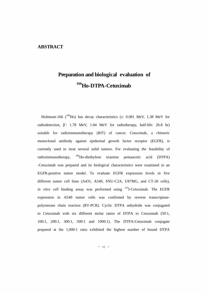
ABSTRACT
Preparation and biological evaluation of
166Ho-DTPA-Cetuximab
Holmium-166 (166Ho) has decay characteristics (γ: 0.081 MeV, 1.38 MeV for
radiodetection, β-: 1.78 MeV, 1.84 MeV for radiotherapy, half-life: 26.8 hr)
suitable for radioimmunotherapy (RIT) of cancer. Cetuximab, a chimeric
monoclonal antibody against epidermal growth factor receptor (EGFR), is
currently used to treat several solid tumors. For evaluating the feasibility of
radioimmunotherapy, 166Ho-diethylene triamine pentaacetic acid (DTPA)
-Cetuximab was prepared and its biological characteristics were examined in an
EGFR-positive tumor model. To evaluate EGFR expression levels in five
different tumor cell lines (A431, A549, SNU-C2A, U87MG, and CT-26 cells),
in vitro cell binding assay was performed using 125I-Cetuximab. The EGFR
expression in A549 tumor cells was confirmed by reverse transcriptase-
polymerase chain reaction (RT-PCR). Cyclic DTPA anhydride was conjugated
to Cetuximab with six different molar ratios of DTPA to Cetuximab (50:1,
100:1, 200:1, 300:1, 500:1 and 1000:1). The DTPA-Cetuximab conjugate
prepared at the 1,000:1 ratio exhibited the highest number of bound DTPA

- vii -
(88.4 molecules). The 166Ho-radiolabeling yield was also the highest in the
same DTPA-Cetuximab conjugate (98.9%). The immunoreactivity and specific
activity of the 166Ho-DTPA-Cetuximab was 60.7 ± 3.0% and 94.0 MBq/mg,
respectively. The 166Ho-DTPA-Cetuximab conjugate was stable in the presence
of serum proteins (>95% for 24 hr at 37℃). In A549 xenograft mice,
166Ho-DTPA-Cetuximab showed the highest tumoral uptake (9.1 ± 1.8% ID/g)
at 72 hr post injection. The conjugate also showed the highest tumor-to-blood
(9.1 ± 2.9) and tumor-to-muscle ratios (9.1 ± 3.7) at 144 hr post injection,
respectively. The selective localization of 166Ho-DTPA-Cetuximab in
EGFR-positive A549 tumor xenografts was confirmed by gamma camera
imaging at 24 hr,. These data showed that the 166Ho-labeled immunoreactive
DTPA-Cetuximab conjugates with a high specific activity would be a
renovated modality of radioimmunotherapeutics for EGFR-expressing tumors.
Keywords : Cetuximab, 166Ho, DTPA, radioimmunotherapy
- 1 -
I. INTRODUCTION
Radioimmunotherapy has been utilized as nuclear medicine for tumor
therapy [1,2,3]. In radioimmunotherapy, monoclonal antibodies with selectivity
for the target cells or tissues are linked to radionuclides with high beta
(iodine-131 and yttrium-90) or alpha (bismuth-213 and astatine-211) emitters
resulting in cell death. In general, beta particles, with a penetration range of
millimeters, are suitable for target-specific tumor therapy, whereas alpha
particles, with a penetration range of a few cell diameters, are suitable for
micrometastasis or circulating tumor cells [1,4].
Cetuximab (MERCK, Germany), an antibody being used therapeutically to
target epidermal growth factor receptor (EGFR) [8,9], was originally reported
to stimulate EGFR internalization and downregulation [10]. Also, Cetuximab
(either alone or in combination with drugs or radioisotopes) has been used for
treatment of several solid tumors, such as non-small cell lung carcinoma,
pancreatic cancer, head and neck cancer, etc. [11,12,13].
The radiolanthanides (ie., samarium-153, lutetium-177, holmium-166, etc.)
are considered as excellent candidates for radiotherapy because their desirable
physical characteristics and availability. The various radiolanthanides exhibit a
variety of nuclear properties yet have very similar chemistry. The
radiolanthanide, holmium-166 (166Ho) is an attractive therapeutic radionuclide
because of its short half-life (26.8 hr), deep penetration range (~9.0 mm in
soft tissue), high beta energy with 1.77 MeV (48%) and 1.85 MeV (51%)

- 2 -
suitable for radiotherapy and a small portion of gamma rays with 0.08 MeV
(6.6%) and 1.38 MeV (0.9%) suitable for imaging. [5,6,7].
A number of conjugation techniques have been developed for modification
of biomolecules. The conjugation groups for attachment of a bifunctional
chelating agent to biomolecules include anhydride, isothiocyanate,
N-hydroxysuccinim- ide (NHS) ester, and maleimide. Anhydrides of diethylene
triamine pentaacetic acid (DTPA) and tetraazacyclododecane tetraacetic acid
(DOTA) have also been used to prepare their bioconjugates [18,19,20]. DTPA
is an organic compound consisting of a diethylenetriamine backbone modified
with five carboxymethyl groups, which has a high affinity for metal cations.
Technetium-99m (99mTc)-labeled Cetuximab has been applied to single
photon emission computed tomography (SPECT) imaging for
radioimmunodetection [14,15]. It has been reported that 99mTc-labeled
Cetuximab has suitable dosimetric properties for a diagnostic nuclear medicine
agent. The study with the long-lived positron emitter zirconium-89 (89Zr) and
other therapeutic radiometals (such as yttrium-90, and lutetium-177) were
performed to predicting the biodistribution of other therapeutic radiometals
(such as yttrium-90, and lutetium-177), when labeled to Cetuximab via
different types of chelator (ie., p-SCN-Bz-DOTA, p-SCN-Bz-DTPA, etc.) [16].
Copper-64 (64Cu)-DOTA-Cetuximab has also been utilized for EGFR imaging
[17].
- 3 -
In this study, DTPA-Cetiximab was prepared and 166Ho was labeled to the
Cetuximab derivatives. 166Ho-labeling conditions were optimized and the
biological characteristics of the 166Ho-labeled conjugate were examined in a
EGFR-positive tumor model to evaluate the clinical applicability of
166Ho-DTPA-Cetuximab as a therapeutic agent.
- 4 -
II. MATERIALS AND METHODS
1. Materials
Cetuximab was purchased from Merck Inc. (Germany). Diethylene triamine
penta acetic acid anhydride (DTPA), Ethylene diamine tetra acetic acid
(EDTA), ammonium acetate, acetic acid, sodium phosphate, and sodium
bicarbonate were obtained from Sigma-aldrich Chemical Co. (USA). Casein
blocker solution was purchased from BioRad (USA). Antibodies and conjugates
were concentrated using Vivaspin-20 centrifugal filter devices (Sartorius,
Germany) and a Allegra X-15R centrifuge (Beckman Coulter, USA). All
solutions were made using distilled deionized water (Milli-Q; Millipore,
Bedford, MA; 18 mΩ/cm resistivity). 166Holmium was provided by Korea
Atomic Energy Research Institute (KAERI, Korea). The radiochemical purity
and radiolabelling yield were analyzed using Instant thin layer chromatography-
silica gel (ITLC-SG, Pall Co., USA) and measured by a Thin-layer
chromatogram scanner (Aloka, Japan). The radioactivity was counted on a
1480 WIZARD well-type gamma counter (PerkinElmer, USA). Gamma camera
images were obtained with a clinical gamma camera (TRIAD TXLT-20,
Trionix, USA).
- 5 -
2. Methods
2.1. Cell culture
Human lung adenocarcinoma A549, human epithelial carcinoma A431 and
human glioma U87-MG cell were obtained from the American Type Culture
Collection (Manassas, USA). Human colorectal adenocarcinoma SNU-C2A and
CT-26 were obtained from Korean Cell Line Bank (Seoul, Korea). A431,
U87-MG and CT-26 were maintained as monolayer cultures in DMEM (Gibco,
USA), A549 and SNU-C2A cells in RPMI 1640 (Gibco, USA) supplemented
with 10% fetal bovine serum (JR scientific Inc., Woodland, CA), 100 units/mL
penicillin and 100 μg/mL streptomycin in a humidified atmosphere of 95% air
and 5% CO2 at 37℃ .
2.2. Evaluation of EGF receptor expression on the surface of
cancer cells
2.2.1 in vitro cell binding assay
To evaluate EGFR expression levels in five different cancer cell lines,
A549, A431, U87-MG, SNU-C2A and CT-26, in vitro cell binding assay was
performed. All steps in the cell binding assay were performed at 4℃ except
incubation at room temperature. The tumor cells were washed 3 times with
phosphate-buffered saline (PBS, pH=7.4 without CaCl2 and MgCl2). Iodination
- 6 -
of Cetuximab was accomplished using Pierce® Pre-Coated Iodination Tubes
(Pierce, USA). Na125I (100 μCi; PerkinElmer, Finland) and 50 mM sodium
phosphate buffer (pH 7.5) were added in pre-coated tube up to 100 μl, which
was then pre-incubated at room temperature for 15 min with vortexing at
every 5 min. Cetuximab (100 μg/100 μl) was added in the Na125I solution and
then incubated at room temperature for 30 min with vortexing at every 5 min.
A radiolabeling yield was determined by instant thin-layer chromatography
using a silica gel-coated sheet (ITLC-SG, Pall Co., USA) and acetone as a
developing solution. One million cells were counted and incubated for 1 hr
with 125I-labeled Cetuximab (100 ng) in the presence or absence of an
100-fold molar excess of cold Cetuximab (10 μg). The cells were washed
three times with a casein blocker solution (1%). Radioactivity of each sample
was counted in a well-type gamma counter (1480 WIZARD, PerkinElmer,
USA).
2.2.2 Reverse transcriptase-polymerase chain reaction (RT-PCR)
A549 and CT-26 cells (5×10⁶ cells) were lysed and total RNA was
extracted by Easy-spin™ total RNA preparation kit (Intron Biotechnology,
Korea). cDNA was synthesized by Superscript kit (invitrogen, Carlsbad, CA,
USA) according to manufacturer's protocol. RT-PCR of β-actin and epithermal
growth factor receptor (EGFR) was performed in the presence of the primer
oligonuclotides (Bionics, KOREA) (Table 1) [21]. The standard cycling
program was as followed: 94 °C for 60 sec; 57 °C for 30 sec; and 72 °C for
90 sec. After 30 cycles of reaction, the PCR products were extended at 72 °C
- 7 -
for 7 min. After completion of the PCR, 10 μL aliquots of the reaction
mixtures were analyzed on a 2% ethidium bromide-stained tris-acetate-EDTA
(TAE) agarose gel.
- 8 -
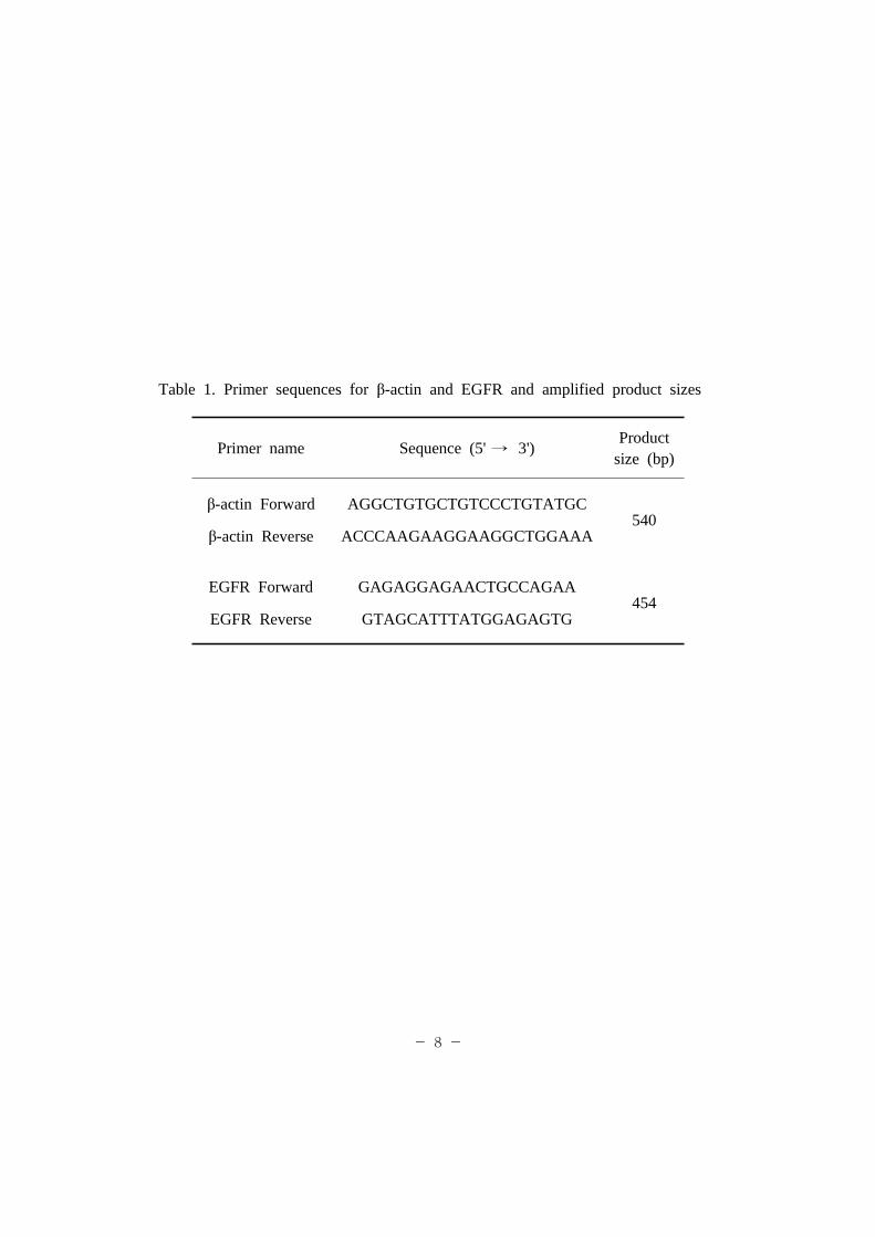
Table 1. Primer sequences for β-actin and EGFR and amplified product sizes
Primer name Sequence (5' → 3')Product
size (bp)
β-actin Forward
β-actin Reverse
AGGCTGTGCTGTCCCTGTATGC
ACCCAAGAAGGAAGGCTGGAAA540
EGFR Forward
EGFR Reverse
GAGAGGAGAACTGCCAGAA
GTAGCATTTATGGAGAGTG454
- 9 -
2.3. Experimental animal models
All animal studies were performed with the approval of the Korea
Institute of Radiological and Medical Science Animal Care and Use
Committee. Female athymic BALB/c-nu/nu (SLC, Japan), aged 5-6 weeks,
were used for animal studies. For tumor implantation, A549 cells in log phase
of growth were harvested with trypsin 0.25% and EDTA 0.02%. The cells
were counted with a hemacytometer, the viability of the cells was confirmed
with the trypan blue-dye exclusion test. Then, the cell number was adjusted to
5×10⁶ cells/mL in the serum-free media. Each mouse was subcutaneously
inoculated with 0.1 mL of the tumor suspension on the left thigh.
2.4. Preparation of DTPA-Cetuximab conjugates
To remove any metal ions in Cetuximab, Cetuximab solution (15 mg) was
incubated for 30 min at room temperature with 75 μL of 0.1 M ammonium
acetate buffer (pH 6) containing 50 mM EDTA and then concentrated with a
centrifugal filter device (MW cut-off 50 K). The buffer of Cetuximab solution
was changed to 0.25 M sodium bicarbonate (pH 8.3) and then concentrated to
1 mg/mL. Cyclic DTPA anhydride was conjugated to Cetuximab (10 mg) at
molar ratios of 50:1, 100:1, 200:1, 300:1, 500:1 and 1000:1 (DTPA:Ab). The
mixtures were incubated at 37℃ for 1 hr, and DTPA-Cetuximab conjugates
were purified and concentrated by Vivaspin-20. The buffer was changed to 0.1
M ammonium acetate buffer. The final concentration of DTPA-Cetuximab was
measured on UV absorbance at 280 nm.
- 10 -
2.5. 166Ho-labeling to DTPA-Cetuximab
The procedure for the production of 166Ho has been described in detail
elsewhere [5]. Briefly, 166Ho was produced at the HANARO (30 MW) research
reactor of KAERI by the neutron irradiation of 165Ho-nitrate [165Ho (n, γ)
166Ho]. Two hundred milligrams of 165Ho(NO3)3․5H2O was weighed and
sealed in a titanium capsule encased in an aluminum capsule and irradiated in
the reactor for 3 days. The initial specific activity of Ho-166 [166Ho(NO3)3]
reached up to 11.7 GBq/mg (315.7 mCi/mg). After 24 hr cooling, the sample
was dissolved in 3 mL of HCl solution (pH 3).
The DTPA-Cetuximab conjugates were labeled with 166Ho, according to a
previous report with minor modifications [22]. The average number of DTPA
chelators per Cetuximab was determined using a previously reported procedure
with minor modifications [23]. Briefly, a defined amount of nonradioactive
HoCl3 in 0.1 M ammonium acetate buffer (pH 6) was added to 0.4 mCi
166HoCl3, and 100 μg of each DTPA-Cetuximab conjugate in 100 μL of 0.1 N
ammonium acetate buffer were added to the above carrier-added 166HoCl3
solution. The reaction mixture was incubated at room temperature for 30 min
with vortexing at every 5 min. After purification of reaction mixture using
Vivaspin-20, the number of DTPA molecules per Cetuximab was calculated as
followed: Ho3+ moles×radiolabeling yield/DTPA-Cetuximab moles
166Ho (13.88 MBq) was added to 1.5 mg of DTPA-Cetuximab conjugates
synthesized at varied molar ratios of DTPA and Cetuximab. The reaction
mixtures were incubated at room temperature for 30 min with vortexing at
every 5 min. The radiolabeling yields and radiochemical purity of
- 11 -
166Ho-DTPA-Cetuximab were assessed by ITLC-SG, developing with 0.15 M
sodium acetate (pH 4.2). The plates were scanned and analyzed with a TLC
scanner (Aloka, Japan).
- 12 -
2.6. Biochemical characterization of 166Ho-DTPA-Cetuximab
conjugates
2.6.1 Immunoreactivity assay of 166Ho-DTPA-Cetuximab
To evaluate the immunoreactivity of 166Ho-DTPA-Cetuximab, in vitro cell
binding assay was performed with 166Ho-DTPA-Cetuximab prepared at
different molar ratio of DTPA and Cetuximab (50:1, 100:1, 200:1, 300:1,
500:1 and 1,000:1), and A549 cells (expressing EGFR). The relative
immunoreactivity was calculated as (cell bound % of
166Ho-DTPA-Cetuximab/cell bound % of 125I-Cetuximab) × 100 (%)
2.6.2 In vitro Stability assay
In vitro stability of 166Ho-DTPA-Cetuximab in serum proteins was
examined for 24 hr. 166Ho-DTPA-Cetuximab (200 μg/200 μL) was mixed with
the same volume of human serum. The mixtures was then incubated in a 37℃water bath for 1, 2, 5, 7, 18, and 24 hr. The stability of
166Ho-DTPA-Cetuximab was assessed by radiochromatography with ITLC-SG as
described earlier.
- 13 -
2.7. Biodistribution studies of 166Ho-DTPA-Cetuximab
conjugates in A549 tumor-bearing nude mice
Biodistribution analysis of 166Ho-DTPA-Cetuximab conjugates was
conducted in A549 tumor-bearing nude mice when the tumor size grew to a
diameter of 1 cm at 4-5 weeks after tumor implantation. Each tumor-bearing
mouse was injected with 0.1 mL of 166Ho-DTPA-Cetuximab conjugate (460
KBq/50 μg) via tail vein. The mice (n=4 per each time point) were sacrificed
at 2, 6, 24, 48, 72, and 144 hr after injection. The blood was collected by
cardiac puncture and the organs of skin, muscle, femur, heart, lungs, liver,
kidneys, spleen, stomach, small intestine, large intestine, and tumors were
excised carefully. The organ samples were weighed and counted in a well-type
gamma counter (1480 WIZARD well-type gamma counter, PerkinElmer,
Finland). The accumulated radioactivity was expressed as a percentage of
injected dose per gram of tissue (% ID/g). Tumor-to-blood (T/B) ratios and
tumor-to-muscle (T/M) ratios were also calculated.

- 14 -
2.8. Gamma camera images of A549 tumor-bearing nude mice
administered with 166Ho-DTPA-Cetuximab conjugates
Gamma camera images of A549 tumor-bearing nude mice were obtained
after intravenous administration of 166Ho-DTPA-Cetuximab conjugates (580
KBq/100 μg) through a tail vein. At 24 hr postinjection, the animals were
anesthetized by intraperitoneal injection of a mixture of ketamine (80 mg/Kg)
and xylazine (12 mg/Kg). Tumor images were acquired in a gamma camera
(TRIAD TXLT-20, Trionix, USA) at a 256 × 256 matrix (1×10⁶ counts).
- 15 -
III. RESULTS
1. Evaluation of EGF receptor expression on the surface of
cancer cells
In vitro Cetuximab cellular binding assay was performed to verify
expression of EGF receptors in A431, A549, SNU-C2A, U87MG, and CT-26
cells. Cellular binding of Cetuximab (%) was calculated by a formula of
(cell-bound radioactivity - non specific binding radioactivity)/total radioactivity
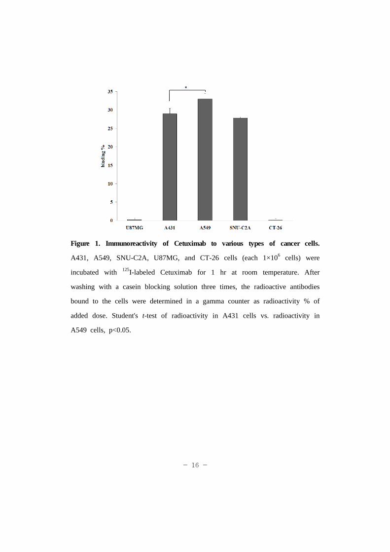
× 100 (%). 125I-Cetuximab was effectively bound to A431, A549, and
SNU-C2A cells while little bound to the U87MG and CT-26 cells. Binding %
of 125I-Cetuximab to A431, A549, SNU-C2A, U87MG, and CT-26 cells was
29.0 ± 1.5, 33.0 ± 1.5, 27.8 ± 0.3, 0.3 ± 0.4 and 0.2 ± 0.3%, respectively
(Figure 1). Presumably A549 cells have the highest expression of EGF
receptors, resulting in the highest binding of Cetuximab to the cells.
cDNA was synthesized from the total RNA extracted from EGFR-positive
A549 cells and EGFR-negative CT-26 cells. The cDNA sequences of EGFR
were amplified with the designated primer oligonucleotides mentioned earlier
(Table 1). According to the electrophoretic analysis of the amplified PCR
product, the A549 cells express a high density of EGFR while the CT-26 cells
do not (Figure 2). Based on these observations, the A549 cells were utilized
as an EGFR-positive tumor model for further in vitro and in vivo studies in
this research.
- 16 -
Figure 1. Immunoreactivity of Cetuximab to various types of cancer cells.
A431, A549, SNU-C2A, U87MG, and CT-26 cells (each 1×10⁶ cells) were
incubated with 125I-labeled Cetuximab for 1 hr at room temperature. After
washing with a casein blocking solution three times, the radioactive antibodies
bound to the cells were determined in a gamma counter as radioactivity % of
added dose. Student's t-test of radioactivity in A431 cells vs. radioactivity in
A549 cells, p<0.05.

- 17 -
Figure 2. Verification of EGFR expression in the A549 and CT-26 tumor cells
by RT-PCR. Total RNA of A549 tumor cells and CT-26 cells were extracted
and the cDNA synthesized. RT-PCR of β-actin and EGFR were performed
with appropriate primers (Table 1). After completion of the PCR, reaction
mixtures were analyzed on a 2% ethidium bromide TAE agarose gel.
- 18 -
2. Preparation of DTPA-Cetuximab conjugate
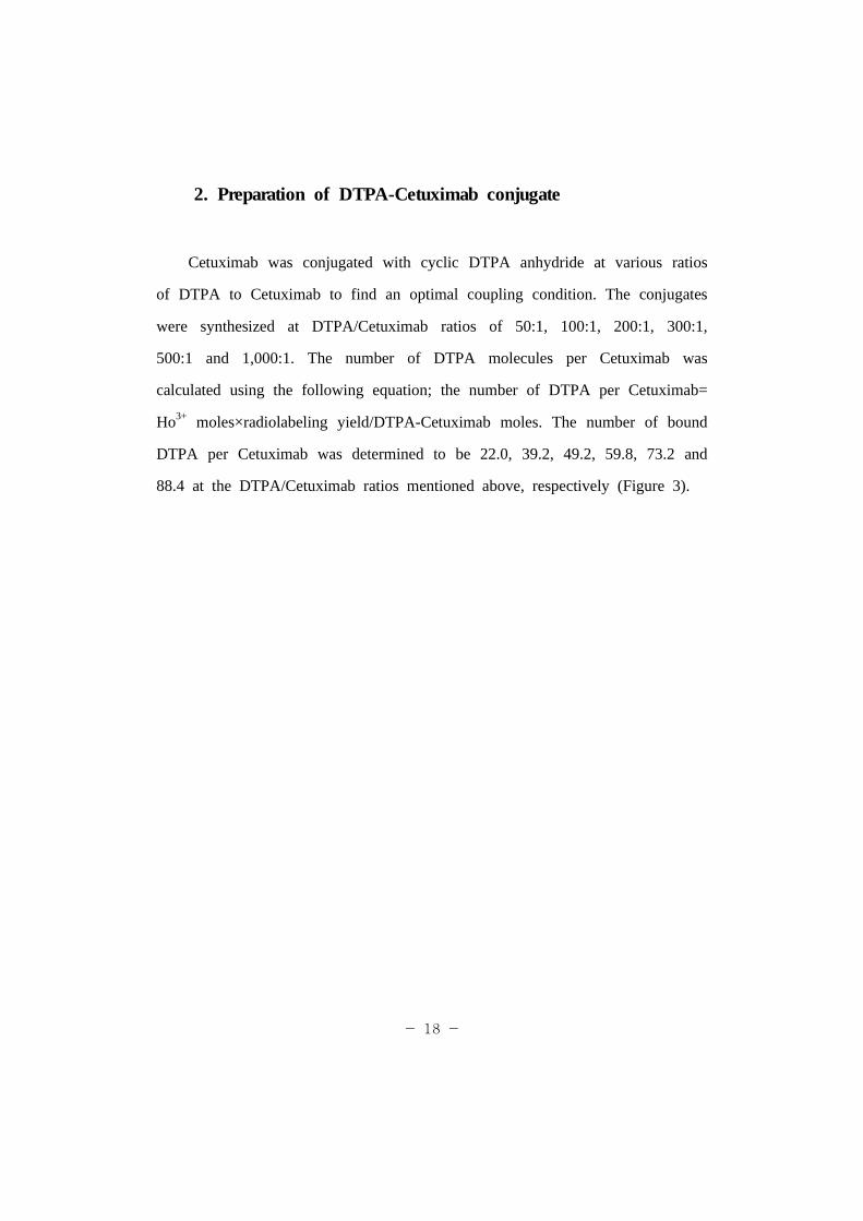
Cetuximab was conjugated with cyclic DTPA anhydride at various ratios
of DTPA to Cetuximab to find an optimal coupling condition. The conjugates
were synthesized at DTPA/Cetuximab ratios of 50:1, 100:1, 200:1, 300:1,
500:1 and 1,000:1. The number of DTPA molecules per Cetuximab was
calculated using the following equation; the number of DTPA per Cetuximab=
Ho3+ moles×radiolabeling yield/DTPA-Cetuximab moles. The number of bound
DTPA per Cetuximab was determined to be 22.0, 39.2, 49.2, 59.8, 73.2 and
88.4 at the DTPA/Cetuximab ratios mentioned above, respectively (Figure 3).
- 19 -
Figure 3. The number of DTPA molecules bound to Cetuximab depending on
reacting their molar ratios. DTPA and Cetuximab were conjugated at varied
their molar ratios. The amount of DTPA chelators bound to the Cetuximab
was calculated by equation; the number of DTPA per Cetuximab= Ho3+
moles×radiolabeling yield/DTPA-Cetuximab moles.
- 20 -
3. Radiolabeling of 166Ho to DTPA-Cetuximab and relative
immunoreactivities of the labeled conjugates
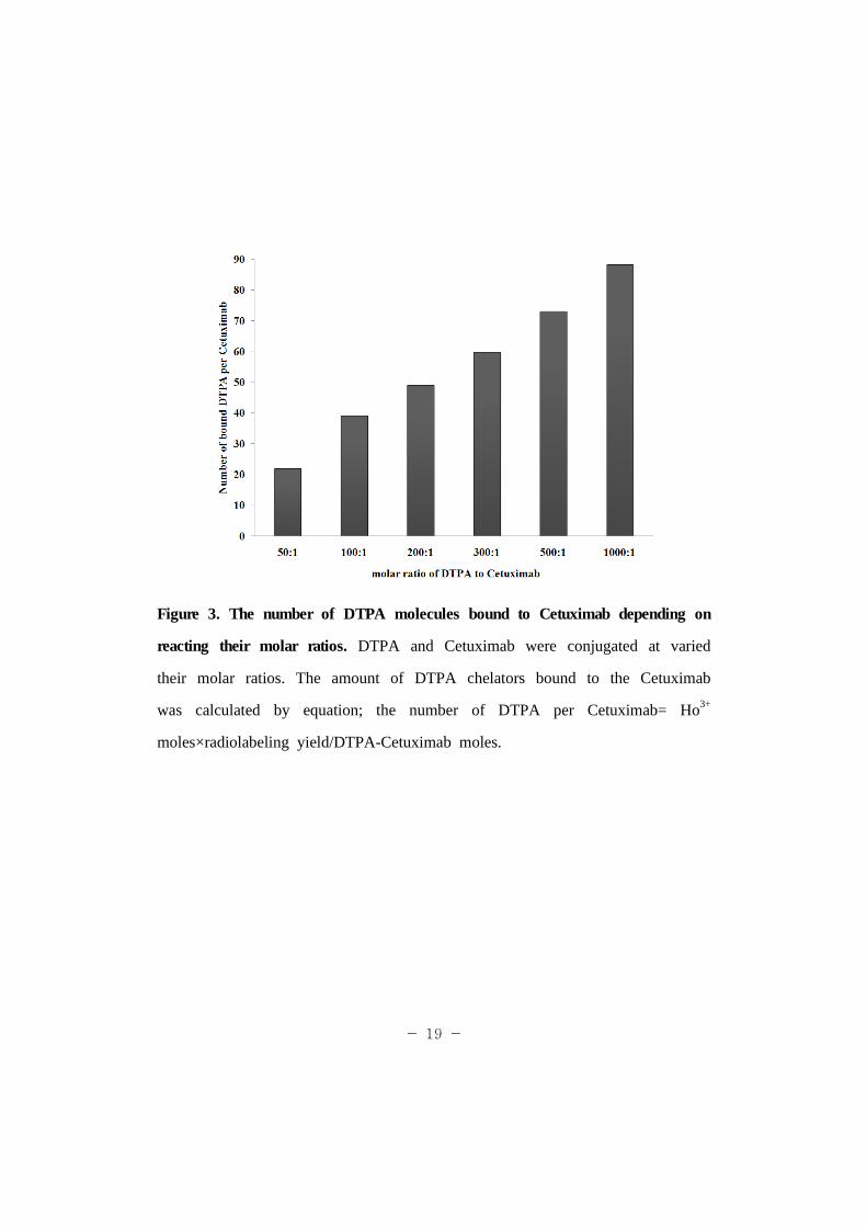
When 0.15 M sodium acetate buffer (pH: 4.2) was used as a mobile
phase, 166Ho-DTPA-Cetuximab remained at the origin (Rf=0), and free 166Ho
migrated with the solvent front (Rf=1) (Figure 4). According to the ITLC-SG
analysis, radiolabeling yields of 166Ho to DTPA-Cetuximab conjugates prepared
at 50:1, 100:1, 200:1, 300:1, 500:1 and 1000:1 molar ratios of DTPA to
Cetuximab were 63.5, 69.4, 97.2, 98.2, 97.9, and 98.9%, respectively (Figure
5, Table 2). The specific activities of the 166Ho-DTPA-Cetuximab conjugates
were 23.3, 41.8, 52.2, 63.6, 77.7, and 94.0 MBq/mg, respectively as well
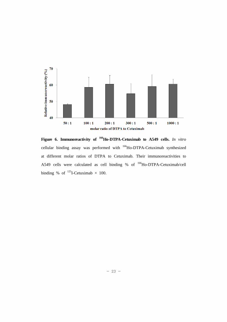
(Table 2). Although the number of DTPA per Cetuximab was different among
the six conjugates, their immunoreactivities to A549 cells were similar to each
other, 48.2 ± 0.4, 58.6 ± 6.3, 60.6 ± 5.3, 54.8 ± 6.1, 59.2 ± 7.2, and 60.7 ±
3.0% at 50:1, 100:1, 200:1, 300:1, 500:1 and 1000:1 molar ratios of DTPA to
Cetuximab (Figure 6). According to these data, the 166Ho-DTPA-Cetuximab
conjugates prepared at the 1000:1 molar ratio of DTPA to Cetuximab were
utilized for further in vitro and in vivo experiments.
- 21 -
Figure 4. Radiochromatogram of 166Ho-DTPA-Cetuximab. ITLC-SG as a
stationary phase and 0.15 M sodium acetate buffer (pH: 4.2) as a mobile
phase are used for radiochromatography. 166Ho-DTPA-Cetuximab synthesized at
the 1000:1 molar ratio of DTPA to Cetuximab remained at the origin (Rf=0),
while free 166Ho migrated with the solvent front (Rf=1).

- 22 -
Figure 5. Radiolabeling yields of 166Ho-DTPA-Cetuximab conjugates. 166Ho was
labeled to DTPA-Cetuximab conjugates synthesized at various molar ratios of
DTPA to Cetuximab. Radiolabeling yields and radiochemical purity were
assessed by ITLC-SG with the developing solvent of 0.15 M sodium acetate
buffer solution (pH 4.2).
- 23 -
Figure 6. Immunoreactivity of 166Ho-DTPA-Cetuximab to A549 cells. In vitro
cellular binding assay was performed with 166Ho-DTPA-Cetuximab synthesized
at different molar ratios of DTPA to Cetuximab. Their immunoreactivities to
A549 cells were calculated as cell binding % of 166Ho-DTPA-Cetuximab/cell
binding % of 125I-Cetuximab × 100.
- 24 -
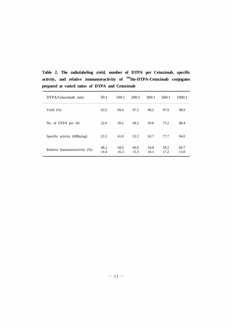
Table 2. The radiolabeling yield, number of DTPA per Cetuximab, specific
activity, and relative immunoreactivity of 166Ho-DTPA-Cetuximab conjugates
prepared at varied ratios of DTPA and Cetuximab
DTPA/Cetuximab ratio 50:1 100:1 200:1 300:1 500:1 1000:1
Yield (%) 63.5 69.4 97.2 98.2 97.9 98.9
No. of DTPA per Ab 22.0 39.2 49.2 59.8 73.2 88.4
Specific activity (MBq/mg) 23.3 41.8 52.2 63.7 77.7 94.0
Relative Immunoreactivity (%)48.2±0.4
58.6±6.3
60.6±5.3
54.8±6.1
59.2±7.2
60.7±3.0
- 25 -
4. In vitro stability of 166Ho-DTPA-Cetuximab in serum
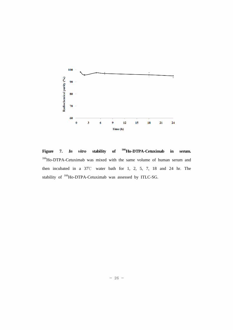
The stability of 166Ho-DTPA-Cetuximab prepared at 1000:1 molar ratio of
DTPA to Cetuximab was evaluated after incubation in the presence of serum
proteins for 1, 2, 5, 7, 18 and 24 at 37℃ . At the designated time points,
intact 166Ho-DTPA-Cetuximab conjugates were separated and analyzed by
ITLC. Radioactivities of the 166Ho-DTPA-Cetuximab conjugates were
maintained 98 ± 0.5, 96 ± 0.5, 98 ± 0.3, 97 ± 0.9, 96 ± 1.8 and 95 ± 1.5%
after incubation for 1, 2, 5, 7, 18 and 24 hr, respectively. According to these
date, it can be concluded that the structure of 166Ho-DTPA-Cetuximab
conjugate is stable in serum proteins.
- 26 -
Figure 7. In vitro stability of 166Ho-DTPA-Cetuximab in serum.
166Ho-DTPA-Cetuximab was mixed with the same volume of human serum and
then incubated in a 37℃ water bath for 1, 2, 5, 7, 18 and 24 hr. The
stability of 166Ho-DTPA-Cetuximab was assessed by ITLC-SG.
- 27 -
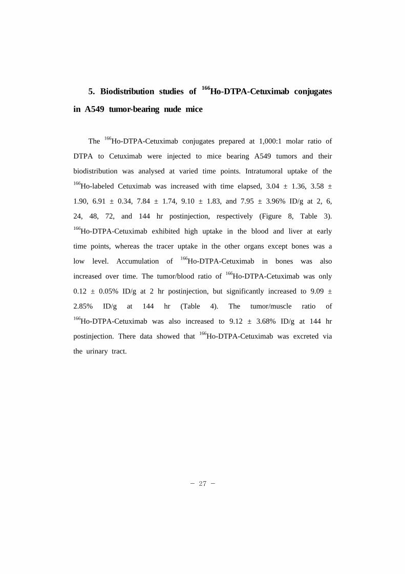
5. Biodistribution studies of 166Ho-DTPA-Cetuximab conjugates
in A549 tumor-bearing nude mice
The 166Ho-DTPA-Cetuximab conjugates prepared at 1,000:1 molar ratio of
DTPA to Cetuximab were injected to mice bearing A549 tumors and their
biodistribution was analysed at varied time points. Intratumoral uptake of the
166Ho-labeled Cetuximab was increased with time elapsed, 3.04 ± 1.36, 3.58 ±
1.90, 6.91 ± 0.34, 7.84 ± 1.74, 9.10 ± 1.83, and 7.95 ± 3.96% ID/g at 2, 6,
24, 48, 72, and 144 hr postinjection, respectively (Figure 8, Table 3).
166Ho-DTPA-Cetuximab exhibited high uptake in the blood and liver at early
time points, whereas the tracer uptake in the other organs except bones was a
low level. Accumulation of 166Ho-DTPA-Cetuximab in bones was also
increased over time. The tumor/blood ratio of 166Ho-DTPA-Cetuximab was only
0.12 ± 0.05% ID/g at 2 hr postinjection, but significantly increased to 9.09 ±
2.85% ID/g at 144 hr (Table 4). The tumor/muscle ratio of
166Ho-DTPA-Cetuximab was also increased to 9.12 ± 3.68% ID/g at 144 hr
postinjection. There data showed that 166Ho-DTPA-Cetuximab was excreted via
the urinary tract.

- 28 -
Table 3. Biodistribution of 166Ho-DTPA-Cetuximab conjugates in A549
tumor-bearing mice
Unit : %ID/g*
Organ 2hr 6hr 24hr 48hr 72hr 144hr
Blood 24.1±2.1 17.9±1.0 8.3±1.6 4.4±0.3 4.2±2.3 0.9±0.3
Liver 12.2±0.7 12.3±0.9 16.6±2.4 17.6±2.3 11.6±2.2 8.0±2.0
Lung 8.8±0.7 6.3±1.0 3.9±0.9 3.0±0.3 3.2±0.6 1.5±0.3
Spleen 5.3±0.7 4.6±0.9 6.3±1.4 9.7±3.2 6.8±1.0 6.4±1.5
Kidney 9.2±1.0 7.1±0.3 5.1±0.5 3.6±0.5 3.1±0.8 1.5±0.3
Stomach 0.9±0.3 0.7±0.4 1.9±0.2 0.5±0.4 0.7±0.1 0.6±0.3
S. Intestine 3.2±0.3 2.2±0.2 2.1±0.7 1.1±0.3 0.8±0.2 0.4±0.1
L. Intestine 2.9±0.2 2.2±0.2 2.5±0.5 1.5±0.8 1.3±0.2 0.4±0.1
Thyroid 4.3±0.5 3.7±0.4 4.0±1.3 2.4±0.3 3.8±0.5 3.2±0.4
Femur 3.6±1.0 5.0±0.1 6.1±2.1 6.3±1.3 9.0±2.3 8.9±0.8
Muscle 2.1±0.6 2.0±0.2 2.8±1.1 1.0±0.1 1.5±0.3 1.1±0.8
A549 3.0±1.4 3.6±1.9 6.9±0.3 7.8±1.7 9.1±1.8 8.0±4.0
*Values at each time point represent the mean ±s.d. of percentage of injected dose per gram
of tissue weight (n=4).

- 29 -
Figure 8. Biodistribution of 166Ho-DTPA-Cetuximab conjugates in A549
tumor-bearing mice. Each tumor-bearing mouse was injected with 0.1 mL of
166Ho-DTPA-Cetuximab conjugates (460 KBq/50 μg) into a tail vein. Collected
organs were weighed and radioactivities of the organs were counted in a
well-type gamma counter. The accumulated radioactivity was expressed as a
percentage of injected dose per gram of tissue (% ID/g).

- 30 -
Table 4. Organ per Tissue ratio of 166Ho-DTPA-Cetuximab conjugates in A549
tumor-bearing mice
Organ/Tissue 2hr 6hr 24hr 48hr 72hr 144hr
Tumor/Blood ratio 0.1±0.1* 0.2±0.1 0.9±0.2 1.8±0.3 2.6±1.1 9.1±2.9
Tumor/Muscle ratio 1.7±1.2 1.8±1.0 2.7±2.4 7.8±2.4 6.2±0.9 9.1±3.7
*Mean ± S.D., n=4.
- 31 -
6. Gamma camera images of A549 tumor-bearing nude mice
administered with 166Ho-DTPA-Cetuximab conjugate
Gamma camera images of A549 tumor-bearing nude mice were acquired
to evaluate in vivo tumor targetability of 166Ho-DTPA-Cetuximab.
166Ho-DTPA-Cetuximab conjugates (580 KBq/100 μg) were administrated
through a tail vein. At 24 hr postinjection, tumor images were acquired in a
256 × 256 matrix (1×10⁶ counts). According to the gamma camera image at
24 hr postinjection, 166Ho-DTPA-Cetuximab was selectively localized in the left
thigh with EGFR-positive A549 tumors, but not in the control right thigh.
However, the resolution of the gamma images was not good enough to
provide clear selective localization of the 166Ho-DTPA-Cetuximab in the A549
xenograft.
- 32 -
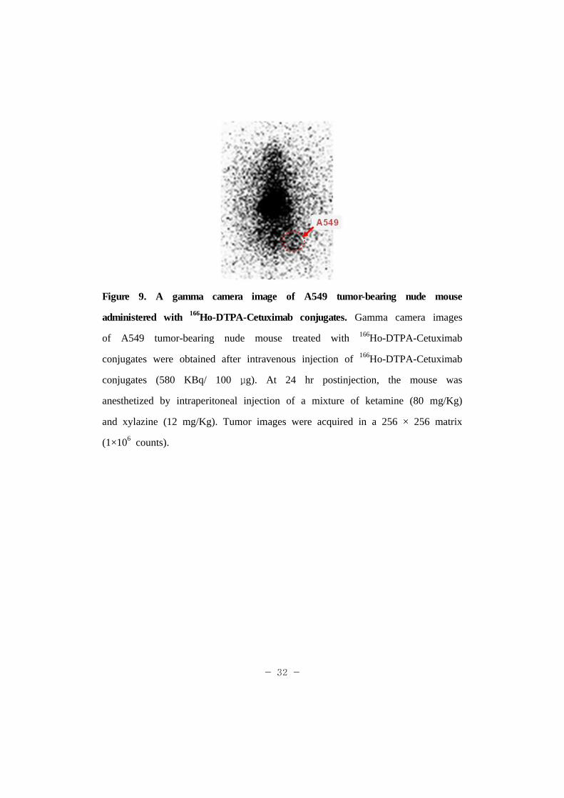
Figure 9. A gamma camera image of A549 tumor-bearing nude mouse
administered with 166Ho-DTPA-Cetuximab conjugates. Gamma camera images
of A549 tumor-bearing nude mouse treated with 166Ho-DTPA-Cetuximab
conjugates were obtained after intravenous injection of 166Ho-DTPA-Cetuximab
conjugates (580 KBq/ 100 μg). At 24 hr postinjection, the mouse was
anesthetized by intraperitoneal injection of a mixture of ketamine (80 mg/Kg)
and xylazine (12 mg/Kg). Tumor images were acquired in a 256 × 256 matrix
(1×10⁶ counts).
- 33 -
IV. DISCUSSION
Many radioisotopes and monoclonal antibodies have been widely used in the
diagnosis and therapy of various types of cancers. Especially, in recent
targeted RIT has been clinically validated and approved for the treatment of
cancer by the US Food and Drug Administration.
To label 166Ho to Cetuximab, first of all chelators for radioisotope, DTPA in
this study, have to be conjugated to Cetuximab molecules. Therefore,
conjugation conditions have to be optimized to have the stable
DTPA-Cetuximab conjugates with a strong immunoreactivity against tumor
cells. DTPA is known to stably chelate metals such as 99mTc [24]. The DTPA
was added to the Cetuximab solution at the DTPA/Cetuximab ratios of 50:1,
100:1, 200:1, 300:1, 500:1 and 1,000:1 used for conjugation reaction. The
higher amount of DTPA was added, the higher number of DTPA was coupled
to Cetuximab molecules. At the 1,000:1 ratio, approximately 88 DTPA
molecules were conjugated to one molecule of Cetuximab antibody.
166Ho-DTPA-Cetuximab conjugated at the 1,000:1 ratio exhibited the highest
radiolabeling yield and the highest specific activity among the conjugates.
Also, the relative immunoreactivity of the 68Ho-labeled Cetuximab was
increased with increase of DTPA number per molecule. Previously it has been
reported that conjugation of bifunctional chelating agents diminishes
immunoreactivity of monoclonal antibodies [25]. However, in this study, the
immunoreactivity of Cetuximab was little affected by the increased number of
DTPA. This suggests that the accessible lysine residues for DTPA conjugation
are fairly away from the complementarity-determining region of Cetuximab. In
- 34 -
addition, the 166Ho-DTPA-Cetuximab was very stable in serum proteins. The
radiochemical purity of 166Ho-DTPA-Cetuximab was reduced only less than 5%
after incubation with human serum at 37℃ for 24 hr.
The immunoreactivity of the 68Ho-labeled Cetuximab was examined in the
A549 tumor model. In order to establish an appropriate tumor model for in
vitro and in vivo experiments, A431, A549, SNU-C2A, U87MG, and CT-26
tumor cells were treated with 125I-labeled Cetuximab. 125I is a convenient
radionuclide for laboratory studies because it has relatively long half-life (60.14
d) and emits a short-range of Auger and Coster-Kronig electrons [26].
According to the immunoreactivity assay with the cancer cell lines, A549 cells
showed the highest affinity to the 125I-labeled Cetuximab than the other types
of tumor cells, implying the highest expression of EGF receptors. The EGFR
expression in A549 cells was also confirmed by RT-PCR.
The 166Ho-DTPA-Cetuximab prepared at the 1,000:1 molar ratio of DTPA to
Cetuximab was able to effectively accumulated in the A549 tumor xenografts
in vivo. The intratumoral uptake of 166Ho-DTPA-Cetuximab was increased with
time elapsed and reached a maximum at 72 hr postinjection. The 166Ho-labeded
Cetuximab exhibited a relatively higher uptake in the blood, liver and bones at
early time points. It is important to note that free 68Ga and 111In tend to
localize in the liver and lungs due to their strong binding capability to
transferrin while 90Y and lanthanide isotopes are readily deposited on the bone
[27]. The tumor/blood ratio and tumor/muscle ratio of 166Ho-DTPA-Cetuximab
reached 9.1 ± 2.9% ID/g and 9.1 ± 3.7% ID/g at 144 hr postinjection. This
result also shows the specific localization of the conjugates in A549 tumors
mediated by Cetuximab.

- 35 -
However, the low resolution of gamma camera images which were acquired
after intravenous injection of 166Ho-DTPA-Cetuximab was rather disappointing.
166Ho-DTPA-Cetuximab appeared to be selectively localized in the A549 tumor
xenograft at 24 hr postinjection, but gave a poor resolution of image. These
results suggest that 166Ho-DTPA-Cetuximab need a further optimization process
before preclinical and clinical applications as a radioimmunotherapeutic agent
against EGFR expressing tumors.
- 36 -
VI. REFERENCE
1. Milenic, D.E., Brady, E.D., Brechbiel, M.W. Antibody-targeted radiation
cancer therapy. Nat Rev Drug Discov 2004; 3: 488-99.
2. Allen, T.M. Ligand-targeted therapeutics in anticancer therapy. Nat Rev
Drug Discov 2002; 2: 750-763.
3. Boswell, C.A., Brechbiel M.W. Development of radioimmunotherapeutic
and diagnostic antibodies: an inside-out view. Nucl Med Biol 2007; 34(7):
757-778.
4. Goldenberg, D.M. Targeted therapy of cancer with radiolabeled antibodies. J
Nucl Med 2002; 43: 693–713.
5. Hong, Y.D., Park, K.B., Jang, B.S., et al. Holmium-166-DTPA as a liquid
source for endovascular brachytherapy. Nucl Med Biol 2002; 29: 833-839.
6. Suzuki, Y.S., Momose, Y., Higashi, N. et al. Biodistribution and kinetics of
Ho-166-chitosan complex in rats and mice. J Nucl Med 1998; 39: 2161-2166.
7. Kassis, A.I., Adelstein, S.J.. Radiobiologic principles in radionuclide
therapy. J Nucl Med 2005; 46: 4S-12S.
- 37 -
8. Rowinsky, E.K. The erbB family: targets for therapeutic development
against cancer and therapeutic strategies using monoclonal antibodies and
tyrosine kinase inhibitors. Annu Rev Med 2004; 55: 433-457.
9. Harding, J., Burtness, B. Cetuximab: an epidermal growth factor receptor
chemeric human-murine monoclonal antibody. Drugs Today(Barc.) 2005; 41:
107–127.
10. Sunada, H., Magun, B.E., Mendelsohn, J., et al. Monoclonal antibody
against epidermal growth factor receptor is internalized without stimulating
receptor phosphorylation. Proc Natl Acad Sci USA 1986; 83: 3825-3829.
11. Thienelt, C.D., Bunn, P.A. Jr., Hanna, N. et al. Multicenter phase I/II
study of cetuximab with paclitaxel and carboplatin in untreated patients with
stage IV nonsmall- cell lung cancer. J Clin Oncol 2005; 23: 8786-8793.
12. Xiong, H.Q., Rosenberg, A., LoBuglio, A., et al. Cetuximab, a monoclonal
antibody targeting the epidermal growth factor receptor, in combination with
gemcitabine for advanced pancreatic cancer: a multicenter phase II trial. J Clin
Oncol 2004; 22: 2610-2616.
- 38 -
13. Bonner, J.A., Harari, P.M., Giralt, J., et al. Radiotherapy plus cetuximab
for squamous-cell carcinoma of the head and neck. N Engl J Med 2006; 354:
567-578.
14. Schechter, N.R., Yang, D.J., Azhdarinia, A. et al. Assessment of epidermal
growth factor receptor with 99mTc-ethylenedicysteine-C225 monoclonal antibody.
AntiCancer Drugs 2003; 14: 49-56.
15. Schechter, N.R., Wendt, R.E. 3rd., Yang, D.J., et al. Radiation dosimetry
of 99mTc-labeled C225 in patients with squamous cell carcinoma of the head
and neck. J Nucl Med 2004; 45: 1683–1687.
16. Perk, L.R., Visser, G.W., Vosjan, M.J., et al. 89Zr as a PET surrogate
radioisotope for scouting biodistribution of the therapeutic radiometals 90Y and
177Lu in tumor-bearing nude mice after coupling to the internalizing antibody
cetuximab. J Nucl Med 2005; 46: 1898–1906.
17. Cai, W., Chen, K., He, L., et al. Quantitative PET of EGFR expression in
xenograft-bearing mice using 64Cu-labeled cetuximab, a chimeric anti-EGFR
monoclonal antibody. Eur J Nucl Med Mol Imaging 2007; 34: 850–858.

- 39 -
18. Pippin, C.G., Parker, T.A., McMurry, T.J., et al. Spectrophotometric
method for the determination of a bifunctional DTPA ligand in
DTPA-monoclonal antibody conjugates. Bioconjug Chem 1992; 3: 342-345.
19. Sherry, A.D., Brown, R.D. III, Gerades, C.F.G., et al. Synthesis and
characterization of the gadolinium(3+) complex of DOTA propylamide: a
model DOTA-protein conjugate. Inorg Chem 1989; 28: 620-622.
20. Sieving, P.F., Watson, A., Rocklage, S.M. Preparation and characterization
of paramagnetic polychelates and their protein conjugates. Bioconjug Chem
1990; 1: 65-71.
21. Bor, M.V., Sørensen, B.S., Rammer, P., and Nexø, E. Calibrated
user-friendly reverse transcriptase-PCR assay: Quantitation of epidermal growth
factor receptor mRNA. Clin Chem 1998; 44: 1154-1160
22. Ekaterina, D., Saed, M., Suzanne, V., et al. Radiolabeling antibodies with
Holmium-166 Appl Radiat Isot 1997; 48: 477-481
23. Meares, C.F., McCall, M.J., Reardan, D.T., et al. Conjugation of antibodies
with bifunctional chelating agents: isothiocyanate and bromoacetamide reagents,
methods of analysis, and subsequent addition of metal ions. Anal Biochem
1984; 142: 68-78.
- 40 -
24. Eckelman, W.C., Levenson, S.M., Radiopharmaceuticals labelled with
technitium. Int J Appl Rad Iso 1977; 28: 67-82.
25. Cai, W., Wu, Y., Chen, K., et al. In vitro and In vivo Characterization of
64Cu-Labeled AbegrinTM, a Humanized Monoclonal Antibody against Integrin αv
β3. Cancer Res 2006; 66(19): 9673-81.
26. Buchsbaum, D.j., Langmuir, V.K., Wessels, B.W. Experimental
radioimmunotherapy. Med Phys 1993; 20: 551-67.
27. Ando, A., Ando, I., Hiraki, T., et al. Relation between the location of
elements in the periodic table and various organ-uptake rates, Nucl Med Biol
1989; 16: 57-80.
- 41 -
국국국문문문 요요요약약약
166Ho-DTPA-Cetuximab의의의 제제제조조조와와와생생생물물물학학학적적적평평평가가가
연세대학교 대학원 임상병리학과
서 형 엽
Ho의 방사성동위원소 166Ho(γ에너지 : 0.081 MeV, 1.38 MeV, β-에너지 :
1.78 MeV, 1.84 MeV, 반감기 : 26.8 hr)은 β-의 큰 에너지를 방출하기 때문에 방사면역치료에 적합한 물리적 특성을 지니고 있으며 γ선에 의한 핵의학 영상이 가능한 장점이 있다 . Cetuximab은 EGFR에 대한 키메릭 단클론항체로서 현재 EGFR을 과발현하는 고형암 치료에 사용되고 있다 . 본 연구에서는 166Ho을 먼저 bifunctional chelating agent인 DTPA와 conjugation 시킨후 Cetuximab에 방사성 표지하여 종양형성 누드마우스에서 생체 분포 및생체 영상을 평가함으로써 방사면역진단 및 치료제로써 이용가능성을 보고자 하였다 . EGFR의 발현 정도를 평가하기 위해서 5가지 종양세포 (A431,
A549, SNU-C2A, U87MG, CT-26 tumor cells)에서 125I-Cetuximab을 이용해 in
vitro cell binding assay를 실시하였다 . A549 종양세포가 다른 나머지 세포들에 비해 높은 EGFR의 발현을 한다는 것을 확인한 후 RT-PCR을 통해A549 세포의 EGFR의 발현을 확인하였다 . Cetuximab에 다양한 몰비 (50:1,
- 42 -
100:1, 200:1, 300:1, 500:1, 1000:1)의 cyclic DTPA를 반응시켜DTPA-Cetuximab을 제조하였다 . Cetuximab에 결합한 DTPA의 수는 DTPA와Cetuximab의 몰비가 1000:1일 때 88.4개로 가장 많은 수가 결합하였고166Ho의 방사성 표지수율 또한 98.9%로 가장 높았다 . 166Ho-DTPA-Cetuximab
의 면역반응성과 비방사능은 각각 60.7 ± 3.0%와 94.0 MBq/mg이었다 .
A549 종양이식 마우스에서 166Ho-DTPA-Cetuximab은 72시간째에 가장 높은종양으로의 집적 (9.1 ± 1.8% ID/g)을 나타냈고 144시간째에 가장 높은 종양대 혈액비 (9.1 ± 2.9)와 종양 대 근육비 (9.1 ± 3.7)를 나타내었다 . 또한166Ho-DTPA-Cetuximab을 A549 종양 형성 누드마우스에 주사하여 24 시간후에 gamma camera 영상에서 종양으로의 집적을 확인할 수 있었다 . 이 결과들을 통해 166Ho을 방사성 표지한 높은 비방사능을 가진 DTPA-Cetuximab
이 EGFR을 과다 발현하는 종양에 대한 방사면역치료제로의 이용가능성을확인하였다 .
핵심어 : Cetuximab, 166Ho, DTPA, 방사면역치료