premature ovarian failure: a critical condition in the reproductive
TRANSCRIPT
1
Review Article
Received: 1 Nov 2012, Accepted: 2 Jun 2013* Corresponding Address: P.O. Box: 1985-717443, Department of Medical Genetics, Faculty of Medicine, Shahid Beheshti University of Medical Sciences, Tehran, IranEmail: [email protected]
Premature Ovarian Failure: A Critical Condition in The Reproductive Potential with
Various Genetic Causes
Farkhondeh Pouresmaeili, Ph.D.1, 2*, Zahra Fazeli, M.Sc.1
1. Department of Medical Genetics, Faculty of Medicine, Shahid Beheshti University of Medical Sciences, Tehran, Iran
2. Infertility and Reproductive Health Research Center (IRHRC), Shahid Beheshti University of Medical Sciences, Tehran, Iran
AbstractPremature ovarian failure (POF) is identified as a heterogeneous disorder leading to amenorrhea and ovarian failure before the age of 40 years. The first known symp-tom of the disease is having irregular menstrual periods. The phenotype appear-ance of POF depends significantly on the variations in hormones. Low levels of gonadal hormones (estrogens and inhibins) and increased level of gonadotropins [luteinizing hormone (LH) and Follicle stimulating hormone (FSH)] (hypergonado-tropic amenorrhea) are well documented as causes of POF. There is an association between the failure of germ cell development and complete ovarian failure, and consistently decreased number of germ cells is more likely associated with partial ovarian failure resulting in secondary amenorrhea. A literature review on recent findings about POF and its association with genomic alterations in terms of genes and chromosomes. POF is a complex heterogeneous disorder. Some of POF cases are carriers of a single gene mutation inherited in an autosomal or X-linked manner while a number of patients suffer from a chromosome abnormality like Turner syn-drome in mosaic form and manifest secondary amenorrhea associated with ovarian dysgenesis. Among many of the known involved genes in POF development, several are prove to be positively associated to the disease development in different popula-tions. While there is a promising association between X chromosome anomalies and specific gene mutations with POF, genome-wide analysis could prove a powerful tool for identifying the most important candidate genes that influence POF mani-festation.
Keywords: Premature Ovarian Failure, Infertility, Amenorrhea
Citation: Pouresmaeili F, Fazeli Z. Premature ovarian failure: a critical condition in the reproductive potential with various genetic causes. Int J Fertil Steril. 2014; 8(1): 1-12.
Royan InstituteInternational Journal of Fertility and Sterility Vol 8, No 1, Apr-Jun 2014, Pages: 1-12
Introduction Premature ovarian failure (POF [MIM 311360])
is an early ovarian malfunction different from men-opause, which disturbs production of follicles re-sulting in amenorrhea under the age of 40 in 1-3% of reproductive age women (1-3). About 10-28% of the patients experience primary amenorrhea and about 4-18% show secondary Amenorrhea (2). Af-fected women show menstrual problems followed
by an elevated level of gonadotropines [follicle stimulating hormone (FSH) ≥40IU/L] and hypoes-trogenism for an average four months (4). Measur-ing serum FSH is a routine diagnosis procedure for the disease (2, 5). Genetic analysis of the early menopause patients showed that a variety of gene defects and chromosome anomalies in-volving X chromosome and autosomes were as-sociated with POF. The studies confirmed that
Int J Fertil Steril, Vol 8, No 1, Apr-Jun 2014 2
Pouresmaeili and Fazeli
the multifactorial and heterogeneous biological events including infection, autoimmune disor-ders and metabolic factors are likely respon-sible for the disorder development. In 90% of observed cases, the etiology is unknown and the disease is defined as idiopathic POF (2, 6-8).
Most POF cases are sporadic and it is sug-gested that between 4-31% of them are familial (9-11). In this regard, the abnormalities of the X chromosome are presented as the most im-portant causes of the disease (12-17), followed by the fragile X mental retardation (FMR1) pre-mutation which is present in POF patients with frequencies of 13 and 6%, respectively (18, 19).
Loss of one X chromosome as X monosomy [Turner syndrome (TS)], the related gene dele-tions and X/autosome translocations, trisomy X, X linked gene mutations and premutations and anomalies of autosomal linked genes have been widely studied in correlation with POF disease. In examining the genetic mutations re-sponsible for POF, each mutation could affect part of the disease phenotype. The diagnosis of the disease could be confirmed by two separate blood tests for FSH (4). Studies in different populations have shown various factors in as-sociation with POF. In addition to the genetic anomalies and chromatin structure of specific genome environment, autoimmune factors and toxins are reported as other important causes of the disease. The exact reason for POF develop-ment still remains unknown in many cases. Cytogenetic analysis for POFChromosome abnormalities
In different reports, chromosomal abnormalities have been recognized as the most common causes of POF disease (12-17, 20), confirming the importance of cytogenetic analysis in reproductive management and genetic counseling for this disease. Investigations in this regard show the association of POF with chro-mosome abnormalities, particularly those of X chro-mosome such as structural anomalies, translocation of X with autosomes, isochromosomes and the related aneuploidies (21-25). Also, translocation of Y chro-mosome heterochromatic regions on derivative X chromosome which affected the X chromosome inactivation was reported (26). Presence of a Y chromosome in a woman’s genome is a clear
sign of chromosome abnormality which mostly causes tumor formation in mosaic karyotypes. Pouresmaeili et al. reported a patient with POF who carried aneuploidy of this kind (27). Un-known X-linked gene imbalance is an expected cause of POF in these patients.
A critical region from Xq13.3 to Xq27 has been characterized for ovarian development and func-tion (28). Studies have shown that deletion of the short arm and the long arm of the X chromosome result in either early primary or secondary amen-orrhea (29). These observations suggested that important genes for normal ovarian function are located on both arms of the X chromosome (30).
Translocational studies between X chromosome and autosomes have been significant in determin-ing the involvement of autosomal as well as X chromosomal genes in the development of POF disease. For example, the association of HS6ST1, HS6ST2, MATER and CHM genes with POF were identified following to the analysis of the POF pa-tients with karyotypes 46, X, der (X) t (X; 19) (p21; q13), 46, X, t (X; 2) (q21; q14), 46, X, der (X)t (X; Y) (q25-26; q11.22), 46, X, t (X; 4) (q21.2; p16.3) respectively (31, 32). One possible explanation for the disease occurrence in these cases was at-tributed to the positional effect of an autosome-X chromosome translocation. In fact, transferred genes to the highly heterochromatic region of the X chromosome tolerate epigenetic effects after a rearrangement which changes their chromatin structure and consequently result in lower expres-sion of genes associated with ovarian function and fertility (33).
Moreover, some studies have reported a Rob-ertsonian translocation (13, 14) in some women with sporadic POF. It was also suggested that the functional changes and interruptions in some criti-cal genes for ovarian function on acrocentric chro-mosomes, due to translocation, could be a possible reason of the disease etiology in this group of pa-tients (20, 34, 35).
Also, there are POF patients with trisomy X who were diagnosed after showing an endocrine disor-der, hypergonadotropic hypogonadism (36). It is suggested that the genes located on the X chro-mosome escaping inactivation could be overex-pressed in 47, XXX patients, resulting in the dis-order revelation (37). It is identified that complete
3
POF Genetics
absence or a segmental deletion of one X chro-mosome (TS) causes abnormalities throughout the reproductive system. Using fluorescent in situ hybridization (FISH) and several specific mark-ers to the short arm of X chromosome including DXS1058, DXS6810, DXS1302 and ZXDB, dele-tion of Xp11.2-p22.1 was introduced as a critical area related to TS and POF (38). Therefore, we un-derstand that the presence of two intact X chromo-somes is indigence vital for normal ovarian func-tion (8, 39-41) and prevents follicle apoptosis and atresia (41). This hypothesis is supportive for the POF etiology when there is a significant difference between the highest and lowest follicle numbers in mosaic Turner syndrome and subjects with 45, X karyotype (42).
Trisomy X with or without Turner’s syndromeChromosome aneuploidy leading to trisomy X is
known as one of the genetic reasons of POF which causes elevated endocrine gonadotropine hormone (FSH) with an incidence rate of 1:1000 female live births. Although the ovarian function is normal in most of trisomy X patients, but the ovarian dys-function in some 47, XXX might manifest as early menopause, secondary amenorrhea and oligomen-orrhea (36).
Mosaic types of the syndrome include 10% of the cases with various karyotypes such as 46, XX/47, XXX or 45, X/47, XXX. The mosaic tri-somy X might be the result of a post-zygotic non-disjunction event or post-zygotic trisomy rescue. The manifestation of symptoms depends on the time at which the causing events occurred.
The cytogenetic analysis done on POF patients indicated that trisomy X (regardless of mosaic or non-mosaic) have low frequency in individuals with POF (30, 37, 43).
The patients carried different symptoms with ab-normalities in genitourinary tract which could be associated with the trisomy status (44). Although some of the cases showed uterine dysgenesis, oth-er cases had no defects in the reproductive system and sexual development (37).
Investigations have shown that the autoimmune thyroid disease is related to many POF cases with trisomy X (45). The ovarian failure in 47, XXX patients (either mosaic or non-mosiac) could be the result of meiotic disorganization of three X
chromosomes (20). However, more studies on 47, XXX patients with POF are required.
POF and gene mutations Numerous studies have identified different genes
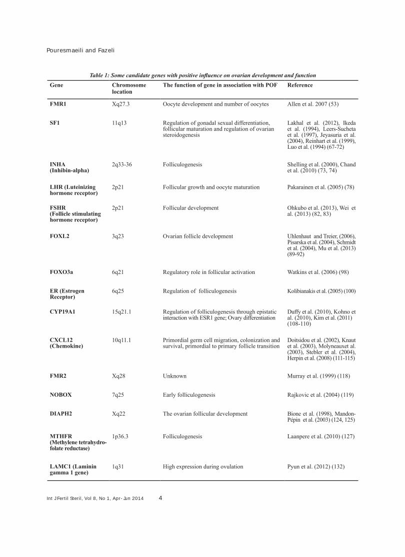
whose functions were significant in ovarian devel-opment and also play a role in POF progression. However, inconsistent results have been observed in different studies which are presumably the re-sult of genetic variability between studied ethnic groups (46-48). Table 1 describes several candi-date genes with possible involvement in ovarian function and POF genesis. All the introduced gen-ic variation as mutations and polymorphisms are thought to affect POF (30).
The FMR1 premutation of CGG repeats with incidence of 1:800 in males and 1:100-200 in women is recognized as the most important gene associated with POF (49-51). The authors mention that carrier women of the premutation are predis-posed to POF disease. Expression study on frag-ile X mental retardation protein has demonstrated that the variation in the level of the gene product (FMRP) could be utilized as a candidate biomarker to evaluate a person with folliculogenesis disrup-tion, heavy follicle atresia and eventual POF. In this study immunocytochemistry was applied on ovarian sections to show different FMRP1 signals in tissues with different CGG repeats. These ex-pression studies showed that the elevated amount of the expressed gene in fetus germ line cells have a negative effect on the number of oocytes and their development (52, 53). The incidence of the disease is about 0.1-1% in normal individuals but 20-28% in the carriers of premutation FMR1, 13 times more than controls, respectively (54-58). Some studies indicated that the intermediate al-leles of FMR1 CGG repeats could also increase the risk of POF development (57, 59-66). SF1, a nuclear receptor which is expressed in vari-ous cell types in fetus and adult, regulates different genes involved in development of the reproductive system, hypothalamic-pituitary-steroidogenesis and familial or isolated POF. The gene polymorphism Gly146Ala resulted from GGG to GCG sequence is known to be associated with POF in either familial or isolated form. Carriers of the 146Ala allele showed a significant decline in plasma stradiol. Therefore, this polymorphism could be a risk marker for POF is some women (67-72).
Int J Fertil Steril, Vol 8, No 1, Apr-Jun 2014 4
Pouresmaeili and Fazeli
Table 1: Some candidate genes with positive influence on ovarian development and functionReferenceThe function of gene in association with POFChromosome
locationGene
Allen et al. 2007 (53)Oocyte development and number of oocytesXq27.3FMR1
Lakhal et al. (2012), Ikeda et al. (1994), Leers-Sucheta et al. (1997), Jeyasuria et al. (2004), Reinhart et al. (1999), Luo et al. (1994) (67-72)
Regulation of gonadal sexual differentiation, follicular maturation and regulation of ovarian steroidogenesis
11q13SF1
Shelling et al. (2000), Chand et al. (2010) (73, 74)
Folliculogenesis2q33-36INHA(Inhibin-alpha)
Pakarainen et al. (2005) (78)Follicular growth and oocyte maturation2p21LHR (Luteinizing hormone receptor)
Ohkubo et al. (2013), Wei et al. (2013) (82, 83)
Follicular development2p21FSHR (Follicle stimulating hormone receptor)
Uhlenhaut and Treier, (2006), Pisarska et al. (2004), Schmidt et al. (2004), Mu et al. (2013) (89-92)
Ovarian follicle development3q23FOXL2
Watkins et al. (2006) (98)Regulatory role in follicular activation6q21FOXO3a
Kolibianakis et al. (2005) (100)Regulation of folliculogenesis6q25ER (Estrogen Receptor)
Duffy et al. (2010), Kohno et al. (2010), Kim et al. (2011) (108-110)
Regulation of folliculogenesis through epistatic interaction with ESR1 gene; Ovary differentiation
15q21.1CYP19A1
Doitsidou et al. (2002), Knaut et al. (2003), Molyneauxet al. (2003), Stebler et al. (2004), Herpin et al. (2008) (111-115)
Primordial germ cell migration, colonization and survival, primordial to primary follicle transition
10q11.1CXCL12(Chemokine)
Murray et al. (1999) (118)UnknownXq28FMR2
Rajkovic et al. (2004) (119)Early folliculogenesis7q25NOBOX
Bione et al. (1998), Mandon-Pépin et al. (2003) (124, 125)
The ovarian follicular developmentXq22DIAPH2
Laanpere et al. (2010) (127)Folliculogenesis1p36.3 MTHFR(Methylene tetrahydro-folate reductase)
Pyun et al. (2012) (132)High expression during ovulation1q31LAMC1 (Laminin gamma 1 gene)

5
POF Genetics
Inhibins are of other POF candidate genes pre-dominantly produced in the ovary at different times of the menstrual cycle and play a regulatory role in folliculogenesis (73, 74). Among these proteins, Inhibin alpha (INHA) gene polymorphisms have been shown to have a significant association with risk of POF in certain ethnic populations (74-77).
The luteinizing hormone (LH) through luteiniz-ing hormone receptor (LHR) plays an important role in the follicular growth and oocyte maturation. Women carrying LHR mutations showed anovula-tion and primary amenorrhea (78-81).
FSH receptor (FSHR) is expressed in the granulosa cells of the ovary and has an impor-tant function in follicular development (82, 83). In different studies on Chinese, Argentinian and British women, it has been revealed that FSH receptor gene mutations are seldom identified in POF patients. First, mutations of FSHR and later sentences are talking about them FSHR gene polymorphisms which are different terms (84-88).
FOXL2 was found in undifferentiated granulosa cells of the ovary which are involved in ovarian follicle development (89-92). The studies have in-dicated that FOXL2 mutations are rarely associ-ated with POF disease (93-95).
The FOX3a is also expressed in the ovary with a regulatory role in the follicular activation (96). Al-though some mutations of the encoding gene have been reported in women with POF, it seems that FOX3a mutations could not be counted as a com-mon cause (97-99).
Estrogen stimulates gonadotropins releasing at the hypothalamus-hypophysis-ovarian axis by act-ing on estrogen receptor-α (ESR1) which enhances folliculogenesis (100). It has been reported that several single nucleotide polymorphisms (SNPs) such as rs2234693, rs9340799 and rs2234693 of ESR1 are associated with the increased risk of POF (101-104). Some studies have suggested an association between Estrogen receptor alpha gene polymorphism, PvuII and XbaI restriction frag-ment length polymorphisms (RFLPs) and low bone mineral density (BMD) (105), while others observed no significant correlation between these polymorphisms with age, menopausal status and BMD (106). Nevertheless, it has been revealed
that baseline BMD and change in menstrual status contributed more to the magnitude of the differ-ence in bone change (107).
The CYP19A1 gene encodes aromatase, the key enzyme in biosynthesis of estrogens. High expres-sion of aromatase during the ovary differentiation has been reported previously (108, 109). Investi-gations on Korean patients with POF have shown a significant association between 3’ UTR SNPs "rs10046" and "rs4646" of CYP19A1 with the dis-ease (110).
It has been demonstrated that CXCL12 through its receptor acts on migration and survival of pri-mordial germ cells (PGCs) (111-115). The asso-ciation between CXCL12 polymorphisms and POF has been observed in the studied populations (116, 117).
The FMR2 gene is another candidate gene for POF manifestation. Microdeletions within FMR2 have been found in some individuals with POF disease. However, the function of FMR2 in oocyte development is still unclear (30, 118).
The newborn ovary homeobox gene (NOBOX) functions as an oocyte-specific gene in early follic-ulogenesis (119). However, the mutation analysis of NOBOX indicated that NOBOX mutations are an uncommon cause of POF (120-122).
In some POF patients, deletions within DIAPH2 have been found along with the breakpoints at Xq22 (123). Researchers believe that the human DIAPH2 influences the ovarian follicular develop-ment (124, 125) and that the gene is a potential candidate for POF manifestation (126).
The variants of methylenetetrahydrofolate re-ductase (MTHFR) gene are evidenced to be as-sociated with folliculogenesis (127). The MTHFR C677T and A1298C polymorphisms are signifi-cantly associated with the elevated risk of POF in different studied populations (128, 129).
Laminin is one of the most abundant compo-nents of the basal lamina. It has been demonstrated that the LAMC1 expression increases during fol-licular development (130, 131). LAMC1 variations presented a significantly association with suscepti-bility to POF (132).
Other genetic variations associated to POF in ad-dition to the discussed genes in table 1, is much
Int J Fertil Steril, Vol 8, No 1, Apr-Jun 2014 6
Pouresmaeili and Fazeli
evidence confirming the involvement of other genes coding for small RNAs (like miRNA) during folliculogenesis (133-137). The exact function of these miRNAs is not clear yet, but germ cell development and maturation is influ-enced by several small RNAs such as piRNA, and siRNA that are supposed to be effective in oocyte maturation (138). The experiments indi-cated that the mutations or alterations of the in-volved genes in miRNA processing, biosynthe-sis or miRNA targets could result in increased susceptibility to sex reversal or infertility (139-143). This effectiveness depends on the gonad-otropin hormone surge (like LH and FSH) and the pathway where a gene is able to diminish or elevate another specific activity of a gene in a certain developmental time and in a specific gonad or specific tissue during embryogenesis or ovarian development (144, 145).
In the recent years, copy number variation ar-ray (CNV array) has been an effective tool to assess the numerical variation (micro-deletion and micro-duplication) of important genes in early menopause (146). It is believed that chro-mosomal alterations could negatively affect germ cell apoptosis through meiotic DNA re-pair disruption (147).
SNPs are genetic variations which in interac-tion with other genes are thought to increase the risk for premature ovarian failure (148). The as-sociation data obtained from analysis of TGFR3, HSD17B4, LAMC1, ESR1, HK3 and BRSK1 are of the recent studies explaining how gene variants could be correlated to the etiology of premature ovarian failure (117, 132, 149).
Diagnosis and treatment A woman is diagnosed for POF if she has
lost her regular menstrual periods for at least 4 months before the age 40. The reduction of antral follicle in POF patients could be exam-ined by Pelvic ultrasonography (150). A new diagnostic method is the measurement of anti-Mullerian hormone (AMH) produced by antral follicles. The AMH secretion is decreased in POF patients (151, 152). The measurement of anti-adrenal, anti-ovarian and anti-thyroid au-toantibodies could be useful in the diagnosis of the immune system deficiency leading to POF
(150, 153). After confirming the diagnosis of POF, karyotyping and analysis of FMR1 premu-tation should be done to exclude major genetic causes (150). The hormone replacement therapy (HRT) is the accepted management for POF pa-tients. Estrogen replacement is recommended to decrease the risk of osteoporosis and cardiovas-cular disease (154). Anxiety and depression in-creases in women with POF, thus, psychologi-cal support could be useful in the management of the disease (155, 156). POF is associated with complete follicular depletion and infertil-ity. Infertility in patients with POF could be re-solved by ovum donation (157). Recent studies have focused on stem cell therapy of POF. One of these investigations used CD44+/CD105+ HuAFCs (human amniotic fluid cells) to treat POF in mice and demonstrated that the cells were valid candidates for stem cell transplanta-tion of POF due to their long half-life in vitro and mesenchymal potential (158).
Conclusion
Premature ovarian failure is a complicated dis-order which inhibits women’s fertility potential years before normal menopause. Most of the cases are idiopathic. Genetic variation, aberrant interac-tion between genes, autoimmune ovarian atrophy, iatrogenic factors, radiotherapy or chemotherapy, various environmental factors like viruses, tox-ins and smoking are recognized as the important agents affecting POF.
Women may encounter POF from the time of menarche and before having babies to the final years of their 30s. Many genes are found to be associated with the development, formation and function of the female reproductive sys-tem. Polymorphisms of these genes are likely to be used for the diagnosis of POF in women with normal karyotypes. What could be useful for screening and early diagnosis of mutations in these genes is the study of the association of gene polymorphisms in a large population of patients and a deeper scan of the genome including entire exons, introns and regulatory upstream and downstream regions, 5'UTR and 3'UTR regions.
Fortunately, it is easy to collect a large num-ber of patients for testing candidate genes due to
7
POF Genetics
the considerable frequency of POF among fertile women (1%). Technology for mutation screening is also improving rapidly, and it will be feasible to screen a large set of candidate genes rapidly in the near future.
It is difficult to find a single candidate gene for a complex disease. Identification of the role of some important genes in POF etiology is la-borious due to their involvement in several bio-logical functions. One of these genes encodes the estrogen receptor, a nuclear factor involved in the pathogenesis of several diseases such as lung cancer, bladder cancer, osteoporosis as well as its critical role in sexual development and reproductive organization (106, 159-161). Certainly, screening a group of the genes in-volved in creating POF becomes easier in the future. Genetic analysis through genome-wide tests using microarray technology may identify candidate genes in patients with POF. This kind of information is helpful and informative for genetic counseling and risk assessment of POF susceptibility in family members of a particular patient.
There are conflicting data about association between some gene variants with POF, sug-gesting the effectivness of interactions between haplotypes of different genes on the disease etiology (75, 76, 162). A variety of analytical tools such as genome-wide association study (GWAS) could be used to find genetic varia-tions associated with the disease (163). Another well-known and useful method is linkage analy-sis which can find the defective chromosomal haplotype associated with POF etiology of non-syndromic POF (164).
It is helpful to screen women at risk for POF in early age to preserve their fertility with new-ly available technologies. Based on the present information, the study of X chromosome abnor-malities is the easiest way to look at the im-mediate genetic cause of POF. Surely, after the identification of POF associated genes in the future, easier and cheaper genetic tests for early diagnosis of POF will improve the livelihood of women.
Acknowledgements We greatly appreciate our colleagues for reading
the manuscript. There is no conflict of interest in this study.
References1. Nelson LM, Covington SN, Rebar RW. An update: spon-
taneous premature ovarian failure is not an early meno-pause. Fertil Steril. 2005; 83(5): 1327-1332.
2. Goswami D, Conway GS. Premature ovarian failure. Hum Reprod Update. 2005; 11(4): 391-410.
3. Santoro N. Mechanisms of premature ovarian failure. Ann Endocrinol(paris). 2003; 64(2): 87-92.
4. Goswami D, Conway GS. Premature ovarian failure. Horm Res. 2007; 68(4): 196-202.
5. Welt CK. Primary ovarian insufficiency: a more accurate term for premature ovarian failure. Clin Endocrinol (Oxf). 2008; 68(4): 499-509.
6. Vujovic S. Aetiology of premature ovarian failure. Meno-pause Int. 2009; 15(2): 72-75.
7. Shelling AN. Premature ovarian failure. Reproduction. 2010; 140(5): 633-641.
8. Laml T, Preyer O, Umek W, Hengstschlager M, Hanzal H. Genetic disorders in premature ovarian failure. Hum Reprod Update. 2002; 8(5): 483-491.
9. Cramer DW, Xu H, Harlow BL. Family history as a predic-tor of early menopause. Fertil Steril. 1995; 64(4); 740-745.
10. Torgerson DJ, Thomas RE, Reid DM. Mothers and daugh-ters menopausal ages: is there a link?. Eur J Obstet Gy-necol Reprod Biol. 1997; 74(1): 63-66.
11. Vegetti W, Grazia Tibiletti M, Testa G, de Lauretis Yankowski, Alagna F, Castoldi E, et al. Inheritance in idi-opathic premature ovarian failure: analysis of 71 cases. Hum Reprod. 1998; 13(7): 1796-1800.
12. Castillo S, Lopez F, Tobella L, Salazar S, Daher V. The cytogenetics of premature ovarian failure. Rev Chil Obstet Ginecol. 1992; 57(5): 341-345.
13. Portnoi MF, Aboura A, Tachdjian G, Bouchard P, Dewailly D, Bourcigaux N, et al. Molecular cytogenetic studies of Xq critical regions in premature ovarian failure patients. Hum Reprod. 2006; 21( 9): 2329-2334.
14. Ceylaner G, Altinkaya SO, Mollamahmutoglu L, Ceylaner S. Genetic abnormalities in Turkish women with prema-ture ovarian failure. Int J Gynaecol Obstet. 2010; 110(2): 122-124.
15. Janse F, Knauff EA, Niermeijer MF, Eijkemans MJ, Laven JS, Lambalk CB, et al. Similar phenotype characteristics comparing familial and sporadic premature ovarian fail-ure. Menopause. 2010; 17(4): 758-765.
16. Lakhal B, Braham R, Berguigua R, Bouali N, Zaouali M, Chaieb M, et al. Cytogenetic analyses of premature ovar-ian failure using karyotyping and interphase fluorescence in situ hybridization (FISH) in a group of 1000 patients. Clin Genet. 2010; 78 (2): 181-185.
17. Baronchelli S, Conconi D, Panzeri E, Bentivegna A, Re-daelli S, Lissoni S, et al. Cytogenetics of premature ovar-ian failure: an investigation on 269 affected women. J Bi-omed Biotechnol. 2011; 2011: 370195.
18. Conway GS, Hettiarachchi S, Murray A, Jacobs PA. Frag-ile X premutations in familial premature ovarian failure. Lancet. 1995; 346(8970): 309-310.
19. Sherman SL. Premature ovarian failure in the fragile X syndrome. Am J Med Genet. 2000; 97(3): 189-194.
20. Jiao X, Qin C, Li J, Qin Y, Gao X, Zhang B, et al. Cy-togenetic analysis of 531 Chinese women with premature ovarian failure. Hum Reprod. 2012; 27(7): 2201-2207.
21. Devi A, Benn PA. X-chromosome abnormalities in wom-
Int J Fertil Steril, Vol 8, No 1, Apr-Jun 2014 8
Pouresmaeili and Fazeli
en with premature ovarian failure. J Reprod Med. 1999; 44(4): 321-324.
22. Schlessinger D, Herrera L, Crisponi L, Mumm S, Per-cesepe A, Pellegrini M, et al. Genes and translocations involved in POF. Am J Med Genet. 2002; 111(3): 328-333.
23. Toniolo D, Rizzolio F. X chromosome and ovarian failure. Semin Reprod Med. 2007; 25(4): 264-271.
24. Bertini V, Ghirri P, Bicocchi MP, Simi P, Valetto A. Molecu-lar cytogenetic definition of a translocation t(X;15) asso-ciated with premature ovarian failure. Fertil Steril. 2010; 94(3): 1097.e5-8.
25. Saranya B, Kavitha Devi D, Chandra RS, Jayashankar M, Santhiya ST. Translocation t(X; 11) (q22; q25) in a woman with premature ovarian failure. Sex Dev. 2013; 7(4): 216-221.
26. Cheng DH, Tan YQ, Di YF, Li LY, Lu GX. Crypt Y chromo-some fragment resulting from an X;Y translocation in a patient with premature ovarian failure. Fertil Steril. 2009; 92(2): 828. e 3-6.
27. Pouresmaeili F, Fallahian M, Azizi F, Azargashb E, Arian N, Shirafkan A, et al. Chromosomal abnormalities in wom-en with premature ovarian failure. J Reprod Fertil. 2007; 8(2): 142-148.
28. Persani L, Rossetti R, Cacciatore C, Bonomi M. Primary ovarian insufficiency: X chromosome defects and autoim-munity. J Autoimmun. 2009; 33(1): 35-41.
29. Sybert VP, McCauley E. Turner’s syndrome. N Engl J Med. 2004; 351(12): 1227-1238.
30. Cordts EB, Christofolini DM, Dos Santos AA, Bianco B, Barbosa CP. Genetic aspects of premature ovarian failure: a literature review. Arch Gynecol Obstet. 2011, 283(3): 635-643.
31. Baronchelli S, Villa N, Redaelli S, Lissoni S, Saccheri F, Panzeri E, et al. Investigating the role of X chromosome breakpoints in premature ovarian failure. Mol Cytogenet. 2012; 5(1): 32.
32. Lorda-Sanchez IJ, Ibañez AJ, Sanz RJ, Trujillo MJ, Ana-bitarte ME, Querejeta ME, et al. Choroideremia, sensori-neural deafness, and primary ovarian failure in a woman with a balanced X-4 translocation. Ophthalmic Genet. 2000; 21(3): 185-189.
33. Rizzolio F, Pramparo T, Sala C, Zuffardi O, Santis LD, Ra-bellotti E, et al. Epigenetic analysis of the critical region I for premature ovarian failure: demonstration of a highly heterochromatic domain on the long arm of the mamma-lian X chromosome. J Med Genet. 2009; 46(9): 585-592.
34. Kopakka N, Dalvi R, Shetty DL, Das BR, Mandava S. Bal-anced autosomal translocation and double Robertsonian translocation in cases of primary amenorrhea in an Indian population. Int J Gynaecol Obstet. 2012; 116(3): 253-257.
35. Hosseini S, Vahid Dastjerdi M, Asgari Z, Samiee H. Pre-mature ovarian failure in a woman with a balanced 15;21 translocation: a case report. J Med Case Rep. 2011; 5: 250.
36. Holland CM. 47, XXX in an adolescent with premature ovarian failure and autoimmune disease. J Pediatr Ado-lesc Gynecol. 2001; 14(2): 77-80.
37. Tartaglia NR, Howell S, Sutherland A, Wilson R, Wilson L. A review of trisomy X (47, XXX). Orphanet J Rare Dis. 2010; 5(11): 8.
38. Zinn AR, Tonk VS, Chen Z, Flejter WL, Gardner HA, Guer-ra R, et al. Evidence for a Turner syndrome locus or loci at Xp11.2-p22.1. Am J Hum Genet. 1998, 63(6): 1757-1566.
39. Davison RM, Davis CJ, Conway GS. The X chromosome and ovarian failure. Clin Endocrinol. 1999; 51(6): 673-679.
40. Conway GS. Premature ovarian failure. Br Med Bull. 2000; 56(3): 643-649.
41. Zinn AR. The X chromosome and the ovary. J Soc Gy-
necol Investig. 2001; 8 Suppl Proceedings 1: S34-S3642. Hreinsson JG, Otala M, Fridström M, Borgström B, Ras-
mussen C, Lundqvist M, et al. Follicles are found in the ovaries of adolescent girls with Turner’s syndrome. J Clin Endocrinol Metab. 2002, 87(8): 3618-3623.
43. Villanueva AL, Rebar RW. Triple-X syndrome and prema-ture ovarian failure. Obstet Gynecol. 1983; 62 Suppl 3: 70s-73s.
44. Lin HJ, Ndiforchu F, Patell S. Exstrophy of the cloaca in a 47, XXX child: review of genitourinary malformations in triple-X patients. Am J Med Genet. 1993; 45(6): 761-763.
45. Goswami R, Goswami D, Kabra M, Gupta N, Dubey S, Dadhwal V. Prevalence of the triple X syndrome in phe-notypically normal women with premature ovarian failure and its association with autoimmune thyroid disorders. Fertil Steril. 2003; 80(4): 1052-1054.
46. Burchard EG, Ziv E, Coyle N, Gomez SL, Tang H, Karter AJ, et al. The importance of race and ethnic background in biomedical research and clinical practice. N Engl J Med. 2003; 348(11): 1170-1175.
47. Sundblad V, Chiauzzi VA, Andreone L, Campo S, Char-reau EH, Dain L. Controversial role of inhibin alpha-subu-nit gene in the aetiology of premature ovarian failure. Hum Reprod. 2006; 21(5): 1154-1160.
48. Marozzi A, Porta C, Vegetti W, Crosignani PG, Tibiletti MG, Dalprà L, Ginelli E. Mutation analysis of the inhibin alpha gene in a cohort of Italian women affected by ovar-ian failure. Hum Reprod. 2002; 17: 1741-1745.
49. Gleicher N, Weghofer A, Barad DH. A pilot study of pre-mature ovarian senescence: I. Correlation of triple CGG repeats on the FMR1 gene to ovarian reserve parameters FSH and anti-Müllerian hormone. Fertil Steril. 2009; 91 (5): 1700-1706.
50. Dombrowski C, Lévesque S, Morel ML, Rouillard P, Mor-gan K, Rousseau F. Premutation and intermediate-size FMR1 alleles in 10572 males from the general population: loss of an AGG interruption is a late event in the genera-tion of fragile X syndrome alleles. Hum Mol Genet. 2002; 11(4): 371-378.
51. Rousseau F, Rouillard P, Morel ML, Khandjian EW, Mor-gan K. Prevalence of carriers of premutation-size alleles of the FMRI gene--and implications for the population ge-netics of the fragile X syndrome. Am J Hum Genet. 1995; 57(5): 1006-1018.
52. Schuettler J, Peng Z, Zimmer J, Sinn P, von Hagens C, Strowitzki T, Vogt PH. Variable expression of the Fragile X Mental Retardation 1 (FMR1) gene in patients with prema-ture ovarian failure syndrome is not dependent on number of (CGG)n triplets in exon 1. Hum Reprod. 2011; 26 (5): 1241-1251.
53. Allen EG, Sullivan AK, Marcus M, Small C, Dominguez C, Epstein MP et al. Examination of reproductive aging mile-stones among women who carry the FMR1 premutation. Hum Reprod. 2007; 22: 2142-2152.
54. Hagerman RJ, Leavitt BR, Farzin F, Jacquemont S, Greco CM, Brunberg JA, et al. Fragile-X-associated tremor/atax-ia syndrome (FXTAS) in females with the FMR1 premuta-tion. Am J Hum Genet. 2004; 74: 1051-1056.
55. Corrigan EC, Raygada MJ, Vanderhoof VH, Nelson LM. A woman with spontaneous premature ovarian failure gives birth to a child with fragile X syndrome. Fertil Steril. 2005; 84(5): 1508.
56. Lin YS, Yang ML. Familial premature ovarian failure in fe-male premutated carriers of fragile X syndrome: a case report and literature review. Taiwan J Obstet Gynecol. 2006; 45(1): 60-63.
57. Sullivan AK, Marcus M, Epstein MP, Allen EG, Anido AE, Paquin JJ, et al. Association of FMR1 repeat size with

9
Premature Ovarian Failure Genetics
ovarian dysfunction. Hum Reprod. 2005; 20(2): 402-412.58. Vilodre LC, Moretto M, Kohek MB, Spritzer PM. Prema-
ture ovarian failure: present aspects. Arq Bras Endocrinol Metabol. 2007; 51(6): 920-929.
59. Bodega B, Bione S, Dalprà L, Toniolo D, Ornaghi F, Vege-tti W, et al. Influence of intermediate and uninterrupted FMR1 CGG expansions in premature ovarian failure man-ifestation. Hum Reprod. 2006; 21(4): 952-957.
60. Bretherick KL, Fluker MR, Robinson WP. FMR1 repeat sizes in the gray zone and high end of the normal range are associated with premature ovarian failure. Hum Gen-et. 2005; 117(4): 376-382.
61. Gleicher N, Weghofer A, Oktay K, Barad D. Relevance of triple CGG repeats in the FMR1 gene to ovarian reserve. Reprod Biomed Online. 2009; 19(3): 385-390.
62. Gleicher N, Weghofer A, Oktay K, Barad DH. Correlation of triple repeats on the FMR1 (fragile x) gene to ovar-ian reserve: a new infertility test?. Acta Obstet Gynecol Scand. 2009; 88(9): 1024-1030.
63. Karimov CB, Moragianni VA, Cronister A, Srouji S, Petrozza J, Racowsky C, et al. Increased frequency of oc cult fragile X-associated primary ovarian insufficiency in infertile women with evidence of impaired ovarian func-tion. Hum Reprod. 2011; 26(8): 2077-2083.
64. Livshyts AB, Kravchenko SA, Berestovoy A, Zinchenko VM, Livshits LA. Allelic polymorphism of the CGG re-peat region in the FMR1 gene in patients with impaired natural and stimulated ovulation. Tsitol Genet. 2010; 44(6): 45-50.
65. Barasoain M, Barrenetxea G, Huerta I, Télez M, Carrillo A, Pérez C, et al. Study of FMR1 gene association with ovarian dysfunction in a sample from the Basque Country. Gene. 2013; 521(1): 145-149.
66. Streuli I, Fraisse T, Ibecheole V, Moix I, Morris MA, de Ziegler D. Intermediate and premutation FMR1 alleles in women with occult primary ovarian insufficiency. Fertil Steril. 2009; 92(2): 464-470.
67. Lakhal B, Ben-Hadj-Khalifa S, Bouali N, Braham R, Hatem E, Saad A. Mutational screening of SF1 and WNT4 in Tu-nisian women with premature ovarian failure. Gene. 2012. 509(2): 298-301.
68. Ikeda Y, Shen WH, Ingraham HA, Parker KL. Develop-mental expression of mouse steroidogenic factor 1, an essential regulator of the steroid hydroxylases. Mol Endo-crinol. 1994; 8(5): 654-662.
69. Leers-Sucheta S, Morohashi K, Mason JI, Melner MH. Synergistic activation of the human type II 3beta-hydrox-ysteroid dehydrogenase/delta5-delta4 isomerase pro-moter by the transcription factor steroidogenic factor-1/adrenal 4-binding protein and phorbol ester. J Biol Chem. 1997; 272(12): 7960-7967.
70. Jeyasuria P, Ikeda Y, Jamin SP, Zhao L, De Rooij DG, Themmen AP, et al. Cell-specific knockout of steroidog-enic factor 1 reveals its essential roles in gonadal function. Mol Endocrinol. 2004; 18(7): 1610-1619.
71. Reinhart AJ, Williams SC, Clark BJ, Stocco DM. SF-1 (steroidogenic factor-1) and C/EBP beta (CCAAT/enhanc-er binding protein-beta) cooperate to regulate the murine StAR (steroidogenic acute regulatory) promoter. Mol En-docrinol. 1999; 13(5): 729-741.
72. Luo X, Ikeda Y, Parker KL. A cell-specific nuclear recep-tor is essential for adrenal and gonadal development and sexual differentiation. Cell. 1994; 77(4): 481-490.
73. Shelling AN, Burton KA, Chand AL, van Ee CC, France JT, Farquhar CM, et al. Inhibin: a candidate gene for pre-mature ovarian failure. Hum Reprod. 2000; 15(12): 2644-2649.
74. Chand AL, Harrison CA, Shelling AN. Inhibin and prema-
ture ovarian failure. Hum Reprod Update. 2010; 16(1): 39-50.
75. Kim H, Chun S, Gu BS, Ku SY, Kim SH, Kim JG. Relation-ship between inhibin-α gene polymorphisms and prema-ture ovarian failure in Korean women. Menopause. 2011; 18(11): 1232-1236.
76. Woad KJ, Pearson SM, Harris SE, Gersak K, Shelling AN. Investigating the association between inhibin alpha gene promoter polymorphisms and premature ovarian failure. Fertil Steril. 2009; 91(1): 62-66.
77. Chand AL, Ooi GT, Harrison CA, Shelling AN, Robertson DM. Functional analysis of the human inhibin alpha subu-nit variant A257T and its potential role in premature ovar-ian failure. Hum Reprod. 2007; 22(12): 3241-3248.
78. Pakarainen T, Zhang FP, Nurmi L, Poutanen M, Huhtanie-mi I. Knockout of luteinizing hormone receptor abolishes the effects of follicle-stimulating hormone on preovulatory maturation and ovulation of mouse graafian follicles. Mol Endocrinol. 2005; 19(10): 2591-2602.
79. Simpson JL. Genetic and phenotypic heterogeneity in ovarian failure: overview of selected candidate genes. Ann N Y Acad Sci. 2008; 1135: 146-154.
80. Toledo SP, Brunner HG, Kraaij R, Post M, Dahia PL, Hayashida CY, et al. An inactivating mutation of the lutein-izing hormone receptor causes amenorrhea in a 46, XX female. J Clin Endocrinol Metab. 1996; 81(11): 3850-3854
81. Puett D, Angelova K, da Costa MR, Warrenfeltz SW, Fanelli F. The luteinizing hormone receptor: insights into structure-function relationships and hormone-receptor-mediated changes in gene expression in ovarian cancer cells. Mol Cell Endocrinol. 2010; 329(1-2): 47-55.
82. Ohkubo M, Yabu T, Yamashita M, Shimizu A. Molecular cloning of two gonadotropin receptors in mummichog Fundulus heteroclitus and their gene expression during follicular development and maturation. Gen Comp Endo-crinol. 2013; 184: 75-86.
83. Wei S, Chen S, Gong Z, Ouyang X, An L, Xie K, et al. Alarelin active immunization influences expression levels of GnRHR, FSHR and LHR proteins in the ovary and en-hances follicular development in ewes. Anim Sci J. 2013; 84(6): 466-475
84. Conway GS, Conway E, Walker C, Hoppner W, Gromoll J, Simoni M. Mutation screening and isoform prevalence of the follicle stimulating hormone receptor gene in women with premature ovarian failure, resistant ovary syndrome and polycystic ovary syndrome. Clin Endocrinol (Oxf). 1999; 51(1): 97-99.
85. Sundblad V, Chiauzzi VA, Escobar ME, Dain L, Charreau EH. Screening of FSH receptor gene in Argentine women with premature ovarian failure (POF). Mol Cell Endocrinol. 2004; 222(1-2): 53-59
86. Tong Y, Liao WX, Roy AC, Ng SC. Absence of mutations in the coding regions of follicle-stimulating hormone receptor gene in Singapore Chinese women with premature ovar-ian failure and polycystic ovary syndrome. Horm Metab Res. 2001; 33(4): 221-226.
87. Woad KJ, Prendergast D, Winship IM, Shelling AN. FSH receptor gene variants are rarely associated with prema-ture ovarian failure. Reprod Biomed Online. 2013; 26(4): 396-399.
88. Du J, Zhang W, Guo L, Zhang Z, Shi H, Wang J, et al. Two FSHR variants, haplotypes and meta-analysis in Chinese women with premature ovarian failure and polycystic ova-ry syndrome. Mol Genet Metab. 2010; 100(3): 292-295.
89. Uhlenhaut NH, Treier M. Foxl2 function in ovarian devel-opment. Mol Genet Metab. 2006; 88(3): 225-234.
90. Pisarska MD, Bae J, Klein C, Hsueh AJ. Forkhead l2 is
Int J Fertil Steril, Vol 8, No 1, Apr-Jun 2014 10
Pouresmaeili and Fazeli
expressed in the ovary and represses the promoter activ-ity of the steroidogenic acute regulatory gene. Endocrinol-ogy. 2004; 145(7): 3424-3433.
91. Schmidt D, Ovitt CE, Anlag K, Fehsenfeld S, Gredsted L, Treier AC, et al. The murine winged-helix transcription fac-tor FOXL2 is required for granulosa cell differentiation and ovary maintenance. Development. 2004; 131(4): 933-942.
92. Mu WJ, Wen HS, He F, Li JF, Liu M, Zhang YQ, et al. Cloning and expression analysis of follicle-stimulating hor-mone and luteinizing hormone receptor during the repro-ductive cycle in Korean rockfish (Sebastes schlegeli). Fish Physiol Biochem. 2013; 39(2): 287-298.
93. Chatterjee S, Modi D, Maitra A, Kadam S, Patel Z, Gokrall J, et al. Screening for FOXL2 gene mutations in women with premature ovarian failure: an Indian experience. Re-prod Biomed Online. 2007; 15(5): 554-560.
94. Bodega B, Porta C, Crosignani PG, Ginelli E, Marozzi A. Mutations in the coding region of the FOXL2 gene are not a major cause of idiopathic premature ovarian failure. Mol Hum Reprod. 2004; 10(8): 555-557.
95. Ni F, Wen Q, Wang B, Zhou S, Wang J, Mu Y, et al. Muta-tion analysis of FOXL2 gene in Chinese patients with pre-mature ovarian failure. Gynecol Endocrinol. 2010; 26(4): 246-249.
96. Castrillon DH, Miao L, Kollipara R, Horner JW, DePinho RA. Suppression of ovarian follicle activation in mice by the transcription factor Foxo3a. Science. 2003; 301(5630): 215-218.
97. Gallardo TD, John GB, Bradshaw K, Welt C, Reijo-Pera R, Vogt PH, et al. Sequence variation at the human FOXO3 locus: a study of premature ovarian failure and primary amenorrhea. Hum Reprod. 2008; 23(1): 216-221.
98. Watkins WJ, Umbers AJ, Woad KJ, Harris SE, Winship IM, Gersak K, et al. Mutational screening of FOXO3A and FOXO1A in women with premature ovarian failure. Fertil Steril. 2006; 86(5): 1518-1521.
99. Vinci G, Christin-Maitre S, Pasquier M, Bouchard P, Fel-lous M, Veitia RA. FOXO3a variants in patients with pre-mature ovarian failure. Clin Endocrinol (Oxf). 2008; 68(3): 495-497.
100. Kolibianakis EM, Papanikolaou EG, Fatemi HM, Devroey P. Estrogen and folliculogenesis: is one necessary for the other?. Curr Opin Obstet Gynecol. 2005; 17(3): 249-253.
101. Bretherick KL, Hanna CW, Currie LM, Fluker MR, Ham-mond GL, Robinson WP. Estrogen receptor alpha gene polymorphisms are associated with idiopathic premature ovarian failure. Fertil Steril. 2008; 89(2): 318-324.
102. Yoon SH, Choi YM, Hong MA, Lee GH, Kim JJ, Im HJ, et al. Estrogen receptor a gene polymorphisms in patients with idiopathic premature ovarian failure. Hum Reprod. 2010; 25(1): 283-287.
103. Yang JJ, Cho LY, Lim YJ, Ko KP, Lee KS, Kim H, et al. Estrogen receptor-1 genetic polymorphisms for the risk of premature ovarian failure and early menopause. J Wom-ens Health (Larchmt). 2010; 19(2): 297-304.
104. Cordts EB, Santos AA, Peluso C, Bianco B, Barbosa CP, Christofolini DM. Risk of premature ovarian failure is as-sociated to the PvuII polymorphism at estrogen receptor gene ESR1. J Assist Reprod Genet. 2012; 29(12): 1421-1425.
105. Albagha OM, Pettersson U, Stewart A, McGuigan FE, MacDonald HM, Reid DM, et al. Association of oestrogen receptor alpha gene polymorphisms with postmenopausal bone loss, bone mass, and quantitative ultrasound prop-erties of bone. J Med Genet. 2005; 42(3): 240-246.
106. Pouresmaeili F, Roohi A, Tehrani MJ, Azargash E, Kazemi B, Tehrani HS, et al. Osteoporosis and its Association with Estrogen Receptor alpha Gene Polymorphism in a popu-
lation of Iranian Women Referring to Loghman Hospital. Int J Endocrinol Metab. 2009; 7(3): 193-199.
107. Melton LJ, Atkinson EJ, O’Connor MK, O’Fallon WM, Riggs BL. Determinants of bone loss from the femoral neck in women of different ages. J Bone Miner Res. 2000; 15(1): 24-31.
108. Duffy TA, Picha ME, Won ET, Borski RJ, McElroy AE, Conover DO. Ontogenesis of gonadal aromatase gene expression in atlantic silverside (Menidia menidia) popula-tions with genetic and temperature-dependent sex deter-mination. J Exp Zool A Ecol Genet Physiol. 2010; 313(7): 421-431.
109. Kohno S, Katsu Y, Urushitani H, Ohta Y, Iguchi T, Guil-lette LJ Jr. Potential contributions of heat shock proteins to temperature-dependent sex determination in the Ameri-can alligator. Sex Dev. 2010; 4(1-2): 73-87.
110. Kim S, Pyun JA, Cha DH, Ko JJ, Kwack K. Epistasis be-tween FSHR and CYP19A1 polymorphisms is associated with premature ovarian failure. Fertil Steril. 2011; 95(8): 2585-2588.
111. Doitsidou M, Reichman-Fried M, Stebler J, Koprunner M, Dorries J, Meyer D, et al. Guidance of primordial germ cell migration by the chemokine SDF-1. Cell. 2002; 111(5): 647-659.
112. Knaut H, Werz C, Geisler R, Nüsslein-Volhard C; Tübin-gen 2000 screen consortium. A zebrafish homologue of the chemokine receptor Cxcr4 is a germ-cell guidance re-ceptor. Nature. 2003; 421(6920): 279-282.
113. Molyneaux KA, Zinszner H, Kunwar PS, Schaible K, Ste-bler J, Sunshine MJ, et al. The chemokine SDF1/ CXCL12 and its receptor CXCR4 regulate mouse germ cell migra-tion and survival. Development. 2003; 130(18): 4279-4286.
114. Stebler J, Spieler D, Slanchev K, Molyneaux KA, Richter U, Cojocaru V, et al. Primordial germ cell migration in the chick and mouse embryo: the role of the chemokine SDF-1/ CXCL12. Dev Biol. 2004; 272(2): 351-361.
115. Herpin A, Fischer P, Liedtke D, Kluever N, Neuner C, Raz E, et al. Sequential SDF1a and b-induced mobility guides Medaka PGC migration. Dev Biol. 2008; 320(2): 319-327.
116. Knauff EA, Franke L, van Es MA, van den Berg LH, van der Schouw YT, Laven JS, et al. Genome-wide associa-tion study in premature ovarian failure patients suggests ADAMTS19 as a possible candidate gene. Hum Reprod. 2009; 24(9): 2372-2378.
117. Wang B, Suo P, Chen B, Wei Z, Yang L, Zhou S, et al. Haplotype analysis of chemokine CXCL12 polymor-phisms and susceptibility to premature ovarian failure in Chinese women. Hum Reprod. 2011; 26(4): 950-954.
118. Murray A, Webb J, Dennis N, Conway G, Morton N. Micro-deletions in FMR2 may be a significant cause of prema-ture ovarian failure. J Med Genet. 1999; 36(10): 767-770.
119. Rajkovic A, Pangas SA, Ballow D, Suzumori N, Matzuk MM. NOBOX deficiency disrupts early folliculogenesis and oocyte-specific gene expression. Science. 2004; 305(5687): 1157-1159.
120. Qin Y, Shi Y, Zhao Y, Carson SA, Simpson JL, Chen ZJ. Mutation analysis of NOBOX homeodomain in Chinese women with premature ovarian failure. Fertil Steril. 2009; 91 Suppl 4: 1507-1509.
121. Wang J, Wang B, Song J, Suo P, Ni F, Chen B, et al. New candidate gene POU5F1 associated with premature ovar-ian failure in Chinese patients. Reprod Biomed Online. 2011; 22(3): 312-316.
122. Zhao XX, Suzumori N, Yamaguchi M, Suzumori K. Mu-tational analysis of the homeobox region of the human NOBOX gene in Japanese women who exhibit premature ovarian failure. Fertil Steril. 2005; 83(6): 1843-1844.
11
Premature Ovarian Failure Genetics
123. Marozzi A, Manfredini E, Tibiletti MG, Furlan D, Villa N, Vegetti W, et al. Molecular definition of Xq common-delet-ed region in patients affected by premature ovarian failure. Hum Genet. 2000; 107(4): 304-311.
124. Bione S, Sala C, Manzini C, Arrigo G, Zuffardi O, Banfi S, et al. A human homologue of the Drosophila melanogaster diaphanous gene is disrupted in a patient with premature ovarian failure: evidence for conserved function in oogen-esis and implications for human sterility. Am J Hum Genet. 1998; 62(3): 533-541.
125. Mandon-Pépin B, Oustry-Vaiman A, Vigier B, Piumi F, Cribiu E, Cotinot C. Expression profiles and chromosomal localization of genes controlling meiosis and follicular de-velopment in the sheep ovary. Biol Reprod. 2003; 68(3): 985-995.
126. Jiang R, Dong J, Joo J, Geller NL, Zheng G. Simple strat-egies for haplotype analysis of the X chromosome with application to age-related macular degeneration. Eur J Hum Genet. 2011; 19(7): 801-806.
127. Laanpere M, Altmäe S, Stavreus-Evers A, Nilsson TK, Yngve A, Salumets A. Folate-mediated one-carbon me-tabolism and its effect on female fertility and pregnancy viability. Folate-mediated one-carbon metabolism and its effect on female fertility and pregnancy viability. Nutr Rev. 2010; 68(2): 99-113.
128. Rah H, Jeon YJ, Choi Y, Shim SH, Yoon TK, Choi DH, et al. Association of methylenetetrahydrofolate reductase (MTHFR 677C>T) and thymidylate synthase (TSER and TS 1494del6) polymorphisms with premature ovarian fail-ure in Korean women. Menopause. 2012; 19(11): 1260-1266.
129. Rosen MP, Shen S, McCulloch CE, Rinaudo PF, Cedars MI, Dobson AT. Methylenetetrahydrofolate reductase (MTHFR) is associated with ovarian follicular activity. Fer-til Steril. 2007; 88(3): 632-638.
130. Lee VH, Britt JH, Dunbar BS. Localization of laminin pro-teins during early follicular development in pig and rabbit ovaries. J Reprod Fertil. 1996; 108(1): 115-122.
131. Irving-Rodgers HF, Rodgers RJ. Extracellular matrix in ovarian follicular development and disease. Cell Tissue Res. 2005; 322(1): 89-98.
132. Pyun JA, Chab DH, Kwacka KB. LAMC1 gene is associ-ated with premature ovarian failure. Maturitas. 2012; 71: 402-406.
133. Juanchich A, Le Cam A, Montfort J, Guiguen Y, Bobe J. Identification of differentially expressed miRNAs and their potential targets during fish ovarian development. Bilo Re-prod. 2013; 88(5): 128.
134. Donadeu FX, Schauer SN, Sontakke SD. Involvement of miRNAs in ovarian follicular and luteal development. J En-docrinol. 2012; 215(3): 323-334.
135. Hossain MM, Sohel MM, Schellander K, Tesfaye D. Char-acterization and importance of microRNAs in mammalian gonadal functions. Cell Tissue Res. 2012; 349(3): 679-690.
136. Yang X, Zhou Y, Peng S, Wu L, Lin HY, Wang S, et al. Differentially expressed plasma microRNAs in premature ovarian failure patients and the potential regulatory func-tion of mir-23a in granulosa cell apoptosis. Reproduction. 2012; 144(2): 235-244.
137. McBride D, Carré W, Sontakke SD, Hogg CO, Law A, Donadeu FX, et al. Identification of miRNAs associated with the follicular-luteal transition in the ruminant ovary. Reproduction. 2012; 144(2): 221-233.
138. Thomson T, Lin H. The biogenesis and function of PIWI proteins and piRNAs: progress and prospect. Annu Rev Cell Dev Biol. 2009; 25: 355-376.
139. Lei L, Jin S, Gonzalez G, Behringer RR, Woodruff TK. The
regulatory role of Dicer in folliculogenesis in mice. Mol Cell Endocrinol. 2010; 315(1-2): 63-73.
140. Murchison EP, Stein P, Xuan Z, Pan H, Zhang MQ, Schultz RM, et al. Critical roles for Dicer in the female germline. Genes Dev. 2007; 21: 682-693.
141. Otsuka M, Zheng M, Hayashi M, Lee JD, Yoshino O, Lin S, Han J. Impaired microRNA processing causes corpus luteum insufficiency and infertility in mice. J Clin Invest. 2008; 118: 1944-1954.
142. Hong X, Luense LJ, McGinnis LK, Nothnick WB, Christen-son LK. Dicer1 is essential for female fertility and normal development of the female reproductive system. Endo-crinol. 2008. 149(12): 6207-6212.
143. Nagaraja AK, Andreu-Vieyra C, Franco HL, Ma L, Chen R, Han DY, et al. Deletion of Dicer in somatic cells of the female reproductive tract causes sterility. Mol Endocrinol. 2008; 22: 2336-2352.
144. Fiedler SD, Carletti MZ, Hong X, Christenson LK. Hormo-nal regulation of MicroRNA expression in periovulatory mouse mural granulosa cells. Biol Reprod. 2008; 79(6): 1030-1037.
145. Yao N, Yang BQ, Liu Y, Tan XY, Lu CL, Yuan XH, et al. Fol-licle-stimulating hormone regulation of microRNA expres-sion on progesterone production in cultured rat granulosa cells. Endocrine. 2010; 38(2): 158-166.
146. McGuire MM, Bowden W, Engel NJ, Ahn HW, Kovanci E, Rajkovic A. Genomic analysis using high-resolution sin-gle-nucleotide polymorphism arrays reveals novel micro-deletions associated with premature ovarian failure. Fertil Steril. 2011; 95(5): 1595-1600.
147. Bolcun-Filas E, Hall E, Speed R, Taggart M, Grey C, de Massy B, et al. Mutation of the mouse Syce1 gene dis-rupts synapsis and suggests a link between synaptone-mal complex structural components and DNA repair. PLoS Genet. 2009; 5(2). e1000393.
148. Christin-Maitre S, Tachdjian G. Genome-wide associa-tion study and premature ovarian failure. Ann Endocrinol (Paris). 2010; 71(3): 218-221.
149. Qin Y, Sun M, You L, Wei D, Sun J, Liang X, et al. ESR1, HK3 and BRSK1 gene variants are associated with both age at natural menopause and premature ovarian failure. Orphanet J Rare Dis. 2012; 7: 5.
150. Maclaran K, Panay N. Premature ovarian failure. J Fam Plann Reprod Health Care. 2011; 37: 35-42.
151. Tehrani FR, Solaymani-Dodaran M, Tohidi M, Gohari MR, Azizi F. Modeling age at menopause using serum concen-tration of anti-mullerian hormone. J Clin Endocrinol Me-tab. 2013; 98(2): 729-735.
152. Méduri G, Massin N, Guibourdenche J, Bachelot A, Fiori O, Kuttenn F, et al. Serum anti-Müllerian hormone expres-sion in women with premature ovarian failure. Hum Re-prod. 2007; 22(1): 117-123.
153. Edassery SL, Shatavi SV, Kunkel JP, Hauer C, Brucker C, Penumatsa K, et al. Autoantigens in ovarian autoimmun-ity associated with unexplained infertility and premature ovarian failure. Fertil Steril. 2010; 94(7): 2636-2641.
154. Park C, Overton C. Premature menopause linked to CVD and osteoporosis. Practitioner. 2010; 254(1727): 21-22.
155. Deeks AA, Gibson-Helm M, Teede H, Vincent A. Prema-ture menopause: a comprehensive understanding of psy-chosocial aspects. Climacteric. 2011; 14(5): 565-572.
156. Liao KL, Wood N, Conway GS. Premature menopause and psychological well-being. J Psychosom Obstet Gy-naecol. 2000; 21(3): 167-174.
157. Beck-Peccoz P, Persani L. Premature ovarian failure. Or-phanet J Rare Dis. 1: 9.
158. Liu T, Huang Y, Guo L, Cheng W, Zou G. CD44+/CD105+ human amniotic fluid mesenchymal stem cells survive

Int J Fertil Steril, Vol 8, No 1, Apr-Jun 2014 12
Pouresmaeili and Fazeli
and proliferate in the ovary long-term in a mouse model of chemotherapy-induced premature ovarian failure. Int J Med Sci. 2012; 9(7): 592-602.
159. Sinchak K, Wagner EJ. Estradiol signaling in the regula-tion of reproduction and energy balance. Front Neuroen-docrinol. 2012; 33(4): 342-363.
160. Song JY, Siegfried JM, Diergaarde B, Land SR, Bowser R, Stabile LP, et al. Genetic variation in ESR2 and estrogen receptor-beta expression in lung tumors. Cancer Epide-miol. 2013; 37(4): 518-522.
161. Kauffman EC, Robinson BD, Downes M, Marcinkiewicz K, Vourganti S, Scherr DS, et al. Estrogen receptor-β ex-pression and pharmacological targeting in bladder cancer. Oncol Rep. 2013; 30(1): 131-138.
162. Yoon SH, Choi YM, Hong MA, Kim JJ, Im HJ, Lee GH, et al. Inhibin α gene promoter polymorphisms in Korean women with idiopathic premature ovarian failure. Hum Re-prod. 2012; 27(6): 1870-1873.
163. Qin Y, Zhao H, Xu J, Shi Y, Li Z, Qiao J, et al. Association of 8q22.3 locus in Chinese Han with idiopathic premature ovarian failure (POF). Hum Mol Genet. 2012; 21(2): 430-436.
164. Oldenburg RA, van Dooren MF, de Graaf B, Simons E, Govaerts L, Swagemakers S, et al. A genome-wide link-age scan in a Dutch family identifies a premature ovarian failure susceptibility locus. Hum Reprod. 2008; 23(12): 2835-2841.