prehospital emergency anaesthesia manual 2.2 · pdf file3 indications for emergency...
TRANSCRIPT

1
GREATER SYDNEY AREA HEMS
PREHOSPITAL EMERGENCY ANAESTHESIA MANUAL
VERSION 2.2 JAN 2016
KAREL HABIG, CLIFF REID, BERNIE HANHRAHAN
VERSION 2.2 EDITED BY ROB CONWAY

2
GREATER SYDNEY AREA HEMS
PREHOSPITAL EMERGENCY ANAESTHESIA MANUAL
FOREWORD
Advanced airway management, including emergency anaesthesia, is a fundamental component of advanced prehospital care. Securing airway patency and protection is an essential skill in caring for the multiply injured patient. It maximises oxygenation of critically injured patients, enables their safe transport to hospital, facilitates neuroprotection as well as rapid in-hospital investigation and surgical care. The extra time spent on scene securing an airway (even by skilled clinicians) is one of the greatest controversies in prehospital care1. The time spent managing the airway is offset by the time saved during the transport and in-hospital phases of resuscitation as long as it is performed safely and expeditiously. Prehospital emergency anaesthesia scene times of less than 20min are achievable and should be the target during training.
Prehospital emergency anaesthesia is potentially riskier than in-hospital general anaesthesia because of the challenges presented by the prehospital environment and therefore every effort must be made to ensure the safety of the procedure. In aviation and military settings it is well accepted, that the higher the acuity of the situation, the greater the need to remove individual procedural preference and the greater the need to adhere to a standard operating procedure.
AIMS
This manual describes the indications for as well as the procedures to be followed for prehospital emergency anaesthesia. The underlying philosophy is to promote a pre-planned laryngoscopy strategy for first look success. This avoids prolonged and multiple attempts and consequent complications. It aims to ensure a safe standardised technique for prehospital emergency anaesthesia while acknowledging the varied circumstances, environments and pathologies encountered in the prehospital setting. The advice given is derived from the combined experience of a large range of clinicians in prehospital trauma care and is evidence-based where possible. The manual outlines the key theoretical and practical competencies which are assessed in the Emergency Anaesthesia Clinical Currency.
Note: This manual describes a system for prehospital emergency anaesthesia; however, many of the principles may be translated directly to in-hospital practice, particularly in skill- or resource-limited settings where retrieval team equipment and expertise may be required to provide the safest and most expedient advanced airway management.
Prehospital,Emergency,Anaesthesia,
Maximises,Oxygena7on,
Facilitates,safe,transfer,Neuroprotec7on,
Allows,expedited,in>hospital,
inves7ga7ons,

3
INDICATIONS FOR EMERGENCY ANAESTHESIA
As with all procedures, the decision to proceed with prehospital emergency anaesthesia must be based on an informed assessment of the risk of the procedure versus the clinical benefits.
The indications for prehospital emergency anaesthesia are:
FAILURE OF AIRWAY PATENCY.
Although simple airway manoeuvres and adjuncts such as ear-to-sternal notch positioning, airway suctioning, chin-lift, oropharyngeal and nasopharyngeal airways may be essential initial measures to open and maintain a non-patent airway, these should be regarded as temporising measures. All patient with non-patent airways will require a secure airway at some point in their resuscitation and ideally this should be performed as soon as possible in the prehospital phase provided it can be done safely and expeditiously.
FAILURE OF AIRWAY PROTECTION.
An unconscious patient with an easily maintained airway and adequate ventilation is still at significant risk of passive regurgitation and aspiration of stomach contents, secretions or blood, particularly if transport times to hospital are prolonged. A patient with an unprotected airway is best defined by their inability to prevent aspiration of secretions, blood or vomitus and is indicated by an absence of spontaneous swallowing and/or failure to spontaneously clear blood, saliva or mucous from the oropharynx. Lack of a gag reflex or GCS <9 as described by EMST/ATLS2 or even GCS motor scores (<4) CANNOT be relied upon as the sole indicator of the need for intubation.
FAILURE OF VENTILATION OR OXYGENATION.
Patients with acute ventilatory failure or failure to maintain adequate oxygen saturation despite supplemental oxygen should be considered for prehospital emergency anaesthesia and intubation. Such patients may have diminished respiratory drive due to head injury or critical chest injuries impairing ventilation. Early intubation is desirable in such patients.
ANTICIPATED CLINICAL COURSE OR HUMANITARIAN REASONS.
This indication refers to the patient who can be predicted to deteriorate (e.g. head injuries, inhalational burns or spinal injuries) or where emergency anaesthesia will be important in removing the work of breathing in the face of multiple major injuries. In the case of major trauma patients, whose management is certain to include a complex and potentially painful series of procedures and diagnostic evaluations as well as the operating theatre, early anaesthesia and intubation should be considered.
Failure(of(airway(patency(,
Failure(of(airway(protec1on,
Failure(of(ven1la1on(or(oxygena1on,
An1cipated(clinical(course(/(humanitarian(reasons,
To(facilitate(safe(transporta1on,

4
TO FACILITATE SAFE TRANSPORTATION.
A sub-group of patients will require emergency anaesthesia to ensure safe transportation particularly in rotary-winged or fixed-wing aircraft and/or where transport times are prolonged. These patients include agitated or uncooperative head injured patients or those with severe psychiatric disturbance.
DECISION TO INTUBATE
The decision to induce anaesthesia and perform intubation is a medical team decision taking the following factors into consideration3:
URGENT COLD INTUBATION
In certain circumstances it will NOT be appropriate to proceed with full preparation and induction of anaesthesia. Patients who are in respiratory or cardiac arrest or who have agonal respiration do not need a full equipment set-up or checklist run-through (see below) and should be intubated without drugs. The abreviated “Cold Intubation Checklist” should be used in these cases. If there is residual muscle tone and clear signs of life but the patient is in extremis consideration should be given to intubation using paralysis only.
Factors(in(favour(of(on<scene(emergency(anaesthesia,• Impaired,airway,maintenance,and/or,protec7on,• Hypoxaemia,or,hypoven7la7on,,or,hyperven7la7on,in,pa7ents,requiring,neuroprotec7on,
• Fluctua7ng,or,deteriora7ng,level,of,consciousness,• Thermal,injury,to,airway,• Penetra7ng,neck,injury,• Long,road,or,air,transfer,with,risk,of,deteriora7on,• Polytrauma,with,requirement,for,mul7ple,interven7ons,and/or,opera7ve,procedures,
• Comba7veness,• High,cervical,lesion,with,diaphragma7c,breathing,
Factors(against(on<scene(emergency(anaesthesia,• Morphology,or,pathology,that,may,hinder,successful,intuba7on,(e.g.,laryngeal,fracture,,morbid,obesity),
• Time,cri7cal,surgical,lesion,(e.g.,penetra7ng,trauma,with,shock),
• Short,distance,from,most,appropriate,hospital,• Hos7le,environment,• Unconducive,team,dynamics,
5
STANDARD PREHOSPITAL EMERGENCY ANAESTHESIA
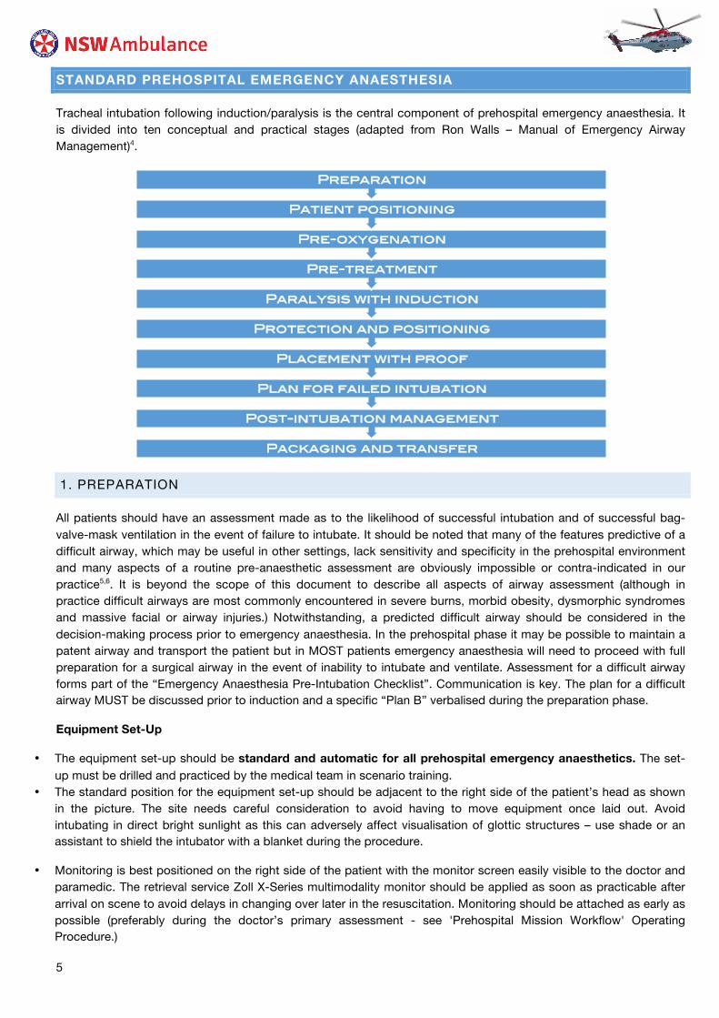
Tracheal intubation following induction/paralysis is the central component of prehospital emergency anaesthesia. It is divided into ten conceptual and practical stages (adapted from Ron Walls – Manual of Emergency Airway Management)4.
1. PREPARATION
All patients should have an assessment made as to the likelihood of successful intubation and of successful bag-valve-mask ventilation in the event of failure to intubate. It should be noted that many of the features predictive of a difficult airway, which may be useful in other settings, lack sensitivity and specificity in the prehospital environment and many aspects of a routine pre-anaesthetic assessment are obviously impossible or contra-indicated in our practice5,6. It is beyond the scope of this document to describe all aspects of airway assessment (although in practice difficult airways are most commonly encountered in severe burns, morbid obesity, dysmorphic syndromes and massive facial or airway injuries.) Notwithstanding, a predicted difficult airway should be considered in the decision-making process prior to emergency anaesthesia. In the prehospital phase it may be possible to maintain a patent airway and transport the patient but in MOST patients emergency anaesthesia will need to proceed with full preparation for a surgical airway in the event of inability to intubate and ventilate. Assessment for a difficult airway forms part of the “Emergency Anaesthesia Pre-Intubation Checklist”. Communication is key. The plan for a difficult airway MUST be discussed prior to induction and a specific “Plan B” verbalised during the preparation phase.
Equipment Set-Up
• The equipment set-up should be standard and automatic for all prehospital emergency anaesthetics. The set-up must be drilled and practiced by the medical team in scenario training.
• The standard position for the equipment set-up should be adjacent to the right side of the patient’s head as shown in the picture. The site needs careful consideration to avoid having to move equipment once laid out. Avoid intubating in direct bright sunlight as this can adversely affect visualisation of glottic structures – use shade or an assistant to shield the intubator with a blanket during the procedure.
• Monitoring is best positioned on the right side of the patient with the monitor screen easily visible to the doctor and paramedic. The retrieval service Zoll X-Series multimodality monitor should be applied as soon as practicable after arrival on scene to avoid delays in changing over later in the resuscitation. Monitoring should be attached as early as possible (preferably during the doctor’s primary assessment - see 'Prehospital Mission Workflow' Operating Procedure.)
Packaging and transfer!
Post-intubation management!
Plan for failed intubation!
Placement with proof!
Protection and positioning!
Paralysis with induction!
Pre-treatment!
Pre-oxygenation!
Patient positioning!
Preparation!
6
• Monitoring will include SpO2, NIBP (set cycling interval to 3min), ECG and ETCO27. Automated NIBP readings are
preferable to manual ones as they avoid prolonged periods without blood pressure readings and result in earlier detection of hypotension. They also facilitate safe post-intubation sedation particularly in head injured patients. The EMMA capnometer is a backup in case of main monitor ETCO2 failure. ETCO2 monitoring must be used whenever a BVM is applied to a patient whether by face-mask, SGA or tracheal tube. It is the most important monitor of ventilation and airway patency. Loss of ETCO2 trace indicates a loss of airway patency until proven otherwise (equipment failure/circuit disconnection etc).
• Oxygen – sufficient oxygen MUST be secured for the pre-oxygenation, extrication and transport phases. A minimum of two full C-size cylinders must be made available. It is essential to prepare this EARLY in the procedure particularly if remote from vehicles.
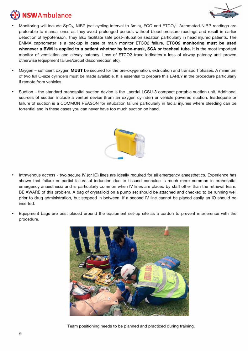
• Suction – the standard prehospital suction device is the Laerdal LCSU-3 compact portable suction unit. Additional sources of suction include a venturi device (from an oxygen cylinder) or vehicle powered suction. Inadequate or failure of suction is a COMMON REASON for intubation failure particularly in facial injuries where bleeding can be torrential and in these cases you can never have too much suction on hand.
• Intravenous access - two secure IV (or IO) lines are ideally required for all emergency anaesthetics. Experience has shown that failure or partial failure of induction due to tissued cannulae is much more common in prehospital emergency anaesthesia and is particularly common when IV lines are placed by staff other than the retrieval team. BE AWARE of this problem. A bag of crystalloid on a pump set should be attached and checked to be running well prior to drug administration, but stopped in between. If a second IV line cannot be placed easily an IO should be inserted.
• Equipment bags are best placed around the equipment set-up site as a cordon to prevent interference with the procedure.
Team positioning needs to be planned and practiced during training.

7
PERSONNEL
• Allocate tasks: 2 or 3 person technique depending on setting.
o Airway Assistant/ELM assistant
o Laryngoscopist
o In-line cervical spine immobilisation operator
• The delegation of roles and appropriate briefing of staff is an essential task. The doctor is responsible for delegation of staff and their briefing but both members of the team must be aware of this procedure.
• The retrieval paramedic is responsible for establishing monitoring and for the equipment set-up. • If there are no features (apart from C-Spine immobilisation) to predict a difficult airway and full pre-oxygenation can
be achieved the first attempt at laryngoscopy may be taken by the retrieval paramedic with the doctor becoming the airway assistant and available as second operator if laryngoscopy proves to be difficult. This enables the doctor to maintain the clinical overview of the procedure. All registrars should take a turn at performing laryngoscopy in the prehospital setting during their term and this should be discussed prior to arrival at the scene.
• Paramedics must be up to date for the Prehospital Emergency Anaesthesia Currency to perform laryngoscopy. • If a difficult airway is anticipated or adequate pre-oxygenation is difficult then the physician should perform the
laryngoscopy. • Non-retrieval service staff should not perform laryngoscopy for emergency anaesthesia regardless of
experience unless they have previously practiced or drilled our procedure and remain current for the procedure. • Staff positioning is shown in the picture with in-line cervical immobilisation on the left of the patient keeping the right
side clear for the equipment set-up, airway assistant and monitoring. • It is important for ancillary staff to be appropriately briefed on their expected role and actions. The in-line cervical
immobilisation operator should be briefed to keep their hands away from the mandible and their arms in line with the long axis of the patient out of the way of the laryngoscopist.
CREW RESOURCE MANAGEMENT ISSUES
• The key to minimizing scene times is ensuring SIMULTANEOUS ACTIVITY AT ALL TIMES during resuscitation and nowhere is this more important than during emergency anaesthesia.
• Always introduce yourselves on arrival and if you can, try to remember the names and designations of staff as you listen to their handover. Most staff are happy to have us there but the service (and the likelihood of our being requested in the future) depends on maintaining good working relationships with on-scene crews.
• It is vital that the on-scene paramedics be involved in the resuscitation. This may well be their only serious trauma case for the year and it is essential to involve them as active participants; always being mindful of their skills.
• Always consider whether other on-scene staff can be utilised for tasks such as splintage, cannulation, setting up fluids or fetching equipment to free up the clinical team to perform tasks only we can perform (such as the equipment set-up or surgical procedures).
• The team need to think several steps ahead and anticipate the need for equipment such as oxygen, suction, a stretcher and the means and route of transport which all of which will be time-consuming if done in a serial fashion.
• There should NEVER be on-scene staff standing around quietly watching – there are MANY things to do at once. Even non-medical staff such as fire crew and police can be utilised to create a cordon, remove curious onlookers or act as a drip stand!

8
2. PATIENT POSITIONING
• Ensure adequate access to the casualty. Intubation is RARELY necessary in the position in which the injured person is found. The first task after deciding to intubate is to locate the most appropriate place to perform the procedure.
• Patients should be extricated to a safe environment (facilitated by analgesia, sedation and regional anaesthetic techniques) e.g. to the roadside on an ambulance stretcher.
• Intubation of patients on the ground is much more difficult and only very rarely necessary.
• Prehospital anaesthesia should usually only be undertaken when the casualty is in the supine position with relatively easy and full access. The ideal position for intubation is on a stretcher or raised platform with 360 o access. (for example on an ambulance or retrieval stretcher with legs retracted (fully down) at the rear of a vehicle. An alternative is on a stretcher lowered to half-height with the intubator in a kneeling position. If the team plans to depart to hospital by helicopter then ideally emergency anaesthesia will occur next to the EC145 or inside the AW 139 (with the stretcher fully extended). Experience has shown that intubation inside an ambulance vehicle or confined space is more difficult and increases the chances of failure and therefore 360o access to the patient should be obtained where possible. A temporary airway adjunct may be useful whilst repositioning.
Positioning the patient just outside the rear of a vehicle has several advantages including access to powered suction, additional lighting and oxygen supplies. A second ambulance stretcher is often available and can be used as an ideal workspace for equipment for an emergency anaesthesia.
A second stretcher acts as an excellent work station
• Complex extrication movements after anaesthesia are often difficult and the focus for management of trapped patients should be on maintaining oxygenation with rapid extrication if the airway is compromised, under airway adjuncts, supraglottic airway (SGA) such as the i-Gel, or in-situ surgical airway.
• Intubation inside an ambulance may occasionally be necessary in severe weather conditions or threatening crowds and this procedure should be practised by all staff.

9
LARYNGOSCOPIST POSITION
• Direct laryngoscopy is the process of bringing the glottis into alignment with the eyes of the operator and as such the doctor or paramedic performing laryngoscopy must maximise their position for laryngoscopy. If the patient cannot be moved from the ground then some of the choices include the following. You should try them to see which suits you best:
o Kneeling – most versatile rough or soiled ground but often difficult to obtain a good view.
o Lying Prone – gets the eye line right but may put the laryngoscopist at a mechanical disadvantage.
o Left lateral Position – provides a good eye line and mechanical advantage and comfort may be improved with padding under the left elbow (such as a SAM splint). These techniques may be best practiced in scenario training.
Lying
Kneeling

10
3. PRE-OXYGENATION
• Pre-oxygenation is essential for safe prehospital emergency anaesthesia. It should proceed throughout the preparation phase above. Pre-oxygenation de-nitrogenates and establishes an oxygen reservoir in the lungs, blood and tissues and, if effective, will allow several minutes of apnoea without O2 desaturation and the need to ventilate the patient. Two nasopharyngeal airways and an oropharyngeal airway with jaw thrust (“Tripod” or “supported Tripod” (with jaw thrust) technique) should be used if there is difficulty maintaining an open airway.
• Head injury is NOT an absolute contra-indication to the CAREFUL placement of a nasopharyngeal airway.
APNOEIC OXYGENATION
• Studies have demonstrated that during apnoea (following muscle relaxant administration in emergency anaesthesia), supplying oxygen to a patent airway can significantly prolong safe apnoea time before desaturation occurs9,10,11,12. This phenomenon is well described in brainstem death testing protocols where oxygenation can be maintained for long periods without ventilation when oxygen is supplied to apnoeic patients via the tracheal tube.
• The physiology is relatively straightforward; during apnoea, oxygen is continuously absorbed into the blood from the FRC at a rate of approximately 250mL/min whilst CO2 continues to accumulate in the blood, only slowly diffusing into the aveoli at a rate of approximately 10mL/min13. This differential creates sub-atmospheric alveolar pressure and a net inflow of gas into the lungs of around 240mL/min in the average anaesthetised human, provided the airway is kept patent. If oxygen (rather than room air) is supplied then the oxygen reservoir in the lungs can be maintained for extended periods.
• In our setting this is best accomplished by administration of high flow oxygen at 15L/min by nasal cannulae just prior to induction (when the Emergency Anaesthesia Pre-Intubation Checklist is completed) and left on until successful tracheal intubation is accomplished. The airway is naturally opened during laryngoscopic attempts and this can be augmented by airway adjuncts or jaw thrust. It is also worth noting that the use of nasal cannulae in addition to a non-rebreather reservoir mask (NRB) or self-inflating bag-valve-mask with reservoir (BVM) for pre-oxygenation has been shown to significantly increase FIO2 in spontaneously breathing patients by washing out ventilatory dead space with oxygen inbetween spontaneous breaths14 and thus increases the effectiveness of pre-oxygenation.
Apnoeic oxygenation set-up via nasal canula

11
There are 2 alternatives for effective prehospital pre-oxygenation in addition to high flow nasal cannulae:
STANDARD APPROACH
AUSTERE ENVIRONMENT APPROACH
In a small number of missions there will not be adequate numbers of staff or oxygen sources to apply the standard approach. Such missions include winch accessed casualties or remote environments and a tight-fitting non-rebreathing mask with reservoir +/- high flow nasal cannulae may be utilised to conserve personnel.
2-handed BVM with PEEP and Apnoeic Oxygenation via NPs
In the majority of missions there will be adequate personnel and oxygen sources to provide standard pre-oxygenation. In our service the standard approach uses a BVM with PEEP valve (set at 5cm H2O) applied tightly to the patient’s face with 2-handed technique with Nasal Prongs running at 4L/min to provide flow between breaths. This technique provides continous positive airway pressure during pre-oxygenation and has been demonstrated to improve alveolar recruitment and increase oxygen reservoir and is of particularly value in patients with shunt physiology following chest trauma.14
PEEP may reduce venous return in shocked patients and this is important to detect prior to full induction. Re-assessment of haemodynamics following application of PEEP allows dose reduction of anaesthetic or blood transfusion where the patient is “PEEP sensitive”
The catheter mount ("goose neck"), HME fliter and ETCO2 sensor should always be placed in the breathing circuit to monitor pre-oxygenation. The ETCO2 waveform becomes a the key measure of ventilation, gives early warning of airway obstruction or leak and indicates onset of paralysis following rocuronium administration. BVM-assisted ventilations are mandatory when the patient has inadequate spontaneous ventilations. Controlled hand ventilation during apnoea to avoid hypoxia may be considered when patient's physiology and suspected injuries make desaturation during apnoea likely.

12
Greater Sydney Area HEMS Prehospital Emergency Anaesthesia Checklist
IVI / Drugs
Fluid connected, runs easily………………….………….…………………….. Check BP cuff on contralateral arm, BP seen…………….…………………………. Check Spare cannula in ……...…………..……………………………………………… Check RSI drugs prepared, doses selected….……………………………………….. Check
Team Brief
C-Spine immobiliser briefed – Collar open…………………………………….. Check ELM plan briefed………….……………………………………….…..……………. Check Difficult laryngoscopy plan briefed….……………………….………………….. Check LMA ………..…………………………………….…………………………………….. Check Crike set ………..……………………………….…………………………………….. Check NP O2 to 15L/min “Checks complete. Anaesthetising at --/--”
Patient position optimised........................................................................... Check
O2 sufficient = 2 bottles.............................................................................. Check
Preox – [BVM, PEEP, Nasal Prongs]…….............…………………………… Check
Suction tested – [second suction considered]........................................... Check
Monitor: ECG, NIBP, SpO2, waveform CO2 seen........................................ Check
Intubation Equipment
BVM inflating……………………………………................................................ Check
OPA & 2 NPAs…………................................................................................... Check
Laryngoscope tested and bright…………………….…………………………. Check
Tube size chosen, cuff tested ………………………..………………………… Check
Alternate Tube ……………………………………………………………..……… Check
Syringe ………………………………………………………………………………. Check
Bougie………………………………………………………………………………… Check
Circuit: catheter mount (gooseneck), filter, capnography..……..………….. Check
Tube tie……………………………………………………………………………….. Check
Other Considerations
• In patients with severe facial injuries with severe airway bleeding consideration should be given to pre-oxygenation in the lateral position to allow airway toilet with movement to supine position immediately after induction.
4. PRE-TREATMENT
• Combative, agitated or uncooperative patients who need intubation are a particular challenge in the prehospital setting.
• Pre-treatment with small titrated doses of ketamine (10-40mg) to a total of 0.5 -1mg/kg can be very effective in order to facilitate further assessment, monitoring and then pre-oxygenation of these patients. This has been described as “Delayed Sequence Intubation”16 or “Titrated Ketamine Induction” by some authors but simply refers to the use of ketamine to sedate a patient in order to facilitate pre-oxygenation for intubation.
• Compelling evidence for atropine pre-treatment in paediatrics is lacking and the incidence of bradycardia seems to be related more to episodes of hypoxia rather than pharmacological or reflex effects17. Atropine is therefore not routinely drawn up for paediatric intubation.
• Fentanyl may have some protective effects against the downstream hypertensive response to ketamine and intubation in haemodynamically stable patients although the clinical significance of this is unknown and the literature is minimal. Its coadministration at induction is an option but tends not to be used by the majority of GSA-HEMS physicians.
• Other pre-treatment regimens such as lidocaine and de-fasciculating doses of paralytics are not well supported by the literature and inevitably delay and complicate standard emergency anaesthesia18, 19.
5. PARALYSIS WITH INDUCTION
• Immediately prior to induction the Emergency Anaesthesia Pre-intubation Checklist should be completed. This is a challenge-response list which aims to identify and The team member who completed the kit dump (usualy the paramedic) reads out the list and the assistant responds to the challenge as each element is identified and checked. Whilst it can feel like a long time to read through the checklist, it rarely takes more than 30 secs and it has proved its effectiveness in trapping errors and preventing threats to patients safety.

13
• All drugs should be given into a running line or flushed in with a bolus of crystalloid by one of the team (usually the doctor).
• IV induction agent- Ketamine (1.5-2mg/kg) is the preferred induction agent for prehospital emergency anaesthesia with a much safer haemodynamic profile than available alternatives20.
• In significantly hypovolaemic patients, induction may unmask compensatory vasoconstriction and they should be given a reduced dose of ketamine (0.5-1 mg/kg) for induction. Blood pressure is often maintained despite significant blood loss in young patients. Signs of hypovolaemia include:
! A reduced pulse pressure with relative diastolic hypertension
! Increased respiratory rate
! Skin pallor or mottling
! Cool skin temperature
! Visibly collapsed veins
• In unconscious peri-arrest patients induction drugs may be omitted and a muscle relaxant only intubation performed, with rocuronium 1.5mg/kg
• If the patient is hypertensive initially then an alternative agent such as thiopentone may be used.
• IV rocuronium 1.5mg/kg is the preferred muscle relaxant for emergency anaesthesia as it may reduce oxygen consumption compared with suxamethonium and maintains paralysis in case of a need for SGA rescue or surgical airway.
• Immediately before induction the nasal prong oxygen source should be turned up to 15L/min.
• Whilst awaiting full relaxation it is useful to warn bystanders of impending cessation of respiration as this can be disturbing to the uninitiated onlooker.
• If Sa02 begins to fall prior to full relaxation then gentle interposed ventilation should be used to maintain oxygenation with ETCO2 waveform scrutinised to ensure effective ventilation. In patients who may be severely acidotic ventilation through the apnoeic phase is recommended.
• Apnoeic oxygenation continues even after the cessation of spontaneous respirations provided an oxygen gradient is maintained between pharynx and alveoli and the airway is open. The team should ensure that the mask is not removed from the patient’s face until the moment of commencement of laryngoscopy, usually marked by apnoea AND the identification of flaccid jaw tone.
• Ensure full relaxation. A common mistake in the enthusiasm to secure the airway is to undertake laryngoscopy prior to adequate relaxation (usually complete at 60 seconds) and the laryngoscopist must ensure full relaxation prior to attempted laryngoscopy by testing jaw tone.

14
6. PROTECTION AND POSITIONING
CRICOID PRESSURE
• Cricoid pressure is not routinely used in the service. There is no evidence that even well applied cricoid pressure (Sellick’s Manoeuvre) prevents passive aspiration. It is most commonly poorly performed. There is evidence that cricoid pressure may reduce tone at the lower oesophageal sphincter21, significantly impair laryngoscopic view and cause unwanted movements of the cervical spine22. It should no longer be routinely applied to patients undergoing prehospital emergency anaesthesia. If the clinician decides to use cricoid pressure they must ensure the cricoid operator is briefed appropriately and cricoid pressure removed if laryngoscopic view is difficult.
• External Laryngeal Manipulation (ELM) (or ‘bimanual laryngoscopy’) has been demonstrated to improve laryngoscopic view23 and should be used whenever initial view is suboptimal as part of the “30 second drills”. The laryngoscopist manipulates the thyroid cartilage to maximize their view and an assistant can be directed to hold the thyroid cartilage in position during intubation. It is important to recognise the difference between this technique and cricoid pressure.
• The cervical collar should be open and the mandible free of any restrictions for intubation. The uncleared C-spine should be routinely protected in all blunt trauma patients and this should be performed by an assistant holding the head from the left side of the patient, facing the laryngoscopist.
• Maintaining the neutral position of the C-spine. In most adult patients, lying supine on a stretcher or extrication board causes significant neck extension and this is exaggerated in those with a large body habitus. This should be corrected by placing a folded towel or SAM splint beneath the occiput to maintain a neutral head position. A neutral position reduces stress on the possibly injured cervical spine, improves the patency of the airway and facilitates direct laryngoscopy. If cervical spine precautions are not needed (for example some burns patients or following submersion incidents) the patient should be positioned in the “Ear to Sternal Notch Postion” which greatly improves laryngoscopic view.
7. PLACEMENT WITH PROOF
• At 60 seconds after muscle relaxant administration, the patient’s jaw should be tested for flaccidity and laryngoscopy attempted. There is always time to perform laryngoscopy gently and carefully.
• The tracheal bougie should be used for ALL prehospital intubations. Use of an intubating bougie is associated with higher success rates, particularly on first attempt24. It reduces the chance of being unable to pass a tube when the glottis is well visualised and may reduce cervical movement required to perform intubation.
• A straight or slightly curved bougie with coude (angled) tip is easiest to use and the bougie should be pre-shaped prior to use.
• The bougie should be used naked with the assistant supporting the free end or pre-loaded with 6-8cm of bougie protruding.
• In the event of a Grade III view which does not improve with 30 second drills a “tactile guided” bougie may be passed under the epiglottis in the midline. Gentle “hold-up” will be felt at around 30 cm and indicates tracheal placement. A tracheal tube should ONLY BE PLACED if “hold-up” is confirmed when manipulating the bougie. Using this approach means an oesophageal intubation should NEVER be performed. Airway audit has shown that “blind” bougie placement through a grade IV view invariably finds the oesophagus.
• Audit shows that the rare grade IV view is almost always improved by changing patient position (improving head elevation with padding) and operator position (removing patient from ground). Meticulous attention should therefore be paid to optimising these prior to the first attempt.
15
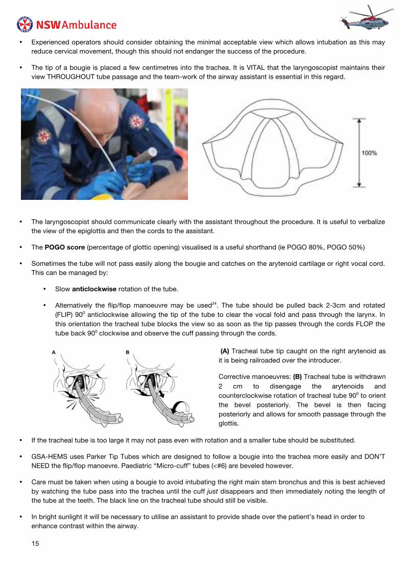
(A) Tracheal tube tip caught on the right arytenoid as it is being railroaded over the introducer.
Corrective manoeuvres: (B) Tracheal tube is withdrawn 2 cm to disengage the arytenoids and counterclockwise rotation of tracheal tube 900 to orient the bevel posteriorly. The bevel is then facing posteriorly and allows for smooth passage through the glottis.
• Experienced operators should consider obtaining the minimal acceptable view which allows intubation as this may reduce cervical movement, though this should not endanger the success of the procedure.
• The tip of a bougie is placed a few centimetres into the trachea. It is VITAL that the laryngoscopist maintains their view THROUGHOUT tube passage and the team-work of the airway assistant is essential in this regard.
• The laryngoscopist should communicate clearly with the assistant throughout the procedure. It is useful to verbalize the view of the epiglottis and then the cords to the assistant.
• The POGO score (percentage of glottic opening) visualised is a useful shorthand (ie POGO 80%, POGO 50%)
• Sometimes the tube will not pass easily along the bougie and catches on the arytenoid cartilage or right vocal cord. This can be managed by:
• Slow anticlockwise rotation of the tube.
• Alternatively the flip/flop manoeuvre may be used24. The tube should be pulled back 2-3cm and rotated (FLIP) 900 anticlockwise allowing the tip of the tube to clear the vocal fold and pass through the larynx. In this orientation the tracheal tube blocks the view so as soon as the tip passes through the cords FLOP the tube back 900 clockwise and observe the cuff passing through the cords.
• If the tracheal tube is too large it may not pass even with rotation and a smaller tube should be substituted.
• GSA-HEMS uses Parker Tip Tubes which are designed to follow a bougie into the trachea more easily and DON’T NEED the flip/flop manoevre. Paediatric “Micro-cuff” tubes (<#6) are beveled however.
• Care must be taken when using a bougie to avoid intubating the right main stem bronchus and this is best achieved by watching the tube pass into the trachea until the cuff just disappears and then immediately noting the length of the tube at the teeth. The black line on the tracheal tube should still be visible.
• In bright sunlight it will be necessary to utilise an assistant to provide shade over the patient’s head in order to enhance contrast within the airway.

16
• Once the tube is in place and the cuff inflated, correct tube placement should be confirmed immediately.
• Beware the tube that does not look far enough (less that 19 to 20 cm) in and has a small but persistent leak. The cuff may have become displaced outside the cords on inflation leaving just the tip between the cords. In this position the tip can be displaced out into the pharynx with the smallest patient movement.
Determination of correct tube placement
See the tube passing through the cords
Palpation of tube movement within the larynx and trachea
Chest wall expansion equally with each ventilation
Auscultation of breath sounds
Absence of epigastric sounds with respiration
End Tidal CO2 monitoring (waveform or quantitative)
• Tube placement MUST always be confirmed by end tidal CO2 detection
EMMA (left), Zoll X-Series (right)

17
8. PLAN FOR FAILED INTUBATION
• The plan for failed intubation should be discussed by the team prior to intubation.
• Actions on first failed intubation during standard emergency anaesthesia:
1. Return to 2-person 2-handed BVM ventilation with adjuncts as required (supported Tripod) to maintain oxygenation if the patient desaturates.
2. Where an adequate view cannot be obtained a further attempt may be undertaken provided deliberate steps have been taken to identify and rectify the problem causing the failure and that oxygenation can be maintained between attempts. These are the 30 sec drills26 (so named because they should be easily performed long before a normal pre-oxygenated patient begins to desaturate).
30 Second Drills
Release cricoid and use bi-manual laryngoscopy (ELM)
Optimise operator position
Optimise patient position (small pad under the head with neck in neutral position)
Suction where secretions or blood block the view
Insert laryngoscope deeply and slowly withdrawn until identifiable anatomy is seen
Consider changing laryngoscope blade size or type
• The key to the management of failed intubation is early recognition of the problem and not persevering in the face of a desaturating patient. See the discussion of Failed Intubation.
In general the following actions have been utilised successfully in our service when initial view has not been ideal.
GRADE IV
GRADE III
• Pull back laryngoscope and slowly re-insert until epiglottis is visualised – “Epiglottoscopy”
• Suctioning of airway secretions or blood
• Provide further head elevation
• ELM to improve position of laryngoscope tip in vallecula
• Lift up floppy epiglottis directly with blade
• Reposition airway alignment “ear to sternal notch”
• Guided bougie under epiglottis in midline feeling for gentle “hold-up” at ~ 30cm
• Insert stylet into tracheal tube in ‘straight to cuff’ configuration and guide tube tip behind epiglottis

18
9. POST INTUBATION MANAGEMENT
• It is important to avoid a post-intubation lull in tempo and vigilance. This is the time when serious errors are most likely. Problems include:
o Tube dislodged,
o Monitoring to become disconnected,
o Ongoing sedation to be forgotten,
o Failure of bag ventilation in the now paralysed patient.
• The team must remain vigilant to avoid such errors.
Be very aware of post anaesthetic complications such as bradycardia (hypoxia) and hypotension (positive pressure ventilation/oversedation), development of tension pneumothorax and inadequate or hyperventilation.
• Consider the following for maintenance of anaesthesia with:
o Fentanyl for analgesia – loading dose of 50-100mcg followed by infusion or incremental small boluses
o Midazolam (infusion or 2mg boluses) or ketamine (infusion or 20mg boluses) for sedation
o Rocuronium for long acting muscle relaxation (0.6mg/kg (~50mg) every 30-40 minutes)
• Analgesia/sedation should be titrated to avoid signs of noxious stimulation and possible awareness (tachycardia, hypertension, eye watering, sweating, symmetrical pupil dilatation).
• Overzealous positive pressure ventilation (particularly in hypotensive patients) by increasing intrathoracic pressure and reducing venous return may reduce cardiac output. Six to eight breaths per minute at tidal volumes of approximately 8-10ml/kg may be adequate to maintain oxygenation without impairing cardiac output28.
• End-tidal CO2 should be kept within physiological values around 30-35mmHg particularly in head injured patients.
• If a patient desaturates following intubation and ventilation, displacement of the tube, obstruction somewhere in the breathing circuit, pneumothorax and equipment failures such as malassembly or malfunction should be sought.
• Gastric decompression with an orogastric tube should also be considered particularly in children and in patients who have had a period of bag-valve-mask ventilation.
10. PACKAGING AND TRANSFER
• All tubes and lines must be absolutely secure and the tracheal tube should be controlled by a member of the team for all transfers, as the risk of tube dislodgment is highest during transfers.
• All airway connections MUST be secured with “Push and twist” technique rather than just being “pushed” together.
• Careful observation of PR, BP, presence or absence of sweating, lacrimation and pupil size and their reaction is required throughout transfer to detect under-sedation.
• Full monitoring should be maintained throughout transfer (the minimum standard is continuous SpO2, intermittent NIBP, continuous ECG and continuous waveform ETCO2 7.
• Monitoring, ventilator infusions and airway must be visualised and accessible throughout retrieval.
19
• Directed observations of the patient from observing skin colour, chest movement and pattern of respiration, following the circuit from the patient to the ventilator and noting monitoring and alarms should be repeated every few minutes.
‘FAILED’ LARYNGOSCOPY
Failed intubation “algorithms” are often used in anaesthetic and emergency practice29 and whilst it is essential that the team has a specific plan for failed intubation this will often be dependent on the patient position, the presence of other injuries (such as chest or facial trauma) and the skills of the operator. As such we present the important issues and encourage the team to think about and discuss the plan for a failed intubation for each case. This forms part of the “Emergency Anaesthesia Pre-Intubation Checklist”.
• The essential recovery action is to maintain oxygenation of the patient.
• BVM ventilation with 2 person technique and full airway adjuncts – guedel and 2 nasopharyngeal airways (Supported Tripod) should be the reflex fall-back position.
• If ventilation is not possible then, in most patients, SGA insertion will be the most appropriate technique. Insertion of the iGel can be facilitated by using the laryngoscope blade as a tongue blade to control the tongue during insertion. An SGA is a temporising measure as it provides little airway protection though if transport times are short and oxygenation is being maintained it may be appropriate to transport the patient with SGA in place.
• For a subgroup of patients insertion of an SGA is likely to be impossible. These patients include those with restricted mouth opening (burns, facial trauma) or where there is obstruction or distortion of the airway.
• Patients with significant chest wall injuries may be impossible to ventilate with an SGA due to the high pressures needed and this can often be predicted during patient assessment.
• The surgical cricothyroidotomy will be the primary mode of securing ventilation and the airway in some patients listed above and the final step in a “can’t intubate/ can’t ventilate” situation. As such the team needs to be well drilled in the procedure and confident of success. The most common mistake in performance of surgical cricothyroidotomy relates to a delay making the decision so that by the time the procedure is performed there has been significant hypoxia and the enhanced stress of falling oxygenation impairs operator performance.
• There is a subgroup of patients for whom surgical cricothyroidotomy may be the primary means of securing the airway without a preceding attempt at emergency anaesthesia. These include full thickness facial/neck burns where neck and mouth movement is severely limited, massive maxillo-facial haemorrhage and the entrapped patient with airway compromise who cannot be extricated and access is impaired.
• A simple surgical technique for performing cricothyroidotomy is described below and experience suggests it may avoid some of the common complications.
• Rarely it is appropriate to allow paralysis to wear off and let the patient wake to pre-anaesthetic status. Most of our patients are intubated for emergent reasons and need their airways securing by another means.
• In young children many sources recommend a needle cricothyroidotomy in preference to a surgical airway. However, evidence from animal studies suggests a high failure rate (40%) and high incidence of perforation of the posterior tracheal wall (42%)30. Note that in infants the cricothyroid membrance is often too small for a tube so a tracheotomy may need to be done with cutting of the anterior part of a tracheal ring to make space. It will bleed and losing control of the trachea is possible, which can be prevented by gaining purchase on the trachea using forceps, sutures or a surgical towel clip. After short training 97% success was achieved in one study using a surgical, as opposed to needle, technique31. These factors should be considered when planning for a can’t intubate/can’t ventilate scenario while preparing for paediatric emergency anaesthesia.

20
SPECIFIC POPULATIONS
PAEDIATRIC EMERGENCY ANAESTHESIA
Prehospital anaesthesia of small children is only rarely required. The risk/benefit equation is altered by the increased complexity of the procedure, tendency to desaturate much earlier and the dangers of drug dosage errors32. Where intubation is needed the anaesthetic doses and equipment sizes should be calculated independently by each team member and results compared. The Paediatric Emergency Reference Cards are helpful in these calculations.
HYPOVOLAEMIC PATIENTS
Hypovolaemic, and particularly frankly hypotensive, patients are at risk of decompensation and cardiac arrest during induction and immediately following positive pressure ventilation. This may relate to loss of vasomotor tone, peripheral vasodilatation, reduced venous return, increased intrathoracic pressures or complications such as tension pneumothorax. The warning signs of compensated hypovolaemia have been discussed in the section on induction. The advice to reduce the dose of induction agent in such patients is reiterated here. A fluid /blood bolus immediately prior to induction should also be considered. In patients who are in extremis, intubation with muscle relaxant only may be necessary.
OBESE PATIENTS
Patients with large body habitus present many problems for resuscitation not least of which is a tendency to desaturate earlier as well as a more difficult airway. The most experienced intubator should make the first attempt at laryngoscopy. Such patients tend to lie supine with significant neck extension in a cervical collar which can make intubation impossible. This problem is also commonly seen in patients with motorcycle leathers or other bulky clothing under them. At least one pad under the occiput to bring the neck into neutral alignment is essential in these patients.
BURNS
Patients with severe burns are challenging both clinically and psychologically. The airway should be secured early in patients with suspected airway burns. Patients with airway burns have a high risk of developing airway swelling which can make subsequent oral intubation impossible. The signs of airway burns include stridor, dysphonia, odynophagia, visible burns or soot deposition in the oropharynx, presence of full thickness facial or neck burns and

21
the presence of inhalational injury. Inhalational injury occurs when superheated gases form in a confined space or very close to the face and are breathed in. This can make preoxygenation and ventilation difficult and consideration should be given to preoxygenation in the sitting position prior to induction. Inability to ventilate may be caused by full thickness burns to the chest requiring on-scene escharotomy. Most patients are completely alert despite severe burns and adequate sedation and analgesia must be given during and following induction of anaesthesia. Ketamine remains an excellent choice for such patients and doses needed may be high. As discussed above, severe full thickness burns of the neck or face can make oral intubation or SGA insertion impossible and a surgical airway may be the primary mode of securing the airway in such patients.
WINCH PRIMARIES
A patient is intubated by our team prior to winch extraction on average once every 2.5 months. Performing emergency anaesthesia following a winch primary with no other staff or equipment present on-scene is fortunately uncommon, but clearly can be necessary. The challenges are many. The increased difficulties include very limited oxygen supplies, difficult environment or terrain, need for stabilisation during the winching procedure where capacity for monitoring or intervening are extremely limited and of course the fact that only 2 personnel are present. If prehospital information suggests the possibility of a winch primary requiring emergency anaesthesia the clinical team must brief their plan, equipment to be winched, lines of communication and the extrication plan in detail. In addition to the usual equipment packs, oxygen should be winched using the winching bag. The Zoll X-Series is winchable but consideration of the risks and benefits of reverting to basic monitoring with finger probe oxygen saturation monitoring, manual BP or pulse checks and EMMA capnography is needed. Scenario training is the best way to practise this challenging contingency.
SURGICAL CRICOTHYROIDOTOMY
The surgical airway equipment should be removed from its pouch when it is anticipated that an airway will be particularly difficult. For example:
o Airway trauma o Difficult anatomy o Burns to face and neck reducing jaw movement o Possible airway burns
The technique of surgical cricothyroidotomy is based on published literature33, animal and cadaver simulations as well as service experience. It is rapid and reliable. It aims to address several problems which may be seen in the prehospital setting which make some other techniques less appropriate.
The most common challenges encountered when performing surgical airway are:
o Delay in decision-making o Significant bleeding from the incision o Inability to instrument the incision with unfamiliar equipment in time-critical situations.

22
It is a one person technique which relies on sense of touch rather than visualisation of structures and which is applicable to any situation requiring surgical access to the airway.
CRICOTHYROIDOTOMY TECHNIQUE
Locate ,,
Grip,
Incise,
Finger,
Bougie,
Tube,
Confirm,
Decision, Incision, Finger, Tube, Cuff, ETCO2,
Once the cricothyroid membrane has been definitively located by surface palpation or palpation through a midline longitudinal incision, a “stabbing/rocking incision is made through the cricothyroid membrane ensuring the size 22 scalpel is fully inserted.
ETCO2 MUST be used to confirm tracheal placement and continuously monitored to ensure the airway remains patent. Sudden loss of ETCO2 suggests displacement of the tube or device related failure.
A size 6.0 tracheal tube (in adults) is then railroaded over the bougie into the trachea, ensuring the cuff is within the trachea and the cuff inflated. The size 22 scalpel is larger than the size 6.0 tube and the finger dilation ensures the tube will fit. The tube can be cut to reduce the chance of displacement. The tube can be tied, taped, or sutured in position.
The larynx must be firmly secured between the thumb and middle finger to prevent movement during the incision. The index finger should be used to locate the cricoid ring and cricothyroid membrane. Any movement of the larynx during the incision must be prevented.
The crico-thyroid membrane must be located with certainty prior to commencing the procedure. This should be done prior to emergency anaesthesia in any patient with a predicted difficult airway. The neck must be fully extended for the cricothyroidotomy usually by placing a blanket under the shoulders. In patients where the anatomy cannot be easily palpated such as obsese patients, those with significant neck soft tissue or a short neck, a long midline longitudinal incision should be made to facilitate accurate identification of the cricothyroid membrane.
The incision is then directly probed using your finger. Index finger or little finger is recommended. The finger performs several important roles: as a highly sensitive probe and dilator and definitively confirms tracheal position prior to insertion of the bougie and tube. The tracheal rings MUST be felt before proceeding further. In the extemely rare setting of a paediatric surgical airway an adult finger may not fit and the bougie can be placed directly through the incision as lotrang as purchase on the trachea is maintained. Confimration of intratracheal bougie placement by confirming hold-up is required as with translaryngeal intubation.
A paediatric tracheal bougie is then placed alongside the finger though the incision and confirmed to be within the trachea by palpation. This is a key step to ensure the bougie is in the trachea.
LOCATE
GRIP
INCISE
FINGER
BOUGIE
TUBE
CONFIRM

23
REFERENCES
1. Wang HE, Yealy DM. Out-of-hospital endotracheal intubation: where are we? Ann Emerg Med
2006;47:532-41.
2. Advanced Trauma Life Support Course Handbook. American College of Surgeons Committee O.
3. Hampshire BASICS MERGENCY ANAESTHESIA SOP
4. Manual of Emergency Airway Management. Ron M. Walls (Ed.). Lippincott Williams & Wilkins, 2000.
5. Kheterpal S et al. Incidence and predictors of difficult and impossible mask ventilation. Anesthesiology 2006 Nov; 105:885-91.
6. M J Reed, M J G Dunn and D W McKeown. Can an airway assessment score predict difficulty at intubation in the emergency department? Emerg. Med. J. 2005;22;99-102
7. Minimum Standards for Intrahospital Transport of Critically Ill Patients ANZCA, JFICM, ACEM.
8. Baraka A et al. Preoxygenation: Comparison of Maximal Breathing and Tidal Volume Breathing Techniques. Anesthesiology. 91(3):612, September 1999.
9. Teller L, Alexander C, Frumin J, Gross J. Pharyngeal insufflation of oxygen prevents arterial desaturation during apnea. Anesthesiology 1988; 69: 980–2.
10. Baraka A, Salem MR, Joseph N. Critical hemoglobin desaturation can be delayed by apneic diffusion oxygenation. Anesthesiology 1999; 90: 332–3.
11. Taha SK, Siddik-Sayyid S, El-Khatib MF, Dagher CM, Hakki M, Baraka AS. Nasopharyngeal oxygen insufflation following preoxygenation by the four deep breath technique. Anaesthesia 2006: 61: 427–30.
12. Ramachandran SK, et al. Apneic oxygenation during prolonged laryngoscopy in obese patients:a randomised, controlled trial of nasal oxygen administration. J Clin Anesth. 2010 May;22(3):164-8.
13. Holmdahl M. Pulmonary uptake of oxygen, acid-base metabolism and circulation during prolonged apnea. Apneic diffusion oxygenation. Acta Chirurgica Scandinavica 1956; 212 (Suppl.): 1–128.
14. Weingart, Scott D, and Richard M Levitan. 2011. Preoxygenation and prevention of desaturation during emergency airway management. Annals of emergency medicine, no. 3 (November 3).
15. Earl J. Delivery of High O2 Abstracts Am Assoc Resp Care 2003
16. Weingart S. Preoxygenation, Reoxygenation, And Delayed Sequence Intubation In The Emergency Department The Journ of Emerg Med. April; 14 (2) 2010
17. Bean A.. Atropine: Re-evaluating its use during pediatric MERGENCY ANAESTHESIA Best Bets BMJ 4/05/2007
18. Clancy M, Halford S, Walls R, et al. In patients with head injuries who undergo rapid sequence intubation using succinylcholine, does pretreatment with a competitive neuromuscular blocking agent improve outcome? A literature review. Emerg Med J 2001; 18:373–375
19. Robinson N and Clancy M. In patients with head injury undergoing rapid sequence intubation, does pretreatment with intravenous lignocaine/lidocaine lead to an improved neurological outcome? A review of the literature. Emerg Med J. 2001 November; 18(6): 453–457.
20. Sehdev R, Symmons D, Kindl K. Ketamine for rapid sequence induction in patients with head injury in the

24
emergency department. Emergency Medicine Australasia. (2006) 18, 37–44
21. Tournadre J-P Cricoid Cartilage Pressure Decreases Lower Esophageal Sphincter Tone Anethesiology 1997;86:7-9 I
22. Ellis DY, Harris T, Zideman D. Cricoid pressure in emergency department rapid sequence tracheal intubations: A risk-benefit analysis. Annals of Emergency Medicine. 2007;50:653-665
23. Levitan RM, Everett WW, Kinkle WC, et al. Pressing on the neck during laryngoscopy:
a comparison of cricoid pressure, backward upward rightward pressure, and external laryngeal
manipulation. Acad Emerg Med 2005;12(Suppl 1):92.
24. McGill J, Vogel EC, Rodgerson JD. Use of the gum elastic bougie as an adjunct for orotracheal
intubation in the emergency department. Acad Emerg Med 2000;7(5):526.
25. Marfin AG, Iqbal R, Mihm F, et al. Determination of the site of tracheal tube impingement during nasotracheal
ntubation. Anaesthesia 2006; 61: 646–50.
26. Srinivasa V, Kodali BS. Caution when using colorimetry to confirm endotracheal intubation. Anesth Analg. 2007 Mar;104(3):738.
27. HEMS London RSI SOP
28. Gentilello LM, Anardi D, Mock C, Arreola-Risa C, Maier RV. J Trauma. 1995 Nov;39(5):846-52. Permissive hypercapnia in trauma patients.
29. Heidegger T, Jörg Gerig H. Algorithms for management of the difficult airway. Curr Opin Anaesthesiol. 2004 Dec ;17 (6):483-4.
30. Stacey J1, Heard AM, Chapman G, Wallace CJ, Hegarty M, Vijayasekaran S, von Ungern-Sternberg BS. The ‘Can’t Intubate Can’t Oxygenate’ scenario in Pediatric Anesthesia: a comparison of different devices for needle cricothyroidotomy Paediatr Anaesth. 2012 Dec;22(12):1155-8
31. Holm-Knudsen RJ1, Rasmussen LS, Charabi B, Bøttger M, Kristensen MS. Emergency airway access in children – transtracheal cannulas and tracheotomy assessed in a porcine model. Paediatr Anaesth. 2012 Dec;22(12):1159-65
32. Gausche M, Lewis RJ, Stratton SJ, Haynes B, Gunter CS, Goodrich SM, Poore PD, McCollough MD, Henderson DP, Pratt FR, Seidel JSS: Effect of out-of-hospital pediatric endotracheal intubation on survival and neurological outcome: A controlled clinical trial. JAMA 2000;283:6:783-790.
33. Paix BR, Griggs WM. Surgical cricothyroidotomy: 24 successful cases leading to a simple 'scalpel-finger-tube' method. Emerg Med Australas. 2012 Feb;24(1):23-30Emergency
Also thanks to
• HEMS London, MAGPAS, Hampshire BASICS