precursors of endometrioid carcinoma of the uterus ... rjz final condensed... · precursors of...
TRANSCRIPT

5/12/2014
1
Precursors of endometrioid carcinoma of the uterus
“State of the Art”*
Richard J. Zaino, MDDept. of Pathology
Hershey Medical CenterPenn State University
Hershey, PA
*and clearly, it is art, not science
Disclosure
Consultant for Becker (NSF International)for cervical cancer screening
Learning objectivesthe participant should understand the
following issues relating to precursors of endometrioid adenocarcinoma
1) Natural history 2) The lexicon
3) Diagnostic reproducibility4) Current practice
5) Recommended terminology WHO 2014
Problems in Defining the Natural History of Hyperplasia
1) pathologic criteria - criteria and diagnostic terminology for the various forms of hyperplasia have changed repeatedly
2) initial sampling - the method of initial diagnosis is biopsy or curettage, which removes some or all of the lesion to be studied

5/12/2014
2
Problems in Defining the Natural History of Hyperplasia
3)coexisting lesions - other lesions such as adenocarcinoma may coexist at the time of diagnosis without our knowledge, since the D&C or biopsy samples only a portion of the endometrium
4)subsequent intervention - hormonal or surgical intervention usually interrupts observations of the natural history of hyperplasia
What do we know?Etiology and Natural History of Hyperplasia
1) endometrial hyperplasia usually occurs in the setting of unopposed estrogen stimulation
2) some hyperplasias regress if the estrogenic stimulation is withdrawn or in response to progestin therapy
3) some hyperplasias progress to adenocarcinoma in time
Etiology and Natural History of Hyperplasia
4) The frequency of hyperplasia is about 20X that of endometrial carcinoma
5) the probability of progression to adenocarcinoma is related to the degree of cytologic atypia in the hyperplasia
6) the majority of adenocarcinomas which arise in a background of hyperplasia are well differentiated, rarely lethal, and often may respond to progestin therapy
Regression of hyperplasia following hormonal therapy
Author lesion response persistence progression duration
Kistner hyper/CIS 100% 0 0 4 weeksSteiner hyper/CIS 100% 0 0 3 days-3 yrsKjorstad atypical 41% 23% 35% 3-10 yearsWentz hyper/atyp 100% 0 0 1-5 yearsWentz hyper/atyp 98% 2% 0 1-4 yearsKurman non-atypical 77% 31% 0 1-26 years
atypical 50% 20% 30% 1-26 yearsGal hyper/atyp 92% 8% 0 .8-9 yearsHuang hyper/atyp 52% 38% 10% 1-12 yearsFerenczy non-atypical 80% 20% 0 2-12 years
atypical 0% 75% 25% 2-12 yearsRandall atypical 94% 6% 0 1-7 years

5/12/2014
3
Progression of hyperplasiato carcinoma
Non atypical hyperplasia 3-12%
Atypical hyperplasia 25-27%
(Retrospective studies, +/- intervening therapy, incomplete follow-up of 1-20 years)
Absolute risk of endometrial carcinoma during 20 year follow-up among women
with endometrial hyperplasia Lacey et al, JCO, 2010
7900 women diagnosed with hyperplasia in a prepaid health plan; 19 years follow-up*
Cumulative progression riskNon-atypical hyperplasia 5%Atypical hyperplasia 28%
*retrospective review, intervening hormonal therapy, D&C
Coexistence of carcinoma with atypical hyperplasia - carcinoma found in
hysterectomies (within 12 weeks of initial diagnosis*)
Author Frequency
Gusberg and Kaplan* 21%
Tavassoli and Kraus* 25%
Kurman and Norris* 17%
Janicek and Rosenshein* 43%
Leitao et al (27% p D&C; 46% p Bx) 34%
(potential bias: these studies were retrospective)
Coexistence of carcinoma with atypical hyperplasia - carcinoma found in
hysterectomies (within 12 weeks of initial diagnosis)
Author FrequencyTrimble et al.* 43%
*prospective Gynecologic Oncology Group study, using community diagnosis of atypical hyperplasia.
Cancer. 2006 Feb 15;106(4):812-9.

5/12/2014
4
Case 1
Case 2

5/12/2014
5
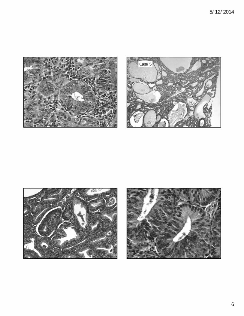
Case 3
Case 4
5/12/2014
6
Case 5
5/12/2014
7
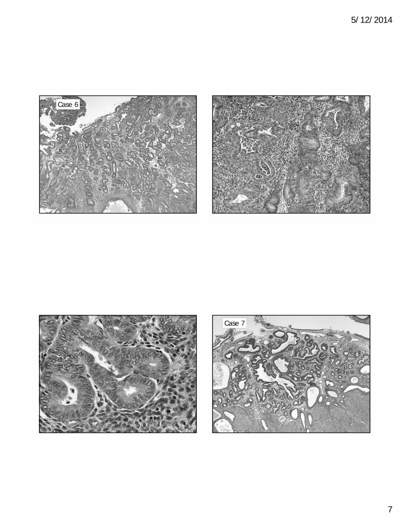
Case 6
Case 7

5/12/2014
8
Typical or atypical?

5/12/2014
9
Diagnostic reproducibility
Skov et al (Scandanavia)Kendall et al (US - Hopkins)
Bergeron et al (Europe)Zaino et al (US - G.O.G.)
Comparison of the Reproducibility of the WHO Classifications of 1975 and
1994 of Endometrial Hyperplasia
B.G. Skov, M.D., H. Broholm, M.D., U. Engel, M.D., M.-B. Franzmann, M.D., A.L. Nielsen, M.D.,
A.F. Lauritzen, M.D., and T. Skov, M.D.
International Journal of Gynecological Pathology 16(1): 33-37, 1997
Overall Agreement and Values for Diagnosis of Endometrial Hyperplasia Using WHO Classifications
of 1975 and 1994 by the Six Observers
Round 1 Round 2 Round 3 Round 4
Overall agreement 0.47 0.45 0.51 0.41 value 0.24 0.25 0.30 0.20
Round 1 and 3, WHO 1975; Round 2 and 4, WHO 1994 classificationkappa below 0.40 – fair to poor agreement, appa values between 0.40 and 0.8 - moderate to good agreement, appa values greater than 0.8 - excellent agreement.
Reproducibility of the Diagnosis of Endometrial Hyperplasia, Atypical
Hyperplasia, and Well-Differentiated Carcinoma
Brian S. Kendall, M.D., Brigitte M. Ronnett, M.D., Christina Isacson, M.D., Kathleen R. Cho, M.D., Lora Hedrick, M.D., Marie Diener-West, Ph.D.,
and Robert J. Kurman, M.D.
The American Journal of Surgical Pathology 22(8):1012-1019, 1998
5/12/2014
10
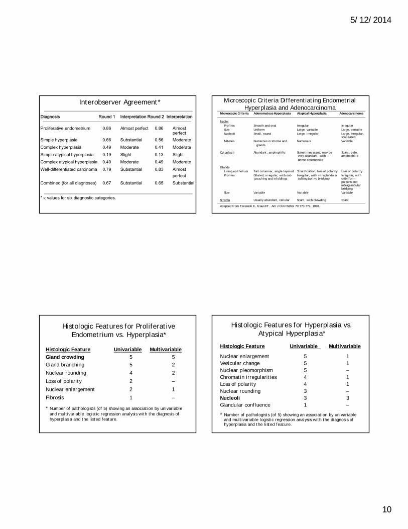
Interobserver Agreement*
Diagnosis Round 1 Interpretation Round 2 Interpretation
Proliferative endometrium 0.86 Almost perfect 0.86 Almost perfect
Simple hyperplasia 0.66 Substantial 0.56 ModerateComplex hyperplasia 0.49 Moderate 0.41 ModerateSimple atypical hyperplasia 0.19 Slight 0.13 SlightComplex atypical hyperplasia 0.40 Moderate 0.49 ModerateWell-differentiated carcinoma 0.79 Substantial 0.83 Almost
perfectCombined (for all diagnoses) 0.67 Substantial 0.65 Substantial
* values for six diagnostic categories.
Microscopic Criteria Differentiating Endometrial Hyperplasia and Adenocarcinoma
Microscopic Criteria Adenomatous Hyperplasia Atypical Hyperplasia Adenocarcinoma
NucleiProfiles Smooth and oval Irregular IrregularSize Uniform Large, variable Large, variableNucleoli Small, round Large, irregular Large, irregular,
spiculatedMitoses Numerous in stroma and Numerous Variable
glands
Cytoplasm Abundant, amphophilic Sometimes scant; may be Scant, pale, very abundant, with amphophilicdense eosinophilia
GlandsLining epithelium Tall columnar, single layered Stratification, loss of polarity Loss of polarityProfiles Dilated, irregular, with out- Irregular, with intraglandular Irregular, with
pouching and infoldings tufting but no bridging cribriform pattern and intraglandularbridging
Size Variable Variable Variable
Stroma Usually abundant, cellular Scant, with crowding Scant
Adapted from Tavassoli F, Kraus FT. Am J Clin Pathol 70:770-779, 1978.
Histologic Features for Proliferative Endometrium vs. Hyperplasia*
Histologic Feature Univariable Multivariable
Gland crowding 5 5
Gland branching 5 2
Nuclear rounding 4 2
Loss of polarity 2 –
Nuclear enlargement 2 1
Fibrosis 1 –
* Number of pathologists (of 5) showing an association by univariable and multivariable logistic regression analysis with the diagnosis of hyperplasia and the listed feature.
Histologic Features for Hyperplasia vs. Atypical Hyperplasia*
Histologic Feature Univariable Multivariable
Nuclear enlargement 5 1Vesicular change 5 1Nuclear pleomorphism 5 –Chromatin irregularities 4 1Loss of polarity 4 1Nuclear rounding 3 –Nucleoli 3 3Glandular confluence 1 –
* Number of pathologists (of 5) showing an association by univariable and multivariable logistic regression analysis with the diagnosis of hyperplasia and the listed feature.
5/12/2014
11
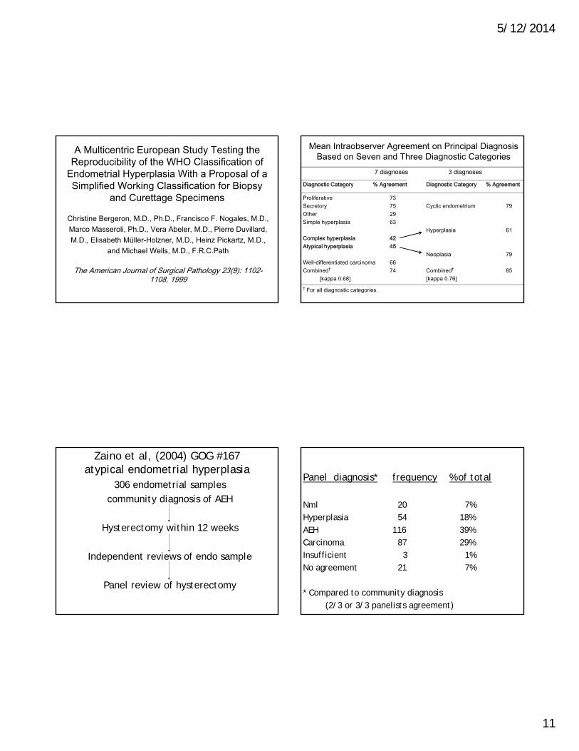
A Multicentric European Study Testing the Reproducibility of the WHO Classification of
Endometrial Hyperplasia With a Proposal of a Simplified Working Classification for Biopsy
and Curettage Specimens
Christine Bergeron, M.D., Ph.D., Francisco F. Nogales, M.D., Marco Masseroli, Ph.D., Vera Abeler, M.D., Pierre Duvillard, M.D., Elisabeth Müller-Holzner, M.D., Heinz Pickartz, M.D.,
and Michael Wells, M.D., F.R.C.Path
The American Journal of Surgical Pathology 23(9): 1102-1108, 1999
Mean Intraobserver Agreement on Principal Diagnosis Based on Seven and Three Diagnostic Categories
7 diagnoses 3 diagnoses
Diagnostic Category % Agreement Diagnostic Category % Agreement
Proliferative 73Secretory 75 Cyclic endometrium 79Other 29Simple hyperplasia 63
Hyperplasia 61Complex hyperplasia 42Atypical hyperplasia 45
Neoplasia 79Well-differentiated carcinoma 66Combined† 74 Combined† 85
[kappa 0.68] [kappa 0.76]† For all diagnostic categories.
Zaino et al, (2004) GOG #167atypical endometrial hyperplasia
306 endometrial samples community diagnosis of AEH
Hysterectomy within 12 weeks
Independent reviews of endo sample
Panel review of hysterectomy
Panel diagnosis* frequency % of total
Nml 20 7%Hyperplasia 54 18%AEH 116 39%Carcinoma 87 29%Insufficient 3 1%No agreement 21 7%
* Compared to community diagnosis (2/3 or 3/3 panelists agreement)

5/12/2014
12
Intra-Panel Agreement
Diagnosis Kappa AssessmentNormal 0.48 moderateHyperplasia 0.38 fairAEH 0.27 fairCarcinoma 0.50 moderateUnable to assess 0.63 substantialOverall 0.39 fair
Diagnostic problems identified
Application of diagnostic criteriaquantitative, qualitative, multiple
criteriaSmall quantity/fragmentation of tissuePoor fixationPoor cryotomyPoor staining
Coexistent carcinoma in the hysterectomy
Trimble et al, Cancer, 2006
Cancer in uterus
Community diagnosis of AEH 43%Panel diagnosis
Nml/typical hyperplasia 18%AEH 43%Carcinoma 65%
GOG Studies Conclusions
1) The reproducibility of the community diagnosis of AEH by a panel of gynpathologists is relatively low
2) Over–estimation and under-estimation of the severity of the lesion is common
3) The reproducibility of the diagnosis of AEH by a panel of gyn pathologists is relatively low
4) The risk of carcinoma associated with the diagnosis of atypical hyperplasia is 43%

5/12/2014
13
Diagnosing endometrial hyperplasia: why is it so difficult to agree?
Allison et al, AJSP, 2008
3 pathologists scored 1800 endometriaFor adequacy, volume of hyperplasia, crowding,
complexity, atypia, metaplasia and diagnosis (using WHO)
Kappas of 0.16 to 0.35 for hyperplasiasDue to:
1) marked diagnostic trends of 2 pathologists, 2) scant tissue3) low volume of hyperplasia
Conclusions of current schemas for endometrial hyperplasia
1) Terminology of hyperplasia is confusing with varying usage of identical terms
2) Diagnostic reproducibility is poor3) Carcinoma frequently coexists in the
uterus with atypical hyperplasia4) There is need for a new conceptual and
practical approach to preinvasive endometrial lesions
In the future, can we distinguish lesions of high risk from those of low
risk for development of cancer?
Morphometry Tumor promoter or suppressor gene mutation
ImmunohistochemistryAnalysis for clonality (neoplasm)
Morphometry - Baak et al
Assessed 22 nuclear and architectural features
Most important Discriminant factors (D-score) in prediction of lesions likely to progress to adenocarcinoma:
1) Volume percentage stroma (VPS)2) Standard deviation of shortest nuclear
axis3) Outer surface density of glands

5/12/2014
14
Morphometry - Baak et al
Lesions with a volume percentage stroma (VPS) of less than 55% are associated with an increased probability of progression to invasive carcinoma
Estimating VPS (http://www.endometrium.org)
70% VPS 60% VPS 40% VPS
gland stroma
Endometrial intraepithelial neoplasia (EIN) (Mutter & Baak, 2000)
Clonality determinations of carcinomas:selected MSI endometrial carcinomas or women
who are heterozygotes at HUMARA (human androgen receptor) on chromosome X
examined adjacent “endometrial hyperplasia” to determine if clonal:
Polyclonal Monoclonal High D-score Low D-scoreVPS >55% VPS <55%(hyperplasia) (EIN)
EIN Criteria (all must be met)
http://www.endometrium.orgArchitecture
Area of Glands>Stroma (VPS<55%) Cytology
Cytology differs between architecturally crowded focus and background.
SizeMaximum linear dimension exceeds 1mm.
Exclude mimicsBenign conditions with overlapping criteria:
basalis, secretory, polyps, repair, etc.. Exclude cancer
Maze-like glands, solid or cribriform growth

5/12/2014
15
Endometrial intraepithelial neoplasia
EIN
prolif

5/12/2014
16
PTENMutter et al
PTEN – tumor suppressor gene, lipid phosphatase crucial in cell survival signal transduction pathway;
most common mutation or deletion in hyperplasia or endometrioid adenocarcinoma (early loss)
Loss of PTENProliferative phase 0% (rare null glands)Disordered pro/hyperplasia 10-40% EIN 40-55%Carcinoma > 60%
EIN
EIN
Disordered proliferative (PTEN) Mutant Gland
5/12/2014
17
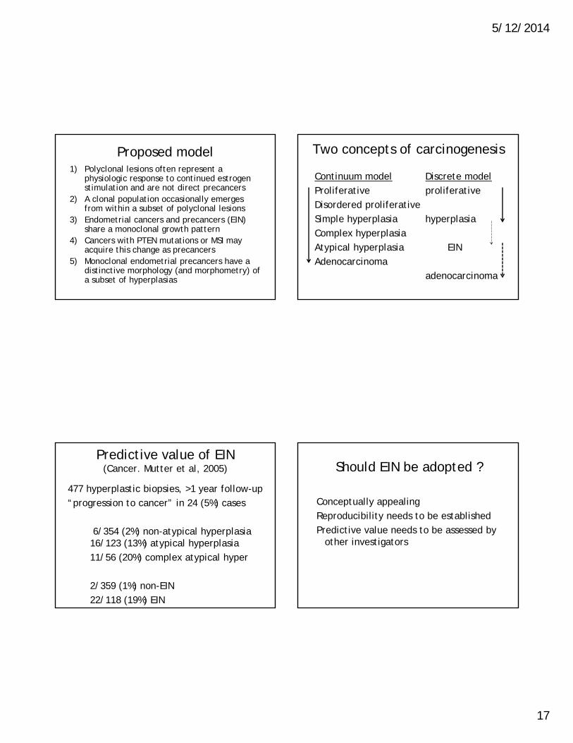
Proposed model1) Polyclonal lesions often represent a
physiologic response to continued estrogen stimulation and are not direct precancers
2) A clonal population occasionally emerges from within a subset of polyclonal lesions
3) Endometrial cancers and precancers (EIN) share a monoclonal growth pattern
4) Cancers with PTEN mutations or MSI may acquire this change as precancers
5) Monoclonal endometrial precancers have a distinctive morphology (and morphometry) of a subset of hyperplasias
Two concepts of carcinogenesis
Continuum model Discrete modelProliferative proliferativeDisordered proliferativeSimple hyperplasia hyperplasiaComplex hyperplasiaAtypical hyperplasia EINAdenocarcinoma
adenocarcinoma
Predictive value of EIN(Cancer. Mutter et al, 2005)
477 hyperplastic biopsies, >1 year follow-up“progression to cancer” in 24 (5%) cases
6/354 (2%) non-atypical hyperplasia 16/123 (13%) atypical hyperplasia11/56 (20%) complex atypical hyper
2/359 (1%) non-EIN22/118 (19%) EIN
Should EIN be adopted ?
Conceptually appealingReproducibility needs to be establishedPredictive value needs to be assessed by
other investigators

5/12/2014
18
Reproducibility of EIN diagnosis is good, but influenced by the diagnostic style of pathologists Usubutun, Mutter, et al, Mod Path, 2012
20 pathologists from Turkey and USKappa 0.58 (benign, EIN, cancer)Disagreements due to a variety of
diagnostic styles which were not associated with experience, practice type, institution, or diagnostic system used in practice.
Reproducibility of current classifications of endometrial endometrioid glandular
proliferations; Ordi et al, Histopathology, 2013
9 expert pathologists from Europe and NA examined 198 endometrial samples
System repro (k) simplified*WHO 0.34 0.59EIN 0.42 0.59EWG 0.53 0.62*reduces to 2 groups (benign, hyperplasia
[without atypia] vs atypical hyperplasia, EIN, carcinoma, neoplasia)
Reproducibility of biopsy diagnoses of endometrial hyperplasia: evidence supporting a simplified classification. (Sherman et al, IJGP, 2008)
WHO 1994 system (209 samples)submitting vs panel diagnosis K = 0.17Panelist 1 vs 2 K = 0.37
Condensed system*Submitting vs panel K = 0.37Panelist 1 vs 2 K = 0.63
* (DP, SH, CH vs AH, CA)
What is the current diagnostic practice in the US?
Hyperplasia – commonEIN – uncommonEIN criteria with hyperplasia terminology
- very common (in the Eastern US)- ? (in the Western US)

5/12/2014
19
Clinical management of AH/EINSGO practice guidelinesTrimble et al, Obstet Gynecol, 2012
Definitive therapyTotal hysterectomy +/- BSO
Non-surgical options*Hormonal therapy (with a progestin)Endometrial ablation not recommended
*for those desiring fertility or poor surgical candidate with co-morbidities
WHO 2014
Uterine Corpus:Epithelial tumors and precursor lesions
Condensed to two diagnostic choices
Hyperplasia without atypiaAtypical hyperplasia/EIN
Hyperplasia without atypia
“An exaggerated proliferation of glands of irregular size and shape, with an associated increase in the gland to stromal ratio compared with proliferative endometrium, without significant cytological atypia”
Syn. Simple or complex non-atypical hyperplasia
Atypical hyperplasia/endometrioid intraepithelial neoplasia
“Cytologic atypia superimposed on endometrial hyperplasia defines atypical hyperplasia/EIN”
Syn. Simple or complex atypical endometrial hyperplasia, endometrial intraepithelial neoplasia

5/12/2014
20
Atypical hyperplasia/EIN (cont.)
“The distinction from hyperplasia without atypia is based on nuclear atypia, which may include enlargement, rounding, loss of polarity, and nucleoli.
As these features are somewhat subjective, intraobserver and interobserver variability remains problematic.”
Atypical hyperplasia/EIN
“The diagnosis of atypia is facilitated by comparison to normal glands or areas of hyperplasia without atypia”
Hyperplasia/EIN conclusions
1) The prior hyperplasia classification methods were not highly reproducible
2) Lesions called hyperplasia include both polyclonal and clonal lesions
3) The binary division into hyperplasia without atypia and atypical hyperplasia/EIN improves diagnostic reproducibility
Hyperplasia/EIN conclusions
4) Clonal lesions (EIN) (many of which resemble AEH) could be considered non-invasive carcinomas
5) ~40% of AEH/EIN on biopsy have invasive carcinoma in the uterus
6) Clinical colleagues should be advised of the relatively low diagnostic reproducibility and high probability of a carcinoma in lesions called AEH

5/12/2014
21
Learning objectivesthe participant should understand the
following issues relating to precursors of endometrioid adenocarcinoma
1) Natural history 2) The lexicon
3) Diagnostic reproducibility4) Current practice
5) Recommended terminology WHO 2014