precision pulse capsulotomy - zepto cataract · live rabbit studies precision pulse capsulotomy...
TRANSCRIPT

Precision Pulse CapsulotomyPreclinical Safety and Performance of a New CapsulotomyTechnology
David F. Chang, MD,1 Nick Mamalis, MD,2 Liliana Werner, MD, PhD2
Purpose: To assess the preclinical safety and performance of a new precision pulse capsulotomy (PPC)method.
Design: Human cadaver eye studies and surgical, slit-lamp, and histopathologic evaluation in a consecutiveseries of 20 live rabbits.
Participants: Human cadaver eyes and New Zealand white rabbits.Methods: Precision pulse capsulotomy uses a highly focused, fast, multipulse, low-energy discharge to
produce a perfectly round anterior capsulotomy instantaneously and simultaneously along all 360�. Capsuloto-mies are performed using a disposable handpiece with a soft collapsible tip and circular nitinol cutting element.Miyake-Apple imaging and scanning electron microscopy (SEM) of PPC were conducted in human cadaver eyes.Surgical, postoperative slit-lamp, and histopathologic assessments of PPC were performed in 20 live rabbits andwere compared with manual continuous curvilinear capsulorrhexis (CCC) in the fellow eye. Anterior chamber (AC)thermocouple temperature measurements were evaluated in a subset of rabbit eyes.
Main Outcome Measures: Capsulotomy edge circularity, SEM morphologic features and zonular move-ment with PPC in human cadaver eyes. Anterior chamber temperature during PPC and grading of ocularinflammation, corneal endothelial damage, anterior capsular opacification (ACO), and posterior capsular opaci-fication (PCO).
Results: Miyake-Apple imaging showed minimal zonular stress, and thermocouple measurements demon-strated negligible AC temperature changes during PPC. Precision pulse capsulotomy produced round, completecapsulotomies in all 20 rabbit eyes, leading to successful in-the-bag intraocular lens (IOL) implantation. Slit-lampexaminations at 3 days and 1, 2, and 4 weeks after surgery showed no significant differences between PPC andCCC in corneal edema, AC inflammatory reaction, capsular fibrosis, ACO, and PCO. Postmortem studies showedno difference in the corneal endothelium between PPC and CCC eyes. All IOLs were well centered in PPC eyes,and histopathologic analysis showed no greater inflammatory infiltrates.
Conclusions: Precision pulse capsulotomy is a new method to automate consistent creation of a perfectlycircular anterior capsulotomy with a disposable handheld instrument that can be used in the normal phaco-emulsification surgical sequence. Compared with CCC in fellow rabbit eyes, PPC was equally safe and showedno greater zonular stress compared with CCC in human cadaver eyes. Human cadaver eye SEM showeda much smoother capsulotomy edge compared to those produced by femtosecond laser. Ophthalmology 2015;-:1e10 ª 2015 by the American Academy of Ophthalmology.
Continuous curvilinear capsulotomy (CCC) is one of themost important components of cataract surgery because ofthe numerous surgical and anatomic advantages it confers.The continuous edge facilitates hydrodissection, corticalaspiration, intraocular lens (IOL) implantation and fixationand renders the capsular bag more resistant to tearing duringsurgery.1,2 Circumferential anterior capsular overlap of theoptic edge optimizes IOL centration, reduces posteriorcapsular opacification, decreases unwanted optical edgedysphotopsias, and may enhance predictability of theeffective lens position.3e6 In contrast, radial anteriorcapsular tears increase the risk of surgical complications,reduce refractive accuracy, and may preclude using certainIOL designs.7,8
� 2015 by the American Academy of OphthalmologyPublished by Elsevier Inc.
The ability to automate a perfectly circular capsulotomyof a consistently precise diameter has driven interest in andadoption of femtosecond laser-assisted cataract surgery.9e12
However, femtosecond laser-assisted cataract surgery ne-cessitates significant capital and per-case costs, alters andslows the normal operative workflow, and cannot be used onevery patient because of affordability or regulatory limita-tions. In addition, there is evidence that a capsulotomycreated with the femtosecond laser may not resist tearing aswell as the manual capsulorrhexis.13e15
We describe a new precision pulse capsulotomy (PPC)technology that is able to create a quick, precise circularcapsulotomy using a disposable handheld instrument calledthe Zepto (Mynosys, Fremont, CA). The PPC Zepto device
1http://dx.doi.org/10.1016/j.ophtha.2015.10.008ISSN 0161-6420/15

Ophthalmology Volume -, Number -, Month 2015
is introduced during surgery in the conventional surgicalsequence and potentially can be centered on the visual axisto produce a capsulotomy of a predetermined diameter.
Methods
Description of Precision Pulse Capsulotomy Device
Precision pulse capsulotomy is performed using a disposablehandpiece and capsulotomy tip called the Zepto (Fig 1A) that isconnected to a control console for operation (Fig 1B). Thecapsulotomy tip consists of a soft, transparent, silicone suctioncup approximately 6 mm in diameter that houses a circular ringelement made of the shape memory alloy nitinol. This nitinolring element has been refined precisely at the micrometer scaleto enable consistent and uniform 360� capsulotomies. Thesuperelastic properties of nitinol allow the capsulotomy tip to bedeformed mechanically into a narrower elongated shape for entrythrough a clear corneal incision. The ring can then re-expandautomatically to its native circular shape within the anteriorchamber (AC). The elongation of the capsulotomy tip is producedby the extension of a push rod, which then is retracted from the tipto allow it to reassume its original circular state.
The PPC handpiece and system were designed throughextensive testing in rabbit eyes, porcine eyes, and in humancadaver eyes from donors in the 50- to 90-year age range. ThePPC handpiece substitutes for capsulorrhexis forceps and isinserted into the AC after it is filled with an ophthalmic visco-surgical device. Handpiece operation is controlled by buttons onthe control console. The PPC methodology in a human cadavereye is illustrated in Figure 2. Precision pulse capsulotomy isbased on a rapid and precisely controlled method of tissuecleavage specifically developed for the efficient cutting of thincollagen membranes such as the human anterior lens capsule.This precision pulse method uses the capsulotomy device’scircular, shape memory alloy nitinol ring element to convert avery brief train of fast electrical pulses efficiently over 4 ms(approximately 1 joule) into mechanical cutting energy (Fig 3).The extremely fast millisecond timeframe of PPC limits anyheat dissipation beyond the layer of water surrounding thenitinol ring. The resulting cutting effect essentially is amechanical one, similar to that with a manual tear. Unlike witha manual or femtosecond laser capsulotomy, however, the
Figure 1. Photographs of the precision pulse capsulotomy (PPC) handpiece capPPC capsulotomy tip from below. A circular ring made of the superelastic shapesuction cup. An extendable and retractable push rod assists in device entry. B, Viserves as to initiate PPC and also acts as an emergency stop.
2
entire circumference of the PPC capsulotomy is created at thevery same instant because of the use of a circular conductingcapsulotomy element. The use of suction delivered through thesuction cup ensures optimal apposition of the nitinol ring withthe lens capsule surface. Small amounts of tilt are compensatedfor automatically. A circular capsulotomy is created, duplicatingthe shape of the circular nitinol element.
Human Cadaver Eye Studies
Cadaver eyes were obtained from eye banks in the United Statesand were used within 72 hours of death for the Miyake-Appleimaging of zonular structures during PPC and for the analysis ofPPC capsulotomy edge morphologic features using scanningelectron microscopy. Precision pulse capsulotomy was performedafter disinserting the iris tissue from cadaver eyes and centering thePPC device at the approximate center of the anterior capsule usingthe limbus as the circumferential boundary.
Zonular forces during performance of PPC were compared withthose with manual CCC using Miyake-Apple imaging in pairedhuman cadaver eyes. The eyes were prepared as previouslydescribed16,17 and placed into an imaging system consisting of ahead model, the underside of which contained a 5-megapixel Uni-versal Serial Bus (USB) video camera. The dissected eye preparationwas mounted into the modified eye socket of the head model, andvideo recordings of PPC or manual CCC were made from below toview the zonular structures during the performance of these 2different capsulotomy methods.
The morphologic features of the anterior capsulotomy edgeproduced by PPC in human cadaver eyes was examined usingscanning electron microscopy. Precision pulse capsulotomy wasperformed on the anterior capsule after open sky eye preparations.The globes then were transferred into a glass vessel and submergedin 0.9% saline for hydrodissection, phacoemulsification, andcortical aspiration. A rim of capsule encompassing the capsu-lotomy opening was dissected free and placed in 2.0% glutaral-dehyde in 0.1 M phosphate buffer, pH 7.4, overnight at 4� C. Thecapsule specimens underwent dehydration using a graded ethanolseries and were placed in between steel filter discs with the cap-sulotomy edge exposed. After processing in a critical point dryer,the specimens were gold-palladium sputtered and imaged using anQuanta 3D FEG scanning electron microscope (FEI, Hillsboro,OR) with the beam voltage set at 5 kV.
sulotomy tip, handpiece, control console, and foot switch. A, View of thememory alloy nitinol is housed within a soft silicone housing that serves as aew of the PPC handpiece, control console, and foot switch. The foot switch

Figure 2. Sequence of photographs illustrating the performance of a precision pulse capsulotomy (PPC) in a human cadaver eye. A, Device is in its nativecircular shape at the start of the procedure. B, Push rod is extended causing the capsulotomy device to assume an elongated shape for entry through thecorneal incision. C, Device is inserted through the corneal incision. D, Device is fully inserted into the anterior chamber containing ophthalmic visco-surgical device. E, As the push rod is retracted, the device begins to re-expand back to its original circular shape. F, Push rod is retracted fully and the deviceis fully circular. The device is centered and gently apposed to the capsular surface; no downward pressure is needed. G, Suction is applied. The suction cupensures even application of suction and apposition of the nitinol ring to the lens capsule without capsular surface distortion. The capsulotomy is performed.H, Suction is reversed automatically. As the capsulotomy tip is manually withdrawn from the anterior chamber, the incision compresses the deformable tipto allow it to exit in its collapsed profile. I, Excised capsule button clings to the underside of the suction cup, from which it is retrieved after device removalfrom the anterior chamber. J, Excised capsule button unfurled for visualization.
Chang et al � Precision Pulse Capsulotomy
Live Rabbit Studies
Precision pulse capsulotomy safety and performance during cata-ract surgery were assessed in a study of 20 New Zealand whiterabbits 11 to 14 weeks of age. Animals were obtained from aUnited States Department of Agricultureeapproved vendor(Western Oregon Rabbit Company, Philomath, OR) in accordancewith the Animal Welfare Act. Rabbit studies complied with theGuide for the Care and Use of Laboratory Animals, as well as withguidelines set by the Association for Research in Vision andOphthalmology. Rabbits received 3 doses of 1% cyclopentolatehydrochloride and 2.5% phenylephrine eye drops at 15-minuteintervals. The rabbits were anesthetized with an intramuscular in-jection of ketamine hydrochloride (50 mg/kg) and xylazine (7 mg/kg)in a 7:1 mixture. One drop of topical proparacaine hydro-chloride anesthetic and 1 drop of Povidone-iodine 5% were placedonto each eye before surgery. A side-port incision was made fol-lowed by injection of the ophthalmic viscosurgical device (Opti-visc; Hoya, China Hills, CA). After placement of a 3.0-mmprimary limbal incision, the PPC capsulotomy tip was insertedthrough this incision into the AC. After performing the anteriorcapsulotomy, the PPC device was removed from the eye. Hydro-dissection was performed, followed by phacoemulsification andcortical aspiration. Each 500 ml of irrigation solution contained 1ml epinephrine 1:1000 and 0.5 ml heparin (10 000 USP units/ml).iSert posterior chamber IOLs were implanted using the iSert Pre-loaded Lens Implantation System (Hoya). The fellow eye in eachrabbit was treated identically except that a manual CCC was per-formed instead of PPC.
Because PPC uses electrical energy, intraocular temperatureswere measured during device use. The surgical procedure other-wise was identical except that a thermocouple was inserted throughthe side-port incision into the AC in 6 of the 20 rabbits beforeinitiating PPC. Continuous temperature measurements were made
at 1 of 2 different locations inside the AC during the capsulotomystep (see Fig 4). At location A, the thermocouple tip was adjacentto the silicone side wall of the PPC device, and as close as possibleto the nitinol ring. At location B, the thermocouple tip waspositioned next to corneal endothelium located closest to thenitinol ring. The thermocouple was removed beforephacoemulsification and IOL implantation.
All study eyes (PPC and CCC) were subjected to gross exam-ination on postoperative day 1 and assessed for infection, inflam-mation, hemorrhage, and wound leak. Dilated slit-lampexaminations were conducted on postoperative day 3 and at 1, 2,and 4 weeks. Conjunctival injection, corneal edema, aqueous cellsand flare, iris vasculature, posterior synechiae, IOL centration, andIOL inflammatory deposits were noted. Anterior and posteriorcapsule opacification were graded on a 0-to-4 scale at each ex-amination. At the end of the 4-week study period, the rabbits wereeuthanized and the globes were harvested and fixed in 10% neutralbuffered formalin. All globes were bisected coronally just anteriorto the equator. Gross examination and photography from theposterior aspect (Miyake-Apple view) were performed. Tissuesections were obtained from all eyes and stained with hematoxylinand eosin for light microscopic examination of inflammatory cells,necrosis, and cell vacuolization in various ocular structures.
Histologic examination was performed to compare morphologicdamage to the corneal endothelium after PPC and CCC. Afterclinical examination on postoperative day 3, 6 study rabbits thatunderwent PPC and manual CCC in their fellow eyes wereeuthanized. The 12 corneas with a rim of surrounding sclera wereremoved from the harvested globes for study. The cornea buttonswere placed endothelial side up and stained using a modification ofa method by Spence and Peyman18 consisting of immersion inTrypan blue 0.25% and Alizarin red 1%. Central corneal buttons8 mm in diameter were punched out using a trephine, and thestained specimens were placed on a microscopic slide,
3
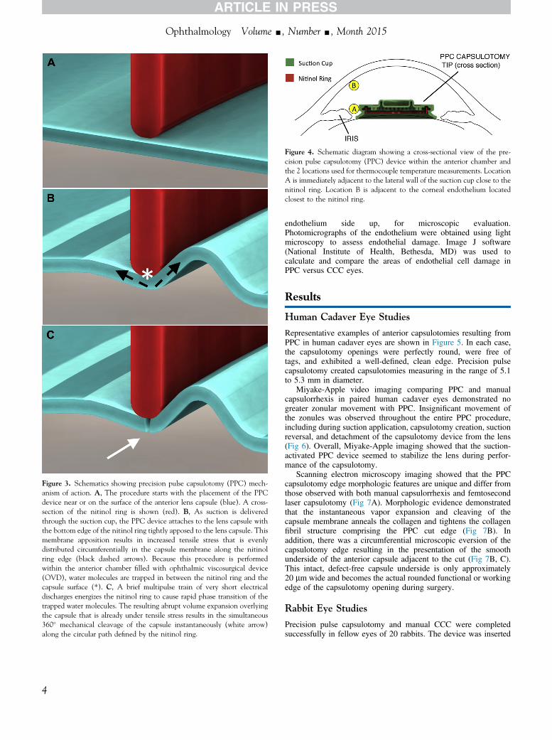
Figure 3. Schematics showing precision pulse capsulotomy (PPC) mech-anism of action. A, The procedure starts with the placement of the PPCdevice near or on the surface of the anterior lens capsule (blue). A cross-section of the nitinol ring is shown (red). B, As suction is deliveredthrough the suction cup, the PPC device attaches to the lens capsule withthe bottom edge of the nitinol ring tightly apposed to the lens capsule. Thismembrane apposition results in increased tensile stress that is evenlydistributed circumferentially in the capsule membrane along the nitinolring edge (black dashed arrows). Because this procedure is performedwithin the anterior chamber filled with ophthalmic viscosurgical device(OVD), water molecules are trapped in between the nitinol ring and thecapsule surface (*). C, A brief multipulse train of very short electricaldischarges energizes the nitinol ring to cause rapid phase transition of thetrapped water molecules. The resulting abrupt volume expansion overlyingthe capsule that is already under tensile stress results in the simultaneous360� mechanical cleavage of the capsule instantaneously (white arrow)along the circular path defined by the nitinol ring.
Figure 4. Schematic diagram showing a cross-sectional view of the pre-cision pulse capsulotomy (PPC) device within the anterior chamber andthe 2 locations used for thermocouple temperature measurements. LocationA is immediately adjacent to the lateral wall of the suction cup close to thenitinol ring. Location B is adjacent to the corneal endothelium locatedclosest to the nitinol ring.
Ophthalmology Volume -, Number -, Month 2015
4
endothelium side up, for microscopic evaluation.Photomicrographs of the endothelium were obtained using lightmicroscopy to assess endothelial damage. Image J software(National Institute of Health, Bethesda, MD) was used tocalculate and compare the areas of endothelial cell damage inPPC versus CCC eyes.
Results
Human Cadaver Eye Studies
Representative examples of anterior capsulotomies resulting fromPPC in human cadaver eyes are shown in Figure 5. In each case,the capsulotomy openings were perfectly round, were free oftags, and exhibited a well-defined, clean edge. Precision pulsecapsulotomy created capsulotomies measuring in the range of 5.1to 5.3 mm in diameter.
Miyake-Apple video imaging comparing PPC and manualcapsulorrhexis in paired human cadaver eyes demonstrated nogreater zonular movement with PPC. Insignificant movement ofthe zonules was observed throughout the entire PPC procedure,including during suction application, capsulotomy creation, suctionreversal, and detachment of the capsulotomy device from the lens(Fig 6). Overall, Miyake-Apple imaging showed that the suction-activated PPC device seemed to stabilize the lens during perfor-mance of the capsulotomy.
Scanning electron microscopy imaging showed that the PPCcapsulotomy edge morphologic features are unique and differ fromthose observed with both manual capsulorrhexis and femtosecondlaser capsulotomy (Fig 7A). Morphologic evidence demonstratedthat the instantaneous vapor expansion and cleaving of thecapsule membrane anneals the collagen and tightens the collagenfibril structure comprising the PPC cut edge (Fig 7B). Inaddition, there was a circumferential microscopic eversion of thecapsulotomy edge resulting in the presentation of the smoothunderside of the anterior capsule adjacent to the cut (Fig 7B, C).This intact, defect-free capsule underside is only approximately20 mm wide and becomes the actual rounded functional or workingedge of the capsulotomy opening during surgery.
Rabbit Eye Studies
Precision pulse capsulotomy and manual CCC were completedsuccessfully in fellow eyes of 20 rabbits. The device was inserted

Chang et al � Precision Pulse Capsulotomy
and subsequently removed through a 3.0-mm incision. All PPCprocedures resulted in a complete tag-free capsulotomy with thecapsule button extracted simultaneously by the PPC device(Table 1). All PPC eyes successfully underwent hydrodissection,phacoemulsification, cortical aspiration, and IOL implantation inthe capsular bag. The geometry of PPC capsulotomies wasconsistently rounder than those obtained using manual CCC asjudged by slit-lamp observation (Fig 8).
Continuous thermocouple temperature measurements duringPPC showed negligible, transient temperature changes at bothpositions A and B within the AC (Fig 4; Table 2). The measuredmean peak temperature rise at location A was only 2� C. Themean measured peak temperature rise at location B was barely1� C. The duration of these minor temperature changes lastedonly several seconds.
In the subset of 6 rabbits (6 PPC eyes and 6 fellow CCC eyes)harvested on postoperative day 3 and evaluated with cornealendothelial cell staining, there was no statistical difference in theamount of morphologic corneal endothelial damage between eyesundergoing PPC or manual CCC (Table 1). The postoperativecourse was otherwise unremarkable for all PPC and manualCCC study eyes (Table 1). Gross examination on postoperativeday 1 noted corneal edema only at the incision site, with nodifference between PPC and manual CCC eyes. Postoperativeday 3 findings also were similar for both groups, and includedstatistically comparable degrees of aqueous cells, irishyperemia, AC fibrin formation, and corneal edema that waspredominantly near the incision. Clinical corneal edema onpostoperative day 3 was statistically similar between bothgroups, as analyzed both by the corneal opacity score and bymultiplying corneal opacity by opacity extent. There were also
Figure 5. AeD, Examples of precision pulse capsulotomy (PPC) procedurevisualization.
no postoperative day 3 differences in terms of aqueous cells,iris hyperemia, and fibrin score. Each of these postoperativeday 3 findings resolved in all eyes (PPC and CCC) by the week2 examination.
The diameter of rabbit capsulotomies tends to increase aftersurgery because of the higher elasticity of the rabbit anteriorcapsule. No difference in capsulotomy expansion was noted be-tween eyes that underwent PPC or manual CCC. Rabbit eyes alsoprovide an accelerated model of anterior capsular opacification andposterior capsular opacification (PCO). There were no differencesbetween the 2 groups in terms of PCO, proliferative lens epithelialcell pearls anterior to the IOL, posterior synechiae, or capsulotomyedge fibrosis at the week 4 examination.
Postmortem examination of all globes at 4 weeks showed thatall IOLs were fixated symmetrically within the capsular bag(Fig 7C, D). There were no differences between the 2 groups ofeyes in terms of central PCO, peripheral PCO, Soemmering’sring formation, anterior capsular opacification, IOL coverage bythe capsulotomy, or IOL decentration (Table 1).
Microscopic histopathologic examination of tissue sectionsfrom eyes of both PPC and CCC groups at 4 weeks showed nosignificant difference in Soemmering’s ring formation, PCO,anterior cortical proliferation, and anterior fibrous metaplasia.There was no evidence of toxicity or excessive inflammation in anyof the PPC or manual CCC eyes.
Discussion
The ideal anterior capsulotomy would be continuous, cir-cular, centered on the visual axis, maximally resistant to
s in human cadaver eyes. The corneas and irides were removed to aid
5

Figure 6. Miyake-Apple imaging of zonular structures during continuous curvilinear capsulotomy (CCC) and precision pulse capsulotomy (PPC) performedin paired cadaver eyes from a 53-year-old donor. A, B, Manual CCC created zonular traction in the sector where the capsular flap was being pulled. C, D, Incontrast, the PPC in the fellow eye showed no significant zonular traction during suction application or creation of the capsulotomy.
Ophthalmology Volume -, Number -, Month 2015
tearing, and of a precise diameter that circumferentiallyoverlaps the IOL optic edge. An inexpensive automatedtechnology that could create such a capsulotomy consis-tently and reproducibly as an integrated surgical step wouldbe attractive to most surgeons. Ideally, it would improverather than decrease surgical efficiency and the cost wouldbe low enough so that it could be used on all patients.Femtosecond laser-assisted cataract surgery is appealing inpart for its ability to automate creation of a perfectly circularcapsulotomy. However, the technology is costly, requiressupplemental patient payment, and disrupts the normaloperative workflow. In addition, several published studieshave raised concerns that the femtosecond laser
6
capsulotomy is less resistant to tearing compared with amanual capsulorrhexis.9e11 The precision pulse capsu-lotomy technology described and evaluated herein wasdeveloped to provide a potentially superior and lessexpensive method to automate creation of a perfectly cir-cular capsulotomy.
Testing in human cadaver eyes as well as in live rabbiteyes showed that the precision pulse technology is able tocreate a perfectly circular capsulotomy without tagsconsistently and instantaneously. The version of the PPCdevice used in these studies was designed to fit through a3.0-mm corneal incision. A newer version of the deviceunder development should fit through a 2.2-mm incision.
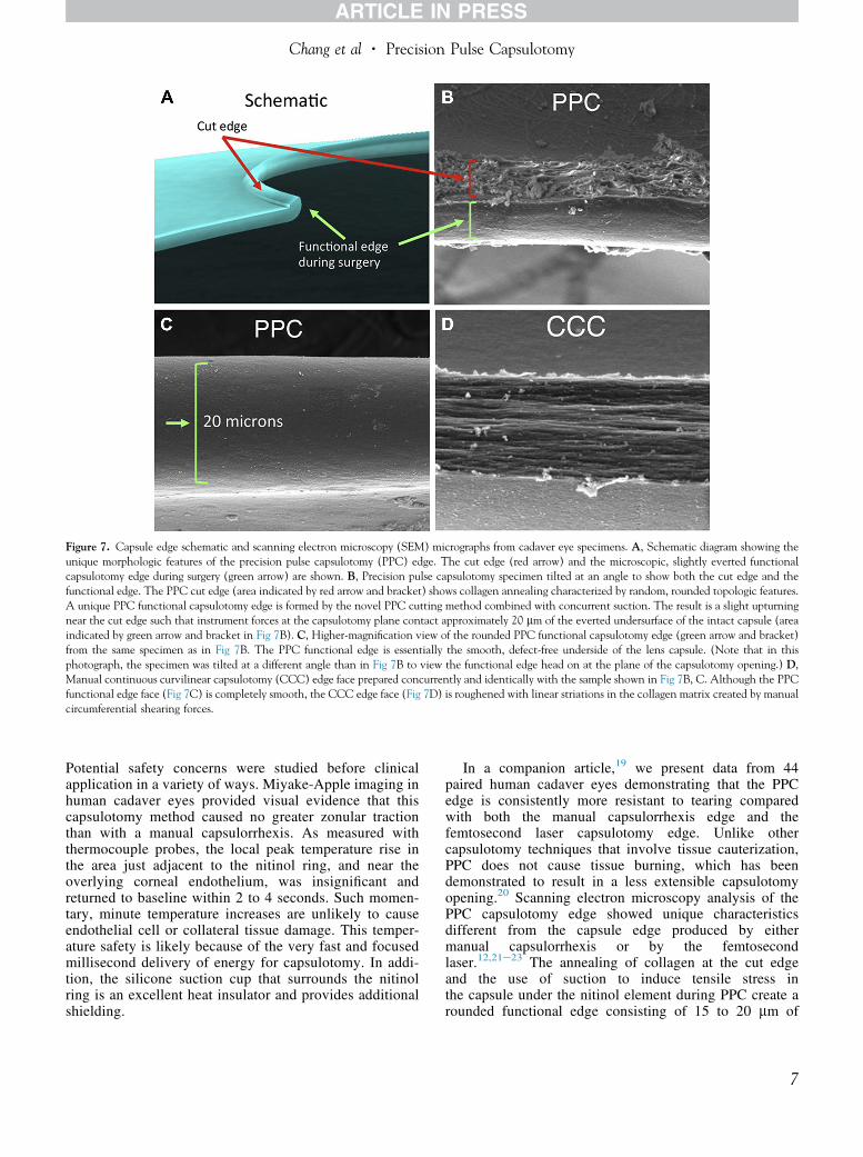
Figure 7. Capsule edge schematic and scanning electron microscopy (SEM) micrographs from cadaver eye specimens. A, Schematic diagram showing theunique morphologic features of the precision pulse capsulotomy (PPC) edge. The cut edge (red arrow) and the microscopic, slightly everted functionalcapsulotomy edge during surgery (green arrow) are shown. B, Precision pulse capsulotomy specimen tilted at an angle to show both the cut edge and thefunctional edge. The PPC cut edge (area indicated by red arrow and bracket) shows collagen annealing characterized by random, rounded topologic features.A unique PPC functional capsulotomy edge is formed by the novel PPC cutting method combined with concurrent suction. The result is a slight upturningnear the cut edge such that instrument forces at the capsulotomy plane contact approximately 20 mm of the everted undersurface of the intact capsule (areaindicated by green arrow and bracket in Fig 7B). C, Higher-magnification view of the rounded PPC functional capsulotomy edge (green arrow and bracket)from the same specimen as in Fig 7B. The PPC functional edge is essentially the smooth, defect-free underside of the lens capsule. (Note that in thisphotograph, the specimen was tilted at a different angle than in Fig 7B to view the functional edge head on at the plane of the capsulotomy opening.) D,Manual continuous curvilinear capsulotomy (CCC) edge face prepared concurrently and identically with the sample shown in Fig 7B, C. Although the PPCfunctional edge face (Fig 7C) is completely smooth, the CCC edge face (Fig 7D) is roughened with linear striations in the collagen matrix created by manualcircumferential shearing forces.
Chang et al � Precision Pulse Capsulotomy
Potential safety concerns were studied before clinicalapplication in a variety of ways. Miyake-Apple imaging inhuman cadaver eyes provided visual evidence that thiscapsulotomy method caused no greater zonular tractionthan with a manual capsulorrhexis. As measured withthermocouple probes, the local peak temperature rise inthe area just adjacent to the nitinol ring, and near theoverlying corneal endothelium, was insignificant andreturned to baseline within 2 to 4 seconds. Such momen-tary, minute temperature increases are unlikely to causeendothelial cell or collateral tissue damage. This temper-ature safety is likely because of the very fast and focusedmillisecond delivery of energy for capsulotomy. In addi-tion, the silicone suction cup that surrounds the nitinolring is an excellent heat insulator and provides additionalshielding.
In a companion article,19 we present data from 44paired human cadaver eyes demonstrating that the PPCedge is consistently more resistant to tearing comparedwith both the manual capsulorrhexis edge and thefemtosecond laser capsulotomy edge. Unlike othercapsulotomy techniques that involve tissue cauterization,PPC does not cause tissue burning, which has beendemonstrated to result in a less extensible capsulotomyopening.20 Scanning electron microscopy analysis of thePPC capsulotomy edge showed unique characteristicsdifferent from the capsule edge produced by eithermanual capsulorrhexis or by the femtosecondlaser.12,21e23 The annealing of collagen at the cut edgeand the use of suction to induce tensile stress inthe capsule under the nitinol element during PPC create arounded functional edge consisting of 15 to 20 mm of
7

Table 1. Postoperative Course and Postmortem Examination Results in Paired Rabbit Eyes Undergoing Precision Pulse Capsulotomyversus Continuous Curvilinear Capsulorrhexis
Precision PulseCapsulotomy (20 Eyes)
Continuous CurvilinearCapsulorrhexis (20 Eyes)
Difference orStatistical Analysis
Intraoperative performanceSuccessful, complete capsulotomy All 20 eyes All 20 eyes No differenceIn-the-bag IOL placement All 20 eyes All 20 eyes No difference
Postoperative coursePostoperative day 1 corneal edema Only at incision site Only at incision site No difference by
examinationPostoperative day 3
Corneal opacity score 1.4�0.72 1.6�1.0 P ¼ 0.57Corneal opacity � extent 2.36�1.8 2.34�2.1 P ¼ 0.94Aqueous cells 1.0�0.0 0.98�0.1 P ¼ 0.32Iris hyperemia 0.33�0.45 0.25�0.40 P ¼ 0.82Fibrin deposits Mild Mild No difference
Postoperative week 2 examination All postoperative day 3findings resolved
All postoperative day 3findings resolved
No difference
Postoperative week 4PCO 2.42�0.86 2.76 � 0.92 P ¼ 0.06LEC pearls anterior to IOL Present Present No differenceSynechiae Present Present No differenceCapsular edge fibrosis Present Present No difference
Corneal endothelial cell damageAssessed by histologic staining of corneal buttons mean (variance)
4.14% (41.1%)mean (variance)4.92% (24.5%)
P ¼ 0.79
Postmortem examination at 4 weeks (gross examination andMiyake-Apple view photographs)
Symmetrically fixated IOLs All 20 eyes All 20 eyes No differenceCentral PCO 1.5�0.9 1.4�0.9 P ¼ 0.76Peripheral PCO 1.9�0.9 2.0�0.9 P ¼ 0.82Soemmering’s formation (intensity � area) 7.0�2.7 7.5�2.1 P ¼ 1IOL coverage by capsulotomy 193�85 176�75 P ¼ 0.82IOL decentration 0.71�0.65 0.77�1.1 P ¼ 0.57
Tissue section or microscopic analysisToxicity or excessive inflammation None None No difference
IOL ¼ intraocular lens; LEC ¼ lens epithelial cell; PCO ¼ posterior capsular opacification.Statistical analyses were performed using the t test: paired 2 sample for means, 2-tailed.
Ophthalmology Volume -, Number -, Month 2015
everted smooth capsular underside. We postulate that thismicroscopic eversion of the PPC edge improves itsbiomechanical strength by approximating the tear resis-tance of smooth, intact, and defect-free capsule. Greatertear resistance of the PPC capsulotomy edge may improvesurgical safety compared with capsulotomies created eithermanually or with the femtosecond laser.
Comparison testing of PPC or manual CCC in pairedeyes of live rabbits showed no differences in postoperativecorneal edema, inflammatory reaction, anterior capsularopacification, or PCO as determined by slit-lamp examina-tion. Histologic analysis of postmortem corneas likewiseshowed no difference in endothelial cell loss. This providespreclinical evidence that the very brief and minor tempera-ture rise, which is spatially confined and insulated by thesilicone housing, should not cause collateral tissue trauma.
A potential advantage of the PPC device is that it offersthe surgeon flexibility in positioning the capsulotomy. Thecircular shape of the PPC capsulotomy tip facilitates
8
centration within the pupil. In cadaver eyes, we also havebeen able to insert the device through a small pupil tocreate a capsulotomy with a larger diameter than the pupil.By design, a clear window in the center of the PPC suctioncup permits patient fixation onto the coaxial microscopelight filament, which theoretically could guide centrationof the capsulotomy on the patient’s visual axis. This mayprove advantageous for refractive IOLs, such as diffractivemultifocals, which should be centered on the patient’s vi-sual axis, rather than the center of the pupil or capsularbag.
In conclusion, a novel PPC technology performed using adisposable handpiece has been developed that can create aprecisely sized, circular capsulotomy as an integrated stepduring conventional phacoemulsification. This technologymay prove able to automate the capsulotomy step and pro-duce a stronger capsulotomy edge, without the higher costs,longer procedural time, and the logistical challenges of us-ing a femtosecond laser.

Figure 8. Postoperative slit-lamp and Miyake-Apple analysis of paired rabbit eyes after precision pulse capsulotomy (PPC) and manual continuouscurvilinear capsulotomy (CCC). A, Slit-lamp photograph obtained on postoperative day 3 showing a PPC. B, Slit-lamp photograph obtained on post-operative day 3 showing the CCC in the fellow eye. The PPCs in this study were noticeably rounder and of a more consistent size than CCCs in the felloweyes. C, Miyake-Apple view of a rabbit eye harvested 4 weeks after PPC and intraocular lens (IOL) implantation. The IOL is well centered within the bag.D, Miyake-Apple view of the fellow eye of the same rabbit as in Figure 8C.
Table 2. Temperature Change in Anterior Chamber during Pre-cision Pulse Capsulotomy
MeasurementLocation
ALocation
B
Duration ofTemperature
Change (Seconds)
PeakTemperatureRise (� C)
1 Device 2.94 1.762 Device 3.59 1.913 Device 3.69 2.01Mean 3.01 1.894 Endothelium 2.69 0.895 Endothelium 4.47 0.536 Endothelium 4.60 1.32Mean 3.92 0.74
Chang et al � Precision Pulse Capsulotomy
References
1. Assia EI, Apple DJ, Tsai JC, Lim ES. The elastic properties ofthe lens capsule in capsulorrhexis. Am J Ophthalmol1991;111:628–32.
2. Krag S, Thim K, Corydon L. Strength of the lens capsuleduring hydroexpression of the nucleus. J Cataract Refract Surg1993;19:205–8.
3. Assia EI, Legler UF, Apple DJ. The capsular bag after short-and long-term fixation of intraocular lenses. Ophthalmology1995;102:1151–7.
4. Ravalico G, Tognetto D, Palomba M, et al. Capsulorhexis sizeand posterior capsule opacification. J Cataract Refract Surg1996;22:98–103.
9

Ophthalmology Volume -, Number -, Month 2015
5. Ram J, Pandey SK, Apple DJ, et al. Effect of in-the-bag intra-ocular lens fixation on the prevention of posterior capsule opa-cification. J Cataract Refract Surg 2001;27:1039–46.
6. Hollick EJ, Spalton DJ, Meacock WR. The effect of capsulor-rhexis size on posterior capsular opacification: one-year results of arandomized prospective trial. Am J Ophthalmol 1999;128:271–9.
7. Aasuri MK, Kompella VB, Majji AB. Risk factors for andmanagement of dropped nucleus during phacoemulsification.J Cataract Refract Surg 2001;27:1428–32.
8. Carifi G, Miller MH, Pitsas C, et al. Complications and out-comes of phacoemulsification cataract surgery complicated byanterior capsule tear. Am J Ophthalmol 2015;159:463–9.
9. Nagy Z, Takacs A, Filkorn T, Sarayba M. Initial clinicalevaluation of an intraocular femtosecond laser in cataractsurgery. J Refract Surg 2009;25:1053–60.
10. Kranitz K, Takacs A, Mihaltz K, et al. Femtosecond lasercapsulotomy and manual continuous curvilinear capsulor-rhexis parameters and their effects on intraocular lens centra-tion. J Refract Surg 2011;27:558–63.
11. Friedman NJ, Palanker DV, Schuele G, et al. Femtosecondlaser capsulotomy. J Cataract Refract Surg 2011;37:1189–98.
12. Palanker DV, Blumenkranz MS, Andersen D, et al. Femto-second laser-assisted cataract surgery with integrated opticalcoherence tomography. Sci Transl Med 2010;2:58ra85.
13. Abell RG, Darian-Smith E, Kan JB, et al. Femtosecond laser-assisted cataract surgery versus standard phacoemulsificationcataract surgery: outcomes and safety in more than 4000 casesat a single center. J Cataract Refract Surg 2015;41:47–52.
14. Abell RG, Davies PE, Phelan D, et al. Anterior capsulotomyintegrity after femtosecond laser-assisted cataract surgery.Ophthalmology 2014;121:17–24.
10
15. Chang JS, Chen IN, Chan WM, et al. Initial evaluation of afemtosecond laser system in cataract surgery. J CataractRefract Surg 2014;40:29–36.
16. Davis BL, Nilson CD, Mamalis N. Revised Miyake-Appletechnique for postmortem eye preparation. J Cataract RefractSurg 2004;30:546–9.
17. Pereira FA, Werner L, Milverton EJ, Coroneo MT. Miyake-Apple posterior video analysis/photographic technique.J Cataract Refract Surg 2009;35:577–87.
18. Spence DJ, Peyman GA. A new technique for the vital stainingof the corneal endothelium. Invest Ophthalmol 1976;15:1000–2.
19. Thompson VM, Berdahl JP, Solano JM, Chang DF. Com-parison of manual, femtosecond laser, and precision pulsecapsulotomy edge tear strength in paired human cadaver eyes.Ophthalmology (Accepted for publication).
20. Wilson ME Jr. Anterior lens capsule management in pediatriccataract surgery. Trans Am Ophthalmol Soc 2004;102:391–422.
21. Ostovic M, Klaproth OK, Hengerer FH, et al. Light micro-scopy and scanning electron microscopy analysis of rigidcurved interface femtosecond laser-assisted and manual ante-rior capsulotomy. J Cataract Refract Surg 2013;39:1587–92.
22. Mayer WJ, Klaproth OK, Ostovic M, et al. Cell death andultrastructural morphology of femtosecond laser-assistedanterior capsulotomy. Invest Ophthalmol Vis Sci 2014;55:893–8.
23. Al Harthi K, Al Shahwan S, Al Towerki A, et al. Comparisonof the anterior capsulotomy edge created by manual capsu-lorrhexis and 2 femtosecond laser platforms: scanning electronmicroscopy study. J Cataract Refract Surg 2014;40:2106–12.
Footnotes and Financial Disclosures
Originally received: August 27, 2015.Final revision: October 3, 2015.Accepted: October 7, 2015.Available online: ---. Manuscript no. 2015-1481.1 Altos Eye Physicians, Los Altos, California.2 John A. Moran Eye Center, University of Utah, Salt Lake City, Utah.
Presented as a poster at: American Academy of Ophthalmology AnnualMeeting, Las Vegas, Nevada, November 2015.
Financial Disclosure(s):The author(s) have made the following disclosure(s): D.F.C.: Consultant -Mynosys; LensAR; AMO
L.W.: Financial support - AMO; AcuFocus; Alcon; Bausch & Lomb;Calhoun Vision; ClarVista Medical; Genisphere; Hoya; LensGen; Myno-sys; Omega; PowerVision; Sharklet; Advisory board - PowerVision
The research sponsor (Mynosys Cellular Devices Inc., Fremont, CA)participated in the design of the study.
Author Contributions:
Conception and design: Chang, Mamalis, Werner
Analysis and interpretation: Chang, Mamalis, Werner
Data collection: Chang, Mamalis, Werner
Obtained funding: none
Overall responsibility: Chang, Mamalis, Werner
Abbreviations and Acronyms:AC ¼ anterior chamber; ACO ¼ anterior capsular opacification;CCC ¼ continuous curvilinear capsulorrhexis; IOL ¼ intraocular lens;PCO ¼ posterior capsular opacification; PPC ¼ precision pulse capsu-lotomy; SEM ¼ scanning electron microscopy.
Correspondence:David F. Chang, MD, Altos Eye Physicians, 762 Altos Oaks Drive, LosAltos, CA 94024. E-mail: [email protected].