pparγ a molecular link between systemic metabolic disease ... · mon molecular pathways disrupted...
TRANSCRIPT

Differentiation 82 (2011) 220–236
Contents lists available at ScienceDirect
Differentiation
0301-46
Join the
doi:10.1
Abbre
syndrom
prostate
DM, dia
density
Sympto
kinases
Prostate
PSA, pro
estradio
waist ton Corr
Tel.: þ1nn Cor
E-m
journal homepage: www.elsevier.com/locate/diff
Invited Review
PPARg: A molecular link between systemic metabolic diseaseand benign prostate hyperplasia
Ming Jiang a,nn, Douglas W. Strand a, Omar E. Franco a, Peter E. Clark a, Simon W. Hayward a,b,n
a Department of Urologic Surgery, Vanderbilt University Medical Center, Nashville, TN, USAb Department of Cancer Biology, Vanderbilt-Ingram Cancer Center, Nashville, TN, USA
a r t i c l e i n f o
Available online 8 June 2011
Keywords:
BPH
LUTS
Co-morbidities
Metabolism
Inflammation
PPARg
81/$ - see front matter & 2011 International
International Society for Differentiation (ww
016/j.diff.2011.05.008
viations: 15-LOX-2, 15-lipoxygenase-2; 15dP
e; AP, anterior prostate; aP2, fatty acid bind
; CAT, catalase; Cdk5, cyclin-dependent kina
betes mellitus; ERK, extracellular signal-regu
lipoprotein; HFD, high-fat diet; IFNg, interfero
m Score; LBD, ligand-binding domain; LDL, lo
; MRI, magnetic resonance imaging; NCoR, nu
Cancer Prevention Trial; PGC1, PPARg co-acti
state specific antigen; PUFA, polyunsaturated
l; TA, transit amplifying; TRUS, transrectal ul
hip ratio
esponding author at: Department of Urologi
615 343 5984; fax: þ1 615 322 8990.
responding author.
ail addresses: [email protected] (M
a b s t r a c t
The emergent epidemic of metabolic syndrome and its complex list of sequelae mandate a more
thorough understanding of benign prostatic hyperplasia and lower urinary tract symptoms (BPH/LUTS)
in the context of systemic metabolic disease. Here we discuss the nature and origins of BPH, examine its
role as a component of LUTS and review retrospective clinical studies that have drawn associations
between BPH/LUTS and type II diabetes, inflammation and dyslipidemia. PPARg signaling, which sits at
the nexus of systemic metabolic disease and BPH/LUTS through its regulation of inflammation and
insulin resistance, is proposed as a candidate for molecular manipulation in regard to BPH/LUTS. Finally,
we introduce new cell and animal models that are being used to study the consequences of obesity,
diabetes and inflammation on benign prostatic growth.
& 2011 International Society of Differentiation. Published by Elsevier B.V. All rights reserved.
Contents
1. Introduction . . . . . . . . . . . . . . . . . . . . . . . . . . . . . . . . . . . . . . . . . . . . . . . . . . . . . . . . . . . . . . . . . . . . . . . . . . . . . . . . . . . . . . . . . . . . . . . . . . . . . . 221
2. Anatomy and development of the prostate . . . . . . . . . . . . . . . . . . . . . . . . . . . . . . . . . . . . . . . . . . . . . . . . . . . . . . . . . . . . . . . . . . . . . . . . . . . . . 221
3. Stromal–epithelial interactions in prostatic development . . . . . . . . . . . . . . . . . . . . . . . . . . . . . . . . . . . . . . . . . . . . . . . . . . . . . . . . . . . . . . . . . . 222
4. Benign prostatic hyperplasia (BPH). . . . . . . . . . . . . . . . . . . . . . . . . . . . . . . . . . . . . . . . . . . . . . . . . . . . . . . . . . . . . . . . . . . . . . . . . . . . . . . . . . . . 222
5. Lower urinary tract symptoms (LUTS) . . . . . . . . . . . . . . . . . . . . . . . . . . . . . . . . . . . . . . . . . . . . . . . . . . . . . . . . . . . . . . . . . . . . . . . . . . . . . . . . . 223
6. Common co-morbidities affecting BPH/LUTS patients . . . . . . . . . . . . . . . . . . . . . . . . . . . . . . . . . . . . . . . . . . . . . . . . . . . . . . . . . . . . . . . . . . . . . 223
6.1. BPH/LUTS and the metabolic syndrome . . . . . . . . . . . . . . . . . . . . . . . . . . . . . . . . . . . . . . . . . . . . . . . . . . . . . . . . . . . . . . . . . . . . . . . . . . 224
6.2. Diabetes and BPH/LUTS . . . . . . . . . . . . . . . . . . . . . . . . . . . . . . . . . . . . . . . . . . . . . . . . . . . . . . . . . . . . . . . . . . . . . . . . . . . . . . . . . . . . . . . 224
6.3. Obesity and BPH/LUTS . . . . . . . . . . . . . . . . . . . . . . . . . . . . . . . . . . . . . . . . . . . . . . . . . . . . . . . . . . . . . . . . . . . . . . . . . . . . . . . . . . . . . . . . 225
7. Mechanisms linking diabetes and obesity to BPH/LUTS . . . . . . . . . . . . . . . . . . . . . . . . . . . . . . . . . . . . . . . . . . . . . . . . . . . . . . . . . . . . . . . . . . . . 226
7.1. Steroid hormones . . . . . . . . . . . . . . . . . . . . . . . . . . . . . . . . . . . . . . . . . . . . . . . . . . . . . . . . . . . . . . . . . . . . . . . . . . . . . . . . . . . . . . . . . . . . 226
7.2. Insulin and IGF/IGFBP axis. . . . . . . . . . . . . . . . . . . . . . . . . . . . . . . . . . . . . . . . . . . . . . . . . . . . . . . . . . . . . . . . . . . . . . . . . . . . . . . . . . . . . 227
7.3. Inflammation . . . . . . . . . . . . . . . . . . . . . . . . . . . . . . . . . . . . . . . . . . . . . . . . . . . . . . . . . . . . . . . . . . . . . . . . . . . . . . . . . . . . . . . . . . . . . . . 227
Society of Differentiation. Published by Elsevier B.V. All rights reserved.
w.isdifferentiation.org)
GJ2, 15-deoxy-D12,14-prostaglandin J2; 15 S-15-HETE, hydroxyeicosatetraenoic acid; AIS, androgen insensitivity
ing protein; AR, androgen receptor; BMI, body mass index; BPH, benign prostate hyperplasia; CaP, carcinoma of the
se 5; COX-2, cyclooxygenase-2; DBD, DNA-binding domain; DHA, docosahexaenoic acid; DHT, dihydrotestosterone;
lated kinase; ERa, estrogen receptor-alpha; GPx, glutathione peroxidase; HDAC3, histone deacetylase-3; HDL, high-
n gamma; IGF-1, insulin-like growth factor-1; IL, interleukin; iNOS, nitric oxide synthase; IPSS, International Prostate
w-density lipoprotein; LUTS, lower urinary tract symptoms; MAPK, mitogen activated protein kinase; MEK, MAPK
clear receptor corepressor; NOD, non-obese diabetic mouse; NSAID, nonsteroidal anti-inflammatory drugs; PCPT,
vator-1; PPARg, peroxisome proliferator-activated receptor gamma; PPRE, peroxisome proliferator response element;
fatty acid; ROS, reactive oxygen species; RXRs, retinoid X receptors; SOD, superoxide dismutase; T/E2, testosterone/
trasound; TZDs, thiazolidinediones; UGM, urogenital mesenchyme; UGS, urogenital sinus; VP, ventral prostate; WHR,
c Surgery, Vanderbilt University Medical Center, A-1302, MCN, 1161 21st Avenue South, Nashville, TN 37232, USA.
. Jiang), [email protected] (S.W. Hayward).

M. Jiang et al. / Differentiation 82 (2011) 220–236 221
8. Peroxisome proliferator-activated receptor gamma (PPARg) signaling . . . . . . . . . . . . . . . . . . . . . . . . . . . . . . . . . . . . . . . . . . . . . . . . . . . . . . . . 228
8.1. Transcriptional regulation of PPARg signaling . . . . . . . . . . . . . . . . . . . . . . . . . . . . . . . . . . . . . . . . . . . . . . . . . . . . . . . . . . . . . . . . . . . . . 228
8.2. Phosphorylation of PPARg protein. . . . . . . . . . . . . . . . . . . . . . . . . . . . . . . . . . . . . . . . . . . . . . . . . . . . . . . . . . . . . . . . . . . . . . . . . . . . . . . 229
8.3. PPARg signaling and oxidative stress . . . . . . . . . . . . . . . . . . . . . . . . . . . . . . . . . . . . . . . . . . . . . . . . . . . . . . . . . . . . . . . . . . . . . . . . . . . . 229
8.4. PPARg signaling in inflammation and immunity . . . . . . . . . . . . . . . . . . . . . . . . . . . . . . . . . . . . . . . . . . . . . . . . . . . . . . . . . . . . . . . . . . . 229
8.5. PPARg, lipotoxicity and diabetes . . . . . . . . . . . . . . . . . . . . . . . . . . . . . . . . . . . . . . . . . . . . . . . . . . . . . . . . . . . . . . . . . . . . . . . . . . . . . . . . 230
9. Application of cell lines and animal models in the studies of pathogenesis of BPH . . . . . . . . . . . . . . . . . . . . . . . . . . . . . . . . . . . . . . . . . . . . . . 231
9.1. Cell lines and tissue recombination–xenografting . . . . . . . . . . . . . . . . . . . . . . . . . . . . . . . . . . . . . . . . . . . . . . . . . . . . . . . . . . . . . . . . . . 231
9.2. Animal models . . . . . . . . . . . . . . . . . . . . . . . . . . . . . . . . . . . . . . . . . . . . . . . . . . . . . . . . . . . . . . . . . . . . . . . . . . . . . . . . . . . . . . . . . . . . . . 231
10. Summary . . . . . . . . . . . . . . . . . . . . . . . . . . . . . . . . . . . . . . . . . . . . . . . . . . . . . . . . . . . . . . . . . . . . . . . . . . . . . . . . . . . . . . . . . . . . . . . . . . . . . . . . 231
Author disclosure statement . . . . . . . . . . . . . . . . . . . . . . . . . . . . . . . . . . . . . . . . . . . . . . . . . . . . . . . . . . . . . . . . . . . . . . . . . . . . . . . . . . . . . . . . . 232
Acknowledgements . . . . . . . . . . . . . . . . . . . . . . . . . . . . . . . . . . . . . . . . . . . . . . . . . . . . . . . . . . . . . . . . . . . . . . . . . . . . . . . . . . . . . . . . . . . . . . . . 232
References . . . . . . . . . . . . . . . . . . . . . . . . . . . . . . . . . . . . . . . . . . . . . . . . . . . . . . . . . . . . . . . . . . . . . . . . . . . . . . . . . . . . . . . . . . . . . . . . . . . . . . . 232
1. Introduction
Benign prostatic hyperplasia (BPH) is a focal enlargement ofthe periurethral region of the prostate seen in most aging men,which results in symptoms requiring clinical intervention inapproximately a third of men over the age of 60. BPH has beentied to a larger collection of symptoms including frequency ofurination, urgency, urinary incontinence, waking up multipletimes at night to void (nocturia), weakened urinary stream,straining to void and a sense of incomplete emptying of thebladder. These morbidities have been grouped together under thegeneral descriptor—lower urinary tract symptoms (LUTS).
Co-morbidities commonly seen in patients with BPH/LUTSinclude obesity and type 2 diabetes. Common co-morbidities havebeen recognized for more than 40 years (Bourke and Griffin, 1966);however, their systematic investigation did not start in earnestuntil much more recently. The likelihood of a BPH patient alsohaving diabetes is elevated, and the progression and severity ofLUTS in diabetic patients is more severe compared to non-diabeticBPH patients (Burke et al., 2006; Michel et al., 2000). Obesity, asmeasured by waist to hip ratio, is also strongly correlated with theincidence and severity of BPH (Kristal et al., 2007).
Both diabetes and obesity result in complex, and in somecases, shared systemic changes. For example both diabetes andobesity alter sex steroid hormone metabolism, and both may beconsidered to be ‘‘pro-inflammatory’’ conditions releasing che-mokines that may well contribute to prostatic growth (Jerde andBushman, 2009). In addition, diabetic and obese patients seesignificant changes in metabolic pathways. In particular, altera-tions in the ability to utilize glucose in diabetic patients result inincreases in insulin-like growth factor-1 (IGF-1) activity. Inflam-mation is a common observation in human BPH and it has beensuggested that the risk of BPH progression and acute urinaryretention is greater in men with prostatic inflammation(McConnell et al., 2003; McVary, 2007).
Within this complex milieu of systemic alterations, we suggestthat cellular management of lipid metabolism and proliferationmay become disrupted. Peroxisome proliferator receptor-acti-vated gamma (PPARg) sits at a key balance point in the regulationof cellular metabolism, differentiation and inflammatoryresponses. Alterations in the activity of this nuclear receptor havepotential roles in both BPH and prostate cancer (Jiang et al.,2010b). PPARg activity is amenable to therapeutic manipulation.Although problems are evident with existing agonists, we spec-ulate that combinatorial approaches that include these pathwaysmay represent a new approach to tackle BPH in the context ofcommon co-morbidities. Furthermore, the identification of com-mon molecular pathways disrupted at a systemic level will likelyyield key insights into the molecular pathogenesis of BPH/LUTS.
Here we will briefly review the nature of benign prostaticenlargement in the context of common co-morbidities that contribute
to a chronic inflammatory environment and thus potentially toprostatic enlargement. The role of cellular stress and metabolism willbe examined as potential areas for clinical intervention.
2. Anatomy and development of the prostate
In 1912 Lowsley (1912) published a classic description of theanatomy and development of the human prostate, starting adebate over the nomenclature used to describe the organ thatcontinued into the 1980s. Lowsley’s work was based on studies offetal glands. He described a dorsal or posterior lobe, a median andtwo lateral lobes, and additionally a ventral lobe that atrophiedafter birth. In the adult human these lobes are fused and cannotbe separated or defined by dissection, giving rise over the years toa number of different views on the anatomic division of thehuman prostate (Franks, 1954; Hutch and Rambo, 1970; McNeal,1984; Tissell and Salander, 1984). The situation is further con-fused by the fact that in most other animals, including otherprimates, the various prostatic lobes are separable in varyingdegrees on anatomical, histological and physiological bases.
The nomenclature now most commonly used to describe thehuman prostate is that pioneered and developed by McNeal(1984). This divides the prostate into three major anatomicallyseparate and histologically distinct areas as shown in Fig. 1. Theseare (1) the non-glandular fibromuscular stroma that surroundsthe organ and (2) two glandular regions termed the peripheraland central zones. Both of these zones contain a complex buthistologically distinct ductal system. In addition, McNeal alsodescribes a third, smaller, glandular region surrounding theprostatic urethra known as the transition zone.
Following McNeal’s description, the peripheral zone ducts exitdirectly laterally from the postero-lateral recesses of the urethralwall. The system is described as consisting of small, simple roundto oval acinar structures emptying into long narrow ductssurrounded by a stroma of loosely arranged and randomlyinterwoven muscular bundles. Ducts and acini are lined withpseudostratified columnar epithelium. This area is the principalsite of prostatitis and carcinoma of the prostate (CaP), althoughnot of benign prostatic hyperplasia (BPH). Within the peripheralzone is included the proximal urethral segment of the prostate.This comprises the region of the prostate between the base of theurinary bladder and the verumontanum (the area where theejaculatory ducts feed into the urethra). The principal feature ofthis region, which comprises around 5% of the total prostate mass,is the preprostatic sphincter. The sphincter is a cylindrical sleeveof smooth muscle that stretches from the base of the bladder tothe verumontanum.
The central zone ducts, in McNeal’s model, course proximally,closely following the ejaculatory ducts. These ducts and acini aredescribed as much larger than those of the peripheral zone and of
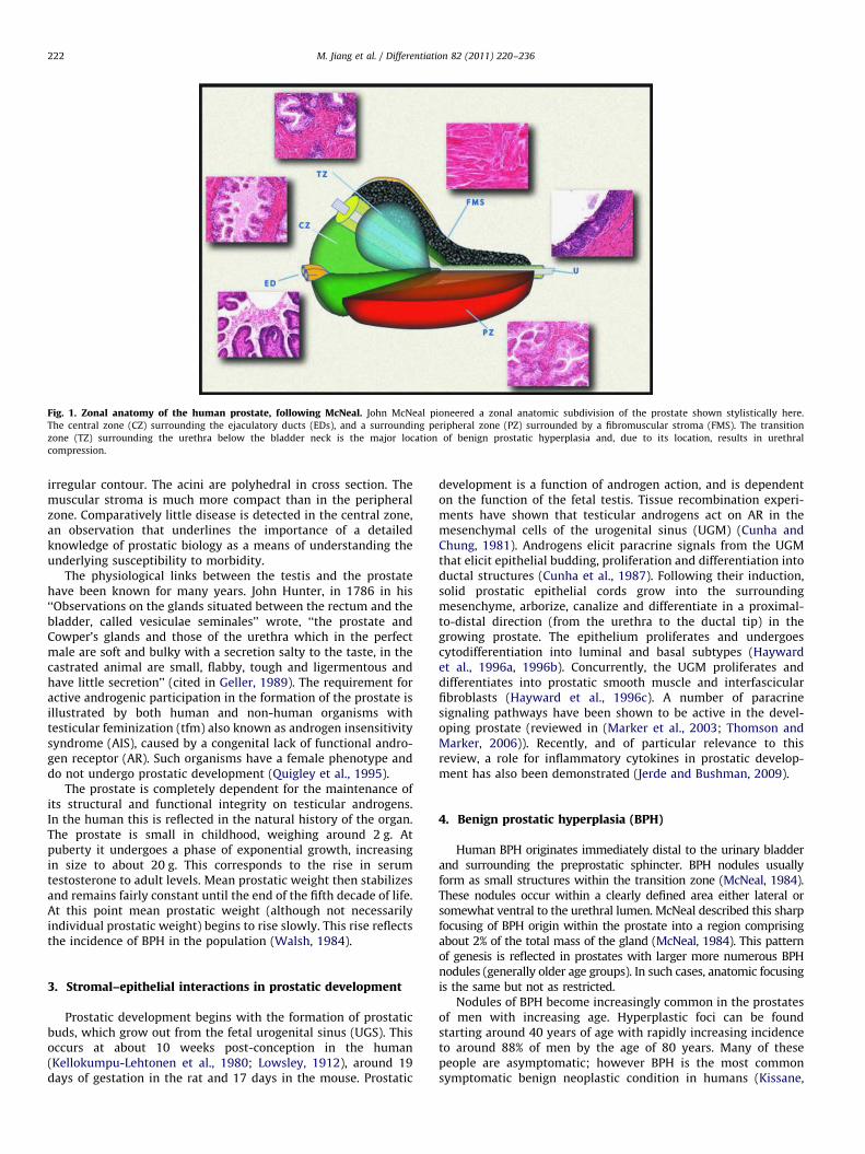
Fig. 1. Zonal anatomy of the human prostate, following McNeal. John McNeal pioneered a zonal anatomic subdivision of the prostate shown stylistically here.
The central zone (CZ) surrounding the ejaculatory ducts (EDs), and a surrounding peripheral zone (PZ) surrounded by a fibromuscular stroma (FMS). The transition
zone (TZ) surrounding the urethra below the bladder neck is the major location of benign prostatic hyperplasia and, due to its location, results in urethral
compression.
M. Jiang et al. / Differentiation 82 (2011) 220–236222
irregular contour. The acini are polyhedral in cross section. Themuscular stroma is much more compact than in the peripheralzone. Comparatively little disease is detected in the central zone,an observation that underlines the importance of a detailedknowledge of prostatic biology as a means of understanding theunderlying susceptibility to morbidity.
The physiological links between the testis and the prostatehave been known for many years. John Hunter, in 1786 in his‘‘Observations on the glands situated between the rectum and thebladder, called vesiculae seminales’’ wrote, ‘‘the prostate andCowper’s glands and those of the urethra which in the perfectmale are soft and bulky with a secretion salty to the taste, in thecastrated animal are small, flabby, tough and ligermentous andhave little secretion’’ (cited in Geller, 1989). The requirement foractive androgenic participation in the formation of the prostate isillustrated by both human and non-human organisms withtesticular feminization (tfm) also known as androgen insensitivitysyndrome (AIS), caused by a congenital lack of functional andro-gen receptor (AR). Such organisms have a female phenotype anddo not undergo prostatic development (Quigley et al., 1995).
The prostate is completely dependent for the maintenance ofits structural and functional integrity on testicular androgens.In the human this is reflected in the natural history of the organ.The prostate is small in childhood, weighing around 2 g. Atpuberty it undergoes a phase of exponential growth, increasingin size to about 20 g. This corresponds to the rise in serumtestosterone to adult levels. Mean prostatic weight then stabilizesand remains fairly constant until the end of the fifth decade of life.At this point mean prostatic weight (although not necessarilyindividual prostatic weight) begins to rise slowly. This rise reflectsthe incidence of BPH in the population (Walsh, 1984).
3. Stromal–epithelial interactions in prostatic development
Prostatic development begins with the formation of prostaticbuds, which grow out from the fetal urogenital sinus (UGS). Thisoccurs at about 10 weeks post-conception in the human(Kellokumpu-Lehtonen et al., 1980; Lowsley, 1912), around 19days of gestation in the rat and 17 days in the mouse. Prostatic
development is a function of androgen action, and is dependenton the function of the fetal testis. Tissue recombination experi-ments have shown that testicular androgens act on AR in themesenchymal cells of the urogenital sinus (UGM) (Cunha andChung, 1981). Androgens elicit paracrine signals from the UGMthat elicit epithelial budding, proliferation and differentiation intoductal structures (Cunha et al., 1987). Following their induction,solid prostatic epithelial cords grow into the surroundingmesenchyme, arborize, canalize and differentiate in a proximal-to-distal direction (from the urethra to the ductal tip) in thegrowing prostate. The epithelium proliferates and undergoescytodifferentiation into luminal and basal subtypes (Haywardet al., 1996a, 1996b). Concurrently, the UGM proliferates anddifferentiates into prostatic smooth muscle and interfascicularfibroblasts (Hayward et al., 1996c). A number of paracrinesignaling pathways have been shown to be active in the devel-oping prostate (reviewed in (Marker et al., 2003; Thomson andMarker, 2006)). Recently, and of particular relevance to thisreview, a role for inflammatory cytokines in prostatic develop-ment has also been demonstrated (Jerde and Bushman, 2009).
4. Benign prostatic hyperplasia (BPH)
Human BPH originates immediately distal to the urinary bladderand surrounding the preprostatic sphincter. BPH nodules usuallyform as small structures within the transition zone (McNeal, 1984).These nodules occur within a clearly defined area either lateral orsomewhat ventral to the urethral lumen. McNeal described this sharpfocusing of BPH origin within the prostate into a region comprisingabout 2% of the total mass of the gland (McNeal, 1984). This patternof genesis is reflected in prostates with larger more numerous BPHnodules (generally older age groups). In such cases, anatomic focusingis the same but not as restricted.
Nodules of BPH become increasingly common in the prostatesof men with increasing age. Hyperplastic foci can be foundstarting around 40 years of age with rapidly increasing incidenceto around 88% of men by the age of 80 years. Many of thesepeople are asymptomatic; however BPH is the most commonsymptomatic benign neoplastic condition in humans (Kissane,
M. Jiang et al. / Differentiation 82 (2011) 220–236 223
1989). BPH results in problems that include gross hematuria,bladder calculi, urinary retention and severe/bothersome LUTS.
Several different types of BPH nodules are recognized histolo-gically. These include those with fibrous and/or muscular stroma,and these may or may not contain an epithelial component.Nodules in the periurethral area are often stromal in characterwith a few small glands penetrating from the periphery. Gland-ular nodules composed of ductal tissue are derived from newlyformed small branches that bud from pre-existing ducts, growinto the adjacent stroma and repeatedly arborize to create a newarchitectural system within the nodule. These changes weredescribed in classical studies by McNeal (1978, 1983, 1984, 1985).
Links between BPH and the testis are well established, basedon the early observation that men castrated before the age of40 years do not develop the condition. The Skoptzys, a Russiansect in which the males underwent ritual castration at 35 years,did not suffer from prostatic enlargement (Zuckerman, 1936).Furthermore it has been shown that the absence of testicularfunction from a young age, either from castration or hypopitui-tarism, prevents the occurrence of BPH in men living into the over55 age group. Post-mortem examination of 28 such patientsshowed no histological evidence of BPH compared to an agematched control group where BPH was found in 50% of thepatients (Moore, 1944).
During the 1970s, debate over the links between the prostate,the testis and BPH centered upon the steroid hormones involvedand on the cellular subtype targeted. A number of reports over thisperiod suggested that levels of dihydrotestosterone (DHT), themost biologically active androgen, were higher in BPH tissue thanin normal prostate (Geller et al., 1976; Hammond, 1978; Krieget al., 1979; Meikle et al., 1978; Siiteri and Wilson, 1970). Thesestudies were unfortunately based on a comparison of BPH tissuethat had been surgically resected with normal prostate tissuederived from cadavers. This debate was quelled to some extentby the publication of data comparing fresh BPH tissue with freshnormal prostate derived from live organ donors. These showed thatthere was no significant difference between the levels of DHT innormal and BPH tissues (Walsh et al., 1983). Work on canine BPHshowed that this condition could be induced with androstanedioland with combinations of androstanediol and estradiol. A com-bined dose of DHT and estradiol was also found to induce thedisease (DeKlerk et al., 1979b; Walsh and Wilson, 1976).
In man, levels of serum testosterone drop by about 35%between the ages of 21 and 85 against a constant level ofestradiol. Thus there is a change in the androgen/estrogen ratio,which may be sufficient to promote the growth of BPH.
McNeal developed the important concept of reawakening ofembryonic inductive potential in BPH stromal cells (McNeal, 1978,1983, 1984, 1985). This idea is based on observations suggestingthat growth of the prostate results from growth factors andchemokines acting between the prostatic epithelium and stroma(Jerde and Bushman, 2009; Marker et al., 2003). In the adultprostate a broadly homeostatic balance of growth promoting andgrowth inhibiting factors is presumed to maintain organ integrity(Hayward and Cunha, 2000). A localized breakdown of such abalance could result in focal re-growth of new prostatic tissues.Adult prostatic epithelium from rats, mice and humans canrespond to prostatic inductive mesenchyme with new growthand development (Chung et al., 1984; Hayashi et al., 1993;Hayward et al., 1998; Norman et al., 1986), providing experimentalsupport for this idea. However, this does not address the under-lying cause of such a stromal to mesenchymal switch. The specificfactors that mediate hyperplastic enlargement of the prostateremain to be determined, although it is likely that these will proveto be the same factors involved in normal prostatic growth giventhe disease does not normally progress to malignancy.
While there are some naturally occurring animal models ofBPH, notably dogs and some non-human primates includingchimpanzees (Steiner et al., 1999), the condition does not natu-rally occur in common laboratory models such as mice. Like itshuman counterpart, canine BPH increases in frequency withage and is restricted to animals with intact testicular function.Canine BPH is histologically and anatomically distinct from thehuman condition. The canine disease is usually diffuse, occurringthroughout the gland (DeKlerk et al., 1979a; McNeal, 1984, 1985)in comparison to the human focal phenotype. A generalizedexpansion of the canine prostate that compresses the rectumand produces constipation is the common presentation. In con-trast, focal growth of transition zone nodules in humans com-presses the urethra and results in urinary retention.
5. Lower urinary tract symptoms (LUTS)
Enlargement of the prostate can alter lower urinary tractdynamics, resulting in a constellation of symptoms often referredto as lower urinary tract symptoms or LUTS. These may includestorage related symptoms such as frequent urination, urgency ofurination, waking up at night to void (nocturia) and urinaryincontinence to voiding related symptoms such as hesitancy,straining to void, poor urinary stream and failure to properlyempty the bladder. The link between BPH and LUTS is likelymultifactorial and may include physical obstruction caused by thehyperplastic nodules impinging on the urethra, altered bladderneck compliance and tone, and/or altered detrusor smoothmuscle physiology and function over time in response to alteredor obstructed outflow. In addition, multiple other disease pro-cesses can also impinge on the storage and voiding function of thelower genitourinary tract that can either mimic or exacerbate thechanges seen with BPH. These include processes such as neuro-logic disorders, malignancies and stricture disease. As a conse-quence, the relationship between the extent/severity of BPH andthe degree of LUTS is not simple or linear and can be influencedby multiple factors in a complex, dynamic relationship.
Since BPH/LUTS is predominantly a disease of the elderly andgiven the progressive aging of the population in the United States,BPH/LUTS represents a significant and growing public healthchallenge. While estimates vary based on the definition used, thenumber of men requiring treatment for LUTS overall is approxi-mately 16/1000 person years overall, increasing from 3.3/1000 formen aged 40–49 and rising up to 30/1000 person years for thoseover age 70 (Jacobsen et al., 1999). In 2005 alone, expenditures fortreatment of BPH/LUTS related problems in the private sector wereestimated at almost $4 billion (Saigal and Joyce, 2005). Untreated,BPH can ultimately give rise to severe sequelae. Permanent bladderdysfunction/acontractility, urinary retention, severe renal dysfunc-tion, end stage renal disease and death are all possible outcomes.However, the incidence of such consequences in the developedworld is extremely low. In practice, men are evaluated and treatedwhen BPH is an annoyance rather than a threat to life. Virtually allof the therapy for BPH/LUTS focuses on symptom managementafter the disease has manifested. Strategies that would preventeither the hyperplastic growth of the prostate or prevent thedownstream manifestations and physiological changes leading toLUTS would be preferable, but are predicated on an in depthunderstanding of the mechanisms underlying these processes. Todate, these mechanisms have not been fully defined.
6. Common co-morbidities affecting BPH/LUTS patients
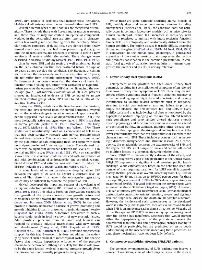
The complex symptomatology of LUTS patients can involve anumber of conditions, some of which may be causal to the disease

Fig. 2. Co-morbidities commonly seen in patients with benign prostatic hyperplasia and lower urinary tract symptoms (BPH/LUTS). Patients commonly, but by no
means always, present with an array of symptoms that are interlinked. These include obesity, which can directly give rise to sleep apnea, erectile dysfunction and diabetes.
BPH and LUTS can arise as a result of a number of these conditions. Sorting out cause and effect is a major clinical problem when trying to determine optimal therapies for
individuals.
M. Jiang et al. / Differentiation 82 (2011) 220–236224
state while others may be a consequence of shared pathophysiolo-gical changes. As shown in Fig. 2 common co-morbidities of patientswith LUTS include the metabolic syndrome, diabetes and obesity.
6.1. BPH/LUTS and the metabolic syndrome
Studies exploring the etiology of prostatic hyperplasia havetraditionally focused on aberrations of the steroid signaling axis asa causal entity. More recently, however, there has been a growingappreciation that other systemic conditions that often co-exist inmen as they age may play a causal role. A prime example of this isthe metabolic syndrome. This syndrome is a profile of findingsincluding impaired glucose metabolism, obesity, altered fat dis-tribution, hypertension, dyslipidemia, markers of systemic inflam-mation such as elevated C-reactive protein and autonomic–sympathetic overactivity, which affects up to 50 million people inthe United States (Kasturi et al., 2006; Rohrmann et al., 2005).A link between the metabolic syndrome and prostatic hyperplasiawas suggested in a series of 158 men with LUTS where those menwith larger prostates had increased incidence of diabetes, hyper-tension and obesity while also having lower levels of low-densitylipoprotein (LDL) and higher serum insulin levels (Hammarstenet al., 1998). A follow up study showed similar findings in a cohortof 250 men with BPH/LUTS (Hammarsten and Hogstedt, 1999). Themen with faster growing prostates had a higher prevalence ofdiabetes and treated hypertension as well as several measuresof obesity and had higher serum insulin levels and lower levels ofhigh-density lipoprotein (HDL). These co-morbidities are all ele-ments of the metabolic syndrome. Several subsequent reports havesubstantiated the notion that at least some elements of themetabolic syndrome are more common in men with BPH/LUTS(Hammarsten and Hogstedt, 2001; Moul and McVary, 2010;Nandeesha et al., 2006a), though this was not universally true(Lekili et al., 2006; Zucchetto et al., 2005). In particular at least twoof the elements linked to the metabolic syndrome, obesity anddiabetes have been repeatedly associated with BPH/LUTS in menacross multiple studies, though whether this relationship is causalremains to be proven.
6.2. Diabetes and BPH/LUTS
Possible links between prostatic hyperplasia and diabeteswere noted as far back as in 1966 in early epidemiologic studies(Bourke and Griffin, 1966, 1968a, b). More recently the studiesfocusing on BPH/LUTS and diabetes can be broken down intothree basic groups. The first are those that looked at the associa-tion with prostatic hyperplasia, typically expressed as radio-graphic measurements of prostate volume by transrectalultrasound (TRUS) or magnetic resonance imaging (MRI) or usinga surrogate for prostatic volume such as serum prostate specificantigen (PSA). The second are those that look at the clinicaldiagnosis of BPH, typically defined either as the need for medicalor surgical therapy for BPH, documented obstruction or lowurinary flow rate on urodynamics or other testing, or assignmentof the diagnosis by a physician. The last looks not at hyperplasiabut at the LUTS themselves most often through validated self-administered questionnaires such as the International ProstateSymptom Score (IPSS) or similar instruments. These groupsrepresent some of the various possible definitions and end pointsthat may be used for studying BPH/LUTS. Many studies haveexamined some or all of these potential end points and in manycases have mixed definitions or failed to clearly delineate whichendpoint was the focus of the study. This represents just one ofseveral reasons why there are some inconsistencies and contro-versies surrounding these associations. The other major problemis variations on what population was used as the control group,studies that were often underpowered and variations on howdiabetes was defined for the purposes of the study. Neverthelessthe weight of the evidence suggests there is a moderate associa-tion of diabetes with BPH and prostatic enlargement and astronger association of diabetes with LUTS, supporting the con-cept that LUTS can be either dependent or independent of BPH.
Several studies have shown a significant relationship betweendiabetes or hyperinsulinemia and larger prostate size (Bergeret al., 2006; Hammarsten and Hogstedt, 1999, 2001; Hammarstenet al., 1998; Nandeesha et al., 2006a; Parsons et al., 2006). Forexample, in one of the largest studies to address this question,

M. Jiang et al. / Differentiation 82 (2011) 220–236 225
Parsons et al. (2006) studied 422 men in the Baltimore Long-itudinal Study of Aging and found that increased fasting bloodglucose levels and diabetes were both risk factors for largerprostates assessed by MRI. Hammarsten and Hogstedt (2001)followed 307 patients with clinical BPH using TRUS and foundthat larger annual prostatic growth was associated with severalcomponents of the metabolic syndrome including diabetes. Theyalso found that total gland volume was positively associated withmore advanced age and higher plasma insulin levels. Otherstudies, such as a study of 2115 men in Olmsted County, havenot shown an association between diabetes and BPH (Burke et al.,2006). This work found that diabetes was associated with higherLUTS as measured by IPSS score and lower peak urinary flowrates, but not with prostate volume. It should be pointed out thatthe proportion of men in this cohort with diabetes was quitesmall, limiting the power of the study even though the overallpopulation was large. Further, lower urinary flow rates and LUTSare used in many studies as criteria for diagnosing BPH for thepurposes of epidemiological studies.
The association between diabetes and the clinical diagnosis ofBPH has been made across a number of different studies (Bergeret al., 2005; Burke et al., 2006; Dahle et al., 2002; Nandeesha et al.,2006a). The largest of these studies looked specifically at the clinicaldiagnosis of BPH (Dahle et al., 2002). Two hundred men who hadundergone surgery for BPH were compared to 302 normal controlmen. The findings were that the men who had undergone surgeryfor BPH had higher plasma insulin levels and increased waist to hipratio, an indication of truncal obesity. In another study, Berger et al.(2005) found that those with evidence for vascular damage due todiabetes were more likely to be diagnosed with BPH/LUTS. Onestudy out of Scandinavia failed to find an association between BPHand diabetes, but this study is subject to significant recall bias sinceall diagnoses were self-reported (Koskimaki et al., 2001).
The strongest association between diabetes and BPH/LUTS hasbeen across studies that have either focused specifically on LUTS orincluded this as part of the definition of BPH (Berger et al., 2005,2006; Burke et al., 2006; Joseph et al., 2003; Michel et al., 2000;
Fig. 3. Potential mechanisms linking obesity and diabetes to benign prostatic hyperrelating to both obesity and diabetes can play a role in the development of BPH/LUTS, a f
than one pathway.
Sarma et al., 2008; Zhang et al., 2008a). One of the largest is thecohort study from Olmsted County mentioned above, but whichwas focused predominantly on Caucasian men (Burke et al., 2006).The association between diabetes and LUTS has also been demon-strated in a cohort of over 700 African American men (Joseph et al.,2003), showing a link between diabetes and LUTS. Michel et al.(2000) analyzed over 9800 men with BPH, including 1290 withdiabetes, and found that the diabetic men had worse IPSS scoresand lower peak urinary flow rates than the non-diabetic men. Theassociation has also been demonstrated when LUTS was moreclosely analyzed using various urodynamic parameters in a studyof 166 men with BPH, 74 of whom also had diabetes (Zhang et al.,2008a). The diabetic men were more likely to demonstrateabnormalities with bladder capacity, decreased bladder sensation,poor bladder compliance and detrusor overactivity, while max-imum detrusor pressure and evidence for bladder outlet obstruc-tion were more common in non-diabetic men with BPH/LUTS.
6.3. Obesity and BPH/LUTS
As with diabetes there is a large body of evidence thatsupports a strong association between obesity and BPH/LUTS(Dahle et al., 2002; Fowke et al., 2007b; Giovannucci et al.,1994; Hammarsten and Hogstedt, 1999, 2001; Hammarstenet al., 1998; Kristal et al., 2007; Lee et al., 2006b, 2009a, b;Maserejian et al., 2009; Parsons, 2007; Parsons et al., 2006;Soygur et al., 1996; Xie et al., 2007); although this finding is notuniversal (Meigs et al., 2001; Seitter and Barrett-Connor, 1992;Zucchetto et al., 2005). This is particularly relevant given theknown ‘‘epidemic’’ of obesity in the United States, which appearsto be getting steadily worse. In addition, obesity is a potentiallypreventable or reversible health condition. If it could be demon-strated that it is causally linked to BPH/LUTS, then targetingobesity would represent a target for disease prevention (seeFig. 3). Such an intervention would also have multiple additionalpositive effects such as decreasing morbidity and mortality
plasia and lower urinary tract symptoms (BPH/LUTS). A number of mechanisms
ew of which are outlined in this figure. Individual patients may be affected by more

M. Jiang et al. / Differentiation 82 (2011) 220–236226
secondary to insulin resistance and cardiovascular disease. As wasthe case with diabetes, reports have varied regarding the endpoints of BPH/LUTS that were studied. An additional layer ofcomplexity in examining the studies of obesity, however, is thatinvestigators have varied in their definition of obesity or exam-ined more than one potential parameter. Examples of definitionsof obesity have included simple body weight, body mass index(BMI), waist circumference, hip circumference and waist to hipratio (WHR). Variations in both the end points of BPH/LUTS usedin the study and the definition(s) of obesity account for some ofthe differences across studies. Nevertheless the preponderanceof the evidence suggests a strong clinical association betweenmeasures of obesity and increased BPH/LUTS in general, andlarger prostate volume in particular.
There has been a strong association found consistently acrossstudies between measures of obesity and increased prostatevolume (Fowke et al., 2007b; Hammarsten and Hogstedt, 1999,2001; Hammarsten et al., 1998; Lee et al., 2006a, 2009a, b; Parsonset al., 2006; Soygur et al., 1996; Xie et al., 2007). Some of the largerstudies have included the 422 men in the Baltimore LongitudinalStudy of Aging, showing that obesity was associated with increasedprostate size based on MRI (Parsons et al., 2006). A larger study byFowke et al. (2007a) found that in 753 men with a negative TRUSprostate biopsy increased prostate volume by TRUS was associatedwith several measures of obesity, including increased BMI andwaist circumference. Similar associations between obesity mea-sures and larger prostate volume by TRUS have also been shownin a cohort of 649 Chinese men (Xie et al., 2007) and 602 Koreanmen (Lee et al., 2009a, b). In the latter study, the association wasstronger in measures of central obesity (waist circumference)rather than overall obesity (such as BMI). The studies byHammarsten and Hogstedt (1999, 2001), Hammarsten et al.(1998) demonstrating an association between more rapid prostategrowth and elements of the metabolic syndrome have also shownthat this includes an association with measures of obesity. Theweight of the evidence strongly suggests, therefore, that obesity ingeneral, and increased central obesity in particular, is associatedwith increased prostatic volume.
In one of the largest epidemiologic studies to address the issueof BPH and obesity the Health Professional Follow Up Study,which included over 25,000 men, found over a two-fold increasedrisk of requiring prostate surgery and a two-fold increased riskqof having LUTS in men with increased waist circumference(a measure of central obesity) (Giovannucci et al., 1994). Anotherlarge-scale study based on the prospective Prostate Cancer Pre-vention Trial (PCPT) found that, among 5667 men who did nothave BPH at baseline, those with a higher WHR were more likelyto develop BPH/LUTS over the seven-year course of the trial(Kristal et al., 2007). Other studies have also corroborated thesefindings (Dahle et al., 2002; Lee et al., 2006b) including a study byDahle et al. (2002) discussed previously, which found that menwho had undergone surgery for BPH had a higher WHR comparedto control men. It should be pointed out that two studies failed tofind an association between obesity and BPH/LUTS (Meigs et al.,2001; Seitter and Barrett-Connor, 1992), but these were bothsmaller trials and therefore may have been underpowered todetect any association that may exist.
As with measures of prostate volume, the association betweenobesity and LUTS is generally a strong one. For example, the studyby Giovannucci et al. (1994) of 25,892 men in the HealthProfessional Follow Up Study found a strong association betweenobesity and increased LUTS. Analysis of the PCPT trial alsodemonstrated increased LUTS in men with higher WHR (Kristalet al., 2007) and a study of Korean men found that waistcircumference was positively associated with increased LUTS(Lee et al., 2009a, b). A small study of 68 men with BPH found
an association between obesity and prostate volume but notworse symptoms, although this was an underpowered studycompared to those discussed previously (Soygur et al., 1996).Finally, approaching the question from a slightly different per-spective, a report from Maserejian et al. (2009) on 1545 men fromthe Boston Area Community Health Survey found that increasedtotal caloric intake was associated with higher LUTS. Overall then,the evidence suggests that direct or indirect measures of obesityare associated with increased prostate volume, LUTS and theclinical diagnosis of BPH/LUTS.
7. Mechanisms linking diabetes and obesity to BPH/LUTS
In considering the systemic effects of diabetes and obesity onBPH and LUTS it is important to differentiate between levels ofeffect. As discussed above, clinical studies have tended to grouppatients broadly. However, the causes of symptoms may wellvary. For example LUTS can result from prostatic enlargement,causing urinary outflow obstruction by a static mechanisminvolving growth of epithelial and stromal elements. This givesrise to urethral compression (Berry et al., 1984; McNeal, 1984),versus an active mechanism in which a1-adrenergic receptors arestimulated resulting in contraction of the urethra (Sarma et al.,2009). Apparently identical LUTS may also arise as a result ofchanges to bladder structure and innervation. In diabetic patients,for example, it can be difficult to differentiate between thesedifferent causes of symptoms. Diabetes can give rise to diabeticcystopathy, resulting in LUTS as a consequence of nerve damage,notably seen in both male and female patients (Hill et al., 2008;Kebapci et al., 2007; Rapidi et al., 2006). As a further complicationthere are also aging-related changes to the musculature andfibrotic nature of the detrusor, potentially also contributing tosymptoms. Both diabetes and obesity can broadly be consideredto be inflammatory states and inflammation is also a possiblemechanism that can give rise to prostatic growth. The specificcauses of similar patient outcomes are not well differentiated atpresent but are certainly important in selecting patients fortherapy. Clearly, treating the prostate of a patient with diabeticcystopathy is unlikely to give rise to the best clinical outcome.
An examination of the various common co-morbidities sug-gests a hierarchy as shown in Fig. 3. Such an arrangementsuggests that problems associated with obesity and its sequelaeare prime drivers of BPH/LUTS. However, it must be acknowl-edged that while this represents the overall situation across thepopulation, individual patient populations do not always fitneatly into such a model. Thus, while it may be that a ‘‘typical’’patient is an overweight diabetic with an aversion to exercise, it isalso true that patients can include slim, fit individuals. In terms oftreatment of this diverse population, a clear understanding of themechanisms underlying disease in a particular patient must beestablished and therapies then need to be selected on a persona-lized basis. Some of the major mechanisms involved in prostaticgrowth that are prospective therapeutic targets include changesin hormonal status, alterations in the insulin/IGF/IGF bindingprotein (IGFBP) axis, inflammation and alterations in cellularmetabolism resulting from persistent abnormal stimulation.
7.1. Steroid hormones
The role of steroid hormones in the pathogenesis of BPH hasbeen debated for several decades. Prostate development is abso-lutely dependent upon androgens, as discussed above, and dis-ruption of the synthesis of dihydrotestosterone is a mainstay ofBPH treatment. However, estrogens also affect prostatic develop-ment and histology. Classic studies by Price (1936) in the 1930s
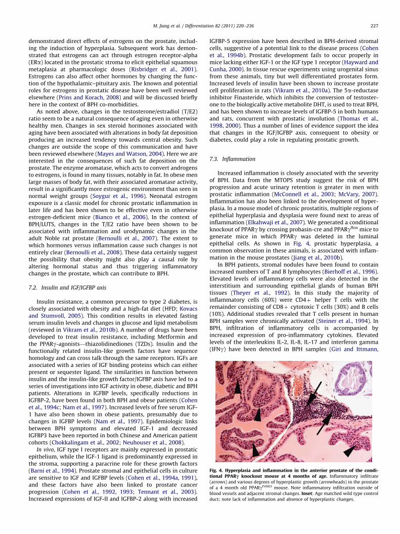
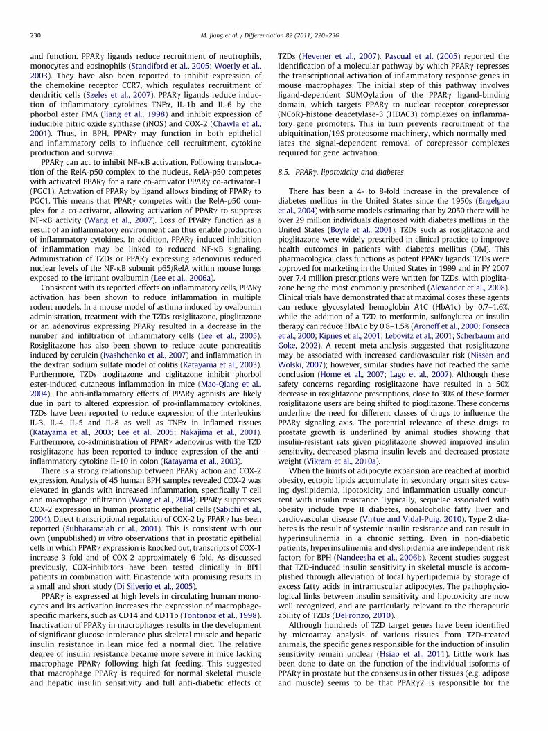
Fig. 4. Hyperplasia and inflammation in the anterior prostate of the condi-tional PPARc knockout mouse at 4 months of age. Inflammatory infiltrate
(arrows) and various degrees of hyperplastic growth (arrowheads) in the prostate
of a 4 month old PPARgPrEKO mouse. Note inflammatory infiltration outside of
blood vessels and adjacent stromal changes. Inset: Age matched wild type control
duct; note lack of inflammation and absence of hyperplastic changes.
M. Jiang et al. / Differentiation 82 (2011) 220–236 227
demonstrated direct effects of estrogens on the prostate, includ-ing the induction of hyperplasia. Subsequent work has demon-strated that estrogens can act through estrogen receptor-alpha(ERa) located in the prostatic stroma to elicit epithelial squamousmetaplasia at pharmacologic doses (Risbridger et al., 2001).Estrogens can also affect other hormones by changing the func-tion of the hypothalamic–pituitary axis. The known and potentialroles for estrogens in prostatic disease have been well reviewedelsewhere (Prins and Korach, 2008) and will be discussed brieflyhere in the context of BPH co-morbidities.
As noted above, changes in the testosterone/estradiol (T/E2)ratio seem to be a natural consequence of aging even in otherwisehealthy men. Changes in sex steroid hormones associated withaging have been associated with alterations in body fat depositionproducing an increased tendency towards central obesity. Suchchanges are outside the scope of this communication and havebeen reviewed elsewhere (Mayes and Watson, 2004). Here we areinterested in the consequences of such fat deposition on theprostate. The enzyme aromatase, which acts to convert androgensto estrogens, is found in many tissues, notably in fat. In obese menlarge masses of body fat, with their associated aromatase activity,result in a significantly more estrogenic environment than seen innormal weight groups (Soygur et al., 1996). Neonatal estrogenexposure is a classic model for chronic prostatic inflammation inlater life and has been shown to be effective even in otherwiseestrogen-deficient mice (Bianco et al., 2006). In the context ofBPH/LUTS, changes in the T/E2 ratio have been shown to beassociated with inflammation and urodynamic changes in theadult Noble rat prostate (Bernoulli et al., 2007). The extent towhich hormones versus inflammation cause such changes is notentirely clear (Bernoulli et al., 2008). These data certainly suggestthe possibility that obesity might also play a causal role byaltering hormonal status and thus triggering inflammatorychanges in the prostate, which can contribute to BPH.
7.2. Insulin and IGF/IGFBP axis
Insulin resistance, a common precursor to type 2 diabetes, isclosely associated with obesity and a high-fat diet (HFD; Kovacsand Stumvoll, 2005). This condition results in elevated fastingserum insulin levels and changes in glucose and lipid metabolism(reviewed in Vikram et al., 2010b). A number of drugs have beendeveloped to treat insulin resistance, including Metformin andthe PPARg-agonists—thiazolidinediones (TZDs). Insulin and thefunctionally related insulin-like growth factors have sequencehomology and can cross talk through the same receptors. IGFs areassociated with a series of IGF binding proteins which can eitherpresent or sequester ligand. The similarities in function betweeninsulin and the insulin-like growth factor/IGFBP axis have led to aseries of investigations into IGF activity in obese, diabetic and BPHpatients. Alterations in IGFBP levels, specifically reductions inIGFBP-2, have been found in both BPH and obese patients (Cohenet al., 1994c; Nam et al., 1997). Increased levels of free serum IGF-1 have also been shown in obese patients, presumably due tochanges in IGFBP levels (Nam et al., 1997). Epidemiologic linksbetween BPH symptoms and elevated IGF-1 and decreasedIGFBP3 have been reported in both Chinese and American patientcohorts (Chokkalingam et al., 2002; Neuhouser et al., 2008).
In vivo, IGF type I receptors are mainly expressed in prostaticepithelium, while the IGF-1 ligand is predominantly expressed inthe stroma, supporting a paracrine role for these growth factors(Barni et al., 1994). Prostate stromal and epithelial cells in cultureare sensitive to IGF and IGFBP levels (Cohen et al., 1994a, 1991),and these factors have also been linked to prostate cancerprogression (Cohen et al., 1992, 1993; Tennant et al., 2003).Increased expressions of IGF-II and IGFBP-2 along with increased
IGFBP-5 expression have been described in BPH-derived stromalcells, suggestive of a potential link to the disease process (Cohenet al., 1994b). Prostatic development fails to occur properly inmice lacking either IGF-1 or the IGF type 1 receptor (Hayward andCunha, 2000). In tissue rescue experiments using urogenital sinusfrom these animals, tiny but well differentiated prostates form.Increased levels of insulin have been shown to increase prostatecell proliferation in rats (Vikram et al., 2010a). The 5a-reductaseinhibitor Finasteride, which inhibits the conversion of testoster-one to the biologically active metabolite DHT, is used to treat BPH,and has been shown to increase levels of IGFBP-5 in both humansand rats, concurrent with prostatic involution (Thomas et al.,1998, 2000). Thus a number of lines of evidence support the ideathat changes in the IGF/IGFBP axis, consequent to obesity ordiabetes, could play a role in regulating prostatic growth.
7.3. Inflammation
Increased inflammation is closely associated with the severityof BPH. Data from the MTOPS study suggest the risk of BPHprogression and acute urinary retention is greater in men withprostatic inflammation (McConnell et al., 2003; McVary, 2007).Inflammation has also been linked to the development of hyper-plasia. In a mouse model of chronic prostatitis, multiple regions ofepithelial hyperplasia and dysplasia were found next to areas ofinflammation (Elkahwaji et al., 2007). We generated a conditionalknockout of PPARg by crossing probasin-cre and PPARgflox mice togenerate mice in which PPARg was deleted in the luminalepithelial cells. As shown in Fig. 4, prostatic hyperplasia, acommon observation in these animals, is associated with inflam-mation in the mouse prostates (Jiang et al., 2010b).
In BPH patients, stromal nodules have been found to containincreased numbers of T and B lymphocytes (Bierhoff et al., 1996).Elevated levels of inflammatory cells were also detected in theinterstitium and surrounding epithelial glands of human BPHtissues (Theyer et al., 1992). In this study the majority ofinflammatory cells (60%) were CD4þ helper T cells with theremainder consisting of CD8þ cytotoxic T cells (30%) and B cells(10%). Additional studies revealed that T cells present in humanBPH samples were chronically activated (Steiner et al., 1994). InBPH, infiltration of inflammatory cells is accompanied byincreased expression of pro-inflammatory cytokines. Elevatedlevels of the interleukins IL-2, IL-8, IL-17 and interferon gamma(IFNg) have been detected in BPH samples (Giri and Ittmann,

M. Jiang et al. / Differentiation 82 (2011) 220–236228
2001; Kramer et al., 2002; Steiner et al., 2003). In addition,expression of IL-15, a cytokine that promotes T cell proliferation,and its receptor IL-15R were higher in BPH epithelial cellscompared to normal prostate (Handisurya et al., 2001). Prostaticinflammatory atrophy also results in increased expression of anumber of cytokines and inflammatory enzymes, includingcyclooxygenase-2 (COX-2; De Marzo et al., 2007; Palapattuet al., 2005). Interleukins have recently been implicated as directregulators of organ growth in the mouse prostate. IL-1 was shownto elicit IGF-dependent prostatic growth and reactive hyperplasiain a murine model (Jerde and Bushman, 2009). Increased cytokineexpression influences the proliferation of non-inflammatory cellsin the prostate, as demonstrated by the cytokines IL-2 and IFNg,which have been shown to increase proliferation of human BPHstromal cell lines (Kramer et al., 2002). Increased inflammationcan result in disruption of epithelial glands, with increased serumlevels of PSA. Agents that reduce inflammation could also beeffective at reducing hyperplasia, thus reducing the symptomsassociated with this disease.
A small number of studies have been conducted and havedemonstrated some efficacy of anti-inflammatory strategies inpatients with BPH (Azzouzi et al., 2006). For example, loxoprofenat 60-mg dose in patients with LUTS resistant to a first-linetreatment showed an improvement of the nocturia in 74% of thecases with better results in more severe cases (Araki et al., 2004).Of particular interest, the combination of a COX-2 inhibitor and a5a-reductase inhibitor resulted in increased apoptosis in BPH(Sciarra et al., 2008). Di Silverio et al. (2005) tested Rofecoxib (aCOX-2 inhibitor) with Finasteride (a 5a-reductase inhibitor)versus Finasteride alone. Their hypothesis was that an anti-inflammatory drug could palliate the lack of efficiency of theFinasteride during the first week of treatment. The authors didfind short-term relief in symptomatic scores. Links betweenspecific mediators of inflammation and cellular metabolism mayrepresent a critical direction in which new approaches for BPHtherapies can be directed. Based upon the data presented above,we would hypothesize that an appropriate anti-inflammatoryapproach could additionally provide a long-term complementaryeffect to the 5a-reductase inhibitor. PPARg is a possible candidatefor managing prostate growth, given its demonstrated efficacy atregulating insulin sensitivity in other insulin target tissues. Todate, there are no retrospective analyses of BPH incidence fordiabetic patients taking TZDs.
8. Peroxisome proliferator-activated receptor gamma(PPARc) signaling
Peroxisome proliferator-activated receptors (PPARs) areligand-activated transcription factors belonging to the nuclearreceptor superfamily (Auwerx et al., 1996; Evans et al., 2004). ThePPAR family is composed of PPARa (NR1C1), PPARb/d (NR1C2)and PPARg (NR1C3) (Barish and Evans, 2004; Evans et al., 2004;Lee et al., 2003; Roberts et al., 2003). PPARs bind as obligateheterodimers with the retinoid X receptors (RXRs) to cognateDNA elements named peroxisome proliferator response elements(PPREs; Evans et al., 2004). PPARs regulate lipid and glucosemetabolism (Argmann et al., 2005; Evans et al., 2004; Formanet al., 1996; Metzger and Chambon, 2007). They also participate incell growth, proliferation and differentiation in adipocytes, hepa-tocytes and keratinocytes as well as prostate epithelial cells(Chambon, 2005; Imai et al., 2001a; Imai et al., 2001b; Jianget al., 2004; Metzger et al., 2005). The PPAR/RXR/PPRE interactionleads to the recruitment of coactivators and the general transcrip-tional machinery, resulting in alterations in gene transcriptionand cell function (Rosen and Spiegelman, 2001). There are two
forms of PPARg, denoted PPARg1 and PPARg2. These representproteins derived from alternative transcriptional start sites in thesame gene, giving rise to two isoforms. PPARg2, using the earlierstart site, has an additional 30 amino acid residues (Fajas et al.,1997; Tontonoz et al., 1994b).
PPARg protein levels are the highest in adipose tissue, wherePPARg ligands induce adipocyte differentiation and regulate lipidstorage and metabolism (Theocharis et al., 2004). In addition,PPARg functions as an important regulator of cell differentiation,proliferation and apoptosis in other types of stromal cells (macro-phages, endothelium and smooth muscle; Barak et al., 1999; Rosenand Spiegelman, 2001; Tontonoz et al., 1994a) and parenchymalepithelial cells of the breast (Yee et al., 2003), colon (Saez et al.,1998; Sarraf et al., 1998) and prostate (Shappell et al., 2001a,2001b). Ligands for PPARg include the eicosanoids 15S-Hydroxyei-cosatetraenoic acid (15-HETE; Bhatia et al., 2003; Shappell et al.,2001b) and the prostaglandin 15-deoxy-D12,14-prostaglandin J2(15dPGJ2; Matsuyama et al., 2005; Soares et al., 2005), docosahex-aenoic acid (DHA; Yu et al., 2008), polyunsaturated fatty acid(PUFA; Calder, 2008) and nonsteroidal anti-inflammatory drugs(NSAID; Romeiro et al., 2008) as well as synthetic thiazolidinedione(TZD) drugs (Rosen and Spiegelman, 2001). The TZDs rosiglitazoneand pioglitazone are currently used in the clinic to treat patientswith type II diabetes, due to their ability to increase insulinsensitivity by stimulating glucose uptake (Smith and Kantoff,2002). However, use of these drugs has been severely curtailed oflate because of cardiovascular side effects and also because clinicaltrials are showing little improvement over more establishedtherapies for diabetes treatments (Lindberg and Astrup, 2007). Toaddress the side effects in off-target tissues, a more thoroughunderstanding of the function, dietary regulation and tissue-specific stoichiometric distribution of each isoform is necessary.
PPARg plays a key role in regulation of cellular lipid metabo-lism, redox status and organelle differentiation in adipose tissueand other organs, including prostate (Jiang et al., 2004; Sugii et al.,2009). The prostaglandin 15dPGJ2 has been described as a PPARgactivator and may potentially play an anti-inflammatory role in aPPARg-dependent manner, decreasing COX-2, PGE2 synthase(PGES) and PGE2 production (Mendez and LaPointe, 2003).Prostate cancer is characterized by increases in pathways thatgenerate pro-inflammatory prostaglandins and by a loss ofenzymes in lipid metabolic pathways such as arachidonate15-lipoxygenase-2 (15-LOX-2), which generate natural ligandsfor PPARg (Shappell et al., 2001b). In contrast, the commonco-morbidities associated with BPH/LUTS elicit changes in theprostatic metabolic environment, specifically with increased lipidavailability and alterations in glucose metabolism consequentto insulin resistance and changes in the IGF/IGFBP axis. Loss ofPPARg function in the prostate leads to a number of conse-quences, including widespread inflammation and hyperplasticgrowth (Jiang et al., 2010b), with more focal premalignantchanges. Due to its central position in balancing cellular metabo-lism and differentiation, and to the existence and current clinicaluse of PPARg agonists, PPARg is an attractive target for manipula-tion in therapeutic strategies to treat prostatic disease.
8.1. Transcriptional regulation of PPARg signaling
PPARg consists of distinct functional domains including anN-terminal transactivation function domain (AF-1), a highlyconserved DNA-binding domain (DBD) and a C-terminal ligand-binding domain (LBD) that contains a ligand-dependent transac-tivation function (AF2). Upon ligand binding the complex ofPPARs and RXRs binds to specific recognition sites on DNA, theperoxisome proliferator response elements (PPREs), and regulatesgene transcription. A PPRE, consisting of an almost perfect direct

M. Jiang et al. / Differentiation 82 (2011) 220–236 229
repeat of the sequence TGACCT spaced by a single base pair, hasbeen identified in the upstream regulatory sequences of genesrelated to metabolic and cell cycle-regulating pathways. Activa-tion of PPARg results in increased expression of many targetgenes involved in lipid metabolism and energy balance such asthe adipocyte fatty acid binding protein (aP2), acyl-CoA oxidase,lipoprotein lipase, acyl-CoA synthase and adiponectin (Memonet al., 2000).
In adipose tissue of obese mice the expression of catalase, ananti-oxidant enzyme, is significantly decreased, potentially result-ing in insufficient elimination of hydrogen peroxide. Adiposecatalase expression is regulated via a novel remote PPARg-responsive region. Treatment of mice with PPARg agonists sig-nificantly enhanced catalase expression in adipose tissue (Okunoet al., 2008). We have demonstrated a similar loss of catalaseexpression resulting from PPARg knockout in mouse prostateepithelium (Jiang et al., 2010b).
8.2. Phosphorylation of PPARg protein
Mitogen-activated protein kinase (MAPK) signaling remains thefocus of intensive research because it plays a pivotal role inmediation of cellular responses to a variety of signaling molecules(Kohno and Pouyssegur, 2003). Among the key signaling pathwaysthat regulate mammalian cell growth and differentiation are theMAPK kinases (MEK)/extracellular signal-regulated kinase (ERK),comprised of MAP kinase kinases, MKK1/2 or MEK1/2, and MAPkinases ERK1/2. MEK1/2 and ERK1/2 are acutely stimulated bygrowth and differentiation factors in pathways mediated by receptortyrosine kinases, heterotrimeric G protein-coupled receptors orcytokine receptors, primarily through p21Ras-coupled mechanisms.These enzymes are ubiquitous and generally expressed at micro-molar levels in mammalian cells (Burgermeister and Seger, 2008;Kohno and Pouyssegur, 2003).
A number of the molecular mechanisms activated in BPHregulate intracellular MAP kinase signaling pathways in epithelialand/or stromal cells. P38 MAP kinase is not normally activated in theprostatic epithelium. However, activation is seen in both BPH andprostate cancer (Royuela et al., 2002). Of particular relevance here,the IGF pathway acts to activate MAP kinase signaling in prostaticepithelial cells as do a number of inflammatory chemokinesproduced by the lymphocytic infiltrate commonly found in BPH(Theyer et al., 1992). MAPK regulation in BPH is also important instromal cells. Estrogenic signaling through the stromal ERa, which isimplicated in the development of BPH, stimulates proliferation ofthe stromal cells via activation of ERK (Zhang et al., 2008b), andflavonoids such as apigenin can inhibit stromal cell proliferation viadecreases in ERK1 and 2 (Bektic et al., 2006). Phosphodiesteraseinhibitors have also recently been shown to elicit clinical responsesin BPH patients (initially seen as a side effect of their use in erectiledysfunction—reviewed in Laydner et al., 2011). This effect has alsobeen shown to result from regulation of stromal MAPK signaling(Zenzmaier et al., 2010). The role of MAPK signaling in the prostatehas been thoroughly reviewed elsewhere (Maroni et al., 2004;Papatsoris and Papavassiliou, 2001)
PPARg is a downstream target of MEK/ERK signaling(Burgermeister and Seger, 2007; Burns and Vanden Heuvel,2007; Diradourian et al., 2005). A unique MAPK phosphorylationsite was mapped at serine 82 in the N-terminal domain in mousePPARg1, which corresponds to serine 112 of mouse PPARg2.Substitution of this serine by alanine led to a loss of PDGF-mediated repression of PPARg activity (Shao et al., 1998). Serine84 of PPARg1 is phosphorylated by ERK2- and JNK-MAPK inhumans (Camp et al., 1999). This phosphorylation is totallyabolished either by mutation of serine 84 to alanine or co-expression of a phosphoprotein phosphatase. Similar to mouse
PPARg, human PPARg1 phosphorylation inhibits both ligand-dependent and -independent transactivating functions whereasan S84A mutant shows an increase in AF-1 transcriptional activityof PPARg (Adams et al., 1997). We performed gene expressionprofiling on both PPARg- and -g2-deficient mouse prostaticepithelial (mPrE) cells and found the up-regulation of numerousgenes in the MAPK signaling pathway (Jiang et al., 2010b),suggesting that MEK/ERK signaling may function as a key med-iator of the phosphorylation status of PPARg protein. It has beenalso reported that PPARg ligands can activate MAPK signaling vianon-genomic signaling (Gardner et al., 2005; Hedvat et al., 2004).However, the precise molecular cross-talk between PPARg andMEK/ERK signaling is still unclear.
Choi et al. (2010) showed that the cyclin-dependent kinase 5(Cdk5) is activated in high-fat-fed obese mice, resulting inphosphorylation of PPARg at serine 273. This modification ofPPARg does not alter its adipogenic capacity but leads to dysre-gulation of a large number of genes whose expression levels arealtered in obesity, including reduced adiponectin. Interestinglythe phosphorylation of PPARg by Cdk5 is blocked by anti-diabeticPPARg ligands such as rosiglitazone and MRL24, and is completelyindependent of classical receptor transcriptional agonism. Theauthors suggest Cdk5-mediated phosphorylation of PPARg maybe involved in the pathogenesis of insulin resistance and presentan opportunity for development of an improved generation ofanti-diabetic drugs working through PPARg.
8.3. PPARg signaling and oxidative stress
Oxidative stress increases with age and may be aggravatedwith any pathological condition that damages tissues, resulting inadditional complications. Chronic inflammation and associatedoxidative stress are commonly associated with BPH and LUTS.Oxidative stress is defined as an imbalance between production offree radicals and reactive metabolites, the so-called oxidants orreactive oxygen species (ROS), and their elimination by protectivemechanisms, referred to as antioxidants. This imbalance leads todamage of important biomolecules and cells, with potentialimpact on the whole organism (Durackova, 2010). Antioxidantsare substances that are able to compete with the other oxidizablesubstrates and thus significantly delay or inhibit their oxidation.Enzymatic antioxidants, such as superoxide dismutase (SOD),glutathione peroxidase (GPx) and catalase (CAT), and non-enzy-matic antioxidants, including glutathione and vitamin C, play arole as scavengers of free radicals. Continued oxidative stress canlead to chronic inflammation, which in turn could mediate manychronic diseases, including cancer, diabetes, cardiovascular, neu-rological and pulmonary diseases, as well as benign prostaticgrowth. Oxidative stress can activate a variety of transcriptionfactors, including p53, PPARg, NFkB, AP-1, HIF-1a, b-catenin/Wntand Nrf2, leading to the altered expression of many genes,including those for growth factors, inflammatory cytokines, cellcycle regulatory molecules, and anti-inflammatory moleculesmany of which are dysregulated in BPH.
8.4. PPARg signaling in inflammation and immunity
PPARg sits at a crucial balance point in regulating oxidativestress within the cell. PPARg expression has been detected inhuman and/or mouse macrophages, dendritic cells, T and B celllymphocytes, natural killer cells, mast cells, neutrophils andeosinophils (Szeles et al., 2007). Activation of PPARg in these cellsnot only results in expression of gene products involved in lipidmetabolism, but also produces anti-inflammatory effects. Theanti-inflammatory responses induced by PPARg ligands appearto be the product of altered immune cell protein expression
M. Jiang et al. / Differentiation 82 (2011) 220–236230
and function. PPARg ligands reduce recruitment of neutrophils,monocytes and eosinophils (Standiford et al., 2005; Woerly et al.,2003). They have also been reported to inhibit expression ofthe chemokine receptor CCR7, which regulates recruitment ofdendritic cells (Szeles et al., 2007). PPARg ligands reduce induc-tion of inflammatory cytokines TNFa, IL-1b and IL-6 by thephorbol ester PMA (Jiang et al., 1998) and inhibit expression ofinducible nitric oxide synthase (iNOS) and COX-2 (Chawla et al.,2001). Thus, in BPH, PPARg may function in both epithelialand inflammatory cells to influence cell recruitment, cytokineproduction and survival.
PPARg can act to inhibit NF-kB activation. Following transloca-tion of the RelA-p50 complex to the nucleus, RelA-p50 competeswith activated PPARg for a rare co-activator PPARg co-activator-1(PGC1). Activation of PPARg by ligand allows binding of PPARg toPGC1. This means that PPARg competes with the RelA-p50 com-plex for a co-activator, allowing activation of PPARg to suppressNF-kB activity (Wang et al., 2007). Loss of PPARg function as aresult of an inflammatory environment can thus enable productionof inflammatory cytokines. In addition, PPARg-induced inhibitionof inflammation may be linked to reduced NF-kB signaling.Administration of TZDs or PPARg expressing adenovirus reducednuclear levels of the NF-kB subunit p65/RelA within mouse lungsexposed to the irritant ovalbumin (Lee et al., 2006a).
Consistent with its reported effects on inflammatory cells, PPARgactivation has been shown to reduce inflammation in multiplerodent models. In a mouse model of asthma induced by ovalbuminadministration, treatment with the TZDs rosiglitazone, pioglitazoneor an adenovirus expressing PPARg resulted in a decrease in thenumber and infiltration of inflammatory cells (Lee et al., 2005).Rosiglitazone has also been shown to reduce acute pancreatitisinduced by cerulein (Ivashchenko et al., 2007) and inflammation inthe dextran sodium sulfate model of colitis (Katayama et al., 2003).Furthermore, TZDs troglitazone and ciglitazone inhibit phorbolester-induced cutaneous inflammation in mice (Mao-Qiang et al.,2004). The anti-inflammatory effects of PPARg agonists are likelydue in part to altered expression of pro-inflammatory cytokines.TZDs have been reported to reduce expression of the interleukinsIL-3, IL-4, IL-5 and IL-8 as well as TNFa in inflamed tissues(Katayama et al., 2003; Lee et al., 2005; Nakajima et al., 2001).Furthermore, co-administration of PPARg adenovirus with the TZDrosiglitazone has been reported to induce expression of the anti-inflammatory cytokine IL-10 in colon (Katayama et al., 2003).
There is a strong relationship between PPARg action and COX-2expression. Analysis of 45 human BPH samples revealed COX-2 waselevated in glands with increased inflammation, specifically T celland macrophage infiltration (Wang et al., 2004). PPARg suppressesCOX-2 expression in human prostatic epithelial cells (Sabichi et al.,2004). Direct transcriptional regulation of COX-2 by PPARg has beenreported (Subbaramaiah et al., 2001). This is consistent with ourown (unpublished) in vitro observations that in prostatic epithelialcells in which PPARg expression is knocked out, transcripts of COX-1increase 3 fold and of COX-2 approximately 6 fold. As discussedpreviously, COX-inhibitors have been tested clinically in BPHpatients in combination with Finasteride with promising results ina small and short study (Di Silverio et al., 2005).
PPARg is expressed at high levels in circulating human mono-cytes and its activation increases the expression of macrophage-specific markers, such as CD14 and CD11b (Tontonoz et al., 1998).Inactivation of PPARg in macrophages results in the developmentof significant glucose intolerance plus skeletal muscle and hepaticinsulin resistance in lean mice fed a normal diet. The relativedegree of insulin resistance became more severe in mice lackingmacrophage PPARg following high-fat feeding. This suggestedthat macrophage PPARg is required for normal skeletal muscleand hepatic insulin sensitivity and full anti-diabetic effects of
TZDs (Hevener et al., 2007). Pascual et al. (2005) reported theidentification of a molecular pathway by which PPARg repressesthe transcriptional activation of inflammatory response genes inmouse macrophages. The initial step of this pathway involvesligand-dependent SUMOylation of the PPARg ligand-bindingdomain, which targets PPARg to nuclear receptor corepressor(NCoR)-histone deacetylase-3 (HDAC3) complexes on inflamma-tory gene promoters. This in turn prevents recruitment of theubiquitination/19S proteosome machinery, which normally med-iates the signal-dependent removal of corepressor complexesrequired for gene activation.
8.5. PPARg, lipotoxicity and diabetes
There has been a 4- to 8-fold increase in the prevalence ofdiabetes mellitus in the United States since the 1950s (Engelgauet al., 2004) with some models estimating that by 2050 there will beover 29 million individuals diagnosed with diabetes mellitus in theUnited States (Boyle et al., 2001). TZDs such as rosiglitazone andpioglitazone were widely prescribed in clinical practice to improvehealth outcomes in patients with diabetes mellitus (DM). Thispharmacological class functions as potent PPARg ligands. TZDs wereapproved for marketing in the United States in 1999 and in FY 2007over 7.4 million prescriptions were written for TZDs, with pioglita-zone being the most commonly prescribed (Alexander et al., 2008).Clinical trials have demonstrated that at maximal doses these agentscan reduce glycosylated hemoglobin A1C (HbA1c) by 0.7–1.6%,while the addition of a TZD to metformin, sulfonylurea or insulintherapy can reduce HbA1c by 0.8–1.5% (Aronoff et al., 2000; Fonsecaet al., 2000; Kipnes et al., 2001; Lebovitz et al., 2001; Scherbaum andGoke, 2002). A recent meta-analysis suggested that rosiglitazonemay be associated with increased cardiovascular risk (Nissen andWolski, 2007); however, similar studies have not reached the sameconclusion (Home et al., 2007; Lago et al., 2007). Although thesesafety concerns regarding rosiglitazone have resulted in a 50%decrease in rosiglitazone prescriptions, close to 30% of these formerrosiglitazone users are being shifted to pioglitazone. These concernsunderline the need for different classes of drugs to influence thePPARg signaling axis. The potential relevance of these drugs toprostate growth is underlined by animal studies showing thatinsulin-resistant rats given pioglitazone showed improved insulinsensitivity, decreased plasma insulin levels and decreased prostateweight (Vikram et al., 2010a).
When the limits of adipocyte expansion are reached at morbidobesity, ectopic lipids accumulate in secondary organ sites caus-ing dyslipidemia, lipotoxicity and inflammation usually concur-rent with insulin resistance. Typically, sequelae associated withobesity include type II diabetes, nonalcoholic fatty liver andcardiovascular disease (Virtue and Vidal-Puig, 2010). Type 2 dia-betes is the result of systemic insulin resistance and can result inhyperinsulinemia in a chronic setting. Even in non-diabeticpatients, hyperinsulinemia and dyslipidemia are independent riskfactors for BPH (Nandeesha et al., 2006b). Recent studies suggestthat TZD-induced insulin sensitivity in skeletal muscle is accom-plished through alleviation of local hyperlipidemia by storage ofexcess fatty acids in intramuscular adipocytes. The pathophysio-logical links between insulin sensitivity and lipotoxicity are nowwell recognized, and are particularly relevant to the therapeuticability of TZDs (DeFronzo, 2010).
Although hundreds of TZD target genes have been identifiedby microarray analysis of various tissues from TZD-treatedanimals, the specific genes responsible for the induction of insulinsensitivity remain unclear (Hsiao et al., 2011). Little work hasbeen done to date on the function of the individual isoforms ofPPARg in prostate but the consensus in other tissues (e.g. adiposeand muscle) seems to be that PPARg2 is responsible for the

M. Jiang et al. / Differentiation 82 (2011) 220–236 231
reduction of lipotoxicity through the metabolism of excesslipotoxic fatty acids into triglycerides and phospholipids(Medina-Gomez et al., 2007) whereas the function of PPARg1seems to be related to mitochondrial biogenesis and branchedchain amino acid metabolism, which are also pathogenicallylinked to lipotoxicity (Hsiao et al., 2011; Sears et al., 2009).
In summary, systemic metabolic disease is associated withlocal inflammation, insulin resistance, dyslipidemia and lipotoxi-city. The array of tissues affected by these sequelae to obesitycontinues to grow and may include prostate. Because the prostateis a known insulin target tissue and TZDs increase insulinsensitivity, a retrospective analysis of diabetic patients takingTZDs or metformin will reveal whether BPH/LUTS should becategorized and treated as a secondary site of insulin resistance/lipotoxicity like liver and muscle or insulin/inflammation-depen-dent growth as a side effect of hyperinsulinemia.
9. Application of cell lines and animal models in the studies ofpathogenesis of BPH
Investigation on the bio-pathogenesis of benign prostatichyperplasia (BPH) has been hampered by the fact that there arefew appropriate cell lines or relevant animal models. The pros-tates of male mammals show quite marked species-specificdifferences in morphology and functions, and at present none ofthe models developed are completely satisfying.
9.1. Cell lines and tissue recombination–xenografting
Prostate research has suffered for many years from a lack ofcell lines representing normal and pathologically identifiabledisease states. In the case of benign prostate, disease cells shouldbe able to recapitulate normal aspects of prostatic development.Recently, two new human adult non-tumorigenic prostaticepithelial cell lines NHPrE1 and BHPrE1 were spontaneouslyimmortalized and characterized in our laboratory (Jiang et al.,2010a). NHPrE1 cells were designed as putative progenitor cells,showing high expression levels of stem cells-associated proteinsCD133, CD44, OCT4 and PTEN detected by immunofluorescence(IF) staining and Western blotting. However BHPrE1 were definedas intermediate or transit amplifying (TA) cells, which highlyexpressed cell cycle regulation-related biomarkers p63, p53,p21/WAF1 and Rb. We have utilized a tissue recombination–xenografting mouse model to functionally compare regenerationof human prostatic glandular tissues in vivo. A series of tissuerecombinants, made by mixing different ratios of human prostaticepithelial progenitor or intermediate cells and inductive raturogenital sinus mesenchyme (UGM), were grafted to the renalcapsule of immunodeficient SCID male mice. Both NHPrE1 andBHPrE1 cell lines were able to regenerate benign secretoryductal–acinar architecture in vivo, containing intact basal andluminal epithelial layers. Prostate differentiation-associated pro-teins, such as AR, PSA, NKX3.1 and 15-LOX-2, were appropriatelyexpressed in the remodeling epithelia. As few as 10 NHPrE1 cellswere able to regenerate benign prostatic glandular structurewhile 200,000 BHPrE1 cells were required to achieve prostaticarchitecture post-grafting at the same time course. These resultssuggest a greater proportion of progenitor/stem cells in NHPrE1cells than in BHPrE1 cells consistent with the idea that in vivo
remodeling of benign human prostatic ductal–acinar architectureis more efficient when initiated from adult tissue-specific puta-tive progenitor cells rather than intermediate or TA cells.
The NHPrE1 and BHPrE1 cell lines are important for a numberof reasons. First, their different molecular characteristics point tothe potentially differing capabilities of cells holding different
stem/progenitor cell-associated abilities to appropriately expresskey biomarkers (including AR, PSA, NKX3.1 and 15-LOX-2) in afunctional tissue recombination–xenografting model. These twocell lines regenerate appropriate benign human prostatic ductal–acinar architecture with both luminal and basal epithelial sub-populations that express the appropriate cytokeratins and pros-tate-related differentiation biomarkers. Such characteristics areeither totally absent, or not appropriately presented in the humanprostatic cell lines that were previously available to the researchcommunity. These cells are fully benign as determined byhistopathologic grading. This also makes them potentially usefulmodels to compare molecular and cellular biological mechanismsin benign and malignant human prostate diseases.
9.2. Animal models
There are a limited number of murine models of BPH. Aprostate-specific 15-LOX-2 transgenic mouse generated usingthe ARR2PB promoter enabled targeted expression of 15-LOX-2or 15-LOX-2sv-b, a splice variant that lacks arachidonic acid-metabolizing activity, and resulted in age-dependent prostatichyperplasia and enlargement of the prostate (Suraneni et al.,2010). This mouse model is consistent with the idea that the15-LOX-2 product, 15-HETE, might be a biological ligand activat-ing PPARg in humans (Shappell et al., 1999, 2001b).
Non-obese diabetic (NOD) mice are used as an animal modelfor type I diabetes. The mice exhibit susceptibility to spontaneousdevelopment of autoimmune insulin dependent diabetes mellitus(IDDM) and were first reported in 1980 (Makino et al., 1980).Histochemical and ultrastructural alterations in the ventral pros-tate (VP) of diabetic mice have been reported. The results showedreduction of the epithelia and increased stroma, with muscularand collagen hypertrophy in the prostatic gland and inflammation(Ribeiro et al., 2006). We have characterized a strong benignhyperplasia phenotype with lipid-like vesicle accumulation in theluminal epithelial cells of the anterior prostate (AP) of NOD/SCIDmice at the age of 6.5 months. The ventral prostate (VP) of NOD/SCID mice also showed epithelial hyperplasia (Fig. 5).
The possibility of a dietary basis for BPH has been suggested bythe disparate prevalence between the Western hemisphere and theFar East (Ranjan et al., 2006). The effects of dietary fatty acidquality on the rat ventral prostate growth, tissue organization andexpression of AR and PPARg suggested that PPARg might representa link between diet, prostate growth and AR expression andfunction (Escobar et al., 2009). The high-fat diet (HFD)-fed mouseis a model for studying mechanisms and treatment of impairedglucose tolerance and type II diabetes. The HFD-treated Sprague-Dawley rats showed prostatic enlargement by nine weeks (Vikramet al., 2010a). Significant increases in the cell proliferation markersalso confirmed the occurrence of cellular hyperplasia in theprostate of hyperinsulinemic rats. Pioglitazone treatment led toimproved insulin sensitivity, decreased plasma insulin level andprostate weight, indicting the role of compensatory hyperinsuli-nemia in prostate growth (Vikram et al., 2010a, 2010b.
10. Summary
In this review we have presented BPH in the context ofcommon co-morbidities. We would suggest that these systemicconditions can contribute significantly to BPH/LUTS and thatin some cases treatments aimed at co-morbidities will havebeneficial side effects on prostatic pathophysiology. Many of theaforementioned metabolic diseases are now accepted co-morbid-ities for BPH, which may implicate the prostate as yet anotherinnocent bystander of obesity, hyperinsulinemia and inflammation.

Fig. 5. Hyperplasia in the diabetic mouse prostate. H&E staining of the anterior (AP) and ventral (VP) prostatic lobes from 6.5 month old NOD/SCID male mice.
A dramatic hyperplasia is seen in the anterior lobe with lipid-like vesicle accumulation in the luminal epithelial cells (arrow) and a milder hyperplasia occurs in the ventral
lobe. Scale bars¼100 mm (upper) and 50 mm (lower) in the panels.
M. Jiang et al. / Differentiation 82 (2011) 220–236232
Current treatments directed at BPH do not address systemicmetabolic disease. In addition, only recently have studies ofco-morbidities for BPH addressed prostate volume. For examplestatins have been widely used to prevent cardiovascular disease,but recent studies show that these drugs also reduce BPH/LUTS asa beneficial side effect (St Sauver et al., 2011). We would suggestthat the pathogenesis of BPH is a molecular subset of co-morbidities within the individual patient and may require uniqueor combinatorial treatments targeting the causative metabolicdysfunction specific to that patient.
We propose here that TZD-mediated regulation of insulinsensitivity, lipotoxicity and inflammation may improve prostatehealth either directly in the prostate or indirectly by modulatingsystemic co-morbidities. However, given the potential side effectsof certain TZDs, more successful targets of the same glucose andlipid metabolic pathways should be explored.
Given the range of underlying systemic and molecular changesthat can result in BPH/LUTS, the best treatment for patients withsimilar symptom profiles may vary. New approaches that treatcommon co-morbidities may well have clinical outcomes thatimpact these secondary symptoms in addition to their primaryintended targets. If appropriately examined the results of suchstudies could potentially make valuable contributions toward thedevelopment of individualized treatment plans for BPH/LUTSpatients, and toward understanding the pathophysiology under-lying these common urologic diseases.
Author disclosure statement
The authors declare that they have no competing financialinterests.
Acknowledgements
This work was supported by Grants 1P20 DK090874 and 2R01DK067049 from NIDDK. D.W.S. is supported through DOD-PCRPFellowship W81XWH-09-1-0222.
References
Adams, M., Reginato, M.J., Shao, D., Lazar, M.A., Chatterjee, V.K., 1997. Transcrip-tional activation by peroxisome proliferator-activated receptor gamma isinhibited by phosphorylation at a consensus mitogen-activated protein kinasesite. J. Biol. Chem. 272, 5128–5132.
Alexander, G.C., Sehgal, N.L., Moloney, R.M., Stafford, R.S., 2008. National trends in treat-ment of type 2 diabetes mellitus, 1994–2007. Arch. Intern. Med. 168, 2088–2094.
Araki, T., Yokoyama, T., Kumon, H., 2004. Effectiveness of a nonsteroidal anti-inflammatory drug for nocturia on patients with benign prostatic hyperplasia:a prospective non-randomized study of loxoprofen sodium 60 mg once dailybefore sleeping. Acta Med. Okayama 58, 45–49.
Argmann, C.A., Chambon, P., Auwerx, J., 2005. Mouse phenogenomics: the fasttrack to ‘‘Systems metabolism’’. Cell Metab. 2, 349–360.
Aronoff, S., Rosenblatt, S., Braithwaite, S., Egan, J.W., Mathisen, A.L., Schneider, R.L.,2000. Pioglitazone hydrochloride monotherapy improves glycemic control inthe treatment of patients with type 2 diabetes: a 6-month randomizedplacebo-controlled dose–response study. The Pioglitazone 001 Study Group.Diabetes Care 23, 1605–1611.
Auwerx, J., Schoonjans, K., Fruchart, J.C., Staels, B., 1996. Transcriptional control oftriglyceride metabolism: fibrates and fatty acids change the expression of theLPL and apo C-III genes by activating the nuclear receptor PPAR. Athero-sclerosis Suppl. S29-37, 124.
Azzouzi, A.R., Fourmarier, M., Desgrandchamps, F., Ballereau, C., Saussine, C.,Haillot, O., Lukacs, B., Devonec, M., de la Taille, A., 2006. Other therapies forBPH patients: desmopressin, anti-cholinergic, anti-inflammatory drugs, andbotulinum toxin. World J. Urol. 24, 383–388.
Barak, Y., Nelson, M.C., Ong, E.S., Jones, Y.Z., Ruiz-Lozano, P., Chien, K.R., Koder, A.,Evans, R.M., 1999. PPAR gamma is required for placental, cardiac, and adiposetissue development. Mol. Cell 4, 585–595.
Barish, G.D., Evans, R.M., 2004. PPARs and LXRs: atherosclerosis goes nuclear.Trends Endocrinol. Metab. 15, 158–165.
Barni, T., Vannelli, B.G., Sadri, R., Pupilli, C., Ghiandi, P., Rizzo, M., Selli, C., Serio, M.,Fiorelli, G., 1994. Insulin-like growth factor-I (IGF-I) and its binding proteinIGFBP-4 in human prostatic hyperplastic tissue: gene expression and itscellular localization. J. Clin. Endocrinol. Metab. 78, 778–783.
Bektic, J., Guggenberger, R., Spengler, B., Christoffel, V., Pelzer, A., Berger, A.,Ramoner, R., Bartsch, G., Klocker, H., 2006. The flavonoid apigenin inhibits theproliferation of prostatic stromal cells via the MAPK-pathway and cell-cyclearrest in G1/S. Maturitas 55S, S37–S46.
Berger, A.P., Deibl, M., Leonhartsberger, N., Bektic, J., Horninger, W., Fritsche, G.,Steiner, H., Pelzer, A.E., Bartsch, G., Frauscher, F., 2005. Vascular damage as arisk factor for benign prostatic hyperplasia and erectile dysfunction. BJU Int.96, 1073–1078.
Berger, A.P., Bartsch, G., Deibl, M., Alber, H., Pachinger, O., Fritsche, G., Rantner, B.,Fraedrich, G., Pallwein, L., Aigner, F., Horninger, W., Frauscher, F., 2006. Athero-sclerosis as a risk factor for benign prostatic hyperplasia. BJU Int. 98, 1038–1042.
Bernoulli, J., Yatkin, E., Talvitie, E.M., Santti, R., Streng, T., 2007. Urodynamicchanges in a noble rat model for nonbacterial prostatic inflammation. Prostate67, 888–899.

M. Jiang et al. / Differentiation 82 (2011) 220–236 233
Bernoulli, J., Yatkin, E., Konkol, Y., Talvitie, E.M., Santti, R., Streng, T., 2008. Prostaticinflammation and obstructive voiding in the adult Noble rat: impact of thetestosterone to estradiol ratio in serum. Prostate 68, 1296–1306.
Berry, S.J., Coffey, D.S., Walsh, P.C., Ewing, L.L., 1984. The development of humanbenign prostatic hyperplasia with age. J. Urol. 132, 474–479.
Bhatia, B., Maldonado, C.J., Tang, S., Chandra, D., Klein, R.D., Chopra, D., Shappell,S.B., Yang, P., Newman, R.A., Tang, D.G., 2003. Subcellular localization andtumor-suppressive functions of 15-lipoxygenase 2 (15-LOX2) and its splicevariants. J. Biol. Chem. 278, 25091–25100.
Bianco, J.J., McPherson, S.J., Wang, H., Prins, G.S., Risbridger, G.P., 2006. Transientneonatal estrogen exposure to estrogen-deficient mice (aromatase knockout)reduces prostate weight and induces inflammation in late life. Am. J. Pathol.168, 1869–1878.
Bierhoff, E., Vogel, J., Benz, M., Giefer, T., Wernert, N., Pfeifer, U., 1996. Stromalnodules in benign prostatic hyperplasia. Eur. Urol. 29, 345–354.
Bourke, J.B., Griffin, J.P., 1966. Hypertension, diabetes mellitus, and blood groups inbenign prostatic hypertrophy. Br. J. Urol. 38, 18–23.
Bourke, J.B., Griffin, J.P., 1968a. Diabetes mellitus in patients with benign prostatichyperplasia. Br. Med. J. 4, 492–493.
Bourke, J.B., Griffin, J.P., 1968b. Diabetes mellitus and prostatic hyperplasia. Br.Med. J. 4, 832–833.
Boyle, J.P., Honeycutt, A.A., Narayan, K.M., Hoerger, T.J., Geiss, L.S., Chen, H., Thompson,T.J., 2001. Projection of diabetes burden through 2050: impact of changingdemography and disease prevalence in the U.S. Diabetes Care 24, 1936–1940.
Burgermeister, E., Seger, R., 2007. MAPK kinases as nucleo-cytoplasmic shuttles forPPARgamma. Cell Cycle (Georgetown, Tex.) 6, 1539–1548.
Burgermeister, E., Seger, R., 2008. PPARgamma and MEK Interactions in Cancer.PPAR Res. 2008, 309469.
Burke, J.P., Jacobson, D.J., McGree, M.E., Roberts, R.O., Girman, C.J., Lieber, M.M.,Jacobsen, S.J., 2006. Diabetes and benign prostatic hyperplasia progression inOlmsted County, Minnesota. Urology 67, 22–25.
Burns, K.A., Vanden Heuvel, J.P., 2007. Modulation of PPAR activity via phosphor-ylation. Biochim. Biophys. Acta 1771, 952–960.
Calder, P.C., 2008. Polyunsaturated fatty acids, inflammatory processes andinflammatory bowel diseases. Mol. Nutr. Food Res. 52, 885–897.
Camp, H.S., Tafuri, S.R., Leff, T., 1999. c-Jun N-terminal kinase phosphorylatesperoxisome proliferator-activated receptor-gamma1 and negatively regulatesits transcriptional activity. Endocrinology 140, 392–397.
Chambon, P., 2005. The nuclear receptor superfamily: a personal retrospect on thefirst two decades. Mol. Endocrinol. 19, 1418–1428.
Chawla, A., Barak, Y., Nagy, L., Liao, D., Tontonoz, P., Evans, R.M., 2001. PPAR-gamma dependent and independent effects on macrophage-gene expressionin lipid metabolism and inflammation. Nat. Med. 7, 48–52.
Choi, J.H., Banks, A.S., Estall, J.L., Kajimura, S., Bostrom, P., Laznik, D., Ruas, J.L.,Chalmers, M.J., Kamenecka, T.M., Bluher, M., Griffin, P.R., Spiegelman, B.M.,2010. Anti-diabetic drugs inhibit obesity-linked phosphorylation of PPAR-gamma by Cdk5. Nature 466, 451–456.
Chokkalingam, A.P., Gao, Y.T., Deng, J., Stanczyk, F.Z., Sesterhenn, I.A., Mostofi, F.K.,Fraumeni Jr., J.F., Hsing, A.W., 2002. Insulin-like growth factors and risk ofbenign prostatic hyperplasia. Prostate 52, 98–105.
Chung, L.W., Matsuura, J., Rocco, A.K., Thompson, T.C., Miller, G.J., Runner, M.N.,1984. A new mouse model for prostatic hyperplasia: induction of adultprostatic overgrowth by fetal urogenital sinus implants. Prog. Clin Biol. Res.145, 291–306.
Cohen, P., Peehl, D.M., Rosenfeld, R.G., 1994a. The IGF axis in the prostate. Horm.Metab. Res. 26, 81–84.
Cohen, P., Peehl, D.M., Lamson, G., Rosenfeld, R.G., 1991. Insulin-like growthfactors (IGFs), IGF receptors, and IGF-binding proteins in primary cultures ofprostate epithelial cells. J. Clin. Endocrinol. Metab. 73, 401–407.
Cohen, P., Peehl, D.M., Baker, B., Liu, F., Hintz, R.L., 1994b. Insulin-like growthfactor axis abnormalities in prostatic cells from patients with benign prostatichyperplasia. J. Clin. Endocrinol. Metab. 79, 1410–1415.
Cohen, P., Graves, H.C., Peehl, D.M., Kamarei, M., Giudice, L.C., Rosenfeld, R.G., 1992.Prostate-specific antigen (PSA) is an insulin-like growth factor binding protein-3protease found in seminal plasma. J. Clin. Endocrinol. Metab. 75, 1046–1053.
Cohen, P., Peehl, D.M., Stamey, T.A., Wilson, K.F., Clemmons, D.R., Rosenfeld, R.G.,1993. Elevated levels of insulin-like growth factor-binding protein-2 in theserum of prostate cancer patients. J. Clin. Endocrinol. Metab. 76, 1031–1035.
Cohen, P., Peehl, D.M., Baker, B., Liu, F., Hintz, R.L., Rosenfeld, R.G., 1994c. Insulin-like growth factor axis abnormalities in prostatic stromal cells from patientswith benign prostatic hyperplasia. J. Clin. Endocrinol. Metab. 79, 1410–1415.
Cunha, G.R., Chung, L.W., 1981. Stromal-epithelial interactions—I. Induction ofprostatic phenotype in urothelium of testicular feminized (Tfm/y) mice.J. Steroid Biochem. 14, 1317–1324.
Cunha, G.R., Donjacour, A.A., Cooke, P.S., Mee, S., Bigsby, R.M., Higgins, S.J.,Sugimura, Y., 1987. The endocrinology and developmental biology of theprostate. Endocr. Rev. 8, 338–362.
Dahle, S.E., Chokkalingam, A.P., Gao, Y.T., Deng, J., Stanczyk, F.Z., Hsing, A.W., 2002.Body size and serum levels of insulin and leptin in relation to the risk ofbenign prostatic hyperplasia. J. Urol. 168, 599–604.
De Marzo, A.M., Platz, E.A., Sutcliffe, S., Xu, J., Gronberg, H., Drake, C.G., Nakai, Y.,Isaacs, W.B., Nelson, W.G., 2007. Inflammation in prostate carcinogenesis.Nat. Rev. 7, 256–269.
DeFronzo, R.A., 2010. Insulin resistance, lipotoxicity, type 2 diabetes and athero-sclerosis: the missing links. The Claude Bernard Lecture 2009. Diabetologia53, 1270–1287.
DeKlerk, D.P., Coffey, D.S., Ewing, L.L., McDermott, I., Reiner, W., Robinson, C., Scott,W., Strandberg, J., Talalay, P., Walsch, P., Wheaton, L., Zirkin, B., 1979a.Comparison of spontaneous and experimentally induced canine prostatichyperplasia. J. Clin. Invest. 64, 842–849.
DeKlerk, D.P., Coffey, D.S., Ewing, L.L., McDermott, I.R., Reiner, W.G., Robinson, C.H.,Scott, W.W., Strandberg, J.D., Talalay, P., Walsh, P.C., Wheaton, L.G., Zirkin, B.R.,1979b. Comparison of spontaneous and experimentally induced canine pro-static hyperplasia. J. Clin. Invest. 64, 842–849.
Di Silverio, F., Bosman, C., Salvatori, M., Albanesi, L., Proietti Pannunzi, L.,Ciccariello, M., Cardi, A., Salvatori, G., Sciarra, A. (2005) Combination therapywith rofecoxib and finasteride in the treatment of men with lower urinarytract symptoms (LUTS) and benign prostatic hyperplasia (BPH). Eur. Urol. 47:72-78(Discussions 78–79).
Diradourian, C., Girard, J., Pegorier, J.P., 2005. Phosphorylation of PPARs: frommolecular characterization to physiological relevance. Biochimie 87, 33–38.
Durackova, Z., 2010. Some current insights into oxidative stress. Physiol. Res.59, 459–469.
Elkahwaji, J.E., Zhong, W., Hopkins, W.J., Bushman, W., 2007. Chronic bacterialinfection and inflammation incite reactive hyperplasia in a mouse model ofchronic prostatitis. Prostate 67, 14–21.
Engelgau, M.M., Geiss, L.S., Saaddine, J.B., Boyle, J.P., Benjamin, S.M., Gregg, E.W.,Tierney, E.F., Rios-Burrows, N., Mokdad, A.H., Ford, E.S., Imperatore, G.,Narayan, K.M., 2004. The evolving diabetes burden in the United States.Ann. Intern. Med. 140, 945–950.
Escobar, E.L., Gomes-Marcondes, M.C., Carvalho, H.F., 2009. Dietary fatty acidquality affects AR and PPARgamma levels and prostate growth. Prostate 69,548–558.
Evans, R.M., Barish, G.D., Wang, Y.X., 2004. PPARs and the complex journey toobesity. Nat. Med. 10, 355–361.
Fajas, L., Auboeuf, D., Raspe, E., Schoonjans, K., Lefebvre, A.M., Saladin, R., Najib, J.,Laville, M., Fruchart, J.C., Deeb, S., Vidal-Puig, A., Flier, J., Briggs, M.R., Staels, B.,Vidal, H., Auwerx, J., 1997. The organization, promoter analysis, and expres-sion of the human PPARgamma gene. J. Biol. Chem. 272, 18779–18789.
Fonseca, V., Rosenstock, J., Patwardhan, R., Salzman, A., 2000. Effect of metforminand rosiglitazone combination therapy in patients with type 2 diabetesmellitus: a randomized controlled trial. J. Am. Med. Assoc. 283, 1695–1702.
Forman, B.M., Chen, J., Evans, R.M., 1996. The peroxisome proliferator-activatedreceptors: ligands and activators. Ann. NY Acad Sci. 804, 266–275.
Fowke, J.H., Motley, S.S., Cookson, M.S., Concepcion, R., Chang, S.S., Wills, M.L.,Smith Jr., J.A., 2007a. The association between body size, prostate volume andprostate-specific antigen. Prostate Cancer Prostatic Dis. 10, 137–142.
Fowke, J.H., Motley, S.S., Cookson, M.S., Concepcion, R., Chang, S.S., Wills, M.L.,Smith Jr., J.A., 2007b. The association between body size, prostate volume andprostate-specific antigen. Prostate Cancer Prostatic Dis. 10, 137–142.
Franks, L.M., 1954. Benign nodular hyperplasia of the prostate: a review. Ann. R.Coll. Surg. 14, 92–106.
Gardner, O.S., Dewar, B.J., Graves, L.M., 2005. Activation of mitogen-activatedprotein kinases by peroxisome proliferator-activated receptor ligands: anexample of nongenomic signaling. Mol. Pharmacol. 68, 933–941.
Geller, J., 1989. Pathogenesis and medical treatment of benign prostatic hyper-plasia. Prostate 2 (Suppl.), 95–104.
Geller, J., Albert, J., Lopez, D., Geller, S., Niwayama, G., 1976. Comparison ofandrogen metabolites in benign prostatic hypertrophy (BPH) and normalprostate. J. Clin. Endocrinol. Metab. 43, 686–688.
Giovannucci, E., Rimm, E.B., Chute, C.G., Kawachi, I., Colditz, G.A., Stampfer, M.J.,Willett, W.C., 1994. Obesity and benign prostatic hyperplasia. Am. J. Epide-miol. 140, 989–1002.
Giri, D., Ittmann, M., 2001. Interleukin-8 is a paracrine inducer of fibroblast growthfactor 2, a stromal and epithelial growth factor in benign prostatic hyperplasia.Am. J. Pathol. 159, 139–147.
Hammarsten, J., Hogstedt, B., 1999. Clinical, anthropometric, metabolic and insulinprofile of men with fast annual growth rates of benign prostatic hyperplasia.Blood Pressure 8, 29–36.
Hammarsten, J., Hogstedt, B., 2001. Hyperinsulinaemia as a risk factor fordeveloping benign prostatic hyperplasia. Eur. Urol. 39, 151–158.
Hammarsten, J., Hogstedt, B., Holthuis, N., Mellstrom, D., 1998. Components of themetabolic syndrome-risk factors for the development of benign prostatichyperplasia. Prostate Cancer Prostatic Dis. 1, 157–162.
Hammond, G.L., 1978. Endogenous steroid levels in the human prostate from birthto old age: a comparison of normal and diseased tissues. J. Endocrinol. 78, 7–19.
Handisurya, A., Steiner, G.E., Stix, U., Ecker, R.C., Pfaffeneder-Mantai, S., Langer, D.,Kramer, G., Memaran-Dadgar, N., Marberger, M., 2001. Differential expressionof interleukin-15, a pro-inflammatory cytokine and T-cell growth factor, andits receptor in human prostate. Prostate 49, 251–262.
Hayashi, N., Cunha, G.R., Parker, M., 1993. Permissive and instructive induction ofadult rodent prostatic epithelium by heterotypic urogenital sinus mesench-yme. Epithelial Cell Biol. 2, 66–78.
Hayward, S.W., Cunha, G.R., 2000. The prostate: development and physiology.Radiol. Clin North Am. 38, 1–14.
Hayward, S.W., Brody, J.R., Cunha, G.R., 1996a. An edgewise look at basal cells:three-dimensional views of the rat prostate, mammary gland and salivarygland. Differ. Res. Biol. Diversity 60, 219–227.
Hayward, S.W., Haughney, P.C., Rosen, M.A., Greulich, K.M., Weier, H.U., Dahiya, R.,Cunha, G.R., 1998. Interactions between adult human prostatic epitheliumand rat urogenital sinus mesenchyme in a tissue recombination model.Differentiation 63, 131–140.

M. Jiang et al. / Differentiation 82 (2011) 220–236234
Hayward, S.W., Baskin, L.S., Haughney, P.C., Cunha, A.R., Foster, B.A., Dahiya, R.,Prins, G.S., Cunha, G.R., 1996b. Epithelial development in the rat ventralprostate, anterior prostate and seminal vesicle. Acta Anat. 155, 81–93.
Hayward, S.W., Baskin, L.S., Haughney, P.C., Foster, B.A., Cunha, A.R., Dahiya, R.,Prins, G.S., Cunha, G.R., 1996c. Stromal development in the ventral prostate,anterior prostate and seminal vesicle of the rat. Acta Anat. 155, 94–103.
Hedvat, M., Jain, A., Carson, D.A., Leoni, L.M., Huang, G., Holden, S., Lu, D., Corr, M.,Fox, W., Agus, D.B., 2004. Inhibition of HER-kinase activation prevents ERK-mediated degradation of PPARgamma. Cancer Cell 5, 565–574.
Hevener, A.L., Olefsky, J.M., Reichart, D., Nguyen, M.T., Bandyopadyhay, G., Leung,H.Y., Watt, M.J., Benner, C., Febbraio, M.A., Nguyen, A.K., Folian, B., Subrama-niam, S., Gonzalez, F.J., Glass, C.K., Ricote, M., 2007. Macrophage PPAR gammais required for normal skeletal muscle and hepatic insulin sensitivity and fullantidiabetic effects of thiazolidinediones. J. Clin. Invest. 117, 1658–1669.
Hill, S.R., Fayyad, A.M., Jones, G.R., 2008. Diabetes mellitus and female lowerurinary tract symptoms: a review. Neurourol. Urodyn. 27, 362–367.
Home, P.D., Pocock, S.J., Beck-Nielsen, H., Gomis, R., Hanefeld, M., Jones, N.P.,Komajda, M., McMurray, J.J., 2007. Rosiglitazone evaluated for cardiovascularoutcomes—an interim analysis. N. Engl. J. Med. 357, 28–38.
Hsiao, G., Chapman, J., Ofrecio, J.M., Wilkes, J., Resnik, J.L., Thapar, D., Subrama-niam, S., Sears, D.D., 2011. Multi-tissue, selective PPARgamma modulation ofinsulin sensitivity and metabolic pathways in obese rats. Am. J. Physiol.Endocrinol. Metab. 300, E164–174.
Hutch, J.A., Rambo, O.N., 1970. A study of the anatomy of the prostate, prostaticurethra and the urinary sphincter system. J. Urol. 105, 443–452.
Imai, T., Jiang, M., Chambon, P., Metzger, D., 2001a. Impaired adipogenesis andlipolysis in the mouse upon selective ablation of the retinoid X receptor alphamediated by a tamoxifen-inducible chimeric Cre recombinase (Cre-ERT2) inadipocytes. Proc. Natl. Acad. Sci. USA 98, 224–228.
Imai, T., Jiang, M., Kastner, P., Chambon, P., Metzger, D., 2001b. Selective ablationof retinoid X receptor alpha in hepatocytes impairs their lifespan andregenerative capacity. Proc. Natl. Acad. Sci. USA 98, 4581–4586.
Ivashchenko, C.Y., Duan, S.Z., Usher, M.G., Mortensen, R.M., 2007. PPAR-gammaknockout in pancreatic epithelial cells abolishes the inhibitory effect ofrosiglitazone on caerulein-induced acute pancreatitis. Am. J. Physiol. Gastro-intest. Liver Physiol. 293, G319–326.
Jacobsen, S.J., Jacobson, D.J., Girman, C.J., Roberts, R.O., Rhodes, T., Guess, H.A.,Lieber, M.M., 1999. Treatment for benign prostatic hyperplasia among com-munity dwelling men: the Olmsted County study of urinary symptoms andhealth status. J. Urol. 162, 1301–1306.
Jerde, T.J., Bushman, W., 2009. IL-1 induces IGF-dependent epithelial proliferationin prostate development and reactive hyperplasia. Science Signaling 2 ra49.
Jiang, C., Ting, A.T., Seed, B., 1998. PPAR-gamma agonists inhibit production ofmonocyte inflammatory cytokines. Nature 391, 82–86.
Jiang, M., Shappell, S.B., Hayward, S.W., 2004. Approaches to understanding theimportance and clinical implications of peroxisome proliferator-activatedreceptor gamma (PPARg) signaling in prostate cancer. J. Cell. Biochem.91, 513–527.
Jiang, M., Strand, D.W., Fernandez, S., He, Y., Yi, Y., Birbach, A., Qiu, Q., Schmid, J.,Tang, D.G., Hayward, S.W., 2010a. Functional remodeling of benign humanprostatic tissues in vivo by spontaneously immortalized progenitor andintermediate cells. Stem Cells 28, 344–356.
Jiang, M., Fernandez, S., Jerome, W.G., He, Y., Yu, X., Cai, H., Boone, B., Yi, Y.,Magnuson, M.A., Roy-Burman, P., Matusik, R.J., Shappell, S.B., Hayward, S.W.,2010b. Disruption of PPARgamma signaling results in mouse prostatic intrae-pithelial neoplasia involving active autophagy. Cell Death Differ. 17, 469–481.
Joseph, M.A., Harlow, S.D., Wei, J.T., Sarma, A.V., Dunn, R.L., Taylor, J.M., James, S.A.,Cooney, K.A., Doerr, K.M., Montie, J.E., Schottenfeld, D., 2003. Risk factors forlower urinary tract symptoms in a population-based sample of African-American men. Am. J. Epidemiol. 157, 906–914.
Kasturi, S., Russell, S., McVary, K.T., 2006. Metabolic syndrome and lower urinarytract symptoms secondary to benign prostatic hyperplasia. Curr. Urol. Rep.7, 288–292.
Katayama, K., Wada, K., Nakajima, A., Mizuguchi, H., Hayakawa, T., Nakagawa, S.,Kadowaki, T., Nagai, R., Kamisaki, Y., Blumberg, R.S., Mayumi, T., 2003. A novelPPAR gamma gene therapy to control inflammation associated with inflam-matory bowel disease in a murine model. Gastroenterology 124, 1315–1324.
Kebapci, N., Yenilmez, A., Efe, B., Entok, E., Demirustu, C., 2007. Bladder dysfunc-tion in type 2 diabetic patients. Neurourol. Urodyn. 26, 814–819.
Kellokumpu-Lehtonen, P., Santti, R., Pelliniemi, L.J., 1980. Correlation of earlycytodifferentiation of the human fetal prostate and Leydig cells. Anat. Rec.196, 263–273.
Kipnes, M.S., Krosnick, A., Rendell, M.S., Egan, J.W., Mathisen, A.L., Schneider, R.L.,2001. Pioglitazone hydrochloride in combination with sulfonylurea therapyimproves glycemic control in patients with type 2 diabetes mellitus: arandomized, placebo-controlled study. Am. J. Med. 111, 10–17.
Kissane, J.M., 1989. Anderson’s Pathology, St. Louis. Mosby.Kohno, M., Pouyssegur, J., 2003. Pharmacological inhibitors of the ERK signaling
pathway: application as anticancer drugs. Prog. Cell Cycle Res. 5, 219–224.Koskimaki, J., Hakama, M., Huhtala, H., Tammela, T.L., 2001. Association of non-
urological diseases with lower urinary tract symptoms. Scand. J. Urol. Nephrol.35, 377–381.
Kovacs, P., Stumvoll, M., 2005. Fatty acids and insulin resistance in muscle andliver. Best Pract. Res. Clin. Endocrinol. Metab. 19, 625–635.
Kramer, G., Steiner, G.E., Handisurya, A., Stix, U., Haitel, A., Knerer, B., Gessl, A., Lee,C., Marberger, M., 2002. Increased expression of lymphocyte-derived cytokines
in benign hyperplastic prostate tissue, identification of the producing celltypes, and effect of differentially expressed cytokines on stromal cell prolif-eration. Prostate 52, 43–58.
Krieg, M., Bartsch, W., Janssen, W., Voigt, K.D., 1979. A comparative study ofbinding, metabolism and endogenous levels of androgens in normal, hyper-plastic and carcinomatous human prostate. J. Steroid Biochem. 11, 615–624.
Kristal, A.R., Arnold, K.B., Schenk, J.M., Neuhouser, M.L., Weiss, N., Goodman, P.,Antvelink, C.M., Penson, D.F., Thompson, I.M., 2007. Race/ethnicity, obesity,health related behaviors and the risk of symptomatic benign prostatichyperplasia: results from the prostate cancer prevention trial. J. Urol. 177,1395–1400 (quiz 1591).
Lago, R.M., Singh, P.P., Nesto, R.W., 2007. Congestive heart failure and cardiovas-cular death in patients with prediabetes and type 2 diabetes given thiazoli-dinediones: a meta-analysis of randomised clinical trials. Lancet 370,1129–1136.
Laydner, H.K., Oliveira, P., Oliveira, C.R., Makarawo, T.P., Andrade, W.S., Tannus, M.,Araujo, J.L., 2011. Phosphodiesterase 5 inhibitors for lower urinary tractsymptoms secondary to benign prostatic hyperplasia: a systematic review.BJU Int. 107, 1104–1109.
Lebovitz, H.E., Dole, J.F., Patwardhan, R., Rappaport, E.B., Freed, M.I., 2001.Rosiglitazone monotherapy is effective in patients with type 2 diabetes.J. Clin. Endocrinol. Metab. 86, 280–288.
Lee, C.H., Olson, P., Evans, R.M., 2003. Minireview: lipid metabolism, metabolicdiseases, and peroxisome proliferator-activated receptors. Endocrinology 144,2201–2207.
Lee, K.S., Park, S.J., Hwang, P.H., Yi, H.K., Song, C.H., Chai, O.H., Kim, J.S., Lee, M.K.,Lee, Y.C., 2005. PPAR-gamma modulates allergic inflammation through up-regulation of PTEN. Faseb J. 19, 1033–1035.
Lee, K.S., Kim, S.R., Park, S.J., Park, H.S., Min, K.H., Jin, S.M., Lee, M.K., Kim, U.H., Lee,Y.C., 2006a. Peroxisome proliferator activated receptor-gamma modulatesreactive oxygen species generation and activation of nuclear factor-kappaBand hypoxia-inducible factor 1alpha in allergic airway disease of mice.J. Allergy Clin. Immunol. 118, 120–127.
Lee, S., Min, H.G., Choi, S.H., Kim, Y.J., Oh, S.W., Park, Y., Kim, S.S., 2006b. Centralobesity as a risk factor for prostatic hyperplasia. Obesity (Silver Spring) 14,172–179.
Lee, S.H., Kim, J.C., Lee, J.Y., Kim, J.H., Oh, C.Y., Lee, S.W., Yoo, S.J., Chung, B.H.,2009a. Effects of obesity on lower urinary tract symptoms in Korean BPHpatients. Asian J. Androl. 11, 663–668.
Lee, S.H., Kim, J.C., Lee, J.Y., Kim, J.H., Oh, C.Y., Lee, S.W., Yoo, S.J., Chung, B.H.,2009b. Effects of components of metabolic syndrome on sexual function inKorean BPH/LUTS patients. J. Sex. Med. 6, 2292–2298.
Lekili, M., Muezzinoglu, T., Uyanik, B.S., Buyuksu, C., 2006. Serum lipid levels inbenign prostatic hyperplasia. World J. Urol. 24, 210–213.
Lindberg, M., Astrup, A., 2007. The role of glitazones in management of type2 diabetes. A dream or a nightmare? Obes. Rev. 8, 381–384.
Lowsley, O.S., 1912. The development of the human prostate gland with referenceto the development of other structures at the neck of the urinary bladder.Am. J. Anat. 13, 299–349.
Makino, S., Kunimoto, K., Muraoka, Y., Mizushima, Y., Katagiri, K., Tochino, Y., 1980.Breeding of a non-obese, diabetic strain of mice. Jikken Dobutsu 29, 1–13.
Mao-Qiang, M., Fowler, A.J., Schmuth, M., Lau, P., Chang, S., Brown, B.E., Moser,A.H., Michalik, L., Desvergne, B., Wahli, W., Li, M., Metzger, D., Chambon, P.H.,Elias, P.M., Feingold, K.R., 2004. Peroxisome-proliferator-activated receptor(PPAR)-gamma activation stimulates keratinocyte differentiation. J. Invest.Dermatol. 123, 305–312.
Marker, P.C., Donjacour, A.A., Dahiya, R., Cunha, G.R., 2003. Hormonal, cellular, andmolecular control of prostatic development. Dev. Biol. 253, 165–174.
Maroni, P.D., Koul, S., Meacham, R.B., Koul, H.K., 2004. Mitogen activated proteinkinase signal transduction pathways in the prostate. Cell Commun. Signaling 2, 5.
Maserejian, N.N., Giovannucci, E.L., McKinlay, J.B., 2009. Dietary macronutrients,cholesterol, and sodium and lower urinary tract symptoms in men. Eur. Urol.55, 1179–1189.
Matsuyama, M., Yoshimura, R., Hase, T., Uchida, J., Tsuchida, K., Takemoto, Y.,Kawahito, Y., Sano, H., Nakatani, T., 2005. Expression of peroxisome prolif-erator-activated receptor-gamma in renal ischemia-reperfusion injury. Trans-plant. Proc. 37, 1684–1685.
Mayes, J.S., Watson, G.H., 2004. Direct effects of sex steroid hormones on adiposetissues and obesity. Obes. Rev. 5, 197–216.
McConnell, J.D., Roehrborn, C.G., Bautista, O.M., Andriole Jr., G.L., Dixon, C.M.,Kusek, J.W., Lepor, H., McVary, K.T., Nyberg Jr., L.M., Clarke, H.S., Crawford, E.D.,Diokno, A., Foley, J.P., Foster, H.E., Jacobs, S.C., Kaplan, S.A., Kreder, K.J., Lieber,M.M., Lucia, M.S., Miller, G.J., Menon, M., Milam, D.F., Ramsdell, J.W., Schenk-man, N.S., Slawin, K.M., Smith, J.A., 2003. The long-term effect of doxazosin,finasteride, and combination therapy on the clinical progression of benignprostatic hyperplasia. N. Engl. J. Med. 349, 2387–2398.
McNeal, J.E., 1978. Origin and evolution of benign prostatic enlargement. Invest.Urol. 15, 340–345.
McNeal, J.E., 1983. Relationship of the origin of benign prostatic hypertrophy toprostatic structure of man and other mammals. In: Hinman, F.J. (Ed.), BenignProstatic Hypertrophy. Springer-Verlag, New York, pp. 152–166.
McNeal, J.E., 1984. Prostate Anatomy and BPH Morphogenisis. Prog. Clin. Biol. Res.145, 27–54.
McNeal, J.E., 1985. Morphology and biology of benign prostatic hyperplasia. In:Bruchovsky, N., Chapdellaine, A., Neumann, F. (Eds.), Regulation of AndrogenAction. Congressdruck R. Bruckner, Berlin, pp. 191–197.
M. Jiang et al. / Differentiation 82 (2011) 220–236 235
McVary, K.T., 2007. A review of combination therapy in patients with benignprostatic hyperplasia. Clin. Ther. 29, 387–398.
Medina-Gomez, G., Gray, S.L., Yetukuri, L., Shimomura, K., Virtue, S., Campbell, M.,Curtis, R.K., Jimenez-Linan, M., Blount, M., Yeo, G.S., Lopez, M., Seppanen-Laakso, T., Ashcroft, F.M., Oresic, M., Vidal-Puig, A., 2007. PPAR gamma2 prevents lipotoxicity by controlling adipose tissue expandability andperipheral lipid metabolism. PLoS Genet. 3, e64.
Meigs, J.B., Mohr, B., Barry, M.J., Collins, M.M., McKinlay, J.B., 2001. Risk factors forclinical benign prostatic hyperplasia in a community-based population ofhealthy aging men. J. Clin. Epidemiol. 54, 935–944.
Meikle, A.W., Stringham, J.D., Olsen, D.C., 1978. Subnormal tissue 3 alpha-andros-tanediol and androsterone in prostatic hyperplasia. J. Clin. Endocrinol. Metab.47, 909–913.
Memon, R.A., Tecott, L.H., Nonogaki, K., Beigneux, A., Moser, A.H., Grunfeld, C.,Feingold, K.R., 2000. Up-regulation of peroxisome proliferator-activated recep-tors (PPAR-alpha) and PPAR-gamma messenger ribonucleic acid expression inthe liver in murine obesity: troglitazone induces expression of PPAR-gamma-responsive adipose tissue-specific genes in the liver of obese diabetic mice.Endocrinology 141, 4021–4031.
Mendez, M., LaPointe, M.C., 2003. PPARgamma inhibition of cyclooxygenase-2,PGE2 synthase, and inducible nitric oxide synthase in cardiac myocytes.Hypertension 42, 844–850.
Metzger, D., Chambon, P., 2007. Contribution of targeted conditional somaticmutagenesis to deciphering retinoid X receptor functions and to generatingmouse models of human diseases, Handbook of Experimental Pharmacology,pp. 511–524.
Metzger, D., Imai, T., Jiang, M., Takukawa, R., Desvergne, B., Wahli, W., Chambon, P.,2005. Functional role of RXRs and PPARgamma in mature adipocytes.Prostaglandins Leukot. Essent. Fatty Acids 73, 51–58.
Michel, M.C., Mehlburger, L., Schumacher, H., Bressel, H.U., Goepel, M., 2000. Effectof diabetes on lower urinary tract symptoms in patients with benign prostatichyperplasia. J. Urol. 163, 1725–1729.
Moore, R.A., 1944. Benign hypertrophy and carcinoma of the prostate; occurrenceand experimental production in animals. Surgery 16, 152–167.
Moul, S., McVary, K.T., 2010. Lower urinary tract symptoms, obesity and themetabolic syndrome. Curr. Opin. Urol. 20, 7–12.
Nakajima, A., Wada, K., Miki, H., Kubota, N., Nakajima, N., Terauchi, Y., Ohnishi, S.,Saubermann, L.J., Kadowaki, T., Blumberg, R.S., Nagai, R., Matsuhashi, N., 2001.Endogenous PPAR gamma mediates anti-inflammatory activity in murineischemia—reperfusion injury. Gastroenterology 120, 460–469.
Nam, S.Y., Lee, E.J., Kim, K.R., Cha, B.S., Song, Y.D., Lim, S.K., Lee, H.C., Huh, K.B.,1997. Effect of obesity on total and free insulin-like growth factor (IGF)-1, andtheir relationship to IGF-binding protein (BP)-1, IGFBP-2, IGFBP-3, insulin, andgrowth hormone. Int. J. Obes. Relat. Metab. Disord. 21, 355–359.
Nandeesha, H., Koner, B.C., Dorairajan, L.N., Sen, S.K., 2006a. Hyperinsulinemia anddyslipidemia in non-diabetic benign prostatic hyperplasia. Clin. Chim. Acta370, 89–93.
Nandeesha, H., Koner, B.C., Dorairajan, L.N., Sen, S.K., 2006b. Hyperinsulinemia anddyslipidemia in non-diabetic benign prostatic hyperplasia. Clin. Chim. Acta:Int. J. Clin. Chem. 370, 89–93.
Neuhouser, M.L., Schenk, J., Song, Y.J., Tangen, C.M., Goodman, P.J., Pollak, M.,Penson, D.F., Thompson, I.M., Kristal, A.R., 2008. Insulin-like growth factor-I,insulin-like growth factor binding protein-3 and risk of benign prostatehyperplasia in the prostate cancer prevention trial. Prostate 68, 1477–1486.
Nissen, S.E., Wolski, K., 2007. Effect of rosiglitazone on the risk of myocardialinfarction and death from cardiovascular causes. N.Engl. J. Med. 356,2457–2471.
Norman, J.T., Cunha, G.R., Sugimura, Y., 1986. The induction of new ductal growthin adult prostatic epithelium in response to an embryonic prostatic inductor.Prostate 8, 209–220.
Okuno, Y., Matsuda, M., Kobayashi, H., Morita, K., Suzuki, E., Fukuhara, A., Komuro,R., Shimabukuro, M., Shimomura, I., 2008. Adipose expression of catalase isregulated via a novel remote PPARgamma-responsive region. Biochem.Biophys. Res. Commun. 366, 698–704.
Palapattu, G.S., Sutcliffe, S., Bastian, P.J., Platz, E.A., De Marzo, A.M., Isaacs, W.B.,Nelson, W.G., 2005. Prostate carcinogenesis and inflammation: emerginginsights. Carcinogenesis 26, 1170–1181.
Papatsoris, A.G., Papavassiliou, A.G., 2001. Molecular ‘palpation’ of BPH: a tale ofMAPK signalling? Trends Mol. Med. 7, 288–292.
Parsons, J.K., 2007. Modifiable risk factors for benign prostatic hyperplasiaand lower urinary tract symptoms: new approaches to old problems. J. Urol.178, 395–401.
Parsons, J.K., Carter, H.B., Partin, A.W., Windham, B.G., Metter, E.J., Ferrucci, L.,Landis, P., Platz, E.A., 2006. Metabolic factors associated with benign prostatichyperplasia. J. Clin. Endocrinol. Metab. 91, 2562–2568.
Pascual, G., Fong, A.L., Ogawa, S., Gamliel, A., Li, A.C., Perissi, V., Rose, D.W., Willson,T.M., Rosenfeld, M.G., Glass, C.K., 2005. A SUMOylation-dependent pathwaymediates transrepression of inflammatory response genes by PPAR-gamma.Nature 437, 759–763.
Price, D., 1936. Normal development of the prostate and seminal vesicles of the ratwith a study of experimental postnatal modifications. Am. J. Anat. 60, 79–127.
Prins, G.S., Korach, K.S., 2008. The role of estrogens and estrogen receptors innormal prostate growth and disease. Steroids 73, 233–244.
Quigley, C.A., De Bellis, A., Marschke, K.B., el-Awady, M.K., Wilson, E.M., French,F.S., 1995. Androgen receptor defects: historical, clinical, and molecularperspectives. Endocr. Rev. 16, 271–321.
Ranjan, P., Dalela, D., Sankhwar, S.N., 2006. Diet and benign prostatic hyperplasia:implications for prevention. Urology 68, 470–476.
Rapidi, C.A., Karandreas, N., Katsifotis, C., Benroubi, M., Petropoulou, K., Theodorou,C., 2006. A combined urodynamic and electrophysiological study of diabeticcystopathy. Neurourol. Urodyn. 25, 32–38.
Ribeiro, D.L., Caldeira, E.J., Candido, E.M., Manzato, A.J., Taboga, S.R., Cagnon, V.H.,2006. Prostatic stromal microenvironment and experimental diabetes. Eur. J.Histochem. 50, 51–60.
Risbridger, G., Wang, H., Young, P., Kurita, T., Wang, Y.Z., Lubahn, D., Gustafsson,J.A., Cunha, G., Wong, Y.Z., 2001. Evidence that epithelial and mesenchymalestrogen receptor-alpha mediates effects of estrogen on prostatic epithelium.Dev. Biol. 229, 432–442.
Roberts, A.W., Thomas, A., Rees, A., Evans, M., 2003. Peroxisome proliferator-activated receptor-gamma agonists in atherosclerosis: current evidence andfuture directions. Curr. Opin. Lipidol. 14, 567–573.
Rohrmann, S., De Marzo, A.M., Smit, E., Giovannucci, E., Platz, E.A., 2005. SerumC-reactive protein concentration and lower urinary tract symptoms in oldermen in the Third National Health and Nutrition Examination Survey (NHANESIII). Prostate 62, 27–33.
Romeiro, N.C., Sant’Anna, C.M., Lima, L.M., Fraga, C.A., Barreiro, E.J., 2008. NSAIDsrevisited: putative molecular basis of their interactions with peroxisomeproliferator-activated gamma receptor (PPARgamma). Eur. J. Med. Chem.43, 1918–1925.
Rosen, E.D., Spiegelman, B.M., 2001. PPARgamma: a nuclear regulator of metabo-lism, differentiation, and cell growth. J. Biol. Chem. 276, 37731–37734.
Royuela, M., Arenas, M.I., Bethencourt, F.R., Sanchez-Chapado, M., Fraile, B.,Paniagua, R., 2002. Regulation of proliferation/apoptosis equilibrium by mito-gen-activated protein kinases in normal, hyperplastic, and carcinomatoushuman prostate. Hum. Pathol. 33, 299–306.
Sabichi, A.L., Subbarayan, V., Llansa, N., Lippman, S.M., Menter, D.G., 2004.Peroxisome proliferator-activated receptor-gamma suppresses cyclooxygen-ase-2 expression in human prostate cells. Cancer Epidemiol. Biomarkers Prev.13, 1704–1709.
Saez, E., Tontonoz, P., Nelson, M.C., Alvarez, J.G., Ming, U.T., Baird, S.M., Thomazy,V.A., Evans, R.M., 1998. Activators of the nuclear receptor PPARgammaenhance colon polyp formation. Nat. Med. 4, 1058–1061.
Saigal, C.S., Joyce, G., 2005. Economic costs of benign prostatic hyperplasia in theprivate sector. J. Urol. 173, 1309–1313.
Sarma, A.V., Parsons, J.K., McVary, K., Wei, J.T., 2009. Diabetes and benign prostatichyperplasia/lower urinary tract symptoms—what do we know? J. Urol. 182,S32–37.
Sarma, A.V., Burke, J.P., Jacobson, D.J., McGree St, M.E., Sauver, J., Girman, C.J.,Lieber, M.M., Herman, W., Macoska, J., Montie, J.E., Jacobsen, S.J., 2008.Associations between diabetes and clinical markers of benign prostatichyperplasia among community-dwelling Black and White men. Diabetes Care31, 476–482.
Sarraf, P., Mueller, E., Jones, D., King, F.J., DeAngelo, D.J., Partridge, J.B., Holden, S.A.,Chen, L.B., Singer, S., Fletcher, C., Spiegelman, B.M., 1998. Differentiation andreversal of malignant changes in colon cancer through PPARgamma. Nat. Med.4, 1046–1052.
Scherbaum, W.A., Goke, B., 2002. Metabolic efficacy and safety of once-dailypioglitazone monotherapy in patients with type 2 diabetes: a double-blind,placebo-controlled study. Horm. Metab. Res. 34, 589–595.
Sciarra, A., Mariotti, G., Salciccia, S., Gomez, A.A., Monti, S., Toscano, V., Di Silverio,F., 2008. Prostate growth and inflammation. J. Steroid Biochem. Mol. Biol. 108,254–260.
Sears, D.D., Hsiao, G., Hsiao, A., Yu, J.G., Courtney, C.H., Ofrecio, J.M., Chapman, J.,Subramaniam, S., 2009. Mechanisms of human insulin resistance and thiazo-lidinedione-mediated insulin sensitization. Proc. Natl. Acad. Sci. USA 106,18745–18750.
Seitter, W.R., Barrett-Connor, E., 1992. Cigarette smoking, obesity, and benignprostatic hypertrophy: a prospective population-based study. Am. J. Epide-miol. 135, 500–503.
Shao, D., Rangwala, S.M., Bailey, S.T., Krakow, S.L., Reginato, M.J., Lazar, M.A., 1998.Interdomain communication regulating ligand binding by PPAR-gamma.Nature 396, 377–380.
Shappell, S.B., Boeglin, W.E., Olson, S.J., Kasper, S., Brash, A.R., 1999. 15-lipox-ygenase-2 (15-LOX-2) is expressed in benign prostatic epithelium and reducedin prostate adenocarcinoma. Am. J. Pathol. 155, 235–245.
Shappell, S.B., Manning, S., Boeglin, W.E., Guan, Y.F., Roberts, R.L., Davis, L., Olson,S.J., Jack, G.S., Coffey, C.S., Wheeler, T.M., Breyer, M.D., Brash, A.R., 2001a.Alterations in lipoxygenase and cyclooxygenase-2 catalytic activity and mRNAexpression in prostate carcinoma. Neoplasia (New York, NY) 3, 287–303.
Shappell, S.B., Gupta, R.A., Manning, S., Whitehead, R., Boeglin, W.E., Schneider, C.,Case, T., Price, J., Jack, G.S., Wheeler, T.M., Matusik, R.J., Brash, A.R., Dubois, R.N.,2001b. 15S-hydroxyeicosatetraenoic acid activates peroxisome proliferator-activated receptor gamma and inhibits proliferation in PC3 prostate carcinomacells. Cancer Res. 61, 497–503.
Siiteri, P.K., Wilson, J.E., 1970. Dihydrotestosterone in prostatic hypertrophy. 1. Theformation and content of dihydrotestosterone in the hypertrophic prostate ofman. J. Clin. Invest. 49, 1737–1745.
Smith, M.R., Kantoff, P.W., 2002. Peroxisome proliferator-activated receptorgamma (PPargamma) as a novel target for prostate cancer. Invest. N. Drugs20, 195–200.
Soares, A.F., Nosjean, O., Cozzone, D., D’Orazio, D., Becchi, M., Guichardant, M.,Ferry, G., Boutin, J.A., Lagarde, M., Geloen, A., 2005. Covalent binding of

M. Jiang et al. / Differentiation 82 (2011) 220–236236
15-deoxy-delta12,14-prostaglandin J2 to PPARgamma. Biochem. Biophys. Res.Commun. 337, 521–525.
Soygur, T., Kupeli, B., Aydos, K., Kupeli, S., Arikan, N., Muftuoglu, Y.Z., 1996. Effectof obesity on prostatic hyperplasia: its relation to sex steroid levels. Int. Urol.Nephrol. 28, 55–59.
St Sauver, J.L., Jacobsen, S.J., Jacobson, D.J., McGree, M.E., Girman, C.J., Nehra, A.,Roger, V.L., Lieber, M.M., 2011. Statin use and decreased risk of benign prostaticenlargement and lower urinary tract symptoms. BJU Int. 107, 443–450.
Standiford, T.J., Keshamouni, V.G., Reddy, R.C., 2005. Peroxisome proliferator-activated receptor-{gamma} as a regulator of lung inflammation and repair.Proc. Am. Thorac. Soc. 2, 226–231.
Steiner, G., Gessl, A., Kramer, G., Schollhammer, A., Forster, O., Marberger, M.,1994. Phenotype and function of peripheral and prostatic lymphocytes inpatients with benign prostatic hyperplasia. J. Urol. 151, 480–484.
Steiner, G.E., Stix, U., Handisurya, A., Willheim, M., Haitel, A., Reithmayr, F., Paikl,D., Ecker, R.C., Hrachowitz, K., Kramer, G., Lee, C., Marberger, M., 2003.Cytokine expression pattern in benign prostatic hyperplasia infiltrating T cellsand impact of lymphocytic infiltration on cytokine mRNA profile in prostatictissue. Lab. Invest.: J. Tech. Methods Pathol. 83, 1131–1146.
Steiner, M.S., Couch, R.C., Raghow, S., Stauffer, D., 1999. The chimpanzee as amodel of human benign prostatic hyperplasia. J. Urol. 162, 1454–1461.
Subbaramaiah, K., Lin, D.T., Hart, J.C., Dannenberg, A.J., 2001. Peroxisome prolif-erator-activated receptor gamma ligands suppress the transcriptional activa-tion of cyclooxygenase-2. Evidence for involvement of activator protein-1 andCREB-binding protein/p300. J. Biol. Chem. 276, 12440–12448.
Sugii, S., Olson, P., Sears, D.D., Saberi, M., Atkins, A.R., Barish, G.D., Hong, S.H.,Castro, G.L., Yin, Y.Q., Nelson, M.C., Hsiao, G., Greaves, D.R., Downes, M., Yu,R.T., Olefsky, J.M., Evans, R.M., 2009. PPARgamma activation in adipocytes issufficient for systemic insulin sensitization. Proc. Natl. Acad. Sci. USA 106,22504–22509.
Suraneni, M.V., Schneider-Broussard, R., Moore, J.R., Davis, T.C., Maldonado, C.J., Li,H., Newman, R.A., Kusewitt, D., Hu, J., Yang, P., Tang, D.G., 2010. Transgenicexpression of 15-lipoxygenase 2 (15-LOX2) in mouse prostate leads tohyperplasia and cell senescence. Oncogene 29, 4261–4275.
Szeles, L., Torocsik, D., Nagy, L., 2007. PPARgamma in immunity and inflammation:cell types and diseases. Biochim. Biophys. Acta 1771, 1014–1030.
Tennant, M.K., Vessella, R.L., Sprenger, C.C., Sikes, R.A., Hwa, V., Baker, L.D.,Plymate, S.R., 2003. Insulin-like growth factor binding protein-related protein1 (IGFBP-rP1/mac 25) is reduced in human prostate cancer and is inverselyrelated to tumor volume and proliferation index in Lucap 23.12 Xenografts.Prostate 56, 115–122.
Theocharis, S., Margeli, A., Vielh, P., Kouraklis, G., 2004. Peroxisome proliferator-activated receptor-gamma ligands as cell-cycle modulators. Cancer Treat. Rev.30, 545–554.
Theyer, G., Kramer, G., Assmann, I., Sherwood, E., Preinfalk, W., Marberger, M.,Zechner, O., Steiner, G.E., 1992. Phenotypic characterization of infiltratingleukocytes in benign prostatic hyperplasia. Lab. Invest.: J. Tech. MethodsPathol. 66, 96–107.
Thomas, L.N., Cohen, P., Douglas, R.C., Lazier, C., Rittmaster, R.S., 1998. Insulin-likegrowth factor binding protein 5 is associated with involution of the ventralprostate in castrated and finasteride-treated rats. Prostate 35, 273–278.
Thomas, L.N., Wright, A.S., Lazier, C.B., Cohen, P., Rittmaster, R.S., 2000. Prostaticinvolution in men taking finasteride is associated with elevated levelsof insulin-like growth factor-binding proteins (IGFBPs)-2, -4, and -5. Prostate42, 203–210.
Thomson, A.A., Marker, P.C., 2006. Branching morphogenesis in the prostate glandand seminal vesicles. Differentiation Res. Biol. Diversity 74 (382-392).
Tissell, L.E., Salander, H., 1984. Anatomy of the human prostate and its threepaired lobes. Prog. Clin. Biol. Res. 145, 55–66.
Tontonoz, P., Hu, E., Spiegelman, B.M., 1994a. Stimulation of adipogenesisin fibroblasts by PPAR gamma 2, a lipid-activated transcription factor. Cell79, 1147–1156.
Tontonoz, P., Hu, E., Graves, R.A., Budavari, A.I., Spiegelman, B.M., 1994b. mPPARgamma 2: tissue-specific regulator of an adipocyte enhancer. Genes Dev.8, 1224–1234.
Tontonoz, P., Nagy, L., Alvarez, J.G., Thomazy, V.A., Evans, R.M., 1998. PPARgammapromotes monocyte/macrophage differentiation and uptake of oxidized LDL.Cell 93, 241–252.
Vikram, A., Jena, G.B., Ramarao, P., 2010a. Increased cell proliferation andcontractility of prostate in insulin resistant rats: linking hyperinsulinemiawith benign prostate hyperplasia. Prostate 70, 79–89.
Vikram, A., Jena, G., Ramarao, P., 2010b. Insulin-resistance and benign prostatichyperplasia: the connection. Eur. J. Pharmacol. 641, 75–81.
Virtue, S., Vidal-Puig, A., 2010. Adipose tissue expandability, lipotoxicity and themetabolic syndrome—-an allostatic perspective. Biochim. Biophys. Acta 1801,338–349.
Walsh, P.C., 1984. Human benign prostatic hyperplasia: etiological considerations.Prog. Clin. Biol. Res. 145, 1–26.
Walsh, P.C., Wilson, J.D., 1976. The induction of prostatic hypertrophy in the dogwith androstanediol. J. Clin. Invest. 57, 1093–1097.
Walsh, P.C., Hutchins, G.M., Ewing, L.L., 1983. Tissue content of dihydrotestoster-one in human prostatic hyperplasia is not supranormal. J. Clin. Invest.72, 1772–1777.
Wang, L.H., Yang, X.Y., Zhang, X., Farrar, W.L., 2007. Inhibition of adhesiveinteraction between multiple myeloma and bone marrow stromal cells byPPARgamma cross talk with NF-kappaB and C/EBP. Blood 110, 4373–4384.
Wang, W., Bergh, A., Damber, J.E., 2004. Chronic inflammation in benign prostatehyperplasia is associated with focal upregulation of cyclooxygenase-2, Bcl-2,and cell proliferation in the glandular epithelium. Prostate 61, 60–72.
Woerly, G., Honda, K., Loyens, M., Papin, J.P., Auwerx, J., Staels, B., Capron, M.,Dombrowicz, D., 2003. Peroxisome proliferator-activated receptors alpha andgamma down-regulate allergic inflammation and eosinophil activation. J. Exp.Med. 198, 411–421.
Xie, L.P., Bai, Y., Zhang, X.Z., Zheng, X.Y., Yao, K.S., Xu, L., Zeegers, M.P., 2007.Obesity and benign prostatic enlargement: a large observational study inChina. Urology 69, 680–684.
Yee, L.D., Guo, Y., Bradbury, J., Suster, S., Clinton, S.K., Seewaldt, V.L., 2003. Theantiproliferative effects of PPARgamma ligands in normal human mammaryepithelial cells. Breast Cancer Res. Treat. 78, 179–192.
Yu, Y.H., Lin, E.C., Wu, S.C., Cheng, W.T., Mersmann, H.J., Wang, P.H., Ding, S.T.,2008. Docosahexaenoic acid regulates adipogenic genes in myoblasts viaporcine peroxisome proliferator-activated receptor gamma. J. Anim. Sci.86, 3385–3392.
Zenzmaier, C., Sampson, N., Pernkopf, D., Plas, E., Untergasser, G., Berger, P., 2010.Attenuated proliferation and trans-differentiation of prostatic stromal cellsindicate suitability of phosphodiesterase type 5 inhibitors for prevention andtreatment of benign prostatic hyperplasia. Endocrinology 151, 3975–3984.
Zhang, K., Xu, Z., Zhang, J., Wang, H., Zhang, D., Shi, B., 2008a. Clinical significanceof urodynamic analysis in patients with benign prostatic enlargement com-plicated with diabetes mellitus. Urol. Int. 81, 149–152.
Zhang, Z., Duan, L., Du, X., Ma, H., Park, I., Lee, C., Zhang, J., Shi, J., 2008b. Theproliferative effect of estradiol on human prostate stromal cells is mediatedthrough activation of ERK. Prostate 68, 508–516.
Zucchetto, A., Tavani, A., Dal Maso, L., Gallus, S., Negri, E., Talamini, R., Franceschi,S., Montella, M., La Vecchia, C., 2005. History of weight and obesity through lifeand risk of benign prostatic hyperplasia. Int. J. Obes. (London) 29, 798–803.
Zuckerman, S., 1936. Endocrine control of the prostate. Proc. R. Soc. Med.29, 1557–1568.