postnatal handbook - cardiff nicu handbook.pdf · the postnatal wards are on maternity 1st floor...
TRANSCRIPT

1
POSTNATAL HANDBOOK
For junior doctors, ANNPS and Midwives
____________________________________________________________________________________
Dr Manjunath Shetthalli
Dr Kalpana Damodaran
Gail Tector (ANNP)
Hazel Tranter (ANNP)
Dr Sybil Barr
Jump to index:
A B C D E F G H I J K L M N O P
Q R S T U V W X Y Z
Please note: This document is organic and will require regular updates. If any inaccuracies are noted
please contact Dr Sybil Barr, Consultant Neonatologist. If reading a paper copy, please cross-reference
the date of issue with the electronic version on the neonatal portal, as this version is the most up to date.
____________________________________________________________________________________
September 2013

2
Table of contents (CTRL+CLICK on the title for corresponding topic)
_____________________________________________________________________________ 1. Examination of Newborn: (Page: 5)
2. Duties of SHO/ANNP (Page: 6)
A: (Page: 7-9)
Alcohol in pregnancy
Accessory digits (See Limb Anomalies)
Ambiguous genitalia
Anticonvulsant use in pregnancy
Auditory testing – at risk infants
Asymmetric crying facies
B: (Page: 10-13)
BCG
Bilious aspirates or vomiting
Birth marks
Breast feeding advice / contraindications
Breast swelling
Bruising
Breech delivery (Clicky hips)- See DDH
C: (Page: 14-17)
CCAM (Congenital cystic adenamatoid lung malformation)
Caput succedaneum – see scalp swellings
Cephalhematoma- See Scalp swelling
Cardiac disease
Cataracts
Cleft lip & palate
Conjunctivitis
D: (Page: 18-20)
Delayed passage of meconium
Developmental dysplasia of Hips
Drug dependant mothers
Dysmorphic features
Downs Syndrome

3
E: (Page: 21)
Erbs Palsy
Ear tags
F: (Page: 22)
Facial Nerve palsy
Femoral pulses; absent/reduced
G: (Page: 23)
Genitalia
H: (Page: 24-27)
Haemoglobinopathy
Haemangiomas- See Birth marks
Hepatitis B vaccination
Herpes in mother
HIV positive mothers
Hydrocele
Hypospadias
Hypoglycemia
I: (Page: 28-29)
IUGR
Imperforate anus
Infant of Diabetic mother: (Insulin dependent / gestational diabetes)
J: (Page: 30-31)
Jaundice (Unconjugated)
Jittery baby
L: (Page: 32)
Large for gestational age
Limb anomalies
M: (Page: 33)
Meconium stained liquor
Myasthenia Gravis

4
N: (Page: 34)
Natal Teeth
Neonatal Abstinence syndrome
P: (Page: 35-39)
Pink / rusty colour staining on nappy
Polycythaemia
Port wine stains
Premature babies on postnatal ward
R: (Page: 38-39)
Rashes (skin)
Red reflex abnormality
Renal tract anomalies
Respiratory distress
S: (Page: 40-41)
Skin tags – See ear tags
Sacral dimple
Subconjunctival haemorrages
Scalp swelling; lesions
T: (Page: 42-43)
Talipes
Thrombocytopenia
Thyroid disorders (maternal)
Tongue tie
U: (Page: 44)
Umbilical cord; redness, discharge, swelling
Undescended testes – unilateral /bilateral
V: (Page: 45)
Vaginal bleeding/ discharge
Vaginal skin tags
Vitamin D deficiency (maternal)
Vitamin K
W: Weight loss >10% (Page: 45)

5
Examination of the Newborn
Every baby should have a thorough examination prior to discharge home. The ideal time is between 6 and 24 hours
of age, but should be completed by 72 hours of age. These examinations are undertaken by SHOs, ANNPs and
midwives.
The purpose of the examination is to:
identify risks – perinatal / family history
follow up problems identified antenatally e.g. antenatally detected renal anomalies
ascertain parental / nursery nurse concerns
reassure parents and
health promotion History
Review the delivery and antenatal notes for any problems that may affect baby
Obtain a family history: Hips; hearing; haemoglobinopathies
Ascertain maternal drug use in pregnancy either prescribed or otherwise. Some medications may lead to NAS,
teratogenic effects etc
Ascertain nursery nurse concerns eg., feeding problems, baby not handling well, parental anxiety etc
Review maternal investigations and results during pregnancy.
Examination
A thorough head to toe examination is essential. You will develop your own order in which you examine baby
over time, but you may wish to follow the suggested order below. Be opportunistic to listen to the heart when the
baby is quiet and check red reflexes when the eyes are open. Babies should be examined undressed.
Look at the general appearance and alertness, movement, facial features and colour
feel anterior fontanelle and sutures
scalp/skull
ears/eyes – red reflex
nose/mouth – visualise hard/soft palate; feel for oral cysts/teeth
neck/clavicles, look for any sinuses
chest – look for respiratory distress, listen to the heart and lungs,
arms/hands; grasp
legs/feet genitalia/anus
Palpate the abdomen
Feel for femoral pulses
Turn baby to prone position and examine back and spine
Place baby supine and examine the hips
Measure the head circumference
Check the Moro reflex
_________________________________________________________________

6
Duties of the postnatal SHO/ANNP/Midwife and NNU Registrar
The postnatal wards are on maternity 1st floor and consist of three wings, North, East and West (transitional care)
and the MLU (Midwifery led unit). Transitional care unit or TCU (west wing) is reserved for babies who have had
antenatal / postnatal risks identified and those who need to remain in hospital for several days. TCU generally
carries a greater proportion of the postnatal workload.
Transitional care is managed by a designated SHO/ANNP carrying bleep 6811 from 9am to 5pm Monday to Friday,
who will review all the babies in transitional care and perform discharge examinations. Newborn examinations in the
other postnatal wards are usually undertaken by midwives. Any concerns or questions can be discussed with the
designated postnatal registrar who is available on bleep 6268 from 9am to 5pm Monday to Friday.
All babies who need follow up should be discussed with the consultant. Please discuss with the neonatal consultant
before referring babies to other specialists.
Follow up / letters:
All neonatal follow up request / referral letters should be written on the proforma. These should be left along with
both the maternal and neonatal notes for typing in the secretaries’ tray behind the receptionist’s desk on the
neonatal unit. Babies who need follow up blood tests should be booked into the blood clinic run by the neonatal out
reach sisters. Please leave babies’ and mothers’ addressographs with contact numbers in the file, clearly stating
what investigations are needed. An appointment letter (usually found in the same file) for the blood clinic should be
filled in at the same time and handed over to the parents.
Tips that may help you:
Prioritise your workload.
Go through the list of babies with the nursery nurses on duty. Remember maternity staff need to free up beds for
delivery suite. Discuss any problems or concerns with the registrar.
Babies whom you know are staying for up to 72 hours because of low birth weight, on neonatal abstinence
syndrome (NAS) observations etc should be seen within the first 24 hours. The nursery nurses take blood samples
for serum bilirubin and blood glucose only. If other biochemistry is required add them onto the request
retrospectively. All other blood samples should be taken by midwifery/medical staff e.g. FBC, Group & DAT.
If a baby requires a septic screen, please ask one of the neonatal unit Drs or the registrar to do this. It is your
responsibility to chase up the results and act accordingly.
Remember to stop for coffee/lunch
Please note: many common postnatal ward problems have guidelines to aid management. Always check the
NNU guideline folder and the neonatal portal for further information.
__________________________________________________________________

7
A:
Alcohol in pregnancy
Ambiguous genitalia
Anticonvulsant use in pregnancy
Auditory testing – at risk infants
Asymmetric crying facies
__________________________________________________________________
Alcohol in pregnancy:
There is no safe threshold for alcohol intake in pregnancy. Confirmed abstinence from alcohol during the entire
pregnancy excludes the diagnosis. A spectrum of clinical manifestations can occur with gestational alcohol exposure
including:
no effect
CNS anomalies/ neurodevelopmental deficits
alcohol related birth defects without growth restriction
Fetal alcohol syndrome (with growth restriction) Investigations: No test to confirm diagnosis
Clinical features:
IUGR
Microcephaly
Smooth philtrum
Small palpebral fissures
Thin vermillion border
Monitoring: Monitor feeding- at risk of hypoglycaemia
Registrar review if other dysmorphism/history unclear
Discharge: Explore social circumstances and ensure place of safety on discharge, please discuss with a senior.
Discuss with neonatal consultant regarding follow up.
___________________________________________________________________________________
Ambiguous genitalia History:
Is there a family history of ambiguous genitalia?
Drugs taken in pregnancy?
Parental consanguinity?
Any history of previous neonatal deaths/still birth Examination:
General examination
Any dysmorphism/ midline defects
Hydration status
Vital signs including BP
Examine genitalia

8
Remember: In preterm female infants, the clitoris, labia minora are relatively prominent; in boys the testes are
usually undescended until 34 weeks. Arrange senior review registrar/Neonatal consultant urgently. ‘DO NOT
ASSIGN SEX- DO NOT GUESS’
Investigations: Please refer to the neonatal guidelines, will need chromosomes
Parents:
It is paramount that clear explanations and investigations are commenced promptly and that no attempt is made to
guess the sex of the baby. Extreme sensitivity is required. Advise parents not to name their baby yet, and to be
careful to whom they tell. They may wish to keep the matter confidential until the gender has been assigned.
Contact Paediatric Endocrinologist team:
SpR bleep 5679 Dr J Warner Ext 46374 Prof J Gregory Ext 42274
_____________________________________________________________________________
Anticonvulsant use in pregnancy:
Drug Drug transfer
into breast milk
Infant plasma
concentrations
Adverse effects in
the child
Breast feeding
recommendations
Benzodiazepines Low transfer High plasma
concentrations of
longer acting
drugs with active
metabolites
Case reports of
CNS depression
reported for
Diazepam
Sporadic use of short acting
benzodiazepines unlikely to
cause adverse effects
Carbamazepine,
Sodium valproate
Low transfer Low plasma
concentrations
Some case reports
of various adverse
effects in the infant
Generally more compatible
with breast feeding
Lamotrigine High transfer High plasma
concentrations
Exposure in first
trimester increases
risk of oral cleft
None able to be made
Phenytoin First trimester
exposure can lead
to foetal hydantoin
syndrome
Compatible with breast feeding
if maternal levels kept within
therapeutic range.
__________________________________________________________________
9
Auditory testing:
All newborn infants should have routine newborn hearing screen. Some (please see below) may need formal
hearing assessment (BSER - brain stem evoked response):
• Infants affected with syndromes where hearing may be affected
• Family h/o congenital sensori-neural hearing loss
• Prolonged Aminoglycoside use with high trough levels
• Meningitis
• Skull fracture
• Hypoxic Ischaemic Encephalopathy
• Birth weight <1.5 kg
• Head and neck malformations
• Severe neonatal jaundice reaching thresholds above exchange level
• Congenital infections
• Maternal anticonvulsant medications
Discuss with senior/Consultant if any of above. Contact Audiology on Ext 44373
_____________________________________________________________________________
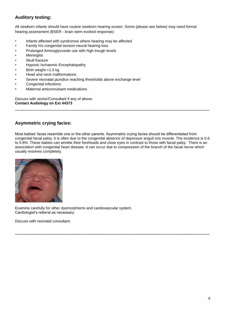
Asymmetric crying facies:
Most babies’ faces resemble one or the other parents. Asymmetric crying facies should be differentiated from congenital facial palsy. It is often due to the congenital absence of depressor anguli oris muscle. The incidence is 0.6 to 0.8%. These babies can wrinkle their foreheads and close eyes in contrast to those with facial palsy. There is an association with congenital heart disease. It can occur due to compression of the branch of the facial nerve which usually resolves completely.
Examine carefully for other dysmorphisms and cardiovascular system. Cardiologist’s referral as necessary. Discuss with neonatal consultant.
__________________________________________________________________

10
B:
BCG
Bilious aspirates or vomiting
Birth marks
Breast feeding advice / contraindications
Breast swelling
Bruising
__________________________________________________________________
BCG:
There is a list of countries (available in Transitional Care) where TB is more prevalent. Babies born to parents from those countries irrespective if parents born in the UK will require BCG vaccination. These babies are identified by the health visitor/GP and will be referred to Llandough hospital or Cardiff Royal Infirmary for BCG vaccination.
_____________________________________________________________________________
Bilious vomiting /aspirates:
Bilious vomiting in a neonate should be evaluated urgently. It is often a non-specific sign of intestinal obstruction but can represent a surgical emergency. A malrotation/ midgut volvulus is a worry and should be excluded. Examine the infant carefully and note if the infant had opened bowels. Identify maternal risk factors for sepsis and review antenatal scans.
Discuss with SpR/ Consultant
Investigations and immediate management:
1. Admit to the neonatal unit
2. IV access – blood for FBC, U/Es, CRP, Glucose, gas, C& S 3. Keep nil by mouth; IV maintenance fluids 4. Pass NG tube
5. AXR
NNU Reg may discuss with paediatric surgeon SpR bleep 5994; SHO 5960 Re: upper GI contrast, antibiotics.
____________________________________________________________________________
Birth Marks:
1. Mongolian blue spot:

11
Mongolian blue spot is an increased area of bluish pigmentation usually over the lower back and buttocks. It is extremely common in Asian and Afro-Caribbean babies. About 10% of Caucasian babies may have a Mongolian blue spot. Later in childhood, a Mongolian blue spot on the body can raise suspicion of a bruise, leading to unnecessary investigation to exclude NAI (non-accidental injury). It is therefore important to document carefully the precise location of any Mongolian spots. Mongolian spots unlike bruises do not undergo colour change or involution.
Occasionally new spots may appear postnatally. 2. Café au-lait spots:
These are uniform tan-brown round, oval or polygonal flat macules with a distinct margin and irregular contour. Most infants are healthy with no other medical problems. Café-au lait spots grow in size proportionally as the infant grows. Presence of multiple spots can be a potential marker for Neurofibromatosis type 1(NF1).However the predictive value of the number or size in newborn infants has not been established.
• They can also be associated with a number of other conditions:
• McCune Albright’s syndrome
• Noonan’s syndrome
• Russel silver syndrome
• Fanconi’s anaemia etc.,
Follow up : Discuss with Consultant
Discuss genetic testing if family history relevant.
3. Salmon patch: (Capillary haemangioma) Otherwise called ’stork bite mark’.
Disappears over time – Reassure parents
4. Naevi:

12
A light to dark brown nevus, present at birth with increased skin markings in comparison to the surrounding skin. At birth it may be flat or raised. The colour spectrum is tan, brown or dark brown. Recently there is less enthusiasm for removal of small naevi as there seems to be no increased risk of melanoma arising from small naevi.
Reassure parents. GP to follow up. If there are changes in appearance, refer to Dermatology.
5. Giant Naevus:
The presence of satellite naevi increases the risk of leptomeningeal involvement, especially when located in the midline of the trunk or on the head. When the naevus cells are present within the leptomeninges, there can be increased intracranial pressure, hydrocephalus, seizures and neurological deficits. Increases risk of melanoma (6.3% life time risk)
Referral to Dermatologist: SpR Bleep 5392; Consultant Dr Katugampola Ext 42190
6. Strawberry haemangioma:
Natural course: New lesions may appear few days after birth. Most rapid growth is during the first 6 months. Lesions may take as long as 7 years to completely disappear. Skin appears pale and softer during the involution phase.
Risks: Ulceration, bleeding, infection
Management: Active non-intervention with clinical observation and follow up. Most peripheral lesions can be followed up by the GP. Serious complications can occur if haemangiomas are located over specific areas such as the lips, eye lids, auditory canal and the air way (may present with stridor).
Specialist referral : Discuss with consultant if involves high risk areas. Dermatologist, Ophthalmologist and ENT
surgeon referrals may be required as necessary.
_____________________________________________________________________________
Breast feeding
Supplementation of breast feeding:
It is usually unnecessary to supplement full term breast fed infants. However in unwell and /or hypoglycaemic infants
supplementation with formula milk or IV dextrose may be needed as a temporary measure. Supplementation may
also be required in newborns with > 10% weight loss or decreased urine out put. (Normal hydration is at least 4-6
wet nappies/ 24 hours).
Mothers should always be encouraged to offer the breast frequently despite formula supplementation.
Please see transitional care guidelines for infants with >10% of loss in birth weight. Contra-indications to breast feeding are few:

13
1. Maternal HIV infection
2. Anti-metabolites eg., methotrexate, 6- mercapto purine
3. Anti-cancer eg, anthracyclines, vinca alkaloids, cytarabine etc.,
4. Anti-psychotics eg., haloperidol, fluphenazine
5. Galactosaemia in the neonate
6. Untreated tuberculosis in the mother (rare in the UK) Drug and Alcohol use: Large quantities of stimulant drugs such as cocaine/crack or amphetamines, heavy drinking, regular use of illicit (street) drugs such as heroin and benzodiazepines: breast feeding is contraindicated SSRIs:
Sertraline and Paroxetine safest for breastfeeding
Fluoxetine and Citalopram:
Potential for toxicity in the infants; however benefits of breastfeeding outweigh the risk
Maternal Hepatitis C:
Transmission of infection via breastfeeding has not been documented. Maternal Hepatitis C is not a contra-indication
for breastfeeding. Maternal Hepatitis B:
Maternal Hepatitis B is not a contra-indication to breastfeeding.
Maternal mastitis/breast abscess:
Breast feeding should continue. Regular expression of milk should be encouraged from the affected breast. Feeding
can be commenced on the affected side once treatment has been commenced.
_____________________________________________________________________________
Breast swelling in the infant:
Breast swelling in newborn infants is common. It is usually due to maternal Oestrogens and subsides as the maternal hormone effect wears off. Occasionally milk discharge can occur. This is not pathological.
Risks: Infection
Reassure parents. Advise to seek medical attention if the swelling gets rapidly bigger/ causing discomfort or if the overlying skin turns red and inflamed.
_____________________________________________________________________________
Bruising:
Babies may get bruised as a result of traumatic delivery. Document areas of bruising (can use a baby body map to accurately describe location) at birth on newborn examination chart. Reassure parents that it will take a few days to disappear.
A sub-conjunctival haemorrhage may take more than 2 weeks to resolve.
Investigations:
If spontaneous bruising occurs in areas not involved in trauma or further new bruising occurs after delivery,
check FBC and clotting.
Monitor SBR as these babies can get jaundiced.
Ensure adequate feeding and hydration.
Check and document if Vitamin K had been administered after birth.
If birth trauma involved, baby may be in pain and irritable. Consider analgesia with oral paracetamol.
__________________________________________________________________
14
C:
CCAM (Congenital cystic adenomatoid lung malformation)
Caput succedaneum/ Cephalhematoma – see scalp swellings
Cardiac disease:
Cataracts
Cleft lip & palate
Conjunctivitis
__________________________________________________________________
CCAM: (Congenital cystic adenomatoid malformation of the lungs) Lung bud malformations include: • CCAM
• Bronchogenic cyst
• Lobar emphysema
• Pulmonary sequestration
Most of these lesions would have been found on antenatal scans and followed up in fetal medicine, but the precise nature of the malformation may not be clear until after delivery.
Examination – infant may have
• Respiratory distress
• Asymmetric breathing
• Pulmonary infection
• High out-put cardiac failure
Investigations:
1. CXR after birth
2. Capillary blood gas as applicable
3. Usually a CT chest is requested in a month’s time; and refer to Respiratory team.
Arrange senior review SpR/Consultant and see neonatal doctors’ guidelines
Referral: 1. Respiratory team SpR bleep 5342
2. Consultant Dr Doull 43530, Dr Forton 44891
3. D/W neonatal consultant re: follow up
____________________________________________________________________________
Cardiac disease:
Antenatally diagnosed cardiac problems/arrhythmias: • Follow plan from Fetal medicine
• Examine baby
• Senior/ Consultant review
Investigations: ECG, oxygen saturations, four limb BP
Refer to Cardiology: SpR bleeps 6343, 5394, 5391
First degree relative with congenital heart disease:
• Well baby, no heart murmur- Refer to Dr Amos Wong's Echo clinic as out patient
• Family h/o Cardiomyopathy - Baby will need Cardiomyopathy screen – Inform cardiologist.

15
Cardiac Murmur:
If less than 24hrs old, review the next day, as the murmur may have disappeared.
If the murmur is audible at the time of newborn examination, and the baby is over 24 hours of age, arrange for
the investigations listed below:
lnvestigations: 4 limb blood pressures
ECG
Pre and post ductal oxygen saturations
Registrar review of above
Echo as inpatient as necessary Follow-up: If above investigations normal book baby into Neonatal Consultants OPD in 6 weeks. If baby has been seen by Cardiology team please refer to their plan for planned follow up. Complete referral letter for secretaries to type. Parents: Explain the findings t o t h e pa r e n ts a n d r eassure them. Explain to parents signs of cardiac failure (poor feeding, snacking feeds, colour change and when to seek medical advice/help. Discuss follow-up arrangement. Useful contact numbers: Paediatric Cardiology SpRs Blp 5394/5391
_____________________________________________________________________________
Cleft lip and Palate:
Always use both methods, digital palpation of the hard palate and visual inspection of the uvula
using torch and spatula/1ml syringe to confirm absence of cleft soft palate and submucus cleft. Cleft lip and palate
may occur as an isolated birth defect or part of a syndrome e.g., Pierre Robin sequence, Strickler's syndrome.
Normal soft palate Cleft lip & palate Cleft soft palate
Cleft soft palate Pierre Robin’s sequence Subucus cleft palate + bifid uvula
Examine carefully for any other associated malformations/dysmorphism, examine the cardiovascular system
Management: Ensure adequate feeding of the infant . Special teats / NG tube feeding may be necessary in some
cases.

16
SpR review
Referral: Cleft lip and Palate team (CLAPA) at Morriston hospital. Contact no: 01792703810
1. Usually a specialist nurse visits within 24 hours of referral.
2. Paediatric Cardiology review if heart murmur present. SpR Bleeps 6343, 5394, 5391
3. Formal written referral to the Plastic surgeon in Swansea.
4. Geneticists referral if syndromic - arrange relevant genetic testing. Ext 42577
5. Refer to Audiology for BSER Ext 44799/ 44373
6. Discuss with neonatal consultant and arrange neonatal follow up.
In babies with no murmur or dysmorphism, routine chromosome testing and pre-operative echo are usually
organised by the Plastic surgeons.
____________________________________________________________________________
Cataracts:
Absence of red reflex should always raise suspicion. No red reflex indicates pathology anterior to the retina Bear in
mind red reflex may not be bright red, rather pink/pale orange in dark skinned infants. Congenital cataracts are
generally rare. Retinoblastoma is an important differential diagnosis
Senior review.
D/W neonatal consultant before making Ophthalmologist referral
Ophthalmology referral SpR Bleep 5529/5525 Or send urgent faxed referral letter to Mr P Watt's secretary. Ext 48583
_____________________________________________________________________________
Conjunctivitis:
The lacrimal (tear) duct is not very well cannulised in newborns, hence conjunctival discharge is a common postnatal
ward finding.
Examination: Purulent discharge from the eyes, inflamed red conjunctiva Swelling of eye lids Investigation: Eye swab C&S Management:
Clean each eye with separate cotton wool dipped in sterile water. If infected, treat with topical Chloramphenicol eye drops 0.5%1 drop QDS for 7 days.
_____________________________________________________________________________
17
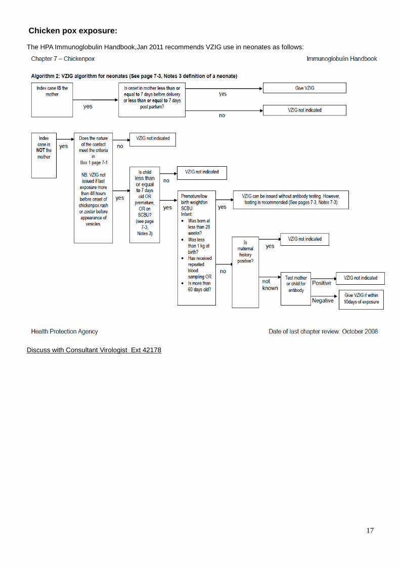
Chicken pox exposure: The HPA Immunoglobulin Handbook,Jan 2011 recommends VZIG use in neonates as follows:
Discuss with Consultant Virologist Ext 42178

18
D:
Delayed passage of meconium
Delayed passage of Urine
Developmental dysplasia of hips
Drug dependant mothers
Dysmorphic features
Down Syndrome
__________________________________________________________________
Delayed passage of meconium:
The first passage of meconium occurs in normal term babies at 7 hours of age. 99.7% of healthy term babies pass meconium by 34 hours of age. Preterm babies may have delayed passage of meconium up to 2 days.(32%).Infants who do not pass meconium by 48 hours of age should be carefully examined and investigated for possible gastro-intestinal tract obstruction such as anal atresia/stenosis, imperforate anus, Hirschsprung’s disease, cystic fibrosis etc.
Discuss with Consultant/SpR Refer to Paediatric surgeon SpR bleep 5994
_____________________________________________________________________________
Delayed passing of urine (> 24hours)
Has the urine been passed but absorbed into a gel nappy? Put cotton wool inside nappy and observe. Apply U-Bag
Examine genitalia and abdomen.
Were there any concerns antenatally regarding amniotic fluid volume – Check USS?
_____________________________________________________________________________
Developmental dysplasia of hips (DDH)
All babies should be assessed to determine if either or both hips are either dislocated or dislocatable. In order to
rule out dislocated hips do the Ortolani Test, the Barlow Test will dislocate a dislocatable hip.
If you have any concerns about unstable or clicky hips please speak to the Neonatal SpR. Check family history for
DDH, presentation of baby in-utero and at birth.
Follow-up:
Dislocated / dislocatable hips: an ultrasound scan should be arranged as an out-patients appointment with Mr
Hemmadi or Miss Carpenter Consultant Orthopaedic Surgeon. This will be arranged by the neonatal secretaries
when they receive referral letter for typing.
We do not currently screen babies with risk factors for DDH, however, this may well change in the future. Babies
with a family history of DDH, breech presentation at birth / in-utero are not routinely referred for USS unless hips are
found to be unstable. If concerned please discuss further with Neonatal Consultant and Miss Carpenter
(OrthoCons).
Advice to parents: Advise parents to nurse baby in double nappies to give extra support to the hips; to avoid lifting baby by feet / legs when changing nappy etc and not to leave baby sitting in car seat for longer than necessary. Discuss follow-up arrangements with parents.
_____________________________________________________________________________

19
Drugs in Pregnancy: Most commonly used maternal medications are compatible with breast feeding and generally the benefits of breast
feeding out weigh the possible risks.
For specific drugs please refer to’ Drugs in Pregnancy and Lactation’ – 15th edition
This book normally lives on the shelf in the doctors’ office in the neonatal unit.
Drug dependant mothers:
History:
Document clearly what drug the mother was using prior to and during pregnancy. Follow NAS protocol for infants:
Most rehabilitation drugs such as Methadone and Subutex are compatible with breast feeding. Check maternal
urinary toxicology in pregnancy. This is usually a fair indicator of drugs used by the mother recently.
Vaccine:
Offer Hepatitis B vaccine to infants of all substance using mothers irrespective of IV drug abuse.
Investigation:
• Please ensure every attempt is made to collect the first void urine from the baby and send for toxicology.
• Cranial USS and echo if mother is a known cocaine user as it can cause vasoconstriction.
Discharge and social: 1. Always explore social circumstances prior to discharge.
2. Check if the newborn infant/siblings are on the child protection register.
3. Discuss with Lois Mortimer – Substance misuse counsellor – on 079623905667.
Follow up
1. Follow Hepatitis B vaccine guidelines and fill in appropriate forms for completion of course by the GP.
2. Arrange Neonatal follow up if baby is IUGR, required treatment for NAS or clinical concerns.
Discuss with consultant.
_____________________________________________________________________________
Dysmorphic features:
Always look at the family as often an infant who appears different may resemble a close member of the family.
Points to remember:
• A thorough history and head to toe examination is important
• Plot growth parameters on the growth chart
• Genetic tests may be useful in the diagnosis
• Careful communication with parents is important
Always involve senior/Consultant to review History:
• Maternal medications, illnesses in pregnancy
• Antenatal scans ? any suspected abnormality?
• Fetal growth and movements
• Liquor volume
• Amniocentesis
• Labour and delivery details • Any resuscitation of the infant required
• Parental consanguinity
• Family history of inherited problems.

20
Investigations:
1. Chromosome and DNA testing based on clinical features/suspected syndrome.
2. Arrange Geneticist review and discuss if sample for CGH( Chromosomal genomic hybridisation) should be sent
Ext 42577
3. Often echo and cranial USS may be required.
4. Request Radiologist (Dr S Morris) to review the kidneys on the antenatal scans
5. Ophthalmologist referral SpR 5529; Mr Watts sec 48583 Communication with parents:
Always involve a senior when speaking to the parents. You may ask ’” who do you think he/she resembles?’ If you
find dysmorphic features, be honest and raise your specific concern to the family saying,’ I feel your baby has some
distinctive facial features which I would like my consultant to have a look. Your baby may need some tests’’.
_____________________________________________________________________________
Down’s syndrome: Down’s syndrome is the commonest chromosomal abnormality. Once the diagnosis has been confirmed, by chromosomal analysis, ensure appropriate investigations and follow up arrangements are made. Issue parents information booklet on Down’s syndrome. This is usually kept by the neonatal out reach team or kept in a brown envelope in the cupboard in parents’ room on the neonatal unit. Monitoring:
• Many infants with will be floppy and may be slow to establish feeds necessitating NG tube feeds.
• Keep baby under observation, if cardiac abnormality is suspected.
• Ensure that meconium was passed within the first 48 hours as the condition can be associated with
Hirschsprung’s disease.
• Daily registrar review.
• Plot growth parameters in the Down’s syndrome growth chart. Investigations:
1. Request cardiologist’s opinion as a baseline record even if there are no immediate cardiac concerns. About 30%
of babies/ children may have a cardiac abnormality. Arrange for baseline ECG, pre and post ductal SpO2 and 4 limb
BP.
2. Arrange for Thyroid function tests in the blood clinic at 10 -14 days of age.
Ensure infant’s and maternal details with telephone numbers are provided in the Out reach team’s folder. Issue the
appointment letter for blood clinic with documented time and date.
Communication:
Document any communication to the parents clearly in the notes. Speak directly with the family and explain your
concerns: “There are some features in your baby that I am concerned about… I will ask my senior colleagues to come
and have a look at your baby”. If the family doesn’t speak/understand English, ensure appropriate interpreter is used
for all communication.
Follow up:
1. Neonatal follow up – some consultants prefer seeing the family once or twice for neonatal follow up,
otherwise follow up is done by the Community paediatrician. Discuss with the Consultant responsible for the
baby. Arrange neonatal out reach follow up ext 43249
2. Refer to the local Community Paediatrician. Please identify the Community Paediatrician assigned to the patient via the patient address and post code.
3. Cardiologist’s follow up depending on echo findings. If unable to obtain echo as an inpatient, in the absence of a murmur, refer to Cardiology outpatient clinic
4. If very young mothers or unusual chromosomal mosaicism, offer referral to the geneticist. If the family agrees, send a referral letter to Dr Sally Davies, Consultant geneticist to arrange follow up and counseling.
Use the usual proforma for the above referrals.
__________________________________________________________________

21
E:
Erbs Palsy
Ear tags or pits
__________________________________________________________________
Erb’s Palsy: Please see doctor’s guidelines for Brachial plexus injury. Parents: Explain to parents regarding diagnosis and document clearly in the notes.
Investigations:
X-ray of the relevant arm, clavicle and chest – to exclude bony fractures and diaphragmatic involvement.
Parents:
Explain to parents regarding diagnosis and document clearly in the notes.
Management:
1. Physiotherapy is the mainstay of treatment.
2. Arrange for urgent Physiotherapist review Ext 46422.
3. Daily SpR review as an inpatient to assess progress.
4. Analgesia – oral paracetamol. The infant may be in pain due to fracture or soft tissue injury.
Follow up: 1. Neonatal consultant follow up in 2-4 weeks- inform respective consultant
2. If baby discharged home on a weekend, send urgent referral to the Neurophysiotherapist on ext 46422 on the
next working day.
_____________________________________________________________________________
Ear tags/Pits/Skin tags:
Reassure. If mother concerned the GP can refer to the plastic surgeons at 1 year of age.
__________________________________________________________________

22
F:
Facial Nerve palsy
Femoral pulses; absent/reduced
__________________________________________________________________
Facial nerve palsy:
Facial palsy in the newborn may be associated with underlying neurological problems, myopathies or due to birth trauma.
History:
Antenatal - Fetal growth, amniotic fluid indices, scans and fetal movements. Delivery – Mode of delivery, shoulder
dystocia, instrumental (forceps).
Examination:
Carefully assess if any other systems involved. Further investigations will depend on associated features at birth.
Arrange senior review.
If unable to shut eyelids, prescribe hypromellose eye drops to prevent exposure keratitis. Monitor feeding on the
postnatal ward Parents:
If isolated facial palsy related to birth trauma the long term outcome is good. If recovery is slow, will need specialist
referral. Follow up: Discuss with neonatal consultant and arrange follow up.
_____________________________________________________________________________
Femoral pulses absent/weak: Probable evolving co-arctation of aorta;
Urgent SpR review
Examine for perfusion in lower limbs and measure urinary output
Discuss with neonatal consultant, may need admission
Investigations:
1. ECG, Ext 43325
2. 4 limb BP
3. SpO2 pre and post ductal
4. Capillary blood gas, Cardiologist review- SpR bleeps: 6343, 5394, 5391
__________________________________________________________________

23
G:
Genitalia
__________________________________________________________________
Genitalia Micropenis can be:
• an incidental finding on the newborn examination
• found in a hypoglycaemic baby.
Normal phallus size is the stretched length from the pubic tubercle to the tip. In a term baby this is usually @3 cm.
Micropenis is a measurement less than 2.2cm . Varies with ethnicity.
A large fat pad at the base of the penis can mimic a micropenis History:
• Family h/o ambiguous genitalia
• Ethnic origin Examination
• Dysmorphism
• Palpate for testes
• Midline defects: Hypertelorism, cleft palate
Differential diagnosis:
• Normal variant
• Anterior pituitary hormone deficiency Investigations
• Pre-feed blood glucose
• Karyotype
Discuss with consultant Referrals:
Endocrinologist SpR Bleep 5679
Prof J Gregory - Ext 42274
Dr J Warner - E x t 46373
Ophthalmologist Mr Watts - Ext 48583
Geneticist referral - Ext 42577
_____________________________________________________________________________
Pigmented scrotum: Common in Asian, African or Middle –Eastern origin
This appearance in a Caucasian baby should raise suspicion of Addison’s disease.
Examination
Does the scrotum look normal?
Are both testes palpable?
Normal variant
CAH (Congenital Adrenal Hyperplasia)
Refer to Ambiguous genitalia guidelines and discuss with SpR
__________________________________________________________________
24
H:
Haemoglobinopathy
Haemangiomas- See Birth marks
Hepatitis B vaccination
Herpes in mother
HIV positive mothers
Hydrocele
Hypospadias
Hypoglycaemia
__________________________________________________________________
Genital Herpes in the mother:
Usually caused by HSV-2 virus and is associated with significant neonatal mortality and morbidity. It is important to establish whether this is a primary infection or a recurrent infection, as primary infection has a 41% risk of neonatal transmission. Discuss with Consultant Virologist Ext 42178 If recurrent Herpes in the mother:
Reassure that the risk of transmitting the infection to her baby is low, even in the presence of active lesions at the
time of delivery. Maternal antibodies will give some protection to the baby. If vaginal delivery did occur in the
presence of HSV lesions; inform the GP and community midwife so that they can monitor for signs of neonatal HSV.
Management of a baby considered to be at risk of neonatal herpes:
• Isolate the patient. Obtain urine and stool cultures and swabs from the oropharynx, eyes and surface sites for viral
culture and typing.
• Intravenous Aciclovir is given by many whilst waiting for the results and is the treatment of choice in confirmed
infection.
• Breast-feeding is recommended unless the mother has herpetic lesions around the nipples. Aciclovir is excreted in
breast milk but there is no evidence of harm.
• Parents should be warned to report any early signs of infection such as poor feeding, lethargy, fever or any
suspicious lesions.
Herpes simplex labialis (lips/cold sores) infection in the mother:
Usually caused by HSV -1. No need to isolate infant from the mother. Careful hand washing of mother after touching
the lesions should be advised. Breast feeding is not contra-indicated. However mother with active lesions around the
mouth should be advised to avoid close contact such as kissing.
_____________________________________________________________________________
Genital Warts in the mother:
Vertical transmission in utero is extremely rare
The most significant manifestation of mother-to-child transmission of the wart virus is juvenile laryngeal papillomatosis. There appears to be an association with maternal genital warts present at the time of delivery.
No immediate intervention needed for the newborn. If in doubt contact Virologist on ext 42178.
_____________________________________________________________________________

25
Haemogloginopathies: (Sickle cell/ Thalassemias) If family h/o haemoglobinopathies, but only one parent has a trait or is affected with the disease, baby will be a carrier only. Sickle cell disease is now looked for on the newborn sreen in Wales.
If mother has disease and the father’s status is unknown or a trait, baby should be tested for haemoglobinopathies
urgently. Investigations:
FBC 1X EDTA
Haemoglobinopathy screen 1 x EDTA
Contact Haemoglobinopathy screening lab for urgent results: Ext 43302 If positive d/w neonatal consultant and refer to Dr Phil Connor, Consultant Paediatric Haematologist for advice and follow up. Ext 44829
_____________________________________________________________________________
Hepatitis B vaccination: Indications: Infants of Hepatitis B positive mothers
Infants of substance misusing mothers.
Hepatitis vaccine given within 12 hours of birth in high risk infants can prevent vertical infection in 75 to 90% cases. It is important to establish the infectivity of the mother in order to decide on Hepatitis B Immunoglobulin.
Please refer to doctor’s guidelines for details. Take consent from mother for immunizations. If mother’s status
unknown, contact Consultant Virologist on Ext 42178
_____________________________________________________________________________
HIV positive mothers: All infants born to HIV positive mothers should be referred to Dr Jennifer Evans. Most mothers are detected antenatally and there should be a clear plan of action for the newborn infant in the maternal notes. Please be aware:
There are specific criteria for infants born to HIV mothers that place them at high risk: gestation, birth weight,
PROM of 4 hours or more, and timing of maternal intrapartum antiviral administration.
Inform consultant Investigations:
Send blood for FBC, LFTS, HIV viral DNA – PCR Management:
• Commence oral Zidovudine or triple therapy as advised by Dr Jennifer Evans.
• Breast feeding and BCG are contra-indicated.
• Baby should stay in hospital until oral medications are well tolerated. Contact:
Neonatal out reach sister Jacqui Evans when baby is born ( 43249; 07768867988
Should any special circumstances arise, please contact Dr J Evans Ext 42273
Follow up: Send referral letter to Dr J Evans for follow up.
Note: Some mothers may choose not to inform GP. Please mark this on the referral letters.
_____________________________________________________________________________

26
Hydrocoele: A hydrocoele is a collection of fluid in the scrotum and is different to an inguinal hernia. The typical hydrocoele is noted shortly after birth as a unilateral or bilateral swelling in the scrotum. The scrotum is enlarged, may be tense with fluid but is usually non-tender; with a bluish appearance. It is non reducible, it is possible to get above the swelling, and it may fluctuate in size.
Trans-illumination is useful but should be interpreted cautiously in small infants as herniated bowel with gas within in can also trans-illuminate. Management:
Observation in the first year of life.
Hydrocoele that persists beyond 2 years of life is unlikely to resolve on its own and will need surgical repair. GP
can follow up during the routine baby checks.
Surgery may be done sooner if difficulty differentiating from a hernia.
Advise to parents:
The swelling may fluctuate in size. If there is rapid increase in size of the swelling/causing discomfort/change in
colour of the skin, urgent medical attention should be sought.
_____________________________________________________________________________
Hypospadias:
Hypospadias consists of three main features;
1. An abnormally placed urinary meatus, placed anywhere from the glans to the peno-scrotal junction
2. Chordee (curvature) of the penis which forces the penis to point towards the scrotum when erect or
3. A deficiency of the foreskin. Normally the foreskin completely encircles the phallus, but in hypospadias the
foreskin is present on the dorsal side only. This gives the appearance of a “hooded” foreskin.
.
Examination: It is important to palpate both testes. If so, then it is XY Karyotype
If only one testis is palpable , a karyotype of XY/XO should be considered.
If no testes are palpable, investigate as ambiguous genitalia
Investigation:
If in doubt, get senior opinion and follow approach for ambiguous genitalia.
Treatment: Isolated hypospadias requires surgical correction between 1& 2 years of age.
Refer to the Paediatric surgical team as outpatient after discussion with Neonatal Consultant.

27
Advise to parents:
Watch for urinary stream prior to discharge. Advise parents against circumcision as the foreskin tissue is used for
reconstruction. Even if there are religious considerations, it can be done once the hypospadias has been repaired.
_____________________________________________________________________________
Hypoglycaemia: It is important to identify infants at risk of hypoglycaemia.
Clear plans for transitional care management should be documented in the notes.
Please follow the Guideline for the prevention and management of hypoglycaemia of the newborn for high risk neonates on the post natal ward
__________________________________________________________________

28
I:
IUGR
Imperforate anus
Infant of Diabetic mother: (Insulin dependent / gestational diabetes)
__________________________________________________________________
IUGR (Intrauterine growth restriction) Identify cause for IUGR and document in the notes
If early onset – usually symmetrical IUGR
If late onset – asymmetrical (usually head spared). Refer to text books for causes Differential diagnosis
Small for gestational age
Investigations: For all IUGR infants
Blood glucose (poor reserves and prone for hypoglycemia). Consider FBC (look for high hematocrit and
thrombocytopenia)
Always take a free flowing venous sample
If no identifiable cause for IUGR/ Twin IUGR perform (in addition to above):
Cranial USS, Urine for CMV
If dysmorphic features present ± h/o previous miscarriages Karyotyping
Monitor on postnatal ward for:
Hypoglycaemia
Hypothermia
Poor feeding
Jaundice
Polycythaemia
IUGR baby with abnormal umbilical Dopplers: If abnormal umbilical artery Dopplers noted in the antenatal scans, provided the baby is clinically well enough to be managed on postnatal ward, there is no need to delay feeds. Breast milk should be given in preference to formula milk.
Follow up:
Depending on clinical picture and birth weight these infants may require follow up. Discuss individual cases with the
consultant.
The growth of healthy IUGR babies can be monitored in primary care.
_____________________________________________________________________________
Infant of diabetic mother: May be gestational diabetes or known IDDM/ Type 2 DM. Note mother’s glycaemic control in pregnancy and what treatment she had been on.
Risks:
Hypoglycaemia
Polycythaemia
Hypocalcaemia
29
Hypomagnesaemia
Transient hyperinsulinism
RDS Congenital
Septal hypertrophy
Septal defects
Sacral agenesis
Management after birth:
Allow to breast feed within an hour of birth.
If mother unwell, formula should be used until mother is able to breast feed.
Follow hypoglycaemia pathway.
Who should be admitted to the neonatal unit?
Babies who remain hypoglycaemic despite full volume feeds and receiving day ahead feed volumes, (Day 1 volume
of feed is 60mls/kg, Day 2 is 90mls/kg and Day 3 is 110mls/kg).
Those who do not tolerate 2 hourly oral/NG feeds on postnatal ward
Those with symptomatic hypoglycaemia should be admitted to the neonatal unit.
SGA (small for gestation) infants of diabetic mothers are at even greater risk.
They are small usually due to maternal vascular disease secondary to diabetes leading to placental insufficiency.
They are at risk of neurodevelopmental sequelae and might require neonatal follow up. Get senior advice
_____________________________________________________________________________
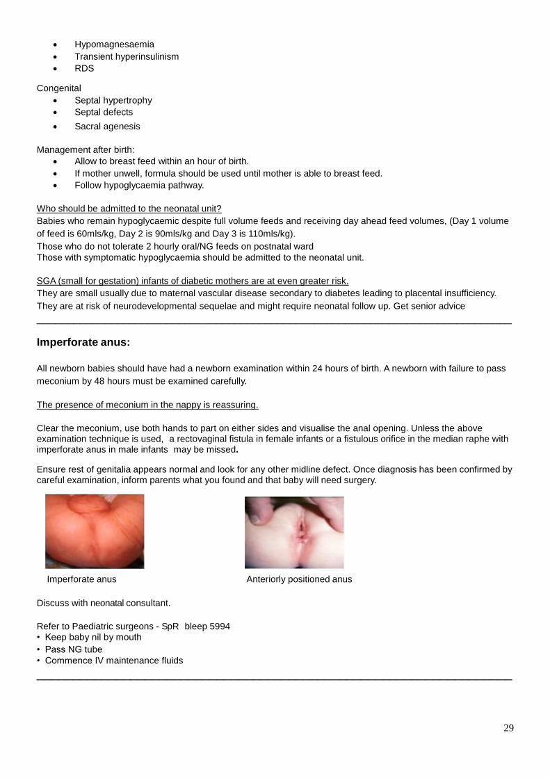
Imperforate anus:
All newborn babies should have had a newborn examination within 24 hours of birth. A newborn with failure to pass
meconium by 48 hours must be examined carefully.
The presence of meconium in the nappy is reassuring.
Clear the meconium, use both hands to part on either sides and visualise the anal opening. Unless the above examination technique is used, a rectovaginal fistula in female infants or a fistulous orifice in the median raphe with imperforate anus in male infants may be missed. Ensure rest of genitalia appears normal and look for any other midline defect. Once diagnosis has been confirmed by careful examination, inform parents what you found and that baby will need surgery.
Imperforate anus Anteriorly positioned anus
Discuss with neonatal consultant.
Refer to Paediatric surgeons - SpR bleep 5994 • Keep baby nil by mouth
• Pass NG tube
• Commence IV maintenance fluids
__________________________________________________________________

30
J:
Jaundice:
Baby <24 hours of age
Baby >24 hours of age
Conjugated hyperbilirubinaemia
Jittery baby
__________________________________________________________________
Jaundice in the newborn Jaundice within 24 hours of birth is always pathological and should be investigated. ABO/Rhesus incompatibility, prematurity, bruising at delivery, sepsis or family h/o haemolytic anaemia are some of the important causes. Jaundice after 24 hours is often physiological (use transcutaneous bilirubinometer) Investigations
• Serum bilirubin (spilt) (follow NICE charts)
• FBC, Reticulocyte count, film
• Urgent Blood group and DAT (particularly if mother is O positive or rhesus negative)
Management • If jaundiced <24 hours, commence double phototherapy, 4-6 hourly SBR
• If > 24 hours, commence phototherapy as per NICE threshold, 8-12 hourly SBR
• Follow exchange transfusion guidelines, if evidence of iso- immunisation
• Ensure adequate feeding
• Babies not tolerating oral/NG feeds will need IV maintenance fluids.
Feeding • A minimum of 4 to 6 wet nappies/24 hours to ensure adequate hydration
• Baby to breast feed at least for 20 minutes initially. Then ask mother to express milk. If sufficient volume of breast milk available, top up the expressed milk by syringe, cup or NG feeds in a poorly sucking infant.
• If EBM insufficient, work out fixed volumes for the day as per schedule and give three hourly formula feeds. Allow breast feeds as extra. Even in this situation, babies should be allowed to breast feed first before giving formula.
• If feeding is adequate there will be an improvement in jaundice, urine out put and weight gain. Reassure that formula milk can be stopped once baby starts latching on properly and mother’s milk production is sufficient.
Advice to parents
• If ABO/Rhesus incompatibility explain the potential for haemolysis and anaemia to occur over a prolonged period
(weeks to months)
• Warn of symptoms of anaemia such as pallor, lethargy, poor feeding, failure to gain weight, infrequent urine output, rapid breathing, sweatiness. If any of the above present, parents to seek urgent medical advice.
Follow up:
• Babies who required phototherapy for physiological jaundice should have a repeat SBR in community within 48
hours.
• Babies with evidence of Rh or ABO incompatibility (DAT +ve) irrespective of requiring phototherapy should be
commenced on Folic acid supplements (500mcg od) for 6 months.
• Arrange to recheck FBC, reticulocyte count and SBR in a week’s time in blood clinic.
• Further tests can be done based on the clinical state and initial results.
• Babies who received IVIG/exchange transfusion will need neonatal follow up.
__________________________________________________________________
31
Jitteriness: It is quite common for newborn babies to become jittery in the first 2 to 3 days of life. Jitteriness involves symmetrical tremors of all four limbs without a fast or slow component. This can occur at rest or on handling. It is extremely stimulus sensitive and usually stops on holding the limb. This is a useful way of distinguishing from clinical seizures where the movements do not stop on holding the limb. There is no facial or autonomic involvement (tachycardia, raised BP, apnoea) associated with jitteriness.
Investigations:
Check observations, including temperature
Check blood glucose and U/Es, Ca, Mg if persistently jittery.
Ensure and document if there is any history of maternal illicit drug use. If above investigations are normal
consider sending urine for toxicology and observe baby for (NAS) neonatal abstinence syndrome.
In otherwise well babies, reassure parents.
__________________________________________________________________

32
L:
Large for gestational age
Limb anomalies
__________________________________________________________________
Large for gestational age: Birth weight > 4.5 kg is macrosomia. Often infants of mothers with poorly controlled diabetes are macrosomic due to the effect of fetal hyperinsulinism secondary to exposure to high maternal glucose. Occasionally LGA can also be associated with increased concentrations of maternal triglycerides and free fatty acids. Complications: • Shoulder dystocia, brachial plexus injury, perinatal asphyxia if traumatic delivery.
• Hypoglycemia(see infant of diabetic mother)
Investigation: • Blood glucose monitoring only if unwell or symptomatic.
_____________________________________________________________________________
Limb anomalies: 1. Polydactyly: (Extra digits)
Pre-axial Post-axial – skin pedicle Post-axial Thorough examination to look for other abnormalities. If abnormal thumb, also examine forearm to feel for the presence or absence of the radius. Pre-axial polydactaly is more likely to be associated with an underlying chromosomal problem. Referral to: Mr Milling, Consultant hand plastic surgeon, Morriston hospital
____________________________________________________________________________________________
2. Syndactyly: (fusion of digits)
Often syndactyly of hands may be associated with syndromes or cardiac anomalies such as ASD. Careful thorough examination of the infant should be performed. Syndactyly of toes may be familial. Usually no treatment is required. Discuss with parents. If parents keen for surgery for cosmetic reasons, a referral to plastic surgeons can be requested through the GP.
Inform the neonatal Registrar Referral to:
Mr Milling, Consultant hand plastic surgeon, Morriston hospital
Paedaitric cardiologist referral as outpatient, – inpatient as indicated. (Bleeps 6343, 5394, 5391)
Geneticist referral if syndromic. Contact Ext: 42577.
__________________________________________________________________

33
M:
Meconium stained liquor
Myasthenia Gravis
__________________________________________________________________
Meconium staining of liquor:
Infants who had meconium staining of liquor at birth should be observed for at least 12 hours. If they cry soon after birth, no resuscitation is required. However they may develop feeding problems due to swallowed meconium.
Investigations:
A baby who develops respiratory distress and had meconium staining of liquor should be investigated for
simultaneous onset of sepsis.
• Check blood C&S, FBC, CRP, capillary blood gas
• Chest X-ray
• Measure SpO2.
Management:
If there is an oxygen requirement or significant respiratory distress the infant should be admitted to the
neonatal unit and commenced on CPAP.
Symptomatic babies should have a partial septic screen and commenced on IV antibiotics
Asymptomatic babies can be discharged home after 12 hours of age, provided observations are reassuring
and feeding is satisfactory
Arrange senior review.
_____________________________________________________________________________
Myasthenia gravis: Affects 20% of infants born to mothers with acquired myasthenia gravis with or with out AchR antibodies (acetylcholine esterase receptor).
Antenatal: Polyhydramnios, reduced fetal movements, arthrogryposis
Neonatal myasthenia:
All infants should be observed for at least 3 days for the following:
Poor sucking and swallowing
Generalised hypotonia
Facial weakness and weak cry
Fatigability during feeds
Respiratory difficulties - may require ventilation
Ptosis and Ophthalmoplegia
Involve physiotherapist if arthrogryposis present (bleep 6422)
Prognosis:
90% recover by 2 months and all by 4 months.
If any symptoms present discuss with senior/consultant. Arrange SCBU admission for monitoring and further investigations. (Neostigmine challenge 0.15mg/kg IM with improvement seen in 15 -30 minutes and effects last up to 3 hours).
_____________________________________________________________________________

34
N:
Natal Teeth
Neonatal Abstinence syndrome
__________________________________________________________________
Natal teeth:
Natal teeth are common in newborns. These should be removed as they may fall out, and pose risk of aspiration or choking in the newborn infant.
Get SpR review. Discuss with consultant and refer to maxillofacial team.
Contact: Maxfax SHO on bleep 5740 or SpR via switch.
_____________________________________________________________________________
Neonatal abstinence syndrome: See NNU guidelines .
History:
Document clear history of maternal drug intake. If on methadone or subutex check dosage. Symptoms usually peak
by 72 hours.
At delivery if respiratory depression present, do not give Naloxone.
Monitoring and investigations
• Observe on NAS chart
• Obtain verbal consent from mother and send urine for toxicology ASAP. Usually the first void urine is preferable.
• If two consecutive scores > 4, needs SCBU admission for monitoring and treatment.
Hepatitis B vaccine Offer Hepatitis B vaccine to all drug using mothers. Infants of IV drug users are at greater risk.
Social:
• Explore social circumstances prior to discharge.
• Contact Substance misuse midwife, Lois Mortimer on 079623905667
• Check if previous children on child protection register
• Discuss social circumstances if there is a social worker already involved.
Discharge and follow up:
• Babies requiring SCBU admission for NAS may need follow up
• IUGR babies / maternal use of Cocaine will need monitoring for neurodevelopmental problems. Discuss with
consultant.
• Letter for GP to complete Hepatitis B vaccination.
• Document destination of discharge.
__________________________________________________________________

35
P:
Pink / rusty colour staining on nappy
Polycythaemia
Port wine stains
Premature babies on postnatal ward
__________________________________________________________________
Pink / rusty staining on the nappy:
Pink staining of nappy often causes maternal anxiety. Reassure mother that it is due to concentrated urine and uric acid formation often gives the distinct colour. Ensure adequate feeding and hydration (minimum 4-6 wet nappies / 24 hours as a rough guide).
_____________________________________________________________________________
Polycythaemia:
Definition: Free flowing venous haematocrit > 65%. Please see NNU guideline.
Risk factors: • Delayed cord clamping > 45 seconds
• Maternal diabetes
• Twin to twin transfusion
• Placental insufficiency
• Small for gestational age / IUGR
• Pre-eclampsia
• Postmaturity
• Dehydration
• Pregnancies at high altitudes
• Chromosome abnormalities
Clinical features: • Plethora • tremors/jitteriness • Lethargy hypotonia • Hypoglycaemia • Hypocalcaemia • Jaundice • Petechiae • Seizures • NEC • Renal failure/renal vein thrombosis • Priapism
Investigations:
Check free flowing venous sample – FBC, U/Es Ca, SBR, blood glucose.

36
Management:
• If haematocrit > 70% or haematocrit 65% to 70% and symptomatic - needs treatment with IV fluids / partial
exchange transfusion (see neonatal doctors’ guidelines)
• If haematocrit >65% and baby is asymptomatic, increase intake of feeds (work out volume of feeds a day ahead)
and give EBM/Formula by bottle, syringe or NG tube.
• Give clear written instructions to the transitional care staff.
• Repeat FBC (venous sample) in 6 hours.
• Manage hypoglycaemia, jaundice and hypocalcemia if present. If Hct persistently elevated despite liberalisation of feeds, discuss with SpR.
_____________________________________________________________________________
Portwine stain:
A portwine stain is a vascular birth mark present at birth with pink, red, or purple discoloration of the skin. This is also known as nevus flammeus. At birth, the portwine stain has a lighter pink color. Over time, it darkens to be red, then purple and becomes elevated. The boundaries of the portwine stain are sharply demarcated. Occasionally, a portwine stain is not visible at birth, but becomes visible within a few hours or days. It is usually an isolated lesion, confined to one area. However, in some affected infants the distribution is over several areas, such as face, neck, chest, arm and buttocks.
The infant with a portwine stain should be evaluated, for the possible association of two more serious disorders:
• Sturge-Weber Syndrome
• Klippel-Trenaunay-Weber Syndrome
The Sturge-Weber Syndrome
Defined as a capillary malformation involving the leptomeninges which overly the cerebral cortex. This is
characterised by a portwine stain involving the distribution of the first branch (ophthalmic) of the trigerminal nerve.
Complications • Epilepsy
• Glaucoma
As to the pattern of the portwine stain that is associated with the Sturge-Weber Syndrome: the stain involves the
upper eyelid and the ipsilateral eye (with glaucoma).
The Klippel-Trenaunay-Weber Syndrome
Characterised by a superficial vascular mark similar to a portwine stain. This is caused by anomalies of the deep
veins, and complications include arteriovenous shunting of the affected limb, and overgrowth of part of or all of an
extremity. The legs are involved in 95 %, the arms in 5 % , and both arms and legs in about 15 %. The portwine stain
of the Klippel- Trenaunay Syndrome does not have the facial mark that is a characteristic of the Sturge-Weber
Syndrome.
Arrange SpR/Consultant review
Referral:
If upper eyelid or ophthalmic division of trigeminal nerve involved, refer to Mr P Watts (Ophthalmologist) Ext 48583
Referral to Dermatogist Dr Katugampola Ext 42109
Discuss with consultant regarding follow up – Will need CT head at a later date.

37
Parents:
If isolated lesion elsewhere on the body – reassure and inform that likely to persist. If possibility of Sturge Weber
syndrome, explain risks of intracranial involvement and potential for seizures. Will be followed up with neuroimaging.
_____________________________________________________________________________
Premature infant on the postnatal ward: Follow transitional care guidelines:
All borderline preterm babies from 34 weeks to < 37 weeks should be observed for at least 72 hours on the postnatal
ward.
Monitor for the following problems: • Respiratory distress
• Hypoglycaemia
• Poor feeding/ sucking – may need NG feeds
• Jaundice
• Hypothermia • Sepsis
• Recheck weight prior to discharge on postnatal ward
Follow up
All borderline preterm babies do not necessarily need neonatal follow up. If there were significant problems during
hospital stay or birth weight<1.8kg, discuss with consultant regarding follow up.
38
R:
Rashes (skin)
Red reflex abnormality
Renal tract anomalies
Respiratory distress
__________________________________________________________________
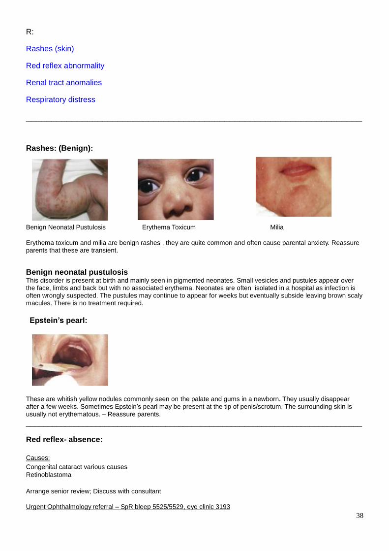
Rashes: (Benign):
Benign Neonatal Pustulosis Erythema Toxicum Milia
Erythema toxicum and milia are benign rashes , they are quite common and often cause parental anxiety. Reassure parents that these are transient.
Benign neonatal pustulosis This disorder is present at birth and mainly seen in pigmented neonates. Small vesicles and pustules appear over the face, limbs and back but with no associated erythema. Neonates are often isolated in a hospital as infection is often wrongly suspected. The pustules may continue to appear for weeks but eventually subside leaving brown scaly macules. There is no treatment required.
Epstein’s pearl:
These are whitish yellow nodules commonly seen on the palate and gums in a newborn. They usually disappear after a few weeks. Sometimes Epstein’s pearl may be present at the tip of penis/scrotum. The surrounding skin is usually not erythematous. – Reassure parents.
_____________________________________________________________________________
Red reflex- absence: Causes:
Congenital cataract various causes
Retinoblastoma
Arrange senior review; Discuss with consultant
Urgent Ophthalmology referral – SpR bleep 5525/5529, eye clinic 3193
39
Investigations:
If congenital cataract,
TORCH screen
Chromosomes
Follow up: Ophthalmologist Mr Watts Neonatal consultant
_____________________________________________________________________________
Renal anomalies:
Most congenital renal problems are picked up antenatally. Review fetal medicine reports. If seen by Paediatric nephrologist antenatally, ensure letter from antenatal clinic is filed in the notes.
Follow plan as advised.
Please refer to the guideline for the management of antenatally detected pelvicalyceal dilatation/ multicystic dysplastic kidneys.
Note: If there is bilateral pelvicalyceal dialatation, especially in male infants; examine carefully and ensure that a
good urinary stream is present.
Commence Trimethoprim prophylaxis as indicated.
Follow up:
Depending on the underlying abnormality detected on the antenatal scan, an urgent USS within the first 48 hours may
be indicated. However, most patients require an USS within 1 to 2 weeks. Follow up should be discussed with the
neonatal consultant. Some complex cases may require follow up in the joint neonatal and renal clinic.
_____________________________________________________________________________
Respiratory distress:
Commonest cause of respiratory distress in the newborn is sepsis. In term infants born by elective LSCS, transient tachypnoea may persist for up to 4 hours. If significant respiratory distress requiring O2 or increased work of breathing at any time, admit to the neonatal unit urgently. Assess for maternal risk factors for sepsis. May be part of NAS.
Investigations and management • Perform partial septic screen
• CXR
• Capillary blood gas
• SpO2 monitoring
• Commence IV antibiotics
• Admission if necessary and commence CPAP
Parents: Communicate to parents clearly the reasons for the above.
__________________________________________________________________

40
S:
Sacral dimple
Subconjunctival haemorrages
Scalp swelling; lesions
__________________________________________________________________
Sacral dimple:
Normal Sacral dimple with a sinus
Sacral dimples in the midline or on either sides of midline are a normal variant.
Examination:
Examine the dimple carefully. If the base is not clearly seen, or a sinus is present, or a tuft of hair is seen, or an
abnormality in the spine is detected by palpation, an USS spine is warranted. These abnormalities may be
associated with underlying spinal cord abnormalities. Examine and document lower limb neurology. Check for good
urinary stream and that the bowels have opened prior to discharge. Discuss with SpR.
Investigation:
Arrange USS spine as outpatient.
Explain to parents that it is important to exclude any underlying spinal cord problems.
Follow up:
Refer to neonatal consultant for follow up.
_____________________________________________________________________________
Subconjuctival haemorrages: Normal after traumatic deliveries. Reassure parents
_____________________________________________________________________________

41
Scalp swellings:
Cephalhaematoma
Cephalhaematoma is bleeding within the scalp that is confined within the sutures lines. Usually present at birth due
to birth trauma.
• Watch for pain and irritability • Jaundice
Parents : Reassure that the swelling may take a few weeks to disappear; and may leave hardened areas due to
calcification of the blood clot within.
No follow up required.
Caput succedaeneum
Scalp swelling that crosses sutures lines. Usually resolves within a few days.
Chignon:
Localised circumscribed swelling from vacuum application at delivery. Resolves within a few days
__________________________________________________________________
42
T:
Talipes
Thrombocytopenia
Thyroid disorders (maternal)
Tongue tie
_____________________________________________________________________________
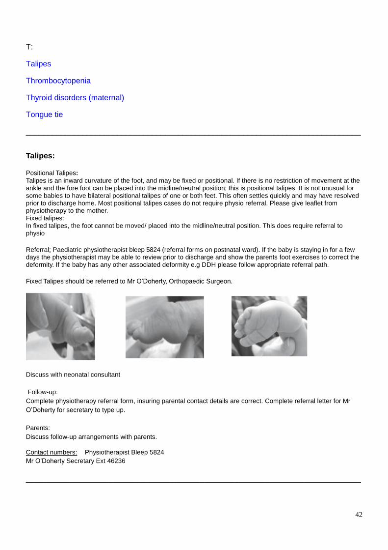
Talipes: Positional Talipes: Talipes is an inward curvature of the foot, and may be fixed or positional. If there is no restriction of movement at the ankle and the fore foot can be placed into the midline/neutral position; this is positional talipes. It is not unusual for some babies to have bilateral positional talipes of one or both feet. This often settles quickly and may have resolved prior to discharge home. Most positional talipes cases do not require physio referral. Please give leaflet from physiotherapy to the mother. Fixed talipes: In fixed talipes, the foot cannot be moved/ placed into the midline/neutral position. This does require referral to physio
Referral: Paediatric physiotherapist bleep 5824 (referral forms on postnatal ward). If the baby is staying in for a few days the physiotherapist may be able to review prior to discharge and show the parents foot exercises to correct the deformity. If the baby has any other associated deformity e.g DDH please follow appropriate referral path.
Fixed Talipes should be referred to Mr O’Doherty, Orthopaedic Surgeon.
Discuss with neonatal consultant
Follow-up:
Complete physiotherapy referral form, insuring parental contact details are correct. Complete referral letter for Mr
O’Doherty for secretary to type up.
Parents:
Discuss follow-up arrangements with parents.
Contact numbers: Physiotherapist Bleep 5824
Mr O’Doherty Secretary Ext 46236
_____________________________________________________________________________

43
Thrombocytopaenia Maternal thrombocytopaenia – causes
Gestational thrombocytopaenia
HELLP syndrome
ITP
Others Neonatal thrombocytopaenia – causes
IUGR
Congenital CMV
Neonatal allo immune thrombocytopaenia
Neonatal auto immune thrombocytopaenia Investigations: Check FBC in the infant – discharge if platelet count > 150 x 10
9/l.
Management: If maternal thrombocytopaenia is present, identify the cause. If the platelet count is less than 20 x 10
9/l the baby may require a platelet transfusion. Neonatal thrombocytopaenia
carries a risk of intracranial haemorrhage. If platelet counts is less that 20 x 109/l IVIG can be considered in cases of
autoimmune thrombocytopaenia. Discuss with neonatal consultant.
_____________________________________________________________________________
Thyroid problems in the mother: It is important to establish the type of thyroid disorder in the mother. Check if TRab and antiTPO antibodies were present in the mother. Check maternal thyroid status in pregnancy as well as maternal medications during pregnancy. Follow the neonatal guidelines.
_____________________________________________________________________________
Tongue tie:
__________________________________________________________________
It is also common in newborns. The short frenulum, however, does not usually interfere with sucking and swallowing, and the frenulum usually lengthens with age. Intervention is only indicated if severe, and interfering with the optimal establishment of feeding. If concerned discuss with breast feeding specialist Reassure parents

44
U:
Umbilical cord; redness, discharge, swelling
Undescended testes – unilateral /bilateral
__________________________________________________________________
Umbilical cord infection: The umbilical cord usually drops off by 7 to 10 days. The base heals with granulation tissue formation. If there is an
offensive discharge from the cord without erythema or induration of surrounding skin, and the baby is otherwise well
treat with oral Flucloxacillin for 7 days.
Umbilical infection Healthy umbilicus
However, if there is surrounding erythema or induration treat with intravenous flucloxacillin and gentamicin as there is potential for the infection to track along the umbilical vein to the liver. Send blood for culture, FBC and CRP. Switch to oral antibiotics to complete 7 days’ course if clinical improvement seen. Frothy discharge from the umbilicus should be considered carefully as it could be a patent vitello-intestinal duct. Although rare, this condition needs surgical review.
_____________________________________________________________________________
Undescended testis:
Bilateral undescended and non-palpable testes needs urgent investigation as for ambiguous genitalia. Look for scrotal pigmentation in a Caucasian infant.
SpR/Consultant review
Investigations:
Urgent Karyotyping and pelvic USS
Unilateral / Bilateral undescended testis but palpable testis in inguinal canal or at the root of scrotum needs no
further investigation. Reassure parents. Review in primary care. Persistently undescended testis after 6 months, should be referred to the Paediatric Surgeons by the GP.
__________________________________________________________________

45
V:
Vaginal bleeding/ discharge
Vaginal skin tags
Vitamin D deficiency (maternal)
Vitamin K
__________________________________________________________________
Vaginal bleeding/ discharge: Vaginal bleeding / white discharge is common in newborns in the first week of life. It is often due to maternal
hormones. Reassure parents. Prolonged persistent bleeding should be investigated.
Vaginal skin tag: Vaginal skin tags are common in newborns. No treatment is usually required and involution occurs as the influence
of maternal hormones subside.
Vitamin D Always look at mother’s antenatal blood results for evidence of maternal vitamin D deficiency. If Mum is vit D
deficient start the baby on Dalivit 0.3ml OD from day 7 until 6 months of age if exclusively breast fed. In formula fed
infants and infants of mothers who receive calcium supplements, Vitamin D supplementation is not necessary.
However, mothers are no longer tested routinely in pregnancy for Vit D levels.
Vitamin K: Vitamin K should be given to all newborn infants. V i t a m i n K h e l p s p r e v e n t Haemorrhagic disease
of the newborn which can be potentially fatal.
RCPCH recommends:
I.M Konakion a single 1mg dose at or soon after birth. If IM vitamin K is declined offer oral vitamin K instead. Oral Konakion MM Paediatric 2mg at birth and between 4 and 7 days. An additional dose at 1month should be given if exclusively breast fed.
Historically there have been concerns regarding IM vitamin K administraton. However, this topic has been extensively researched in several countries and there is no proof that IM vit K causes harm. Moreover, there is a definite well recognized risk of haemorrhagic disease of the newborn if Vitamin K is not administered.
__________________________________________________________________
W:
Weight loss >10%
Weight loss more than 10%:
See transitional care guidelines used on postnatal ward.
__________________________________________________________________

46
References:
1. Roberton’s Text book of Neonatology.
2. Avery’s diseases of the newborn – 9th edition (2011).
3. Common malformations by Lewis B Holmes (2012).
4. Practical Neonatal Endocrinology by Amanda Ogilvy- Stuart & Paula Midgley (2006).
5. Health Protection Agency recommendations.
6. Nursing and Midwifery Council- Midwives Rules 2013