postnatal development of peyer’s patches in albino …€¦ · web viewanatomia, histologia,...
TRANSCRIPT

New York Science Journal 2017;10(8) http://www.sciencepub.net/newyork
Postnatal Development of Peyer’s Patches in Albino Rats (Histological and Morphometric Study)
Mohammed Mokhtar El-Assaly, Hamdino Mohamed Attia, Ahmed Kamal El Banna and Mostafa Rezk Mager
Medical Anatomy & Embryology Department,Faculty of Medicine, Al-Azhar Unviersity, [email protected]
Abstract: Background: Peyer’s patches (PPs) are essential elements of the gut-associated lymphatic tissue that are involved in defense against pathogens that may be colonizing the gut and are also involved in oral food tolerance otherwise food allergy occurs. Objective: This work aims to study the development of Peyer’s patches in postnatal period of albino rats, to shed the light on structural and correlated functional modifications that occur during the postnatal development of Peyer’s patches. Material and Methods: Eighty albino rats were used in the present study. Animals were divided into eight equal groups (ten animals each) according to age (Group I - one day, Group II - one week, Group III - two weeks, Group IV - three weeks, Group V - four weeks, Group VI- eight weeks, Group VII- eighteen weeks and Group VIII- thirty six weeks ). The animals were scarified, terminal parts of their ileum were dissected out and histological sections were prepared for light microscopic examination by different techniques of histological and histochemical staining. Morphometric and statistical analysis were done for results of each group. Results: Observations in the present study showed that by the age of one day, PPs appeared as small epithelial elevations overlying small aggregates of lymphocytes in their connective tissue cores. By the age of one week, Spherical accumulations of lymphoid cells were observed under this dome-shaped epithelium and were consistent with primary lymphoid follicles. These follicles were separated by diffusely dispersed lymphocytes. From the age of two weeks onward the size of these follicle and lymphocytic density increased gradually to reach maximum by the age of Eighteen weeks followed by decrease in lymphocytic cellular density by the age of thirty six weeks. Germinal centers within lymphoid follicles started to appear by the age of three weeks. PPs reached the adult structure by the age of eight weeks and now consisted of well-defined four portions; follicular area (containing lymphoid follicles with distinct germinal centers) interfollicular area, sub epithelial dome and follicle associated epithelium. the mucous membranes at the Peyer’s patches areas in all studied age groups, were found lacking the presence of villi and crypts, instead dome shaped elevations were found. Follicle associated epithelium at the age of one day onwards showed significantly less population of goblet cells (when compared to neighboring villi). Conclusion: Based on the results of the present study, it is believed that many structural changes occurring during the development of Peyer's patches follow a plan of structure-function adaptation. This might explain appearance of germinal centers following the time of weaning due to strong or long exposure of Peyer's patch's lymphoid cells to intestinal antigens. Moreover, the absence of villi and crypts in the covering epithelium of Peyer's patches, in addition to reduced number of goblet cells indicates weak absorptive and secretory functions of Peyer's patches and its adaption for the immunological function when compared to other areas of the intestinal mucosa.[Mohammed Mokhtar El-Assaly, Hamdino Mohamed Attia, Ahmed Kamal El Banna and Mostafa Rezk Mager. Postnatal Development of Peyer’s Patches in Albino Rats (Histological and Morphometric Study). N Y Sci J 2017;10(8):213-223]. ISSN 1554-0200 (print); ISSN 2375-723X (online). http://www.sciencepub.net/newyork. 25. doi:10.7537/marsnys100817. 25 .
Key words: Peyer’s Patches, Postnatal Development, Rats.
1. IntroductionPeyer's patches are macroscopic lymphoid
aggregates that are found in the mucosa and the submucosa along the length of the small intestine, they were named after the Swiss anatomist Hans Conrad Peyer in the 17th century. In humans, there are up to 200 oval lumps of lymphoid tissue along the length of the small intestine. Each is an accumulation of up to 60 lymphoid follicles composed of B lymphocytes with T lymphocytes in the interfollicular areas (Debard et al., 1999).
Morphologically, Peyer's patches are separated into three main domains: the follicular area, the interfollicular area and the follicle-associated
epithelium. The follicle is surrounded by the corona, or subepithelial dome (Jung et al., 2010).
The follicle-associated epithelium (FAE) differs from the epithelium of the villus mucosa: the production of mucus is weak, infiltrated by a large number of lymphocyt with presence of M-cells that act as antigen sampling cells allowing the passage of gut antigens through them (Ermund et al., 2013).
Peyer's patches are involved in defense against pathogens that may be colonizing the gut but they are also involved in oral tolerance. Food antigens are foreign and strictly speaking the immune system should recognize food as an antigenic, potentially threatening challenge to the body's survival. However,
1

New York Science Journal 2017;10(8) http://www.sciencepub.net/newyork
humans are clearly able to absorb food antigens through the gut into the blood stream with no obvious ill effects. Some people may be intolerant to certain food antigens such as lactose in milk or wheat gluten but for the most part foreign food molecules pass through human bodies unnoticed by the immune system (Pabst & Mowat, 2012).
The Peyer's patches decide what are the dangerous gut pathogens, to which the body should respond, and what are the food antigens which the body should accept. The lymphoid tissue is somehow capable of telling the lymphocytes that they should not respond to food antigens. The lymphocytes are tolerized and become non-responsive to these antigens (Pabst & Mowat, 2012).
Information regarding embryogenesis and postnatal development of PPs in rats is less extensive than for mice, but the basic features have been described (Chen et al. 1995 and Parker et al., 2015) Shifts in the cell populations of Peyer's patches are known to take place as the organs mature during the early postnatal period.
A histological and Immunohistochemical study on Peyer's patches formation in rats revealed that on postnatal day 0, the small intestinal mucosa had small cellular aggregates that were consistent with developing Peyer's patches. On Postnatal day 7, the developing Peyer's patches were more pronounced and had distinct follicles, but germinal centers were not present. Peyer's patches became progressively more cellular on Postnatal day 14 and Postnatal day 21. On Postnatal day 21, the majority of Peyer's patches had germinal centers, though the germinal centers were not as developed as those seen at later time points. On Postnatal day 28, the Peyer's patches were fully developed histologically, with prominent follicles and distinct germinal centers. All thesis changes are controversial because they follow a plan of structure– function adaptation (Parker et al., 2015).
2. Materials And MethodsEighty male albino rats were used in this study.
Rats were kept under good hygienic conditions with free access to food and water ad libitum. The study was conducted according to the guidelines of care and use of laboratory animals. Albino rats were divided into 8 groups which were chosen representing different stages of postnatal development (Quinn, 2005). Each group contains 10 albino rats as follow:
Group I: at postnatal day ( neonatal period). Group II: at one week age (early childhood). Group III: at two weeks age (childhood). Group IV: at three weeks age (childhood). Group V: at four weeks age (late childhood). Group VI: at eight weeks age (adolescence).
Group VII: at eighteen weeks age (adolescence).
Group VIII: at thirty six weeks age (adulthood).
The unweaned rats (below the age of 3 weeks) were not separated from their nursing mothers till they were killed. At the specified age of each group, the animals were killed; the terminal part of the ileum was excised and processed for light microscopic examinations. The specimens were fixed in 10% formol saline, and processed for paraffin sectioning (5 mm thick). The sections were stained with:
A) Histological staining Hematoxylin and eosin for normal histology. May-Grunwald Giemsa stain for determining
the lymph follicles and germinal centers contained in Peyer's patch.
B) Histochemical staining Periodic acid-Schiff stain (PAS) for goblet
cells.Quantitative morphometric measurements:Slides were photographed using Olympus®
digital camera installed on Olympus® microscope with 1/2 X photo adaptor, using 40 X objective. The result images were analyzed on Intel® Core I3® based computer using Video Test Morphology® software (Russia) with a specific built-in routine for automated objects analysis. All measurements are calibrated against a micrometer slide which had been photographed with the same system under the same magnification. That enables us to obtain the measurements in um and um2 instead of pixels and square pixels respectively. Lymphocytes were counted, and consequently cellular density was estimated in H & E stained sections. The number of goblet cells was counted per unit length of epithelial surface in PAS stained sections.
A. Total lymphocytes cellular density (expressed in n/10002).
B. Goblet cell density (expressed in n/100).Statistical Analysis:
Measured variables were collected, tabulated, and statistically analyzed in comparison to the preceding age group and/or within the same age group. SPSS (Statistical package for social science) version 22.0 was used for data entry and analysis. Values were presented as Mean ± Standard error mean (SEM). the significance of difference was tested using Student's t-test (Paired) to compare between means of two related groups of numerical (parametric) data and the results were significant when P value was less than 0.05.
3. ResultsMacroscopic observation:
2

New York Science Journal 2017;10(8) http://www.sciencepub.net/newyork
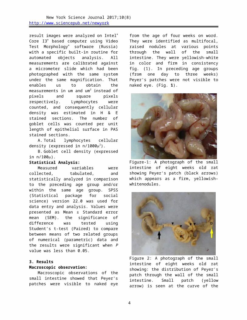
Macroscopic observations of the small intestine showed that Peyer's patches were visible to naked eye from the age of four weeks on word. They were identified as multifocal, raised nodules at various points through the wall of the small intestine. They were yellowish-white in color and firm in consistency fig. (1). In preceding age groups (from one day to three weeks) Peyer's patches were not visible to naked eye. (Fig. 1).
Figure-1: A photograph of the small intestine of eight weeks old rat showing Peyer's patch (black arrows) which appears as a firm, yellowish-whitenodules.
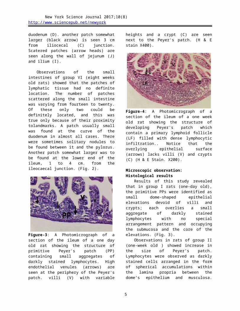
Figure 2: A photograph of the small intestine of eight weeks old rat showing: the distribution of Peyer's patch through the wall of the small intestine. Small patch (yellow arrow) is seen at the curve of the duodenum (D). another patch somewhat larger (black arrow) is seen 3 cm from iliocecal (C) junction. Scatered patches (arrow heads) are seen along the wall of jejunum (J) and ilium (I).
Observations of the small intestines of group VI (eight weeks old rats) showed that the patches of lymphatic tissue had no definite location. The number of patches scattered along the small intestine was varying from fourteen to twenty. Of these only two
could be definitely located, and this was true only because of their proximity tolandmarks. A patch usually small was found at the curve of the duodenum in almost all cases. There were sometimes solitary nodules to be found between it and the pylorus. Another patch somewhat larger was to be found at the lower end of the ileum, 1 to 4 cm. from the ileocaecal junction. (Fig. 2).
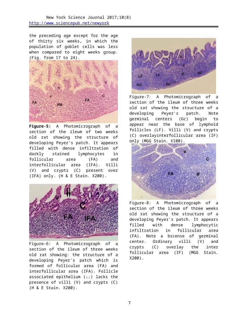
Figure-3: A Photomicrograph of a section of the ileum of a one day old rat showing the structure of primitive Peyer's patch (PP) containing small aggregates of darkly stained lymphocytes. High endothelial venules (arrows) are seen at the periphery of the Peyer's patch. villi (V) with variable heights and a crypt (C) are seen next to the Peyer's patch. (H & E stain X400).
Figure-4: A Photomicrograph of a section of the ileum of a one week old rat showing the structure of developing Peyer's patch which contain a primary lymphoid follicle (LF) filled with dense lymphocytic infiltration.. Notice that the overlying epithelial surface (arrows) lacks villi (V) and crypts (C) (H & E Stain. X200).
Microscopic observation:Histological results
Results of this study revealed that in group I rats (one-day old), the primitive PPs were identified as small dome-shaped epithelial elevations devoid of villi
3
New York Science Journal 2017;10(8) http://www.sciencepub.net/newyork
and crypts; each overlies a small aggregate of darkly stained lymphocytes with no special arrangement pattern and occupying the submucosa and the core of the elevations. (Fig. 3).
Observations in rats of group II (one-week old ) showed increase in the size of Peyer’s patch. Lymphocytes were observed as darkly stained cells arranged in the form of spherical accumulations within the lamina propria between the dome’s epithelium and musculosa. These accumulations were consistent with early primary lymphoid follicles. (Fig. 4).
Peyer's patches in rats of group III (two-weeks old) rats showed increase in the size and in the number of lymphoid follicles when compared to the previous age group (one week old). Moreover, Peyer's patches at this age became more pronounced and had distinct follicles separated from each other by distinct interfollicular areas. (Fig. 5).
In group IV (three-weeks old rats), All examined sections showed the appearance of lymphoid follicles separated by interfollicular areas. Some sections showed Germinal centers (Gs) within lymphoid follicles and other sections didn't show (Gs) within the lymphoid follicles denoting at this age they started to change from primary to secondary follicles. (Fig. 6 & 7 & 8).
In Group V (Four- weeks old rats), an increase in the patch size was noticed, with increase in the density of lymphocytic infiltration especially in follicular areas. Most lymphoid follicles in this age group showed the appearance of germinal centers. (Fig. 9 & 10).
Peyer's patches in rats of group VI (eight-weeks old) showed apparent increase in the size of lymphoid follicles, with increase in the density of lymphocytic infiltration in both follicular and inter-follicular areas of the Peyer's patches.. Nearly all lymphoid follicles at this age group showed well developed germinal centers. (Fig. 11 & 12).
In group VII (Eighteen- weeks old rats), Peyer's patches showed that apart from the apparent increase in size and cellular population of Peyer's patches, no remarkable differences were noted between the results of group V (eight weeks rats) and that of group VI (eighteen weeks rats). (Fig. 13 & 14).
Peyer's patches in rats of group VIII (thirty six- weeks old) showed no marked chang in the size of Peyer's patches as compared to the previous study group (group VII), but lymphocytic density was apparently decreased in both follicular and inter-follicular areas of the Peyer's patches. (Fig. 15 & 16).
The results of the current study showed that the mucous membrane at the Peyer’s patches areas at all studied age groups, was found lacking the presence of villi and crypts, instead dome shaped elevations were found., villi and crypts were found next to PPs and
above para follicular area. High endothelial venules were detected in all studied age groups of the present study starting from the youngest age of one day old.Histochemical results:
The results of the present study in sections stained with Periodic acid-Schiff stain (PAS) revealed that the epithelium overlying the developing Peyer's patches in all studied age groups showed less population of goblet cells (when compared to neighboring villi). While the population of goblet cells in the epithelium overlying the Peyer's patches and neighboring villi was increased progressively in all age groups when compared with the preceding age except for the age of thirty six weeks, in which the population of goblet cells was less when compared to eight weeks group. (Fig. from 17 to 24).
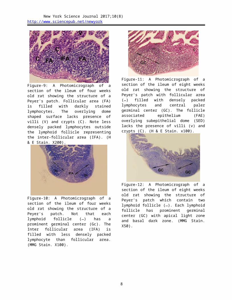
Figure-5: A Photomicrograph of a section of the ileum of two weeks old rat showing the structure of developing Peyer's patch. It appears filled with dense infiltration of darkly stained lymphocytes in follicular area (FA) and interfollicular area (IFA). Villi (V) and crypts (C) present over (IFA) only. (H & E Stain. X200).
Figure-6: A Photomicrograph of a section of the ileum of three weeks old rat showing: the structure of a developing Peyer's patch which is formed of follicular area (FA) and interfollicular area (IFA). Follicle associated epithelium (↓↓) lacks the presence of villi (V) and crypts (C) (H & E Stain. X200).
4
New York Science Journal 2017;10(8) http://www.sciencepub.net/newyork
Figure-7: A Photomicrograph of a section of the ileum of three weeks old rat showing the structure of a developing Peyer's patch. Note germinal centers (Gc) begin to appear near the base of lymphoid follicles (LF). Villi (V) and crypts (C) overlayinterfollicular area (IF) only (MGG Stain. X100).
Figure-8: A Photomicrograph of a section of the ileum of three weeks old rat showing the structure of a developing Peyer's patch. It appears filled with dense lymphocytic infiltration in follicular area (FA). Note a bscense of germinal center. Ordinary villi (V) and crypts (C) overlay the inter follicular area (IF) (MGG Stain. X200).
Figure-9: A Photomicrograph of a section of the ileum of four weeks old rat showing the structure of a Peyer's patch. Follicular area (FA) is filled with darkly stained lymphocytes. The overlying dome shaped surface lacks presence of villi (V) and crypts (C). Note less densely packed lymphocytes outside the lymphoid
follicle representing the inter-follicular area (IFA). (H & E Stain. X200).
Figure-10: A Photomicrograph of a section of the ileum of four weeks old rat showing the structure of a Peyer's patch. Not that each lymphoid follicle (↔) has a prominent germinal center (Gc). The Inter follicular area (IFA) is filled with less densely packed lymphocyte than follicular area. (MMG Stain. X100).
Figure-11: A Photomicrograph of a section of the ileum of eight weeks old rat showing the structure of Peyer's patch with follicular area (↔) filled with densely packed lymphocytes and central paler germinal center (GC). The follicle associated epithelium (FAE) overlying subepithelial dome (SED) lacks the presence of villi (v) and crypts (C). (H & E Stain. x100).
Figure-12: A Photomicrograph of a section of the ileum of eight weeks old rat showing the structure of Peyer's patch which contain two lymphoid follicle (↔). Each lymphoid follicle has prominent germinal center (GC) with apical light zone and basal dark zone. (MMG Stain. X50).
5

New York Science Journal 2017;10(8) http://www.sciencepub.net/newyork
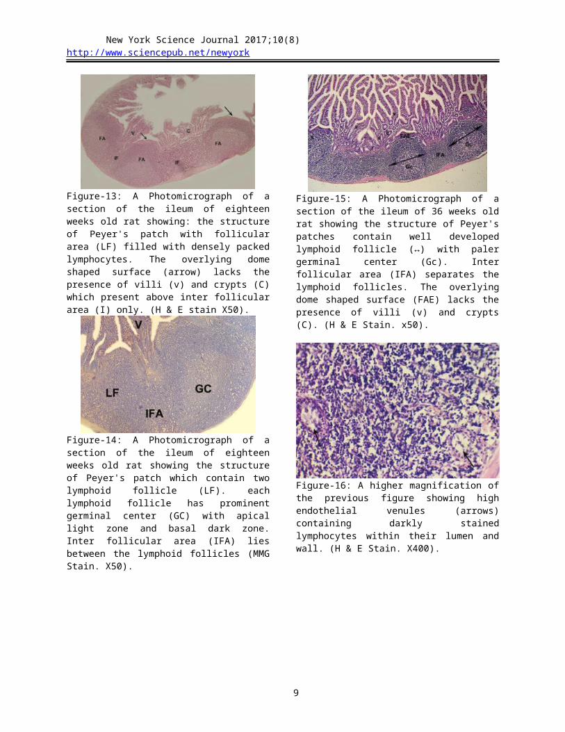
Figure-13: A Photomicrograph of a section of the ileum of eighteen weeks old rat showing: the structure of Peyer's patch with follicular area (LF) filled with densely packed lymphocytes. The overlying dome shaped surface (arrow) lacks the presence of villi (v) and crypts (C) which present above inter follicular area (I) only. (H & E stain X50).
Figure-14: A Photomicrograph of a section of the ileum of eighteen weeks old rat showing the structure of Peyer's patch which contain two lymphoid follicle (LF). each lymphoid follicle has prominent germinal center (GC) with apical light zone and basal dark zone. Inter follicular area (IFA) lies between the lymphoid follicles (MMG Stain. X50).
Figure-15: A Photomicrograph of a section of the ileum of 36 weeks old rat showing the structure of Peyer's patches contain well developed lymphoid follicle (↔) with paler germinal center (Gc). Inter follicular area (IFA) separates the lymphoid follicles. The overlying dome shaped surface (FAE) lacks the presence of villi (v) and crypts (C). (H & E Stain. x50).
Figure-16: A higher magnification of the previous figure showing high endothelial venules (arrows) containing darkly stained lymphocytes within their lumen and wall. (H & E Stain. X400).
Figure-17: A Photomicrograph of a section of the ileum of one day old rat showing the presence of PAS +ve goblet cells (↓) in both epithelial surface overlying the Peyer's patches (PP) and also the epithelial covering of villi (V). (PAS. X400).
Figure-18: A Photomicrograph of a section of the ileum of one week old rat showing the presence of PAS +ve goblet cells (↓) in both the epithelial surface overlying follicular area (FA) of Peyer's patches and also in epithelial covering of the nearby villi (V). in the interfollicullar area n (IFA) (PAS. X400).
6
New York Science Journal 2017;10(8) http://www.sciencepub.net/newyork
Figure-19: A Photomicrograph of a section of the ileum of two weeks old rat showing the presence of few PAS +ve goblet cells (↓) in the epithelium overlying the Peyer's patch, and in the epithelium covering villi. (PAS. X400).
Figure-20: A Photomicrograph of a section of the ileum of three weeks old rat showing the presence of PAS +ve goblet cells (↓) in the epithelium (FAE) overlying follicular area (FA) of Peyer's patch, and in the epithelium covering villi (V) and crypts (C). (PAS. X400).
Figure-21: A Photomicrograph of a section of the ileum of four weeks old rat showing the follicle associated epithelium (FAE) of the mature Peyer's patch with very few goblet cells (↓), while numerous in the epithelial covering of the surrounding villi and epithelial lining of crypts. (PAS. X400).
Figure-22: A Photomicrograph of a section of the ileum of eight weeks old rat showing the follicle associated epithelium (FAE) of the mature Peyer's patch with very few goblet cells, while numerous in the epithelial covering of the surrounding villi (v) and epithelial lining of crypts (C). (PAS. X200)
Figure-23: A Photomicrograph of a section of the ileum of eight weeks old rat showing the follicle associated epithelium (FAE) of the mature Peyer's patch with very few goblet cells (↑), While numerous in the epithelial covering of the surrounding villi (v) and epithelial lining of crypts (C). (PAS. X100).
Figure-24: A Photomicrograph of a section of the ileum of 36 weeks old rat showing the follicle associated epithelium (FAE) of the Peyer's patch with very few goblet cells (↑), while numerous in the epithelial covering of the surrounding villi and epithelial lining of crypts. (PAS. X100).
7
New York Science Journal 2017;10(8) http://www.sciencepub.net/newyork
Morphometric Results1. Cellular density: Cells were counted per unit
area of Peyer's patches in different age groups and cellular density was expressed in n/10002.
Statistical analysis revealed that the mean count of cells per unit area of Peyer's patches was significantly increased (P < 0.05) in all age groups when compared with the preceding age groups except for the age of thirty six weeks, in which cellular density was significantly decreased (P > 0.05) when compared with the preceding age group (eighteen weeks old rats) Histogram (1).
2. Count of goblet cells: Goblet cells were counted per unit length of epithelial surface in the epithelium overlying the Peyer's patches and also in villi in different age groups and cellular density of each type were expressed in n/100.
Statistical analysis revealed that the total count of goblet cells in the epithelium overlying the Peyer's patches and villi was increased progressively in all age
groups when compared with the preceding age except for the age of thirty six weeks, in which the number of goblet cells was high significantly decreased (P < 0.01) when compared to eight weeks group Histogram (2).
Within the same age group there was a high statistically significant decrease (P < 0.01) in the count of the goblet cells at the epithelium covering of Peyer's patches when compared with the epithelium covering the surrounding villi Histogram (3).
Histogram-1:- Mean count of cells per unit area of Peyer's patches in different age groups (n/1000 2).
8
New York Science Journal 2017;10(8) http://www.sciencepub.net/newyork
Histogram-2:- Mean count of goblet cells per unit length of epithelium over Peyer's patches (PP) and villi in different age groups (n/100 ).
Histogram-3:- Mean count of goblet cells per unit length ofepithelium over Peyer's patches (PP) vs villi in different age groups (n/100 ).
4. DiscussionIn the current study, Peyer’s Patches had no
definite location as regard to their distribution along the wall of small intestine. this was in agreement with Schuurman et al., (1994), who found that Peyer’s patches were randomly distributed throughout the mucosa and submucosa of the gastrointestinal tract but were of greatest density in the jejunum and the ilium, and were oriented along the anti-mesenteric border.
In the present study, in eight weeks old rats, the number of visible Peyer’s Patches scattered along the small intestine was varying from fourteen to twenty. this was in agreement with Awang-Hazmiet al., (2006), who reported that the average number of Peyer’s patches in Spraque-Dawley rats weighing 200-250g was 16.33.
The current study revealed that PPs were composed by aggregated lymphoid follicles surrounded by a particular epithelium, the follicle-
associated epithelium (FAE) that was devoid from villi and crypts. These lymphoid follicles were separated from each other by interfollicular area and from FAE by Sub epithelial dome.
the present study was in agreement with Rebold and Cyster (2016), who stated that PPs were organized into three major regions; a series of large B-cell follicles, the overlying follicle-associated epithelium (FAE) and associated subepithelial dome (SED) that was between the follicles and the FAE, and the small T-cell zones that were situated adjacent between the B-cell follicles.
In the current work, we observed specialized venules referred tohigh endothelial venules (HEV) or postcapillary venules. They were noticed in the lamina propria at the periphery of Peyer's patches and within interfollicular areas. This was in agreement withthe results of Abe and Ito, (1978). They observed in Peyer's patches of mice that the parafollicular area
9

New York Science Journal 2017;10(8) http://www.sciencepub.net/newyork
were surrounding the follicular area and were containing postcapillary venules with high endothelium.
In the present study high endothelial venules were observed containing darkly stained lymphocytes within their lumen. This result was in agreement with Beyaz and Asti, (2004), who found specialized venules referred to HEVs were found to be located with lymphocytes in its wall or lumen within inter-follicular areas of Peyer's patches in Bovine foetus.
High endothelial venules were detected in all studied age groups of the present study starting from the youngest age of one day old. This was in agreement with Ogino et al., (2004), who stated that high endothelial venules are important structures necessary for the development of Peyer's patches by facilitating the passage of T lymphocytes across their walls to reach their habitat in the lamina propria.
The current study revealed that in the newly born rats (one day old), the primitive Peyer's patches could be easily identified by light microscope as small dome shaped epithelial elevations overlying small aggregates of lymphocytes with no special arrangement pattern. These findings were in accordance with the results of previous studies done by Sminia et al., (1983) and Parker et al., (2015), who demonstrated the presence of small clusters of lymphocytes in developing Peyer's patches in newly born rats.
In agreement with our study, Peyer’s patches were even detected earlier in the embryonic life in different mammalian species as stated by Recher et al., (2001) and Campbell & Butcher, (2002) in ratsand Yasuda et al., (2005) in claves. They studied the pre-natal development of Peyer's patches using different techniques including immuno-histochemical staining by mono-clonal antibodies specific for, MHC class-II, CD3, CD4, and CD8 for detection of lymphoid cells (T lymphocytes) and non-lymphoid cells (dendritic reticular cells) in rat and bovine fetuses respectively.
In the present study, in one week old rats, the area of Peyer’s patch within the lamina propria which contained darkly stained lymphocytes showed marked increase in the size; In addition to the density of lymphocytes infiltrating this area was apparently increased. These findings go hand in hand with those of Sminia et al., (1983) and Chen et al., (1995), who found progressively increasing lymphocytic
In the present study, Peyer's patches of two weeks old rats showed further marked increase in patch size and number of lymphoid follicles, this was in agreement with Chen et al., (1995), who reported that at Day 5, each Peyer's patch contained 6-8 lymphoid follicles, with the lymphoid follicles
subsequently increasing in size and number with the age.
In the current study, Peyer's patches of two weeks old rats showed the earliest appearance of primary lymphoid follicles separated by diffuse lymphoid cells. These results were matched with those described in previous researches on the rat Peyer's patches done by Ermak and Owen, (1987) and Chen et al., (1995). They stated that B and T lymphocytes were localized in defined regions; the follicular area and inter-follicular area, respectively; at ages ranging from 9 to 14 days after birth.
In agreement with our study for Peyer’s patch of two weeks old rats, Parker et al., (2015) found that on postnatal day 7, the developing PPs were more pronounced and had distinct follicles, but germinal centers were not present.
In three weeks old rats, we found increase in the size of Peyer’s patches and number of lymphoid follicles per patch, with significant increase in the density of lymphocytic infiltration especially in the follicular areas. Moreover, some of the lymphoid nodules started to show germinal centers denoting at this age they begin to change from primary to secondary follicles. These findings were in accordance with those reported by Sminia et al., (1983), who observed the earliest appearance of secondary lymphoid follicles in Peyer’s patches in 18 days old rats.
These results in our study agree with Eikelenboom et al., (1979) who noted thatfrom three to four weeks large pyroninophilic cells with mitotic figures were present in the base of the follicles adjacent to the tunica muscularis.
Pabst et al., (2012) reported similar results and explained the appearance of germinal centers by the strong or sustained exposure of B lymphocytes in the follicular areas of Peyer's patchs to intestinal antigens following time of weaning.
In four weeks old rats, our study revealed that most of Peyer's patchs showed well developed lymphoid follicles containing germinal centers. These findings in our study were nearly similar to the results of Parker et al., (2015) who stated that on postnatal day 21, the majority of PPs had germinal centers, though the germinal centers were not as developed as those seen at later time points. On postnatal day 28, the PPs were fully developed histologically, with prominent follicles and distinct germinal centers.
On the other hand, the results of the present study disagree with those reported by Nakamura et al., (1988), who found recognizable secondary lymphoid follicles (with clear germinal centers) in the lamina propria by the age of seven days. They added that the Peyer's patches were structurally complete by the age of two weeks.
10

New York Science Journal 2017;10(8) http://www.sciencepub.net/newyork
This minor difference between observations in our study and observations reported by Nakamura et al., (1988), may be due todifferences in rat strains, different observational criteria or may reflect differences in the level of antigenic stimulation from the intestinal contents.
In the current work, Apart from increased size of Peyer's patch, the results of group VII (adult rats eighteen weeks old) were entirely identical to those found in the eight weeks old rats. These findings were in agreement with previous studies reported that completion of development of Peyer's patches and their acquisition of their adult structure was reached by the age of 28 days Sminia et al., (1983) and Parker et al., (2015), or 21 days (Nakamura et al., 1988).
In the present study, Peyer's patchs of eighteen weeks old rats showed marked increase in the size when compared to all previous age groups. This result was in agreement with Chen et al., (1995) who reported that Between 3 and 18 weeks after birth, lymphoid follicles increased constantly in size, so that by 18 weeks they had become threefold greater in breadth, i. e. roughly 27 times greater in volume, than at 3 weeks.
In the current work, the results of the last group (thirty six weeks old rats) showed significant decrease in the density of lymphocytic infiltration as compared to previous group (eighteen weeks old rats). These findings were in accordance with results reported by Kelsall, (2004). who found a substantial aging-associated decline in the number of lymphoid cells in the mesenteric lymph nodes (MLN) and spleen, and especially in the Peyer's patches (PP), but not in the lamina propria (LP).
The results of the current study showed that the mucous membrane at the Peyer’s patches areas in all studied age groups, was found lacking the presence of villi and crypts, instead dome shaped elevations were found. This finding was in accordance with almost all the previous studies performed on the development and structure of Peyer's patches in nearly all mammalian species including human Honda et al, (2001) and Parker et al., (2015).
The results of the present study revealed that the epithelium overlying the developing Peyer's patches at the age of one day onwards showed significantly less population of goblet cells (when compared to neighboring villi). These results go hand in hand with the results of Onori et al., (2001). They stated that the number of goblet cells was markedly decreased at the age of three weeks till adulthood in the epithelial covering of the Peyer's patches of the rat.
Reference
1. Abe K and Ito T (1978). Qualitative and quantitative morphologic study of Peyer's patches of the mouse after neonatal thymectomy and hydrocortisone injection. Developmental Dynamics, 151(2): 227-237.
2. Awang-Hazmi A J and Saw P (2006). The Response of Gut Associated Lymphoid Tissues (GALT) Following Intranasal Administration of P. Multocida B2 in Rats. Journal of Animal and Veterinary Advances, 5(11): 1029-1034.
3. Beyaz F and Asti R N (2004). Development of ileal Peyer's patches and follicle associated epithelium in bovine foetuses. Anatomia, histologia, embryologia, 33(3): 172-179.
4. Campbell D J and Butcher E C (2002). Rapid acquisition of tissue-specific homing phenotypes by CD4+ T cells activated in cutaneous or mucosal lymphoid tissues. Journal of Experimental Medicine, 195(1): 135-141.
5. Chen D, Hoshi H, Tanaka K and Murakami G (1995). Postnatal development of lymphoid follicles in rat Peyer's patches, with special reference to increased follicle number. Arch Histol Cytol, 58(3): 335-43.
6. Debard N, Sierro F, and Kraehenbuhl J P (1999). Development of Peyer's patches, follicle-associated epithelium and M cell: lessons from immunodeficient and knockout mice. Semin Immunol, 11(3): 183-91.
7. Eikelenboom P, Levenbach M G, Van De Brink H R and Streefkerk J G. (1979). Development of T and B cell areas in peripheral lymphoid organs of the rat. The Anatomical Record, 194(4): 523-537.
8. Ermak T H and Owen R L (1987). Phenotype and distribution of T lymphocytes in Peyer's patches of athymic mice. Histochemistry, 87(4): 321-325.
9. Ermund A, Gustafsson J, Hansson G and Keita V. (2013). Mucus properties and goblet cell quantification in mouse, rat and human ileal Peyer's patches. PloS one, 8(12): 83-88.
10. Jung C, Hugot J Pand Barreau F (2010). Peyer's patches: the immune sensors of the intestine. International journal of inflammation, 183 (2): 56-71.
11. Kelsall B L (2004). Peyer’s Patch Dendritic Cells Process Viral Antigen from Apoptotic Epithelial Cells in the Intestine of Reovirus-infected Mice. The Journal of Experimental Medicine, 200(2): 235-245.
12. Honda K, Nakano H, Yoshida H, Nishikawa S, Rennert P, Ikuta K and Nishikawa S I (2001). Molecular basis for hematopoietic mesenchymal interaction during initiation of Peyer's patch organogenesis. Journal of Experimental Medicine, 193(5): 621-630.
13. Nakamura S, Sumi Y and Nagura H (1988). Ontogenic development of gut-associated lymphoid tissue in the rat. An immuno-histochemical study. Acta Pathol Jpn, 38(10): 167-183.
11

New York Science Journal 2017;10(8) http://www.sciencepub.net/newyork
14. Ogino T, Miura S, Komoto S, Hara Y, Hokari R, Tsuzuki Y et al., (2004). Senescence-associated decline of lymphocyte migration in gut-associated lymphoid tissues of rat small intestine. Mechanisms of ageing and development, 125(3): 191-199.
15. Onori P, Franchitto A, Sferra R, Vetuschi A and Gaudio E (2001). Peyer's Patches Epithelium in the Rat. Digestive diseases and sciences, 46(5): 1095-1104..
16. Pabst O and Mowat A M (2012). Oral tolerance to food protein. Mucosal immunology, 5(3): 232-239.
17. Parker G A, Picut C A, Swanson C and Toot J D (2015). Histologic features of postnatal development of immune system organs in the Sprague-Dawley rat. Toxicologic pathology, 43: 794-815.
18. Quinn R (2005). Comparing rat’s to human’s age: how old is my rat in people years?. Nutrition, 21(6): 775-777.
19. Recher S, Raccurt M, Lambert A, Lobie P E, Mertani H C and Morel z (2001). Prenatal and adult growth hormone gene expression in rat lymphoid organs. Journal of Histochemistry & Cytochemistry, 49(3): 347-354.
20. Schuurman H J, Kuper C F and Vos J G (1994). Histopathology of the immune system as a tool to assess immunotoxicity. Toxicology, 86(3): 187-212.
21. Sminia T, Janse E M and Plesch B C (1983). Ontogeny of Peyer's patches of the rat. The Anatomical Record, 207(2): 309-316.
22. Yasuda M, Ogawa D, Nasu T, Yamaguchi T and Murakami T (2005). Kinetics and distribution of bovine γδ T-lymphocyte in the intestine: γδ T cells accumulate in the dome region of Peyer's patch during prenatal development. Developmental & Comparative Immunology, 29(6): 555-564.
8/25/2017
12