portrait sculpting - csu chico department of · pdf fileanatomy & aging the skull frontal...
TRANSCRIPT

On the front cover:On the back cover:
On the title page:
The Fisherman's Daughter, Philippe Faraut, 2002.Creating the Skin of the Neck, Philippe Faraut. 2003.Creating the Wrinkles of the Eyes, Philippe Faraut. 2003.Mask of a Smile, Philippe Faraut. 2003.Mask of Disgust, Philippe Faraut. 2003.Mask of Fear, Philippe Faraut. 2003.Mutiny, in progress, Philippe Faraut. 2004.
PORTRAIT SCULPTING
Copyright © 2004 by Philippe and Charisse FarautFirst published in 2004 by PCF Studios, Inc.
All rights reserved. No part of this publication may be reproduced or used in any form or by any means—graphic, electronic,or mechanical, including photocopying, recording, taping, or information storage and retrieval systems—without writtenpermission from the publisher.
Library of Congress Control Number: 2004093850
Publisher's Cataloging-in-Publication(Provided by Quality Books, Inc.)
Faraut, Philippe.Portrait sculpting : anatomy & expressions in clay /
Philippe & Charisse Faraut. — 1st ed.p. cm.Includes bibliographical references and index.LCCN 2004093850ISBN-10: 0-9755065-0-1ISBN-13: 978-0-9755065-0-9
1. Head in art. 2. Modeling. 3. Sculpture-Technique. I. Faraut, Charisse. II. Title.NB1932.F37 2004 731'.74
QB104-200263
First Edition, 2004Printed and bound in the United States of America on acid-free paper
Second Printing, 2006
Visit us at www.pcfstudios.com for information on seminars and supplies for artists.
Attention colleges and universities: Quantity discounts are available on bulk purchases of this book for educational purposes.For information, please contact:
PCFSTUDIOS, INC.PO Box 722 • Honeoye, NY 14471585-229-2976 • 585-229-2865 [email protected] • www.pcfstudios.com
Anatomy & Expressions In ClayP H I L I P P E & C H A R I S S E F A R A U T
PCFSTUDIOS, INC.

ANATOMY & AGING
The Skull
frontal bone
line
f
orbital emit)-
mandible
BONES OF THE SKULL
An understanding of the bone structure and musclemasses of the human head is the foundation of portraitsculpture. There are tremendous differences betweenskulls, depending on gender, age and race. In generalthe male skull is larger than the female skull; the jawis squarer; the chin, more pronounced; the brow ridge,more prominent; and the forehead, more sloping. Maleteeth are often a bit larger. Some of these differencescan easily be seen in the diagram below which comparesa male and a female skull. The photos on the facingpage illustrate the surprising diversity found in skullsfrom various regions.
Recognizing these differences and examining three-dimensional models with this in mind facilitatescomparative study and the refinement of observationalskills required to successfully capture the shape andfeatures of the sculpted model.
The actual sculpting of different skulls reinforces thisknowledge and develops the ability to build volumesbased on observation only and is highly recommended.This chapter focuses on this type of exercise.
The less familiar we are with a shape, the easier it is to
12 BY FARAUT
duplicate it in three-dimension. This is because we areforced to focus on form and volumes themselves ratherthan on preconceived ideas of what the shape shouldbe. For the majority of us, the intricate shapes of askull, when observed closely, are surprisingly unfamiliar,making this exercise a very productive experience. Totake full advantage of this exercise, a three-dimensionaland anatomically correct model is needed in order toaccurately see every nuance of the skull - something aphoto or a drawing cannot provide. Casts of humanskulls are available to purchase. Suppliers are listed inAppendix B at the end of this book.
MALE AND FEMALE SKULL COMPARISON
Peruvian Male Skull Australian Male Skull
^
African Male Skull European Female Skull
13

ANATOMY & AGING
Demonstration 1: Modeling a Skull
1. A ball of newspaper is wrapped and taped around thedowel to form the core of the sculpture. The newspaperwill absorb some of the moisture from the clay, makingthe center more firm and stable. Tape is kept awayfrom the dowel so the sculpture will be able to rotate onthe armature.
2. An even layer of clay is built around the paper.
3. The ball is rounded with a metal scraper.
14 PORTRAIT SCULPTING BY FARAUT
4. A rectangular piece of clay is positioned to create the 5. The clay is then pulled back on each side to form thevolume of the forehead. temporal planes.
6. A coil of clay is applied on the top of the head andshaped to define the profile of the cranium.
7. Volume is then built on each side.
15

ANATOMY & AGING
8. To build the volume of the mandible, a horseshoe-shaped piece of clay is wrapped around the base andflattened on each side to form the planes of the ramus.
9. The depression between the mandible and thefoundation is filled and smoothed, preparing the volumefor the maxilla.
10. The orbital cavities' location is determined byobservation of the model and measurement withcalipers.
16 PORTRAIT SCULPTING BY FARAUT
11. The zygomatic arch is a bone originating from theedges of the orbital cavity, stretching to the externalauditory meatus.
12. To define the zygomatic bone (cheekbone) and the upper part of the maxilla, a depression is created under the
orbital cavity.
17

ANATOMY & AGING
13. The nasal cavity is carved out with a wooden tool,leaving a ridge at the center called the vomer.
14. The width of the ramus needs to be measured andindicated before pushing in the clay on both sides todefine the volume in the back of the maxilla.
15. The zygomatic arch is suspended in its center,leaving space for the temporalis which covers thetemporal bone and connects with the coronoid processof the mandible.
18 PORTRAIT SCULPTING BY FARAUT
16. The mastoid process is a projection of bone behindthe ear. It begins to form only after the age of two.The external auditory meatus is a hole in the bone justbehind the temporal mandibular joint.
17. The teeth are added one at a time paying specialattention to their symmetry.
18. The planes of the temporal bone are refined. Thesealso define the temporal lines.
19. After refining all the volumes with loop tools, abristle brush is used to blend them together.
19

ANATOMY & AGING
20. The teeth are first refined with a stiff brush. A soft brush is used for thefinal texture.
21. A coarse sponge is used torefine the final shape.
22. A soft sponge is used to create the surface texture. The cranial suturesare engraved with a thin metal tool.
20 PORTRAIT SCULPTING BY FARAUT 21

ANATOMY & AGING
The Muscles
depressor labii inferioris
depressor anguli oris
memalis
MUSCLES OF THE FACE
The function of the muscles throughout the body isalways to contract, or pull, usually one bone towardanother, creating for example, a movement of rotationaround a joint. With the exception of the masseter andthe temporalis, most of the muscles of the face havethe peculiarity of not connecting one bone to another.Instead, they attach at one end, directly or indirectly, tothe skull, and at the other, into the skin or into anothermuscle connected to the skin. Facial muscle functionsare discussed in more detail in Chapter 7.
In the face, the pulling action from a fixed origin toan area very close to the skin creates depressions andfolds that are the signatures of different expressions.Furthermore, the repeated action of some muscles ispartly responsible for slow, permanent changes to both
the surface of the skin and the shape of underlyingbone. For example, a person with the habit of chewingon only one side of the mouth will have a strengtheningof the masseter on that side, giving it more volume. Itwill also, over time, create some degree of distortion inthe mandible, pulling it toward the same side. Carefulobservation will also reveal thickening of the temporalis.
The next demonstration is designed to illustrate theorigin and placement of the major facial muscles.Facial muscles, having very little mass, can be appliedwithout the help of a three-dimensional model if noneis available, because they closely follow the shape of theskull. A good anatomy book can be sufficient.
Demonstration 2: Modeling Facial Muscles
1. The fan shaped temporalis muscle attaches tothe temporal bone of the cranium, passes under thezygomatic arch, and connects to the coronoid process ofthe mandible. Thin coils of clay following the directionof the fibers are applied and then flattened.
2. The fibrous texture of the muscle is rendered with abristle brush.
3. Other musclesare applied oneat a time in thefollowing order:levator anguli oris,levator labii alaequenasi, levator labiiand the orbicularisoris.
22 PORTRAIT SCULPTING BY FARAUT 23

ANATOMY & AGING
4. Next, the mentalis, depressor anguli oris, depressor labii, buccinator, masseter, zygomaticus minor, zygomaticusmajor and risorius are added.
24 PORTRAIT SCULPTING BY FARAUT
5. The frontalis, orbicularis oculi and eyeball are placed.
6. Small coils of clay create the volumes of the eyelids.
7. The eyeball is an average oftwenty-five millimeters in diameterand is centered in the orbital cavity.The outer point of the cornea istangent to a line drawn from thecenter of the superior and inferioredges of the orbit.
25
ANATOMY & AGING
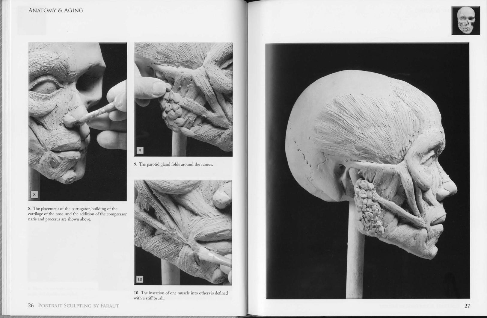
9. The parotid gland folds around the ramus.
8. The placement of the corrugator, building of thecartilage of the nose, and the addition of the compressornaris and procerus are shown above.
10. The insertion of one muscle into others is dennedwith a stiff brush.
26 IAIT SCULPTING BY FARAUT 27

ANATOMY & AGING
The Neck
suprastemal notch— cla\icular head of the sternocleidomastoid
MUSCLES OF THE NECK
The most visible bones of the neck are the clavicle andthe sternum in the front. The scapula, the seventhcervical vertebra, and the first thoracic vertebra are themost visible bones of the upper back.
The most prominent muscles of the neck arethe sternocleidomastoid and the trapezius. Thesternocleidomastoid originates from the sternum andfrom the medial third of the clavicle and inserts intothe mastoid process and the superior nuchal line of theoccipital bone. The tendons of the sternal attachment
of the sternocleidomastoid form the two lateral ridgesof the suprasternal notch.
The trapezius is a large muscle that covers the shoulder,the upper part of the back and the back of the neck. Itoriginates from the superior nuchal line at the base ofthe skull, from the ligamentum nuchea and from thespines of the twelve thoracic vertebrae. It inserts intothe lateral third of the clavicle and the upper border ofthe spine of the scapula.
28 TRAIT SCULPTING BY FARAUT
ligamentum nuchae
spine of the scapula
firstthoracicvertebra
MUSCLES OF THE UPPER BACK29