bones and cavities of the facial cranium. tmj anterior skull frontal bone supraorbit al foramen...
TRANSCRIPT
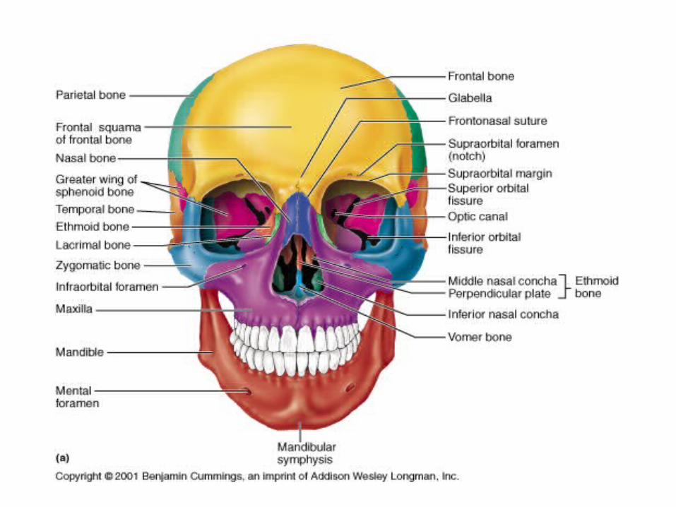
Bones and cavities of the facial cranium
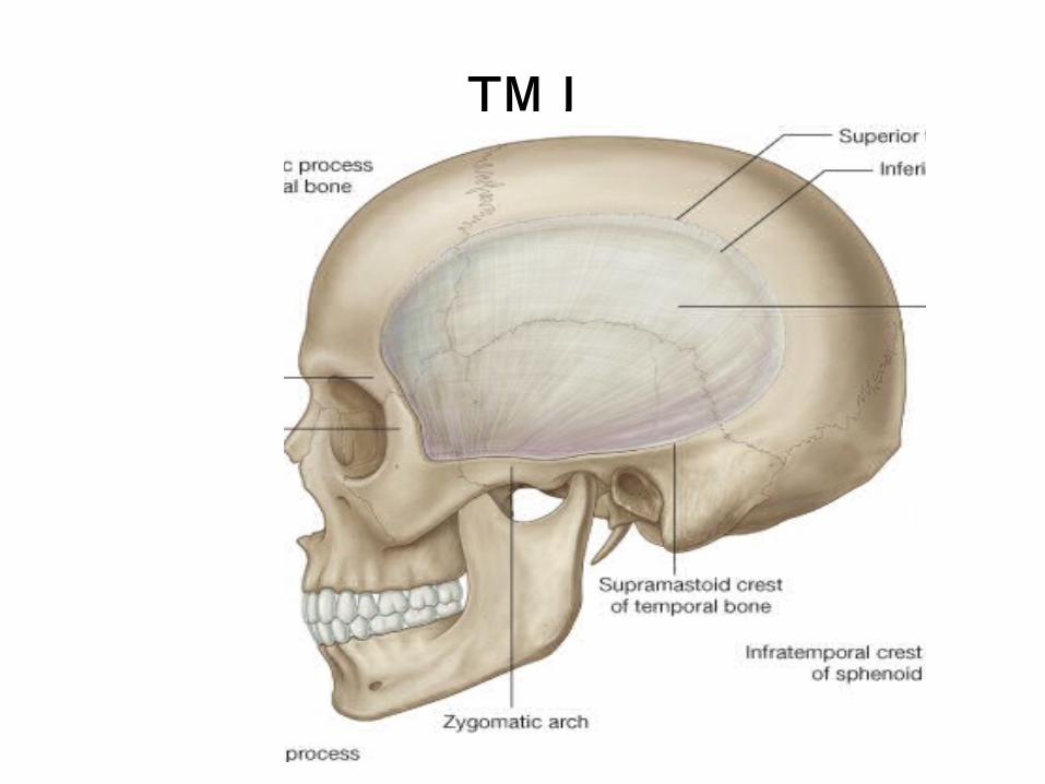
TMJ
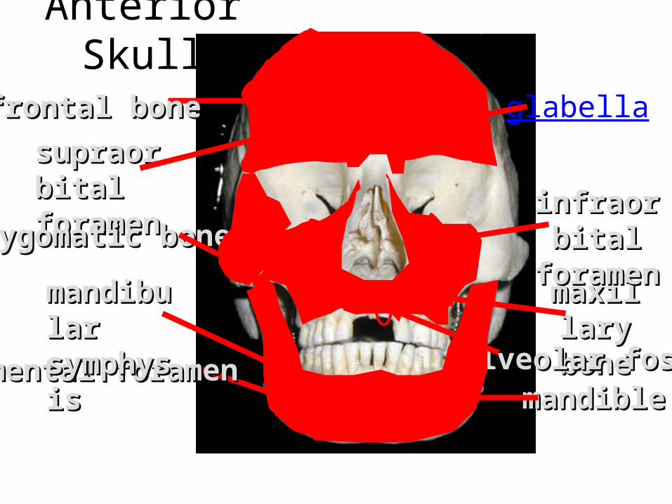
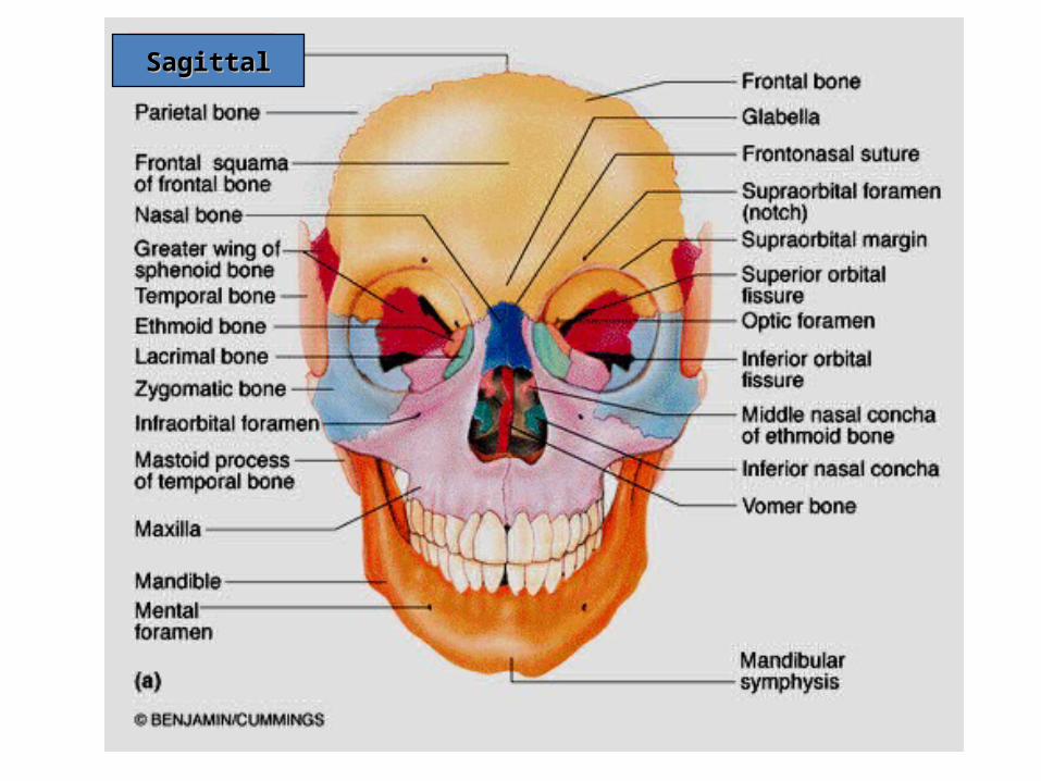
Anterior Skull
frontal bonefrontal bone
supraorbitsupraorbital al foramenforamenzygomatic bonezygomatic bone
maxillarmaxillary boney bone
alveolar fossaalveolar fossa
infraorbitainfraorbital foramenl foramen
glabella
mental foramenmental foramenmandiblemandible
mandibulmandibular ar symphysissymphysis
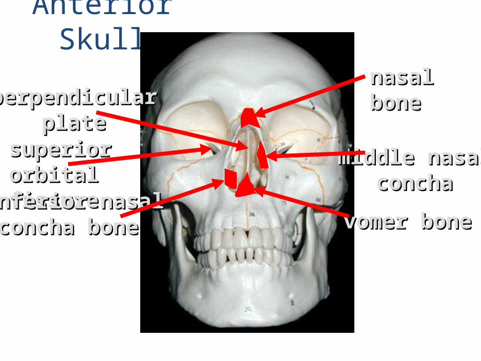
Anterior Skull
nasal nasal boneboneperpendicularperpendicular
plateplate
middle nasalmiddle nasalconchaconcha
vomer bonevomer bone
superior superior orbital fissureorbital fissureinferior nasalinferior nasal concha boneconcha bone
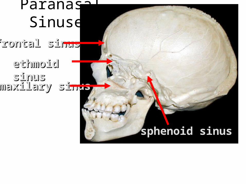
Paranasal Sinuses
frontal sinusfrontal sinus
ethmoid ethmoid sinussinusmaxilary sinusmaxilary sinus
sphenoid sinussphenoid sinus
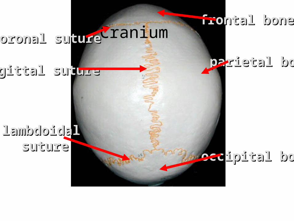
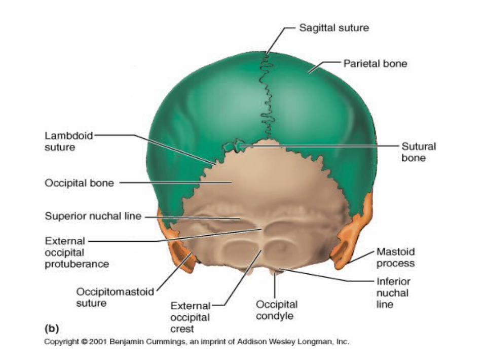
Craniumfrontal bonefrontal bone
parietal boneparietal bone
occipital boneoccipital bone
lambdoidallambdoidal suturesuture
sagittal suturesagittal suture
coronal suturecoronal suture
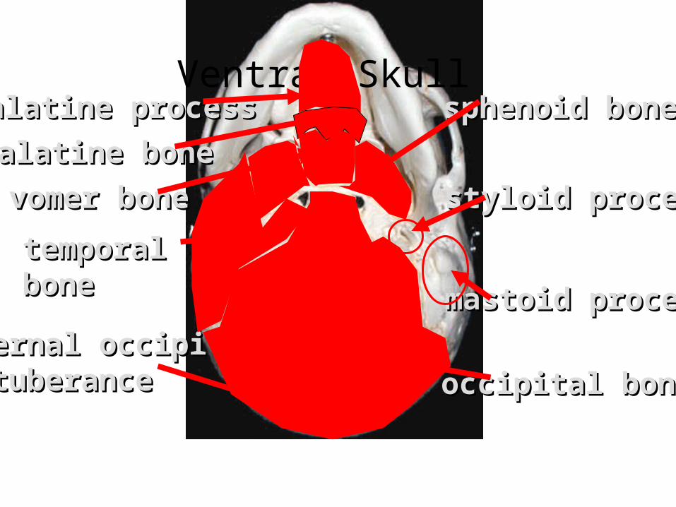
Ventral Skullpalatine processpalatine process
palatine bonepalatine bone
vomer bonevomer bone
mastoid processmastoid process
styloid processstyloid process
external occipitalexternal occipitalprotuberanceprotuberance
sphenoid bonesphenoid bone
temporal temporal bonebone
occipital boneoccipital bone
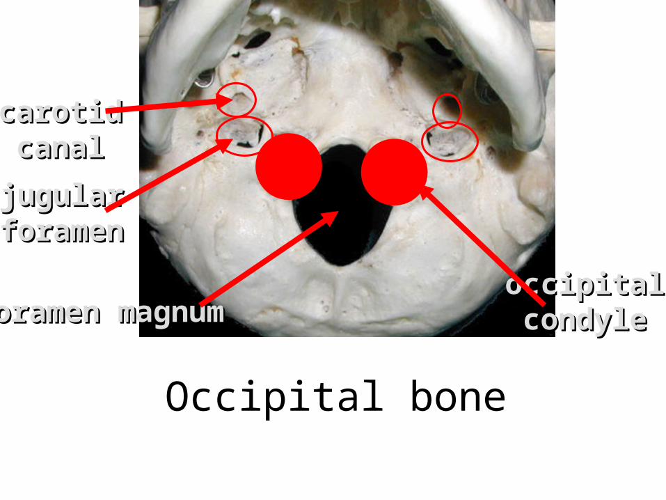
Occipital bone
occipitaloccipitalcondylecondyle
jugularjugularforamenforamen
carotidcarotidcanalcanal
foramen magnumforamen magnum
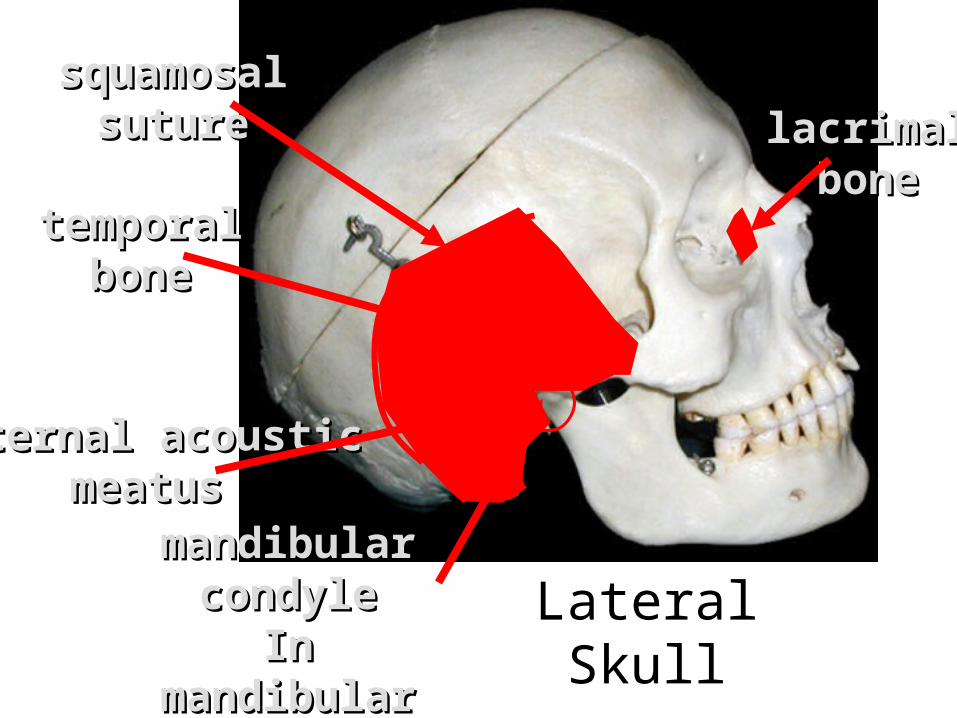
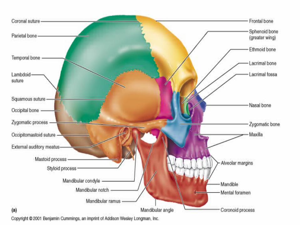
Lateral Skull
lacrimallacrimalbonebone
temporaltemporalbonebone
squamosalsquamosalsuturesuture
mandibular mandibular condylecondyle
In mandibular In mandibular fossafossa
(TMJ joint)(TMJ joint)
external acousticexternal acousticmeatusmeatus
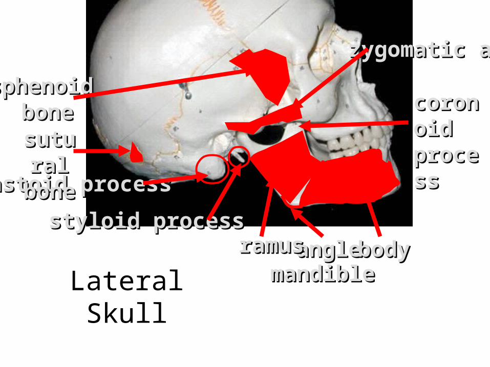
angleangle
coronoicoronoid d processprocess
zygomatic archzygomatic arch
mastoid processmastoid process
styloid processstyloid process
sphenoid sphenoid bonebone
bodybodyramusramusmandiblemandible
Lateral Skull
sutural sutural bonebone
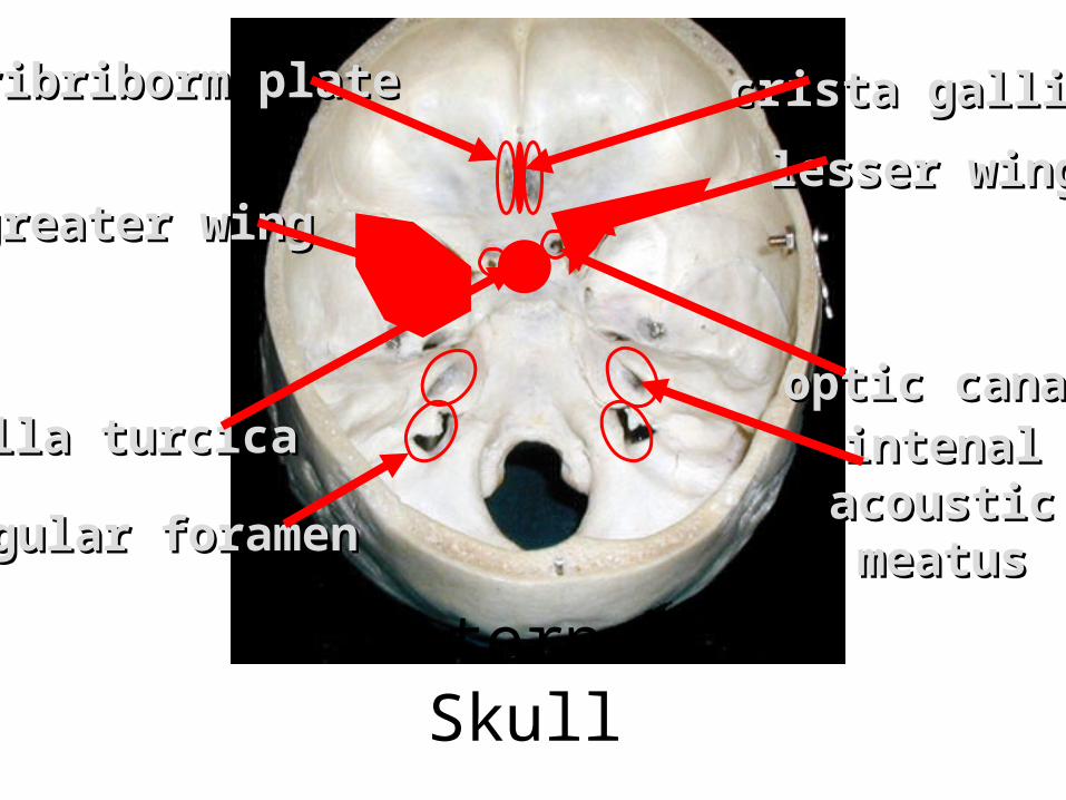
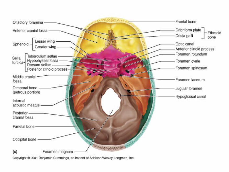
crista gallicrista gallicribriborm platecribriborm plate
intenal intenal acoustic acoustic meatusmeatus
greater winggreater winglesser winglesser wing
optic canaloptic canalsella turcicasella turcica
jugular foramenjugular foramen
Internal Skull
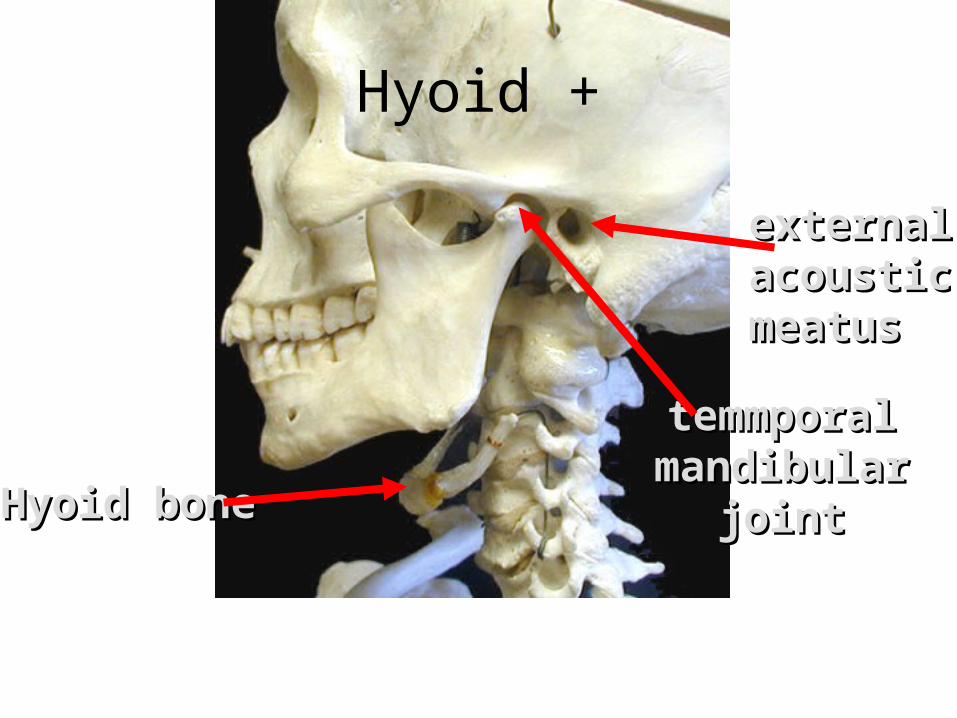
Hyoid boneHyoid bone
temmporaltemmporalmandibularmandibular
jointjoint
external external acousticacousticmeatusmeatus
Hyoid +
________________________________________________SagittalSagittal
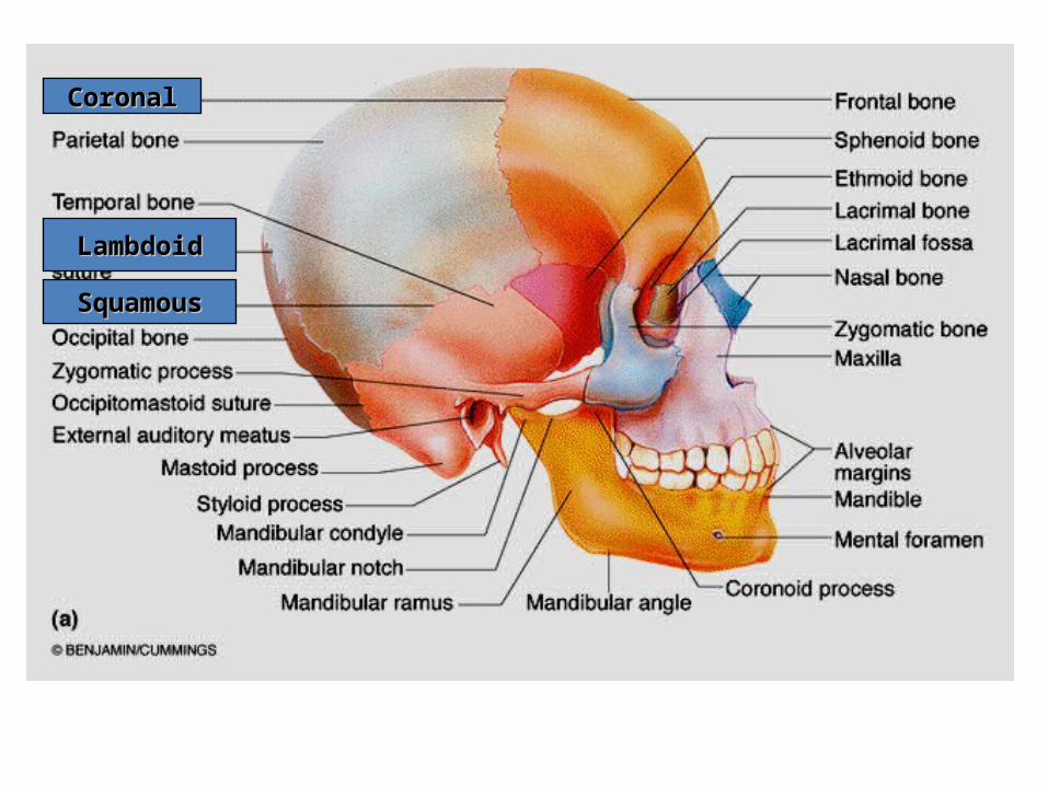
CoronalCoronal
LambdoidLambdoid
SquamousSquamous
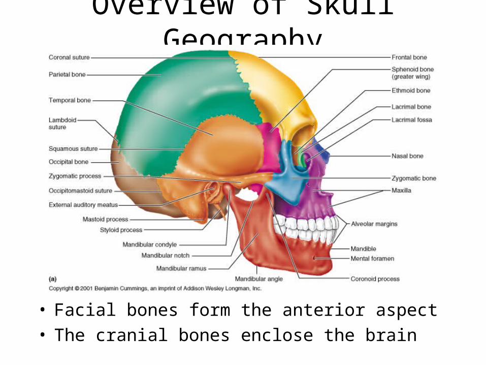
Overview of Skull Geography
• Facial bones form the anterior aspect• The cranial bones enclose the brain
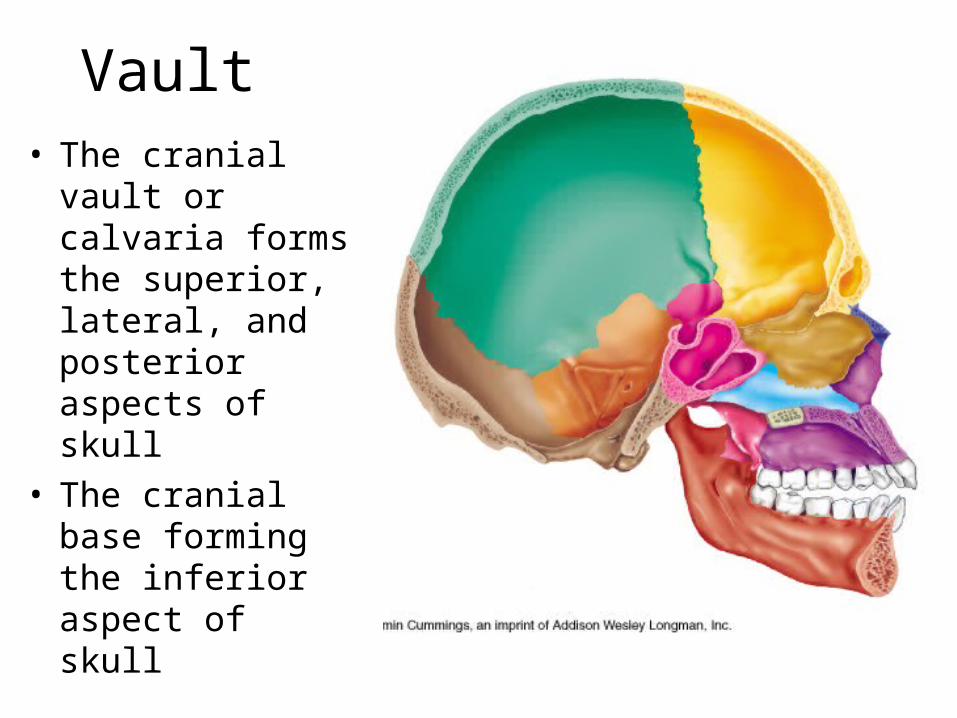
Vault• The cranial vault
or calvaria forms the superior, lateral, and posterior aspects of skull
• The cranial base forming the inferior aspect of skull
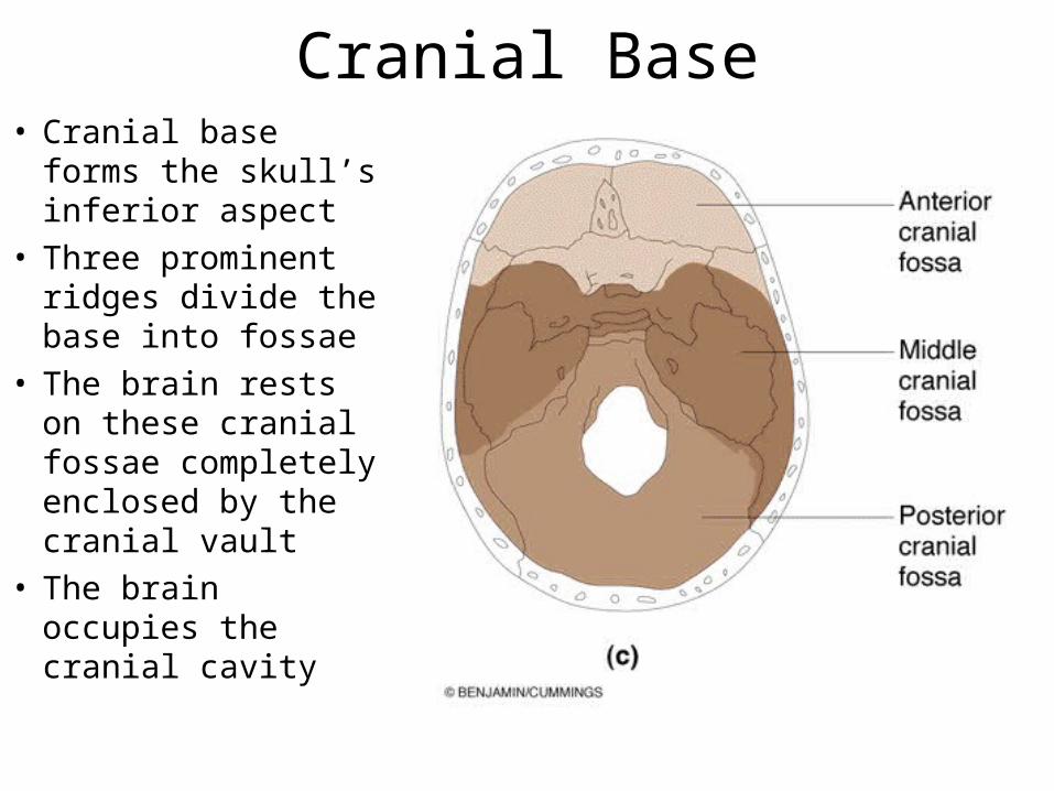
Cranial Base• Cranial base forms
the skull’s inferior aspect
• Three prominent ridges divide the base into fossae
• The brain rests on these cranial fossae completely enclosed by the cranial vault
• The brain occupies the cranial cavity
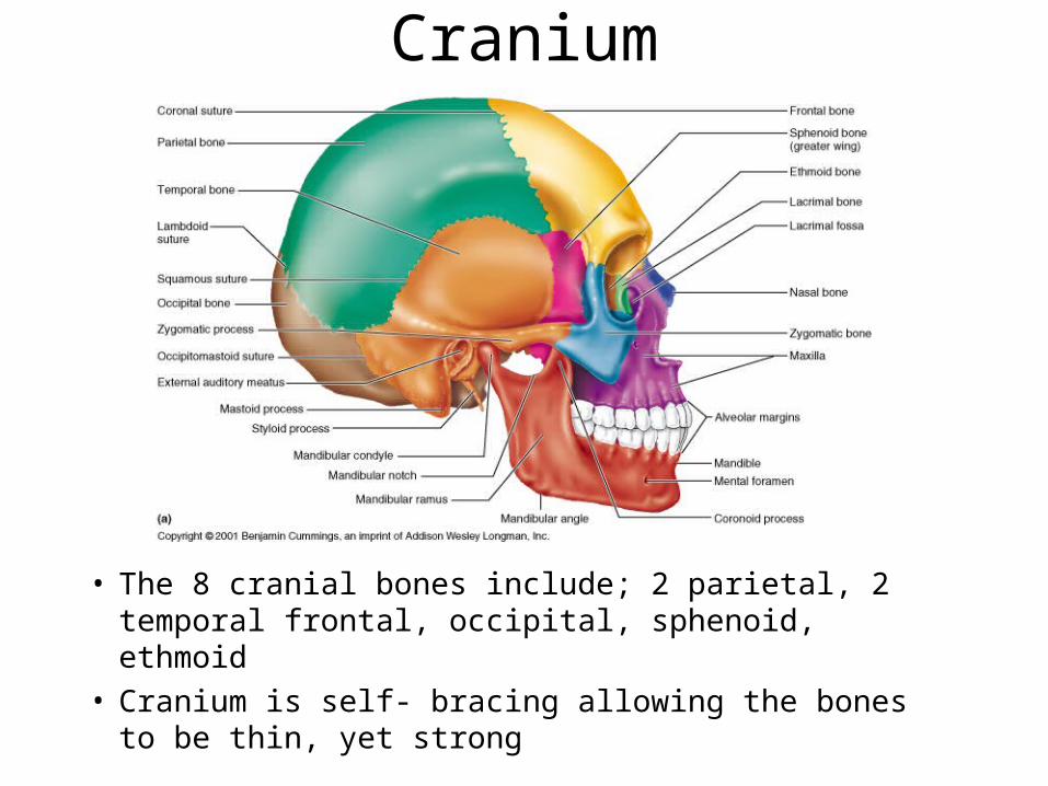
Cranium
• The 8 cranial bones include; 2 parietal, 2 temporal frontal, occipital, sphenoid, ethmoid
• Cranium is self- bracing allowing the bones to be thin, yet strong
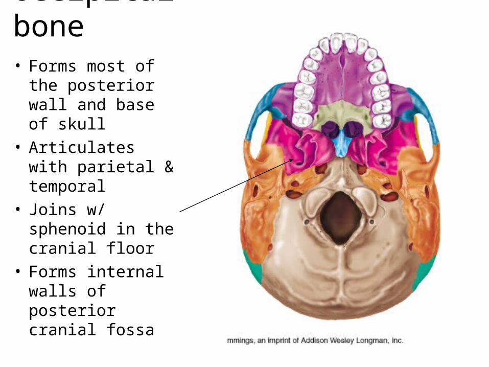
Occipital bone• Forms most of the
posterior wall and base of skull
• Articulates with parietal & temporal
• Joins w/ sphenoid in the cranial floor
• Forms internal walls of posterior cranial fossa
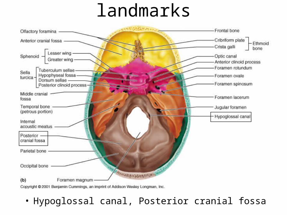
Occipital bone - Int. landmarks
• Hypoglossal canal, Posterior cranial fossa
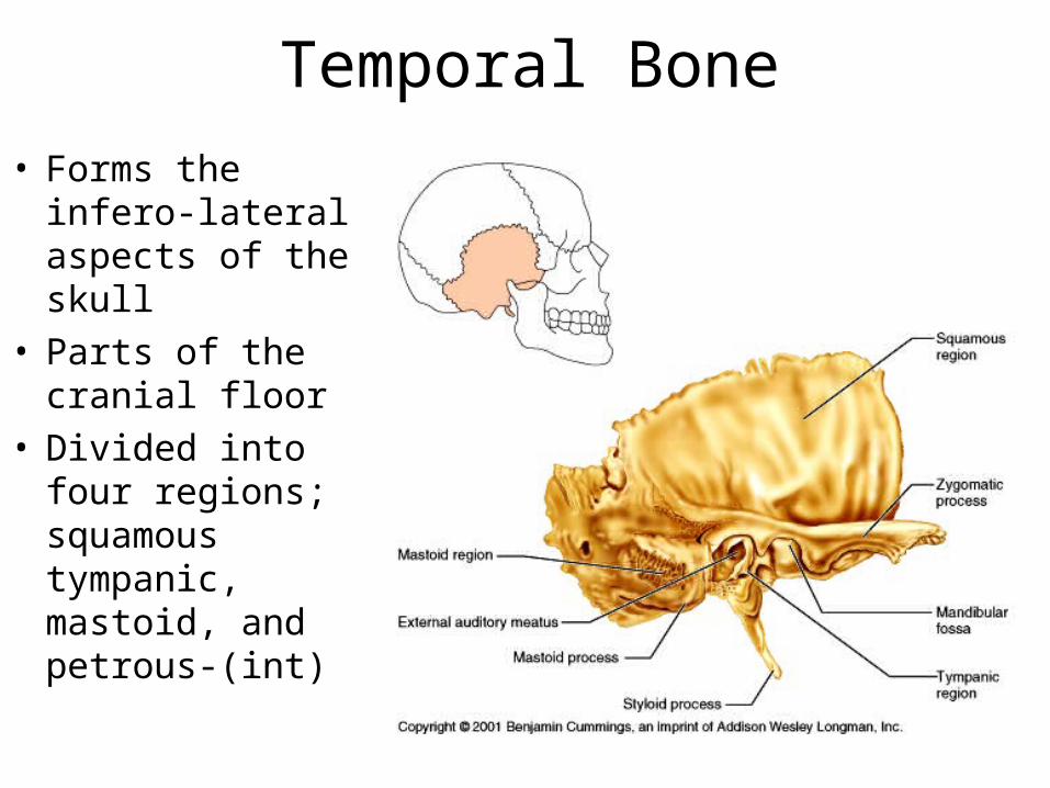
Temporal Bone• Forms the infero-
lateral aspects of the skull
• Parts of the cranial floor
• Divided into four regions; squamous tympanic, mastoid, and petrous-(int)
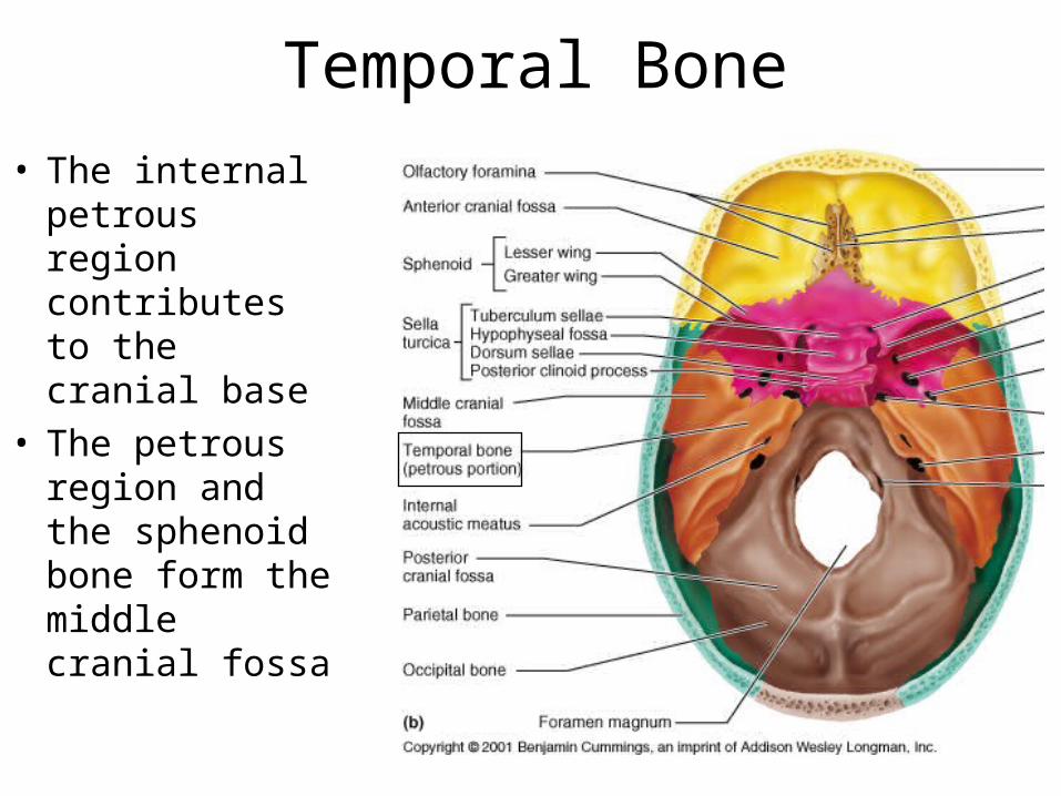
Temporal Bone• The internal
petrous region contributes to the cranial base
• The petrous region and the sphenoid bone form the middle cranial fossa
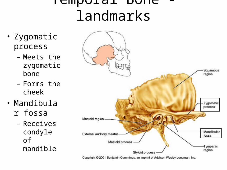
Temporal Bone - landmarks• Zygomatic
process– Meets the
zygomatic bone
– Forms the cheek
• Mandibular fossa– Receives
condyle of mandible
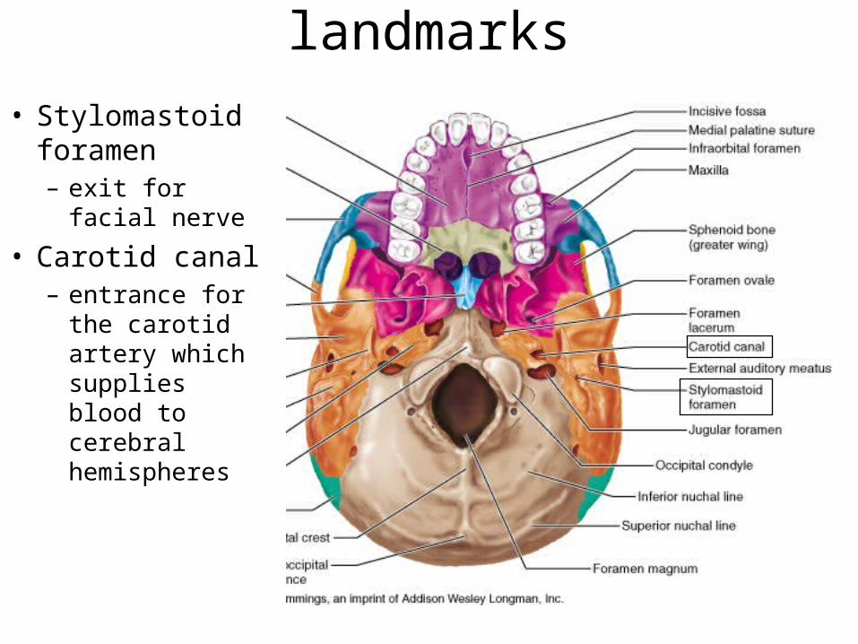
Temporal bones - landmarks• Stylomastoid
foramen– exit for facial
nerve
• Carotid canal– entrance for
the carotid artery which supplies blood to cerebral hemispheres
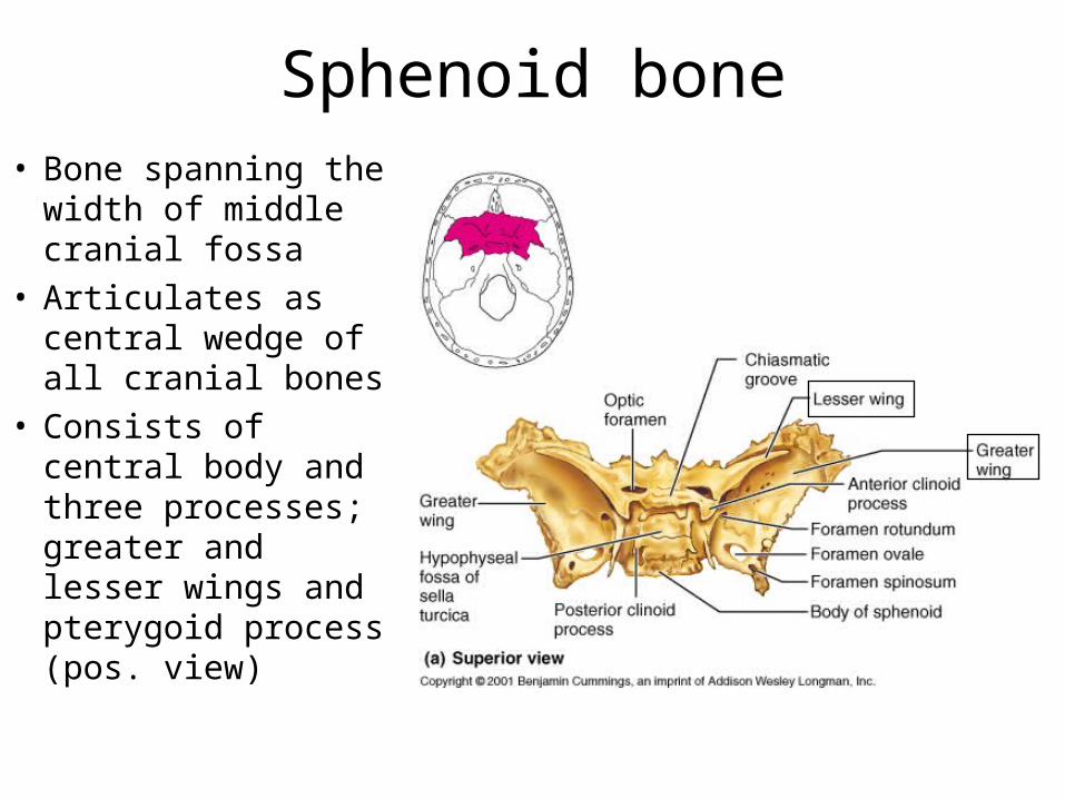
Sphenoid bone• Bone spanning the
width of middle cranial fossa
• Articulates as central wedge of all cranial bones
• Consists of central body and three processes; greater and lesser wings and pterygoid process (pos. view)
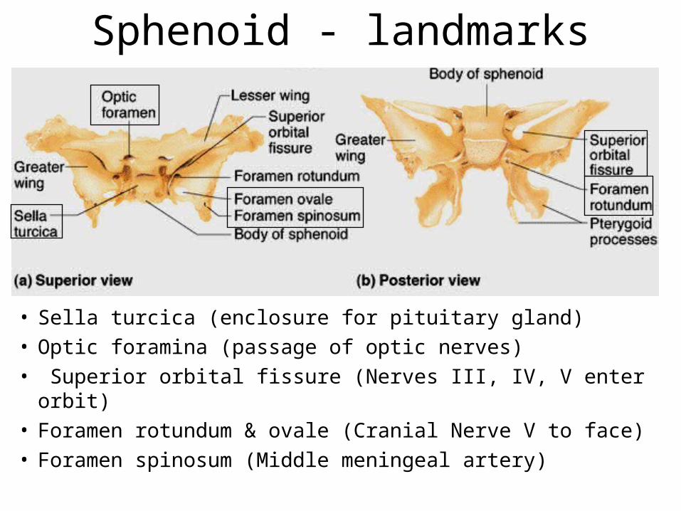
Sphenoid - landmarks
• Sella turcica (enclosure for pituitary gland)• Optic foramina (passage of optic nerves)• Superior orbital fissure (Nerves III, IV, V enter orbit)• Foramen rotundum & ovale (Cranial Nerve V to face) • Foramen spinosum (Middle meningeal artery)
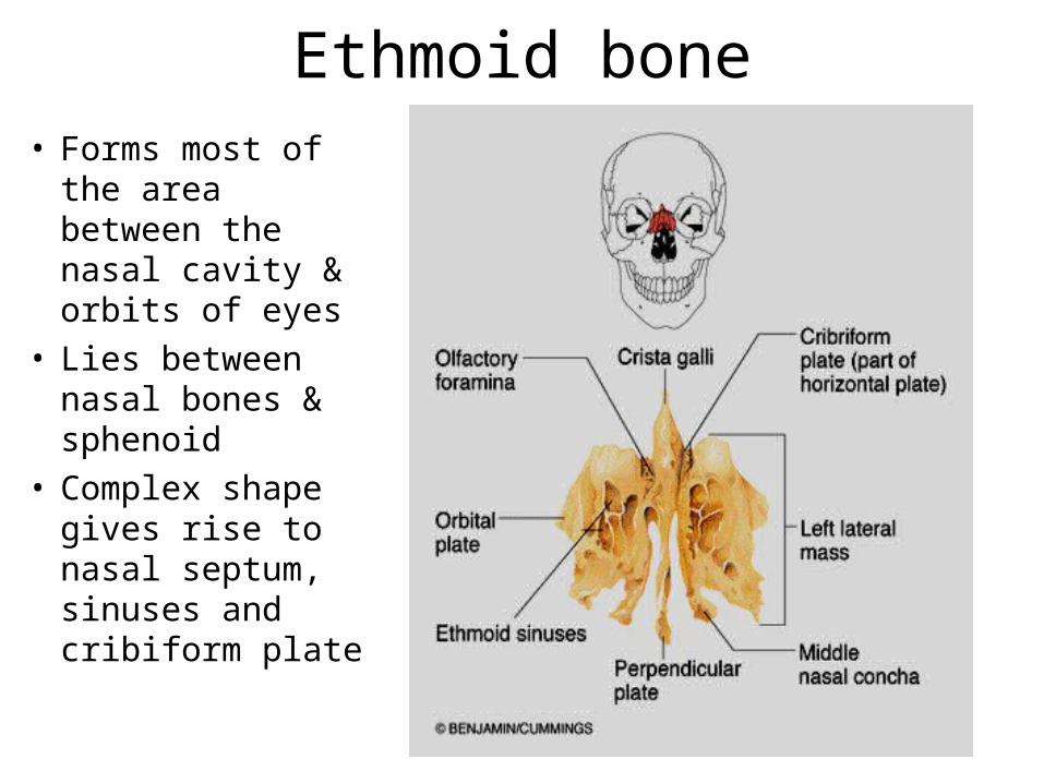
Ethmoid bone• Forms most of the
area between the nasal cavity & orbits of eyes
• Lies between nasal bones & sphenoid
• Complex shape gives rise to nasal septum, sinuses and cribiform plate
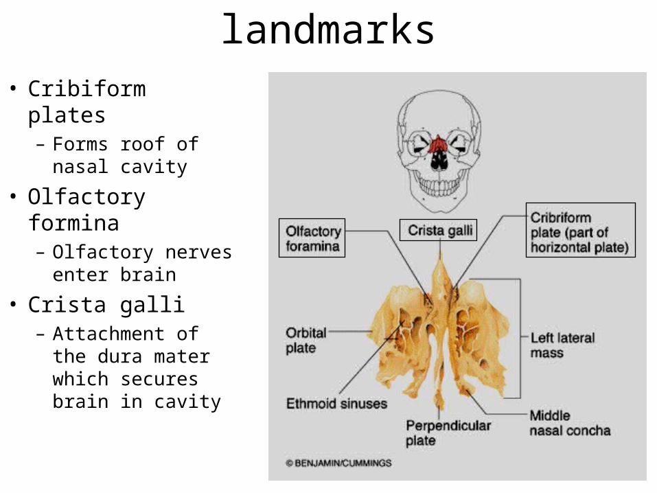
Ethmoid bone - landmarks• Cribiform plates– Forms roof of nasal
cavity
• Olfactory formina– Olfactory nerves
enter brain
• Crista galli– Attachment of the
dura mater which secures brain in cavity
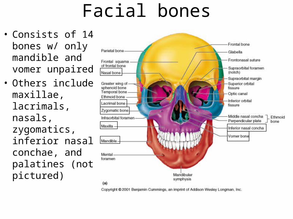
Facial bones• Consists of 14
bones w/ only mandible and vomer unpaired
• Others include maxillae, lacrimals, nasals, zygomatics, inferior nasal conchae, and palatines (not pictured)
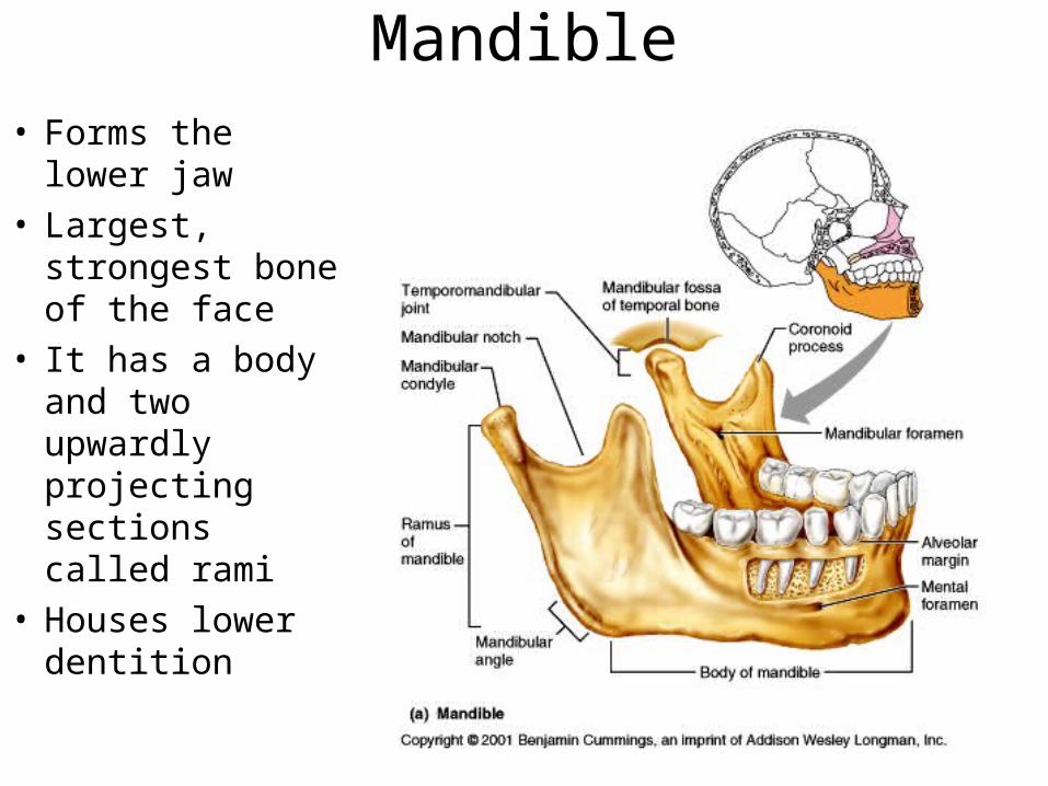
Mandible• Forms the lower
jaw• Largest, strongest
bone of the face• It has a body and
two upwardly projecting sections called rami
• Houses lower dentition
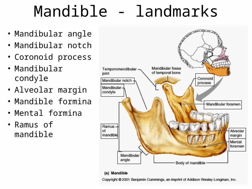
Mandible - landmarks• Mandibular angle• Mandibular notch• Coronoid process• Mandibular
condyle• Alveolar margin• Mandible formina• Mental formina• Ramus of mandible
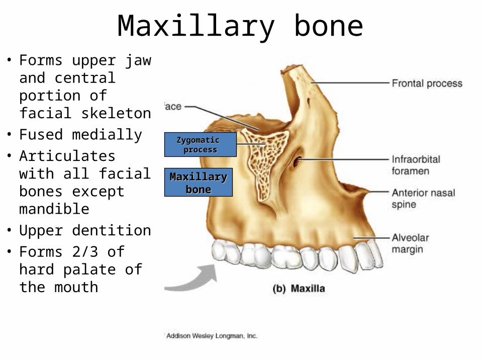
Maxillary bone• Forms upper jaw
and central portion of facial skeleton
• Fused medially• Articulates with
all facial bones except mandible
• Upper dentition• Forms 2/3 of
hard palate of the mouth
MaxillaryMaxillarybonebone
Zygomatic Zygomatic processprocess
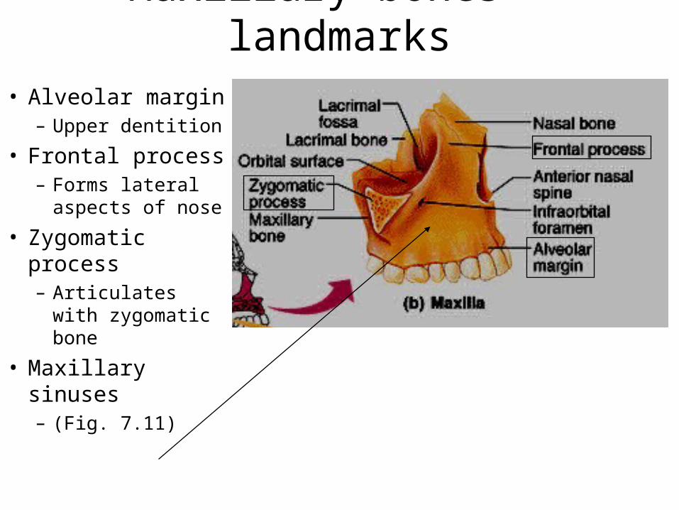
Maxillary bones - landmarks• Alveolar margin– Upper dentition
• Frontal process– Forms lateral
aspects of nose
• Zygomatic process– Articulates with
zygomatic bone
• Maxillary sinuses – (Fig. 7.11)
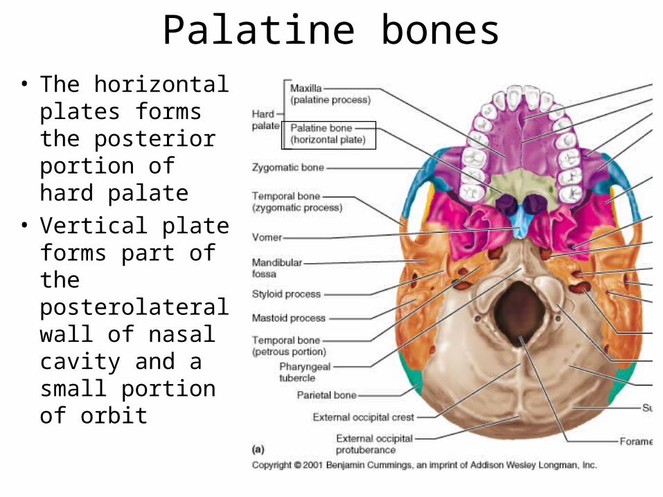
Palatine bones• The horizontal
plates forms the posterior portion of hard palate
• Vertical plate forms part of the posterolateral wall of nasal cavity and a small portion of orbit
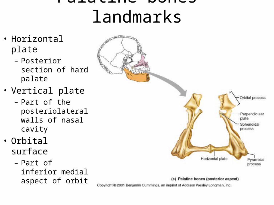
Palatine bones - landmarks• Horizontal plate– Posterior section
of hard palate
• Vertical plate– Part of the
posteriolateral walls of nasal cavity
• Orbital surface– Part of inferior
medial aspect of orbit
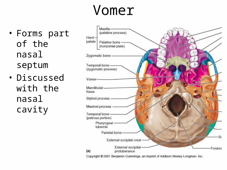
Vomer• Forms part of
the nasal septum
• Discussed with the nasal cavity
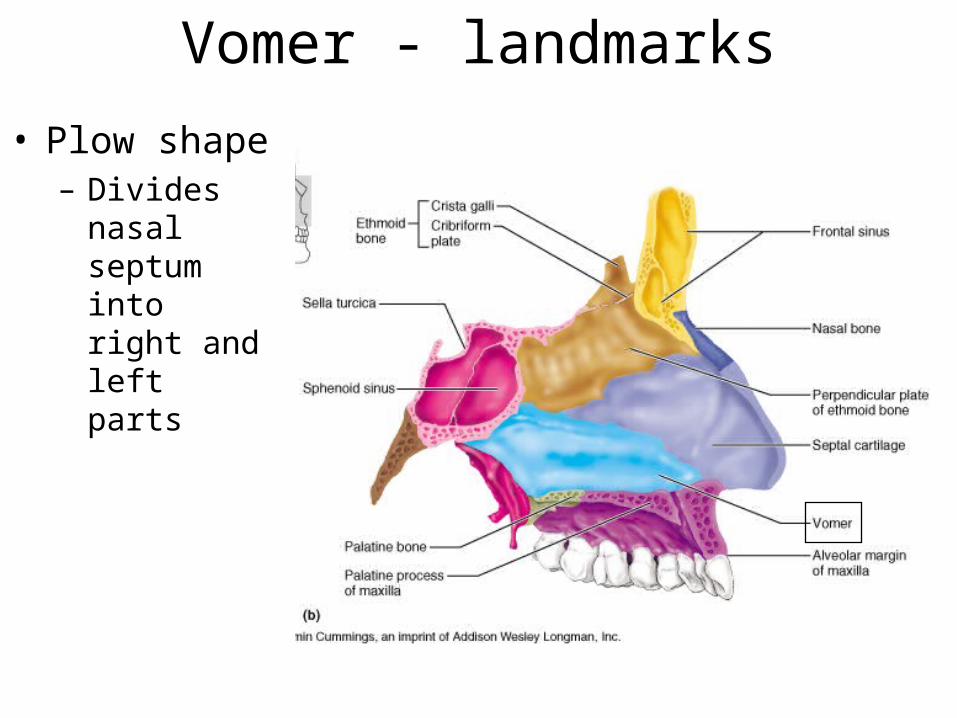
Vomer - landmarks• Plow shape– Divides nasal
septum into right and left parts

Inferior Nasal Conchae - Landmark
• The Inferior nasal conchae is just one of three in the nasal cavity
• Superior and middle concha are on the Ethmoid bone
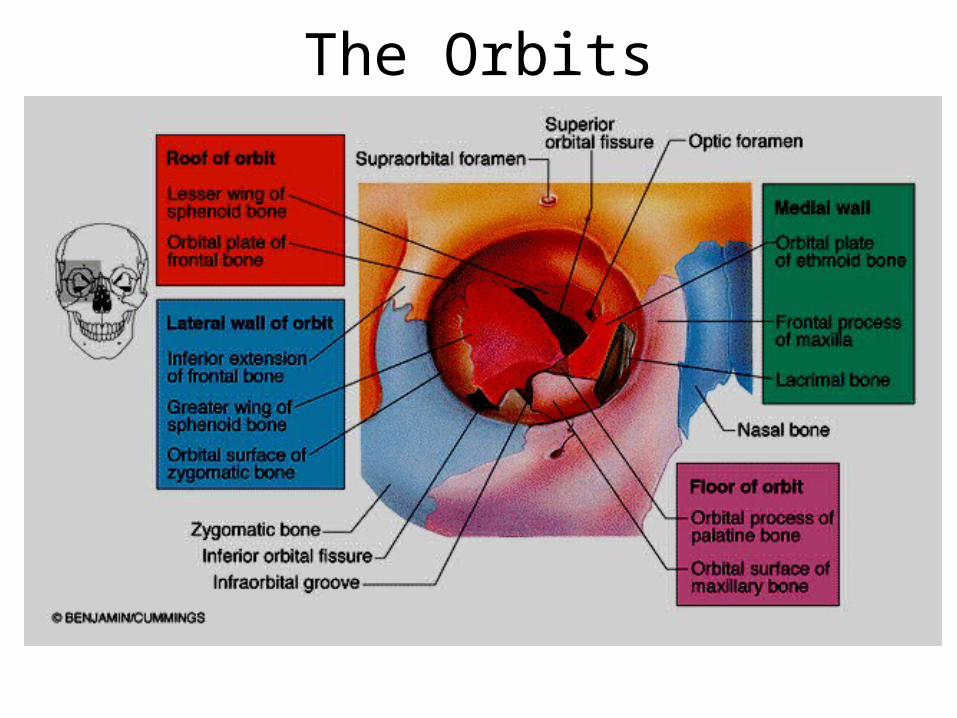
The Orbits
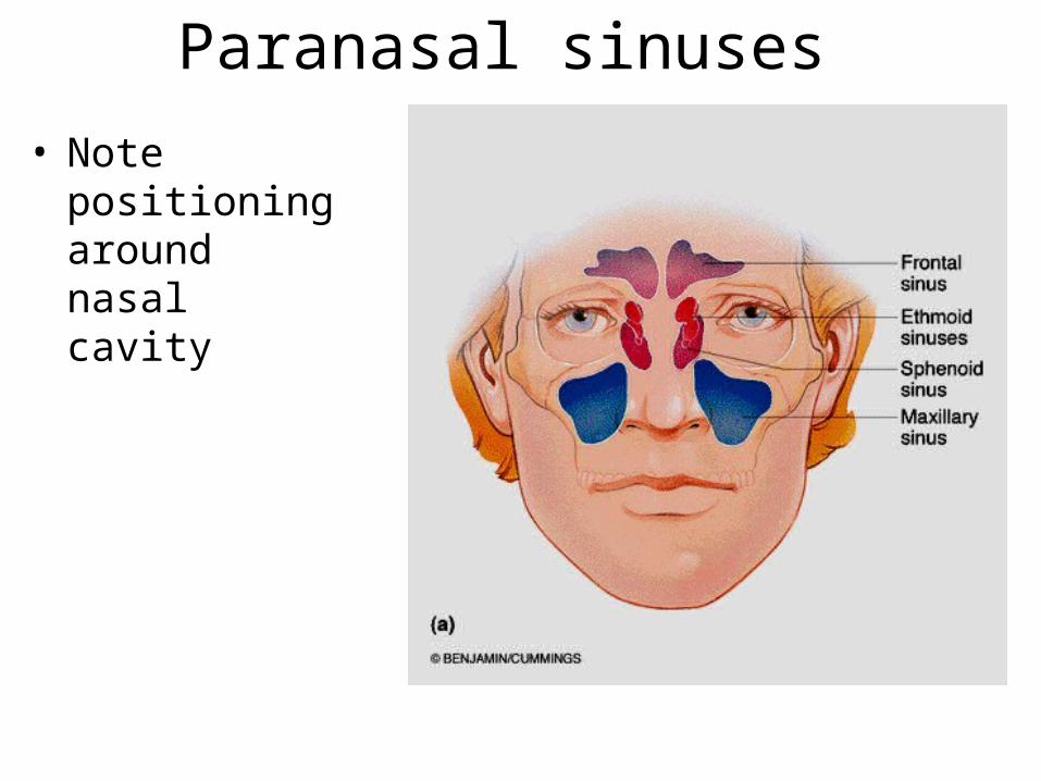
Paranasal sinuses • Note positioning
around nasal cavity
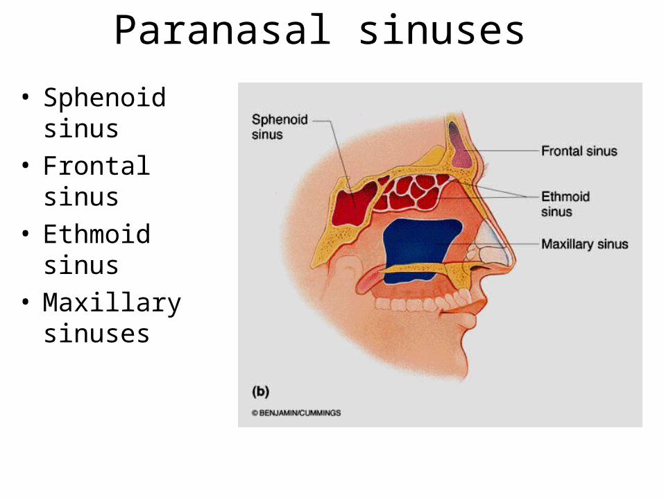
Paranasal sinuses • Sphenoid sinus• Frontal sinus• Ethmoid sinus• Maxillary sinuses
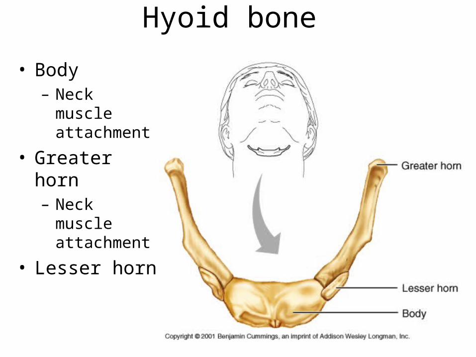
Hyoid bone
• Body– Neck muscle
attachment
• Greater horn– Neck muscle
attachment
• Lesser horn
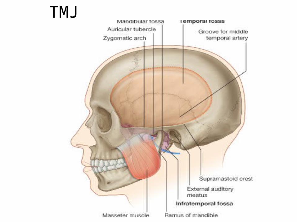
TMJ
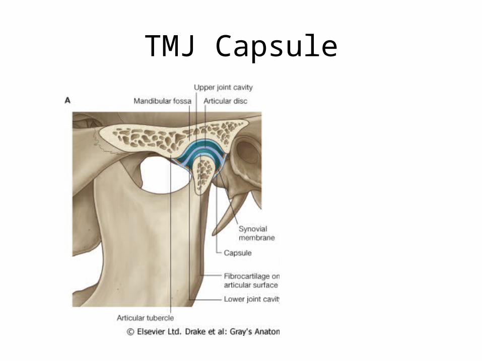
TMJ Capsule
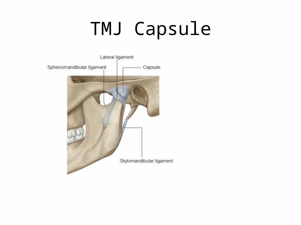
TMJ Capsule
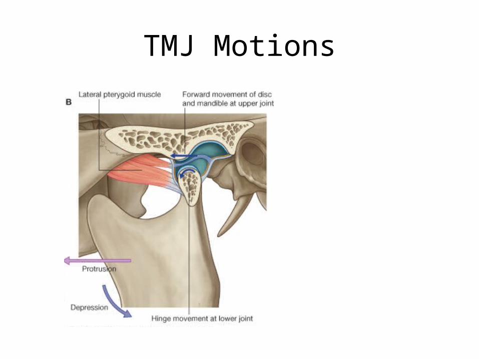
TMJ Motions
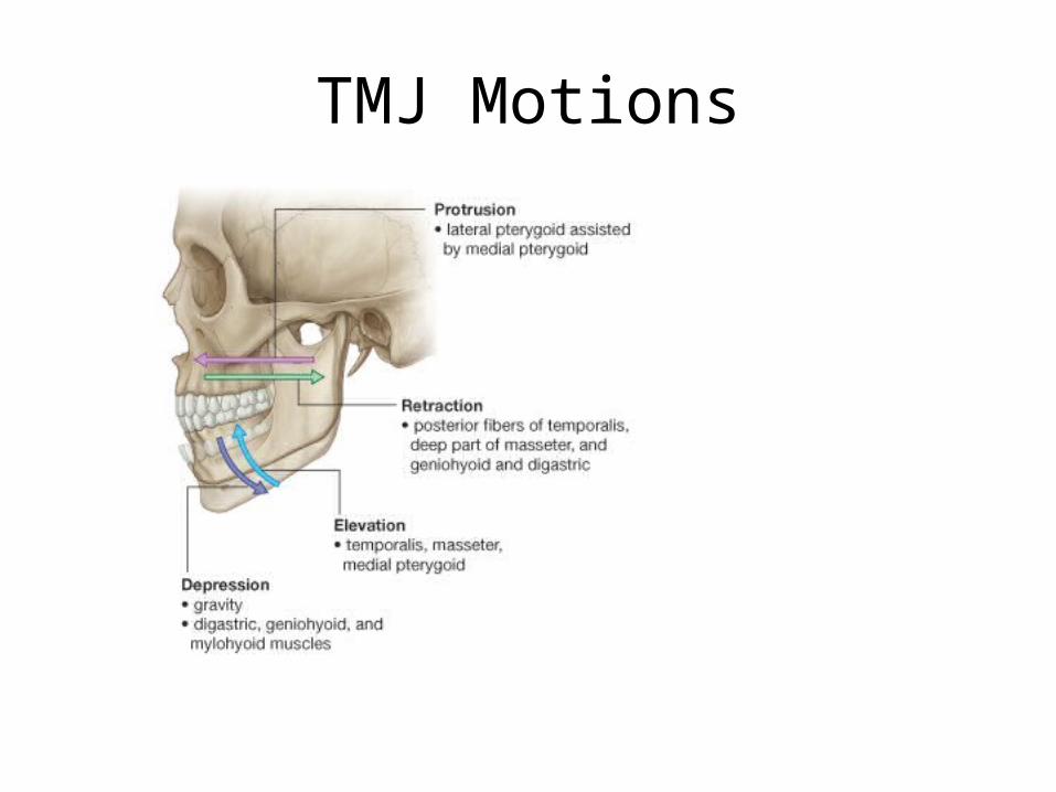
TMJ Motions
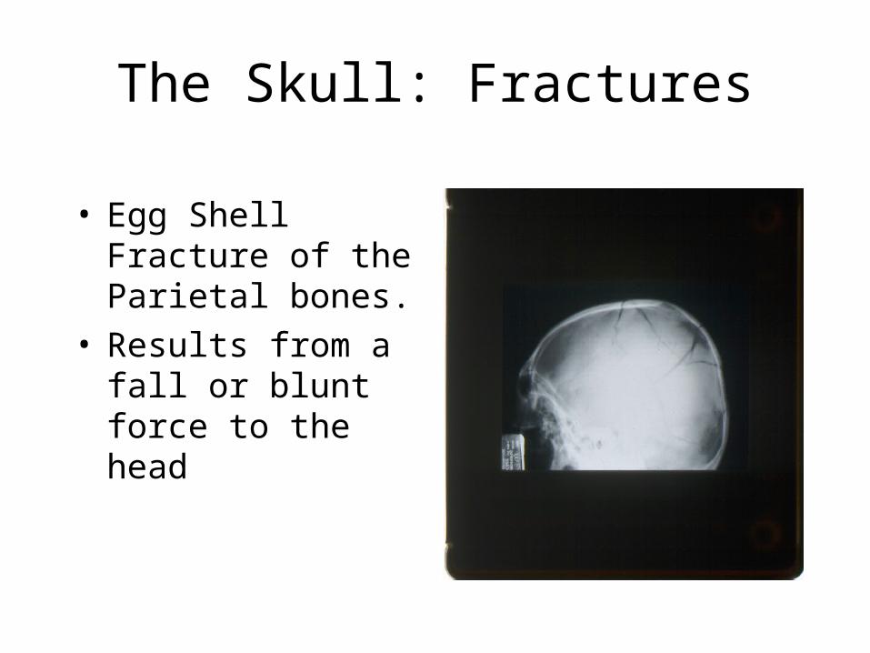
The Skull: Fractures
• Egg Shell Fracture of the Parietal bones.
• Results from a fall or blunt force to the head
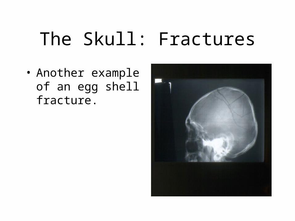
The Skull: Fractures
• Another example of an egg shell fracture.
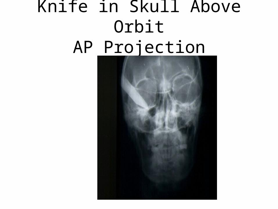
Knife in Skull Above OrbitAP Projection